Embed Size (px)
DESCRIPTION
Chapter 6: Skeletal Tissues: Bones, Ligaments, Cartilage. FUNCTIONS OF skeletal tissues. SUPPORT : B o nes form the FRAMEWORK of the body and contribute to the shape, alignment, and positioning of body parts; LIGAMENTS help hold bones together, - PowerPoint PPT Presentation
Citation preview
Chapter 6: Skeletal Tissues:
Bones, Ligaments, Cartilage
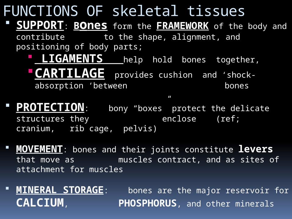
FUNCTIONS OF skeletal tissues SUPPORT: Bones form the FRAMEWORK of the body
and contribute to the shape, alignment, and positioning of body parts;
LIGAMENTS help hold bones together, CARTILAGE provides cushion and ‘shock-absorption
‘between bones
PROTECTION: bony “boxes” protect the delicate structures they enclose (ref; cranium, rib cage, pelvis)
MOVEMENT: bones and their joints constitute levers that move as muscles contract, and as sites of attachment for muscles
MINERAL STORAGE: bones are the major reservoir for CALCIUM, PHOSPHORUS, and other minerals
HEMATOPOIESIS: blood cell formation is carried out by Myeloid tissue, which is located
in the bone marrow
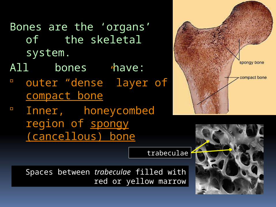
Bones are the ‘organs’ of the skeletal system.
All bones have: outer “dense” layer of
compact bone Inner, honeycombed
region of spongy (cancellous) bone
Spaces between trabeculae filled with red or yellow marrow
trabeculae
TYPES OF BONES Five major types of structural bones
Long bones Short bones Flat bones Irregular bones Sesamoid bones develops within a tendon ( ex.: patella)
Bones serve various needs, and their size, shape, and appearance vary to meet those needs
Bones vary in the proportion of compact and cancellous (spongy)
bone; COMPACT BONE is dense and solid in
appearance, whereas CANCELLOUS BONE is characterized by open space partially filled with needle-like structures,
called spicules, or trabeculae
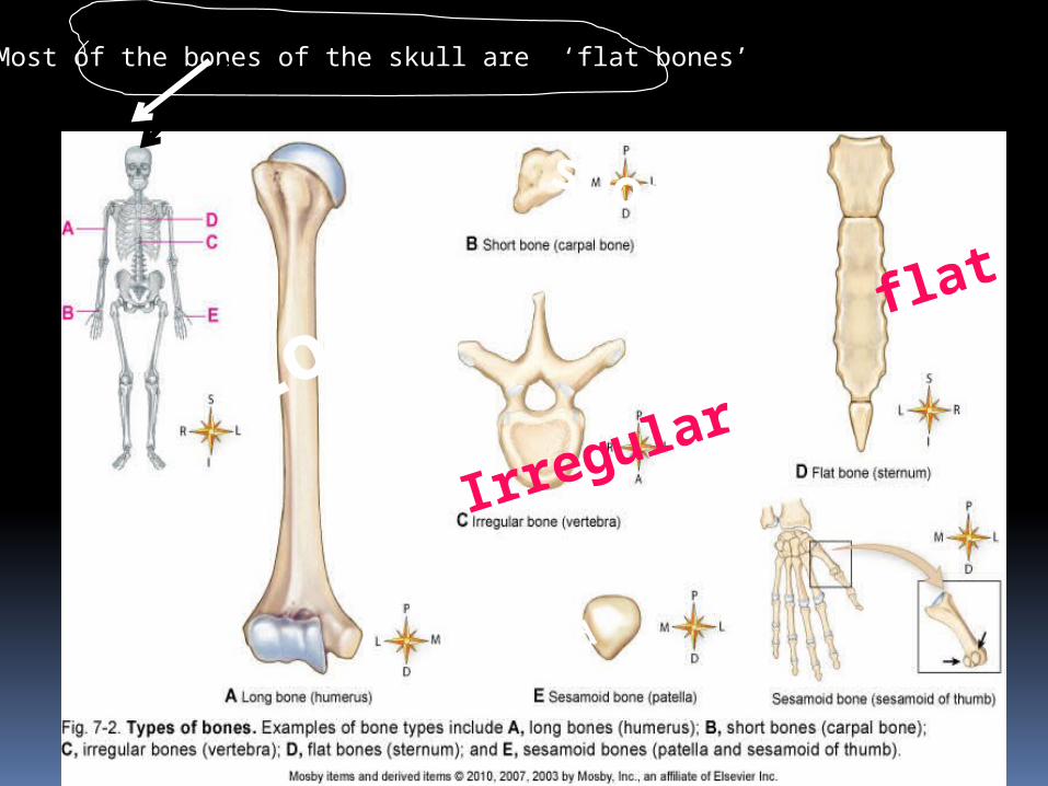
Most of the bones of the skull are ‘flat bones’
Longshort
Irregular
flat
sesamoid
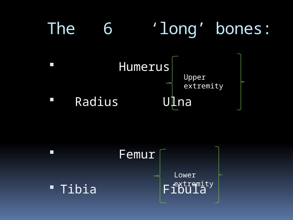
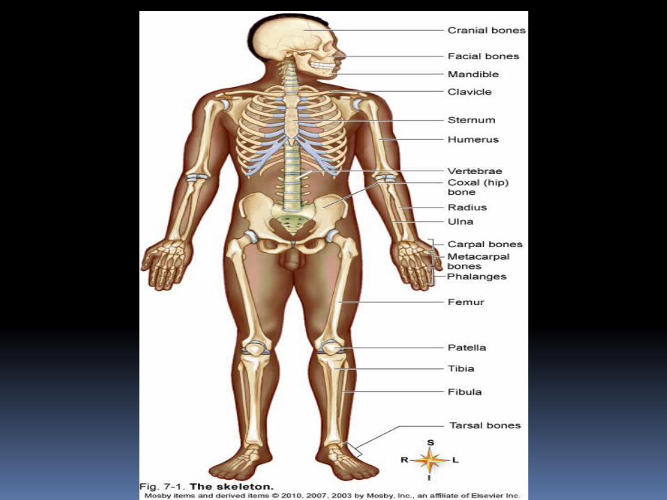
The 6 ‘long’ bones:
Humerus
Radius Ulna
Femur
Tibia Fibula
Upperextremity
Lowerextremity
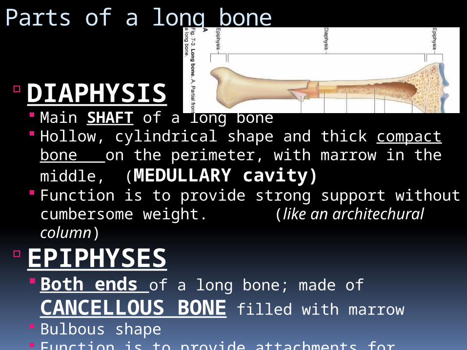
Parts of a long bone
DIAPHYSIS Main SHAFT of a long bone Hollow, cylindrical shape and thick compact bone on
the perimeter, with marrow in the middle, (MEDULLARY cavity)
Function is to provide strong support without cumbersome weight. (like an architechural column)
EPIPHYSES Both ends of a long bone; made of CANCELLOUS BONE filled with marrow
Bulbous shape Function is to provide attachments for muscles and
give stability to joints
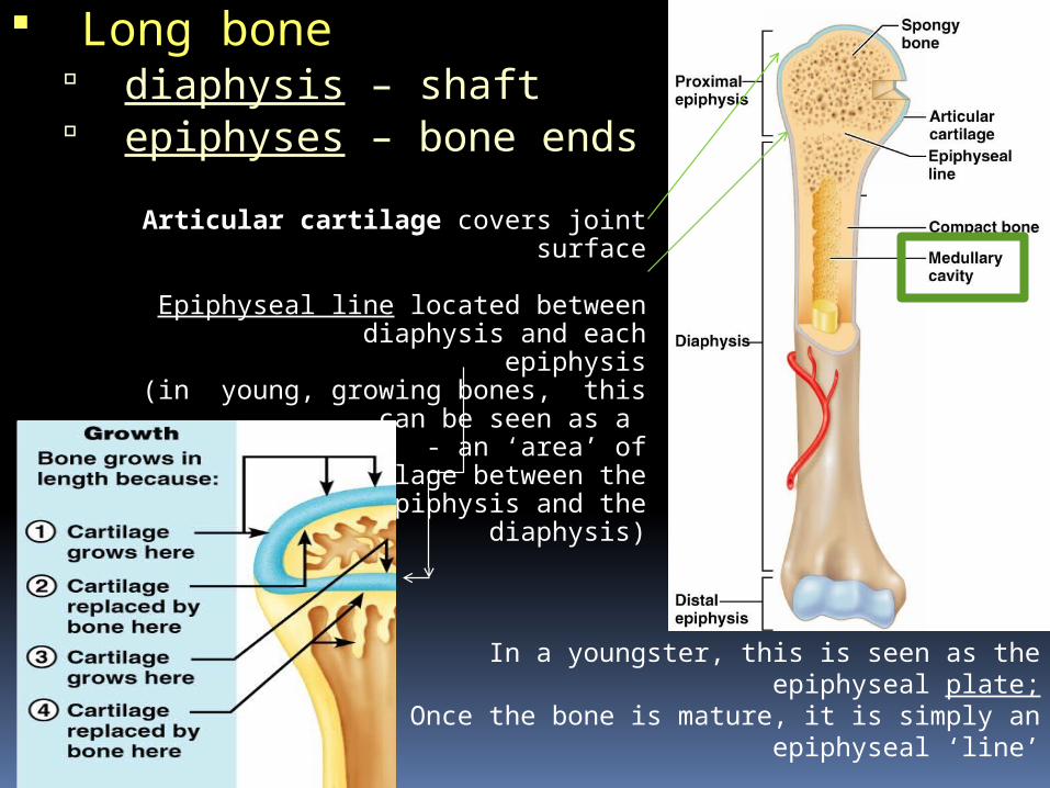
Long bone diaphysis – shaft epiphyses – bone ends
Articular cartilage covers joint surface
Epiphyseal line located between diaphysis and each epiphysis
(in young, growing bones, this can be seen as a METAPHYSIS - an ‘area’
of cartilage between the epiphysis and the diaphysis)
In a youngster, this is seen as the epiphyseal plate;Once the bone is mature, it is simply an epiphyseal
‘line’
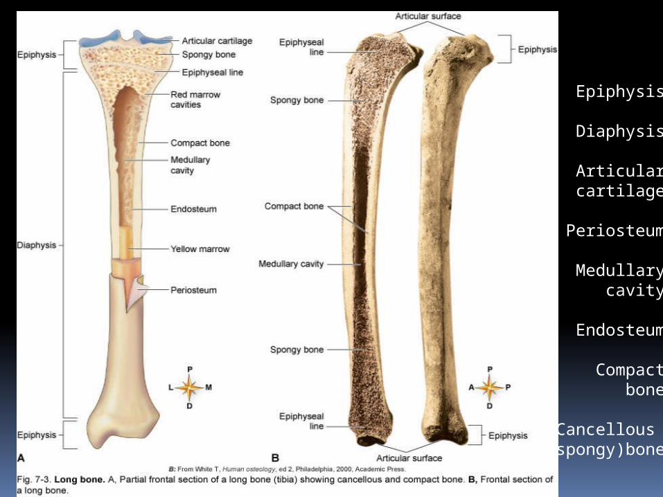
Epiphysis
Diaphysis
Articular cartilage
Periosteum
Medullary cavity
Endosteum
Compact bone
Cancellous (spongy)bone
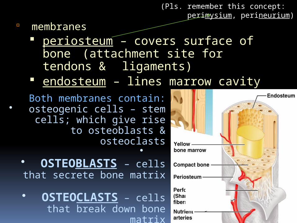
membranes periosteum – covers surface of
bone (attachment site for tendons & ligaments)
endosteum – lines marrow cavityBoth membranes contain:
• osteogenic cells – stem cells; which give rise to osteoblasts &
osteoclasts•
• OSTEOBLASTS – cells that secrete bone matrix
• OSTEOCLASTS – cells that break down bone matrix
(Pls. remember this concept: perimysium, perineurium)
More on the components of bones Articular cartilage
Layer of hyaline cartilage that covers the articular surface of epiphyses
Function is to cushion jolts and blows
Periosteum Dense, white fibrous membrane that
covers bone Attaches tendons firmly to bones Contains cells that form and destroy bone Contains blood vessels important in
growth and repair Contains blood vessels that send branches
into bone Essential for bone cell survival and bone
formation
Components of bone
Medullary (or marrow) cavityTubelike, hollow space in the diaphysis
Filled with yellow marrow in adults . (Yellow marrow is fatty, can be called upon to become active hematopoietic tissue if needed)
Endosteum: thin, fibrous membrane that lines the medullary cavity
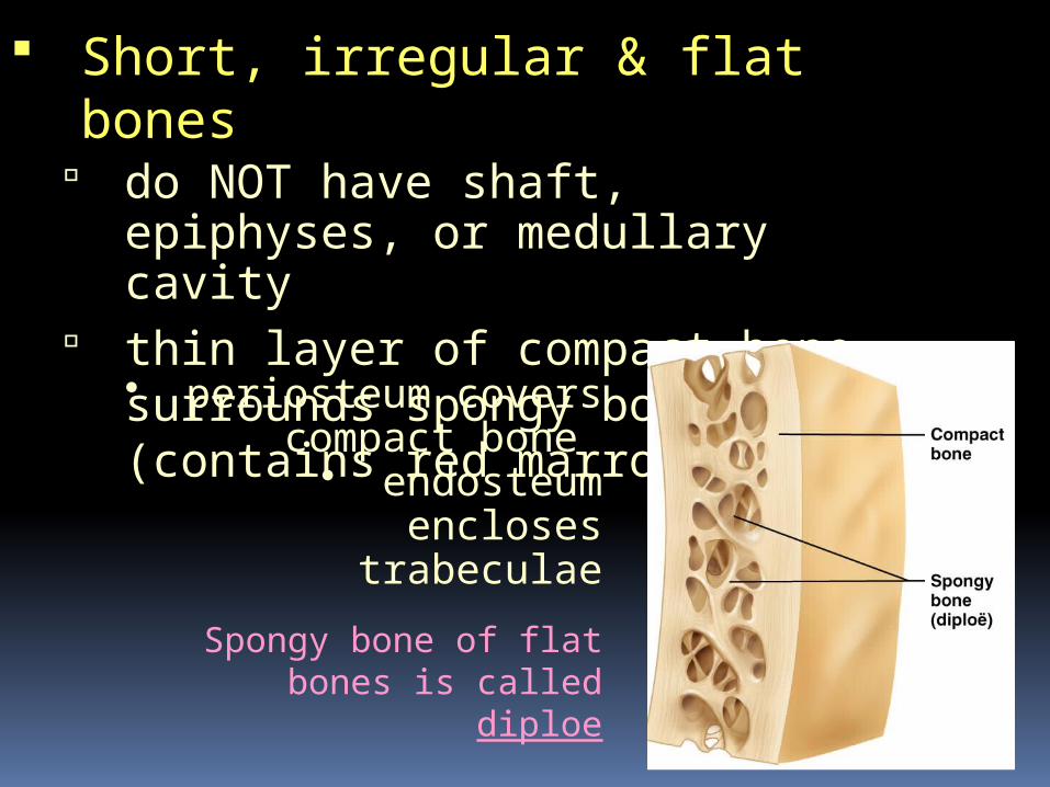
Short, irregular & flat bones do NOT have shaft, epiphyses, or
medullary cavity thin layer of compact bone
surrounds spongy bone center (contains red marrow)
• periosteum covers compact bone
• endosteum encloses trabeculae
Spongy bone of flat bones is called diploe
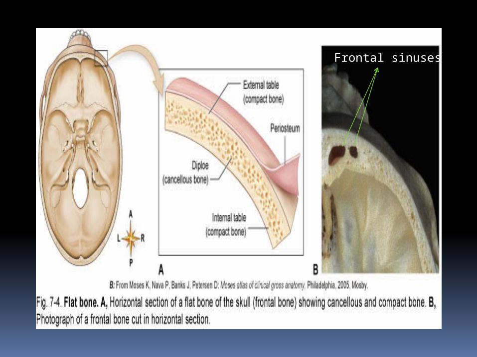
Frontal sinuses
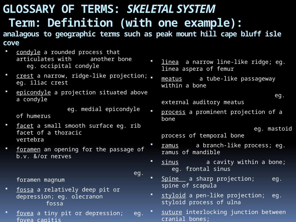
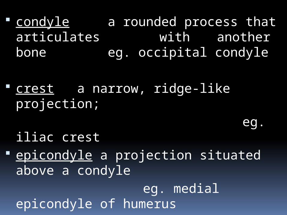
GLOSSARY OF TERMS: SKELETAL SYSTEM Term: Definition (with one example):analagous to geographic terms such as peak mount hill cape bluff isle cove condyle a rounded process that articulates
with another bone eg. occipital condyle
crest a narrow, ridge-like projection; eg. iliac crest
epicondyle a projection situated above a condyle
eg. medial epicondyle of humerus facet a small smooth surface eg. rib facet of
a thoracic vertebra
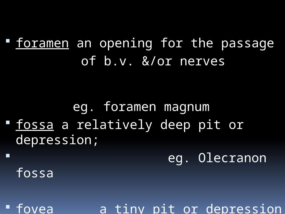
foramen an opening for the passage of b.v. &/or nerves
eg. foramen magnum
fossa a relatively deep pit or depression; eg. olecranon
fossa fovea a tiny pit or depression; eg. fovea
capitis head an enlargement at the end of a bone; eg. femoral
head
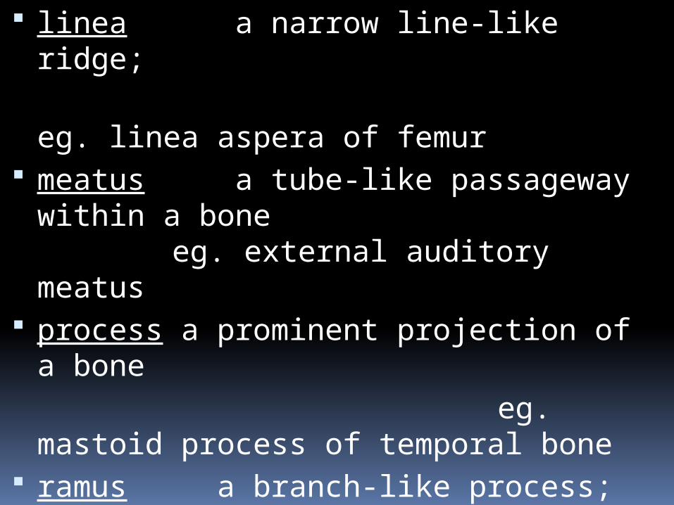
linea a narrow line-like ridge; eg. linea aspera of femur
meatus a tube-like passageway within a bone
eg. external auditory meatus
process a prominent projection of a bone eg. mastoid process of
temporal bone ramus a branch-like process; eg. ramus of
mandible sinus a cavity within a bone; eg.
frontal sinus Spine a sharp projection; eg. spine of
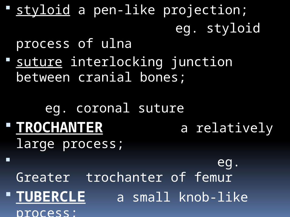
scapula styloid a pen-like projection; eg. styloid
process of ulna suture interlocking junction between cranial
bones; eg.
coronal suture trochanter a relatively large process; eg. Greater trochanter of
femur tubercle a small knob-like process; eg.
tubercle of rib tuberosity a knob-like process larger than a
tubercle; eg. tibial tuberosity
condyle a rounded process that articulates with another bone eg. occipital condyle
crest a narrow, ridge-like projection; eg. iliac crest epicondyle a projection situated above a
condyle eg. medial epicondyle of
humerus facet a small smooth surface eg. rib facet of a thoracic
vertebra
foramen an opening for the passage of b.v. &/or nerves
eg. foramen magnum
fossa a relatively deep pit or depression; eg. Olecranon fossa
fovea a tiny pit or depression; eg. fovea capitis
head an enlargement at the end of a bone;
eg. femoral head
linea a narrow line-like ridge; eg. linea aspera of
femur meatus a tube-like passageway
within a bone eg. external auditory meatus
process a prominent projection of a bone
eg. mastoid process of temporal bone
ramus a branch-like process; eg. ramus of mandible,
Pubic ramus sinus a cavity within a bone; eg.
frontal sinus spine a sharp projection; eg. spine
of scapula
styloid a pen-like projection; eg. styloid process of ulna suture interlocking junction between
cranial bones; eg. coronal suture
TROCHANTER a relatively large process;
eg. Greater trochanter of femur
TUBERCLE a small knob-like process; eg. tubercle of rib TUBEROSITY a knob-like process
larger than a tubercle; eg. tibial tuberosity
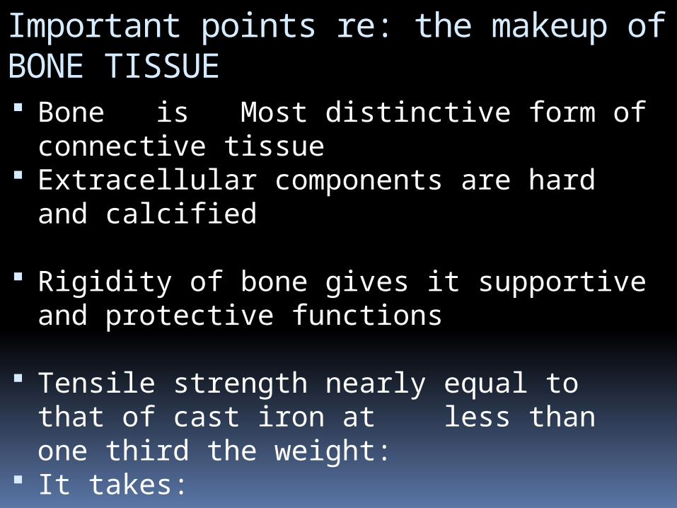
Important points re: the makeup of BONE TISSUE Bone is Most distinctive form of
connective tissue Extracellular components are hard and
calcified
Rigidity of bone gives it supportive and protective functions
Tensile strength nearly equal to that of cast iron at less than one third the weight:
It takes: GRIT and GLUE TO
MAKE BONE:, MINERAL and MATRIX >>>>>>>
see next slide
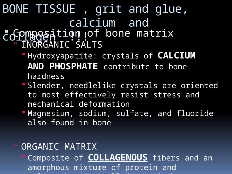
BONE TISSUE , grit and glue, calcium and collagen !!! Composition of bone matrix
INORGANIC SALTS Hydroxyapatite: crystals of CALCIUM AND PHOSPHATE contribute to bone hardness
Slender, needlelike crystals are oriented to most effectively resist stress and mechanical deformation
Magnesium, sodium, sulfate, and fluoride also found in bone
ORGANIC MATRIX Composite of COLLAGENOUS fibers and an
amorphous mixture of protein and polysaccharides called ground substance
Ground substance is secreted by connective tissue cells
Adds to overall strength of bone and gives some degree of resilience to bone
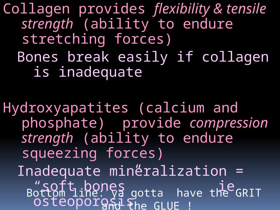
Collagen provides flexibility & tensile strength (ability to endure stretching forces)
Bones break easily if collagen is inadequate
Hydroxyapatites (calcium and phosphate) provide compression strength (ability to endure squeezing forces)
Inadequate mineralization = “soft bones” ie osteoporosisBottom line: ya gotta have the GRIT and the GLUE !
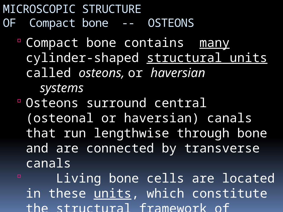
MICROSCOPIC STRUCTURE OF Compact bone -- OSTEONS
Compact bone contains many cylinder-shaped structural units called osteons, or haversian systems
Osteons surround central (osteonal or haversian) canals that run lengthwise through bone and are connected by transverse canals
Living bone cells are located in these units, which constitute the structural framework of compact bone
Osteons permit delivery of nutrients and removal of waste products
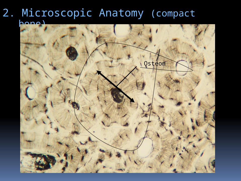
2. Microscopic Anatomy (compact bone)
Osteon
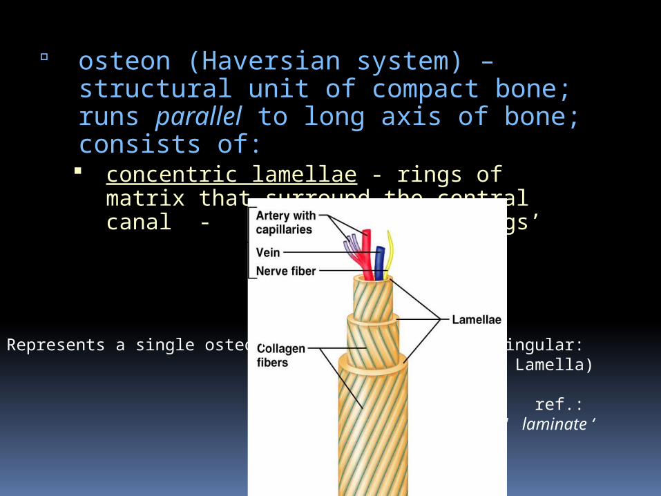
osteon (Haversian system) – structural unit of compact bone; runs parallel to long axis of bone; consists of: concentric lamellae - rings of matrix that
surround the central canal - like ‘wrappings’
Represents a single osteon (Singular: Lamella)
ref.: ‘ laminate ‘
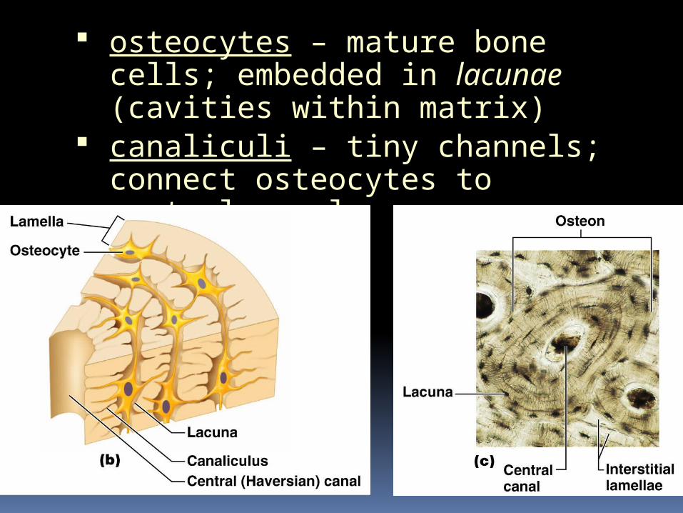
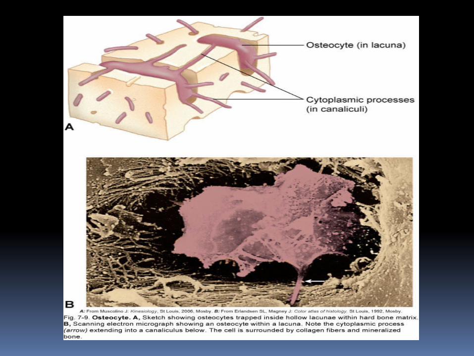
osteocytes – mature bone cells; embedded in lacunae (cavities within matrix)
canaliculi – tiny channels; connect osteocytes to central canal
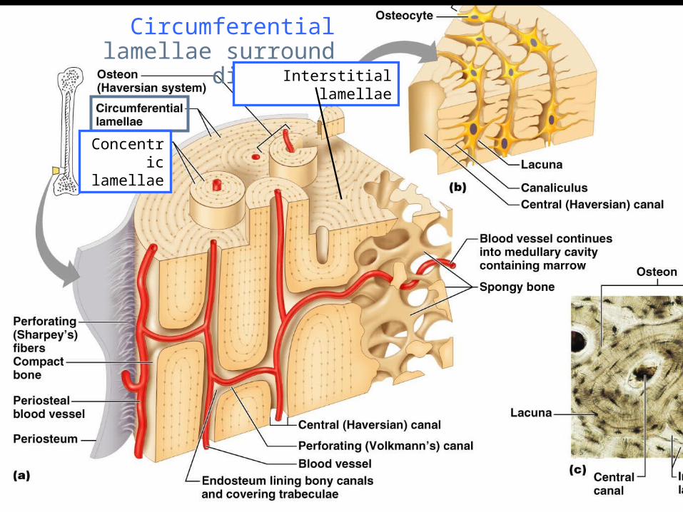
Circumferential lamellae surround diaphysis
Interstitial lamellae
Concentric lamellae
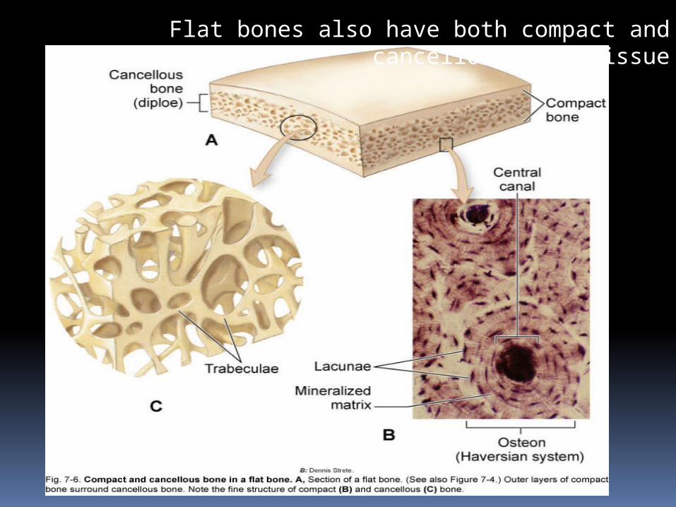
Flat bones also have both compact and cancellous bone tissue
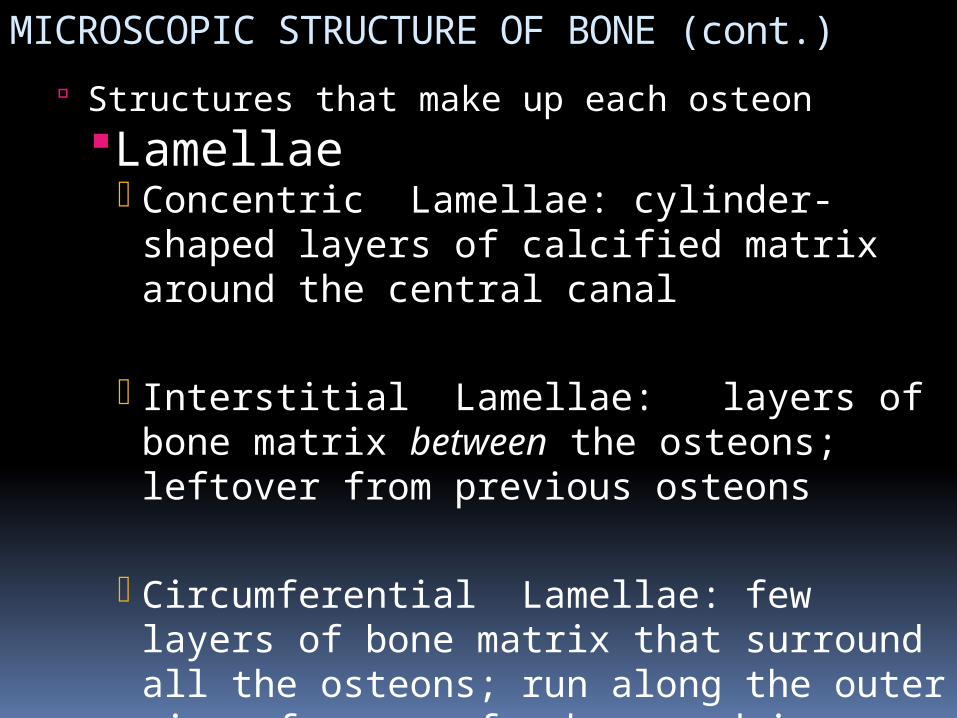
MICROSCOPIC STRUCTURE OF BONE (cont.) Structures that make up each osteonLamellae
Concentric Lamellae: cylinder-shaped layers of calcified matrix around the central canal
Interstitial Lamellae: layers of bone matrix between the osteons; leftover from previous osteons
Circumferential Lamellae: few layers of bone matrix that surround all the osteons; run along the outer circumference of a bone and inner circumference (boundary of medullary cavity) of a bone
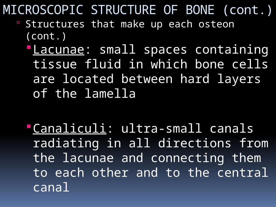
MICROSCOPIC STRUCTURE OF BONE (cont.) Structures that make up each osteon (cont.)
Lacunae: small spaces containing tissue fluid in which bone cells are located between hard layers of the lamella
Canaliculi: ultra-small canals radiating in all directions from the lacunae and connecting them to each other and to the central canal
Central (osteonal or Haversian) canal: extends lengthwise through the center of each osteon; contains blood vessels and lymphatic vessels
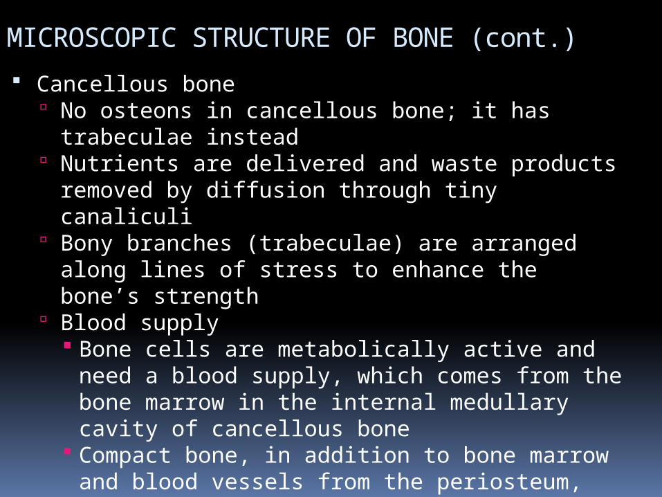
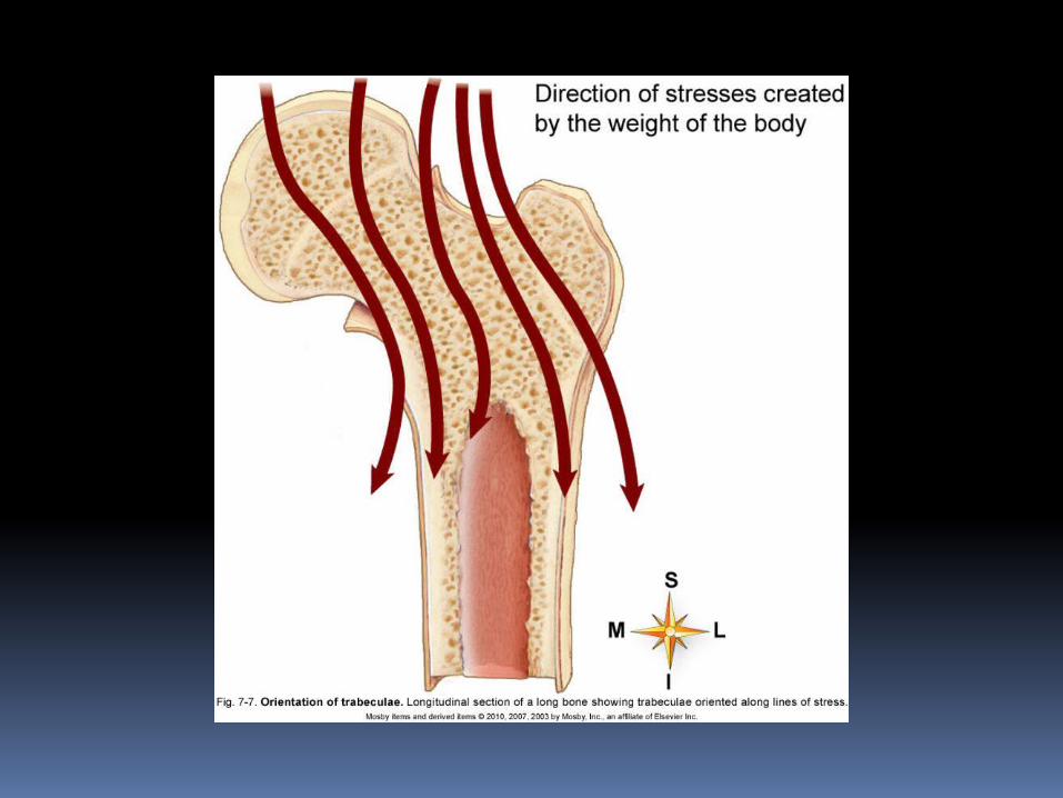
MICROSCOPIC STRUCTURE OF BONE (cont.) Cancellous bone
No osteons in cancellous bone; it has trabeculae instead
Nutrients are delivered and waste products removed by diffusion through tiny canaliculi
Bony branches (trabeculae) are arranged along lines of stress to enhance the bone’s strength
Blood supply Bone cells are metabolically active and need a
blood supply, which comes from the bone marrow in the internal medullary cavity of cancellous bone
Compact bone, in addition to bone marrow and blood vessels from the periosteum, penetrates the bone and then, by way of transverse (Volkmann) canals, connects with vessels in the central canals of osteons
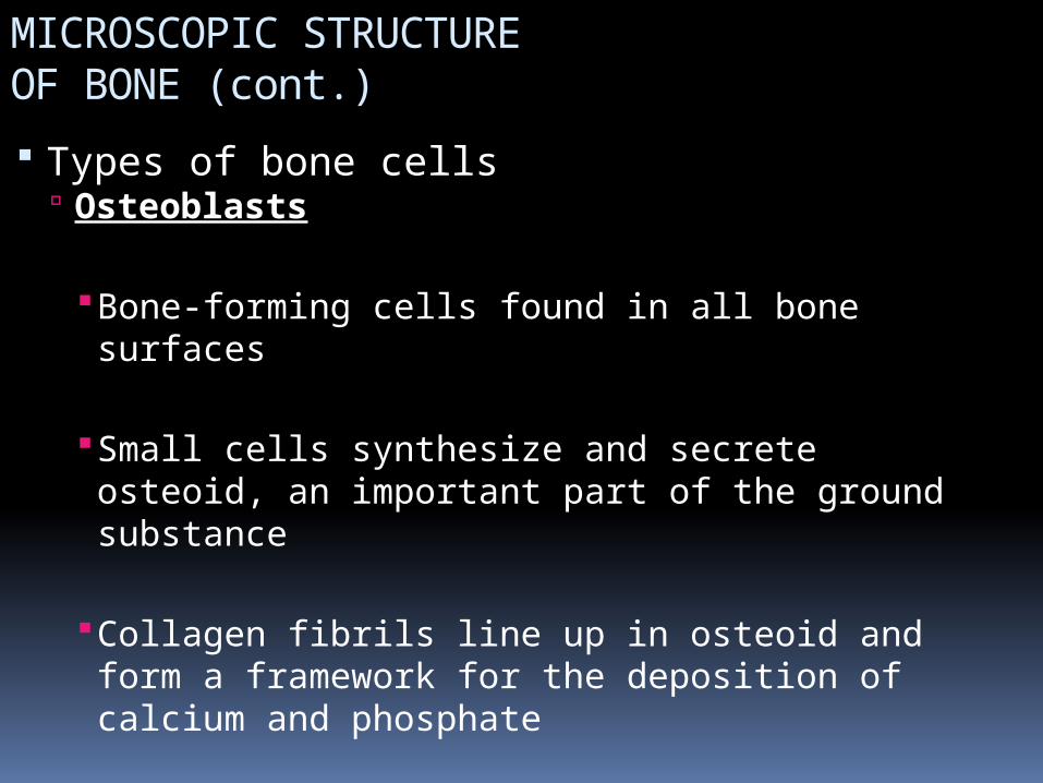
MICROSCOPIC STRUCTURE OF BONE (cont.) Types of bone cells
Osteoblasts
Bone-forming cells found in all bone surfaces
Small cells synthesize and secrete osteoid, an important part of the ground substance
Collagen fibrils line up in osteoid and form a framework for the deposition of calcium and phosphate
MICROSCOPIC STRUCTURE OF BONE (cont.) Types of bone cells
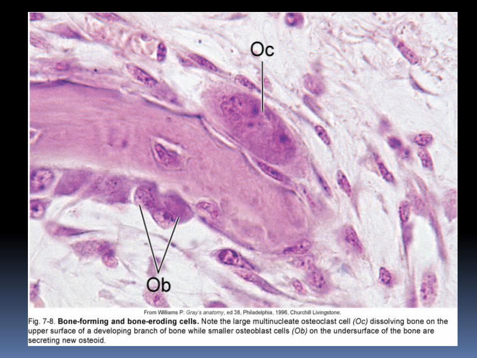
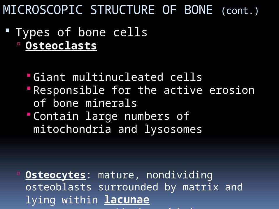
Osteoclasts
Giant multinucleated cells Responsible for the active erosion of bone
minerals Contain large numbers of mitochondria
and lysosomes
Osteocytes: mature, nondividing osteoblasts surrounded by matrix and lying within lacunae
((retirees of the bone, - alive, but relativley inactive, except for ‘maintenance’ - they maintain the
matrix surrounding them)
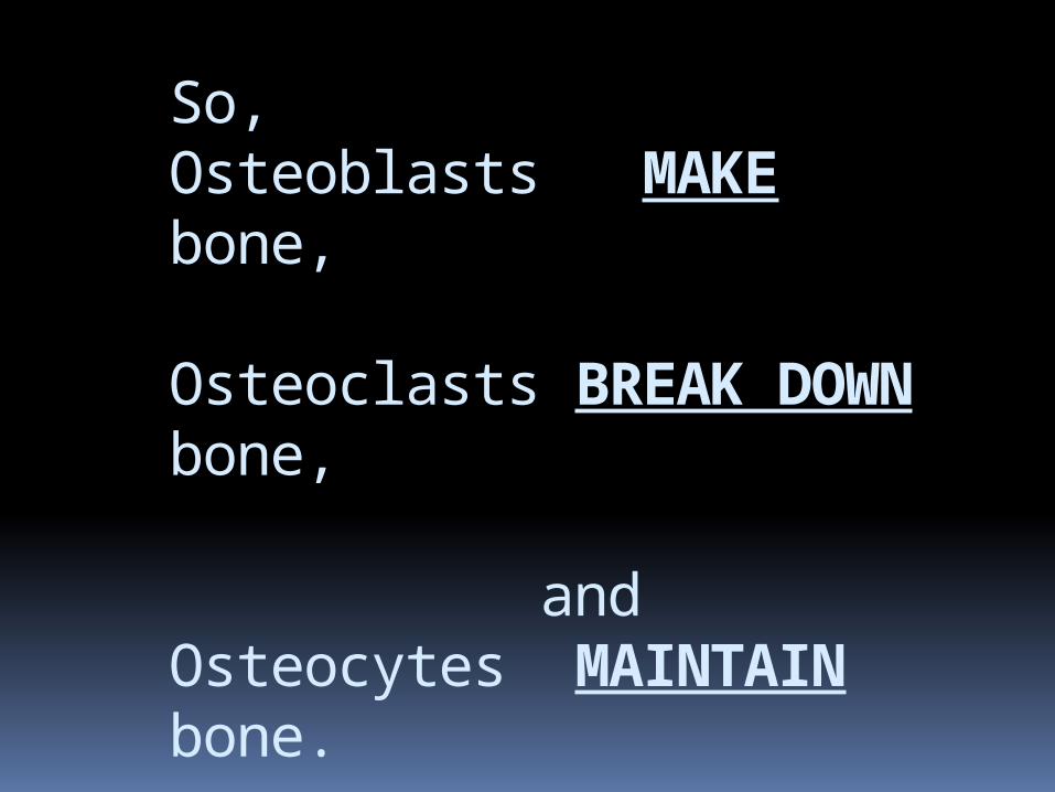
So,Osteoblasts MAKE bone,
Osteoclasts BREAK DOWN bone, and Osteocytes MAINTAIN bone.

question:
What is a ‘GIGABYTE’?
Slide 40

Answer:
AGGIE FAST FOOD DIET
(GIG A BITE)
Slide 41
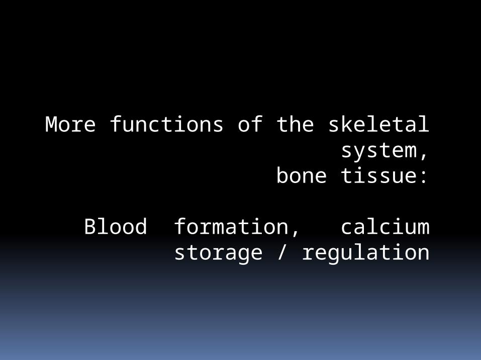
More functions of the skeletal system, bone tissue:
Blood formation, calcium storage / regulation
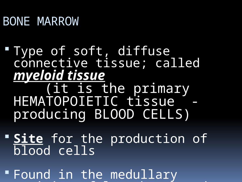
BONE MARROW
Type of soft, diffuse connective tissue; called myeloid tissue
(it is the primary HEMATOPOIETIC tissue - producing BLOOD CELLS)
Site for the production of blood cells
Found in the medullary cavities of long bones and in the spaces of spongy bone
BONE MARROW (cont.) Two types of marrow occur during a
person’s lifetime RED MARROW
Found in virtually all bones in an infant’s or child’s body
Produces red blood cells
YELLOW MARROW As an individual ages, red marrow is replaced
by yellow marrow Marrow cells become saturated with fat and
are no longer active in blood cell production
BONE MARROW (cont.)
The main bones in an adult that still contain red marrow include the ribs, bodies of the vertebrae, humerus, pelvis, and femur
Yellow marrow can change to red marrow during times of decreased blood supply, such as anemia, exposure to radiation, and certain diseases
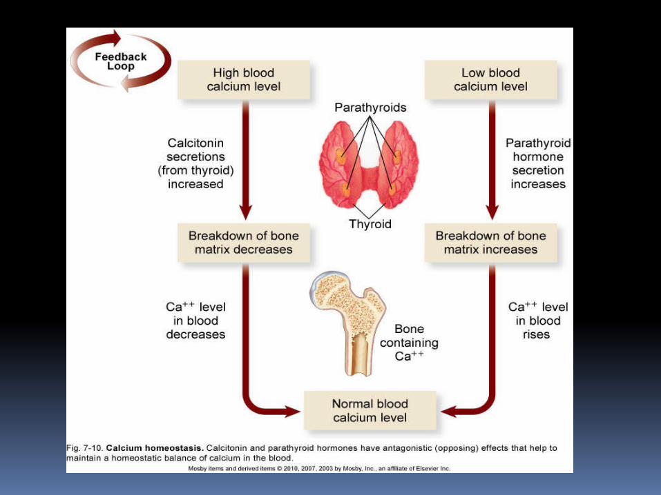
REGULATION OF BLOOD CALCIUM LEVELS Skeletal system is a storehouse for about
98% of body calcium reserves Helps maintain constancy of blood calcium
levels Calcium is mobilized and moves in and out of
blood during bone remodeling
During bone formation, OSTEOBLASTS REMOVE CALCIUM from blood and lower circulating levels
During breakdown of bone, OSTEOCLASTS RELEASE CALCIUM into blood and increase circulating levels
REGULATION OF BLOOD CALCIUM LEVELS (cont.)
Homeostasis of calcium ion concentration
is essential for the following:
Transmission of nerve impulses
Blood clotting
Bone formation, remodeling, and repair
Maintenance of skeletal and cardiac muscle contraction
REGULATION OF BLOOD CALCIUM LEVELS (cont.)
Mechanisms of calcium homeostasis Parathyroid hormone
Primary regulator of calcium homeostasis
Stimulates osteoclasts to initiate breakdown of bone matrix and increase blood calcium levels
Increases renal absorption of calcium from urine
Stimulates vitamin D synthesis
REGULATION OF BLOOD CALCIUM LEVELS (cont.)
Mechanisms of calcium homeostasis CALCITONIN
Protein hormone produced in the thyroid gland
Produced in response to high blood calcium levels
Stimulates bone deposition by osteoblasts
Inhibits osteoclast activity
Far less important in homeostasis of blood calcium levels than is parathyroid hormone
CARTILAGE
CARTILAGE Characteristics
Avascular connective tissue Fibers of cartilage are embedded in a firm
gel Has the flexibility of firm plastic
No canal system or blood vessels Chondrocytes receive oxygen and nutrients
by diffusion
Perichondrium: fibrous covering of the cartilage
Cartilage types differ because of the amount of matrix present and the amounts of elastic and collagenous fibers
CARTILAGE (cont.) Types of cartilage (Figure 7-21)
HYALINE CARTILAGE
Most common type
Covers the articular surfaces of bones
Forms the costal cartilages, cartilage rings in the trachea, bronchi of the lungs, and the tip of the nose
Forms from special cells in chondrification centers, which secrete matrix material
Chondrocytes are isolated into lacunae
CARTILAGE (cont.) Types of cartilage
Elastic cartilage Forms external ear, epiglottis, and eustachian
tubes Large number of elastic fibers confers elasticity
and resiliency
Fibrocartilage Occurs in pubic symphysis and intervertebral disks, and the meniscii of the knee joints
Small quantities of matrix and abundant fibrous elements
Strong and rigid
CARTILAGE (cont.) Functions
Tough, rubberlike nature permits cartilage to sustain great weight or serve as a shock absorber
Strong yet pliable support structure
Permits growth in length of long bones
Next few slides are of INTEREST - not to be tested upon, sit back and relax , watch, listen.
Bone development, fractures and repair:
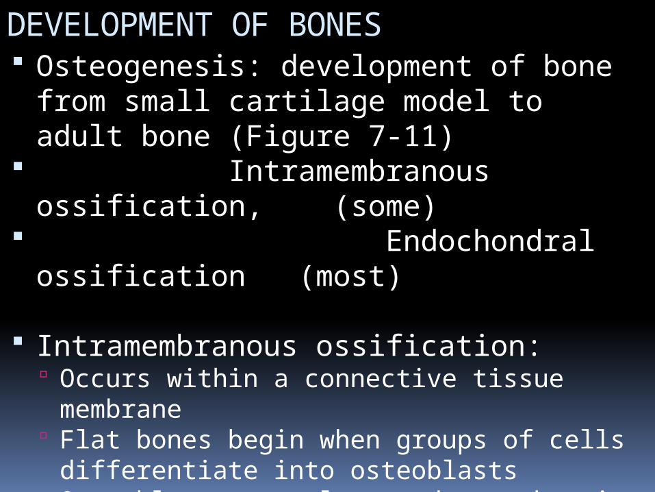
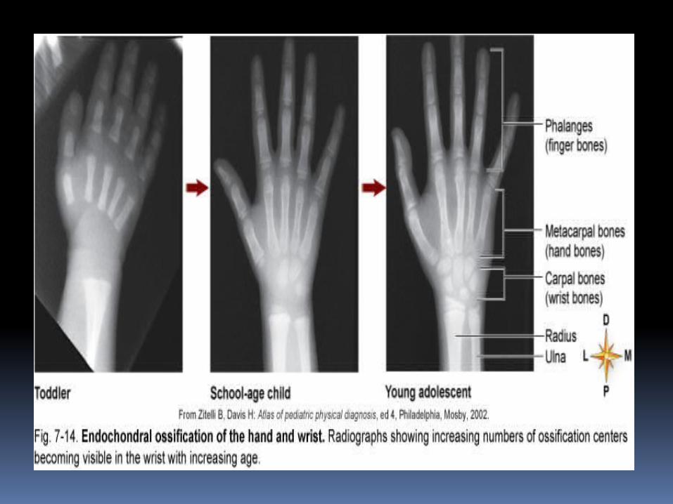
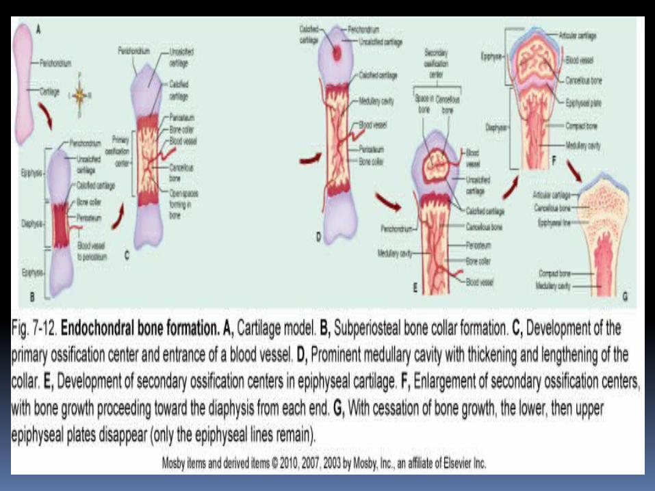
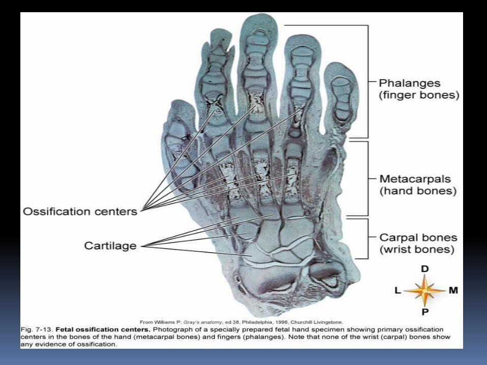
DEVELOPMENT OF BONES Osteogenesis: development of bone
from small cartilage model to adult bone (Figure 7-11)
Intramembranous ossification, (some)
Endochondral ossification (most)
Intramembranous ossification: Occurs within a connective tissue membrane Flat bones begin when groups of cells
differentiate into osteoblasts Osteoblasts are clustered together in
ossification center Osteoblasts secrete matrix material and
collagenous fibrils
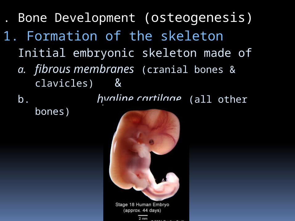
. Bone Development (osteogenesis)1. Formation of the skeleton
Initial embryonic skeleton made ofa. fibrous membranes (cranial bones &
clavicles) &b. hyaline cartilage (all other bones)
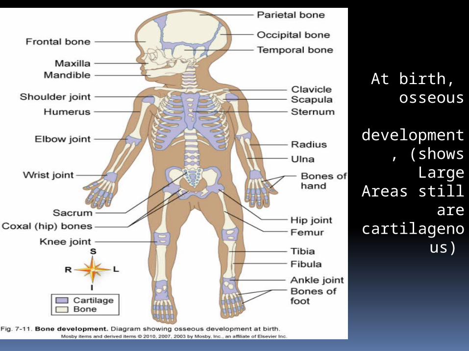
At birth, osseous
development, (shows
LargeAreas still are
cartilagenous)
DEVELOPMENT OF BONES (cont.) Intramembranous ossification
Large amounts of ground substance accumulate around each osteoblast
Collagenous fibers become embedded in the ground substance and constitute the bone matrix
Bone matrix calcifies when calcium salts are deposited
Trabeculae appear and join in a network to form spongy bone
Appositional growth occurs by adding osseous tissue
(like a tree grows in diameter)
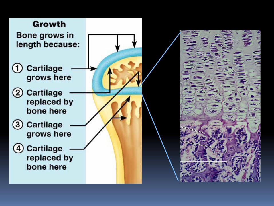
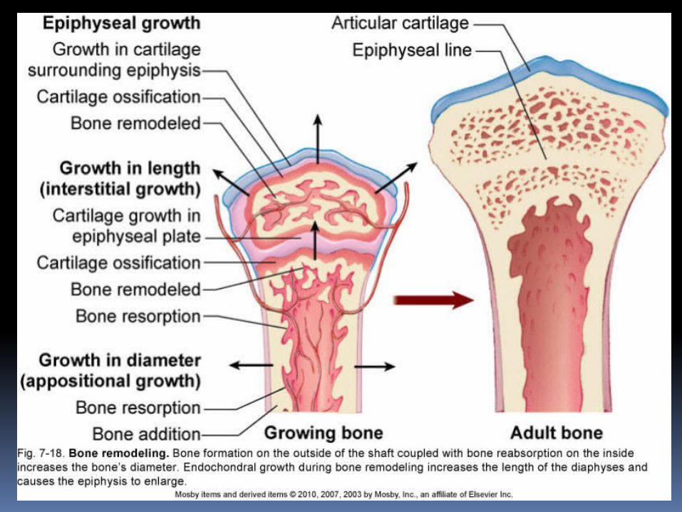
DEVELOPMENT OF BONES (cont.) Endochondral ossification
Most bones begin as a cartilage model with bone formation spreading essentially from the center to the ends
Periosteum develops and enlarges to produce a collar of bone
Primary ossification center forms
Blood vessel enters the cartilage model at the midpoint of the diaphysis
Bone grows in length as endochondral ossification progresses from the diaphysis toward each epiphysis
Secondary ossification centers appear in the epiphysis, and bone growth proceeds toward the diaphysis
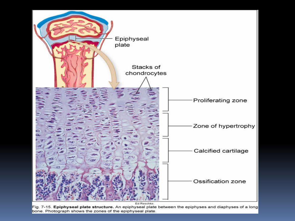
Epiphyseal plate remains between the diaphysis and each epiphysis until bone growth in length is complete
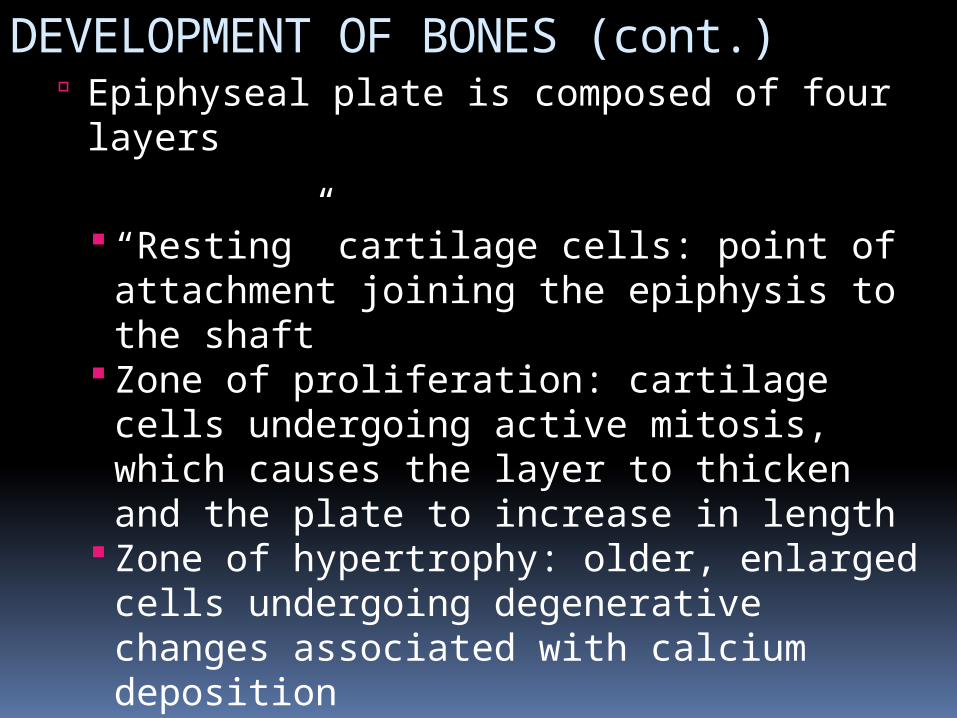
DEVELOPMENT OF BONES (cont.) Epiphyseal plate is composed of four layers
“Resting” cartilage cells: point of attachment joining the epiphysis to the shaft
Zone of proliferation: cartilage cells undergoing active mitosis, which causes the layer to thicken and the plate to increase in length
Zone of hypertrophy: older, enlarged cells undergoing degenerative changes associated with calcium deposition
Zone of calcification: dead or dying cartilage cells undergoing rapid calcification

DEVELOPMENT OF BONES (cont.)
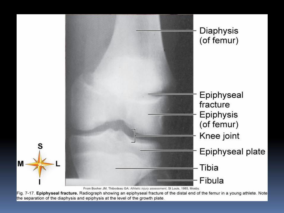
Epiphyseal plate can be a site for bone fractures in young people
Long bones grow in both length and diameter
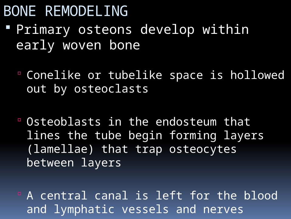
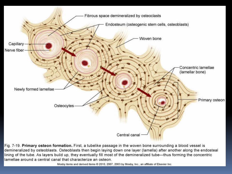
BONE REMODELING Primary osteons develop within early
woven bone Conelike or tubelike space is hollowed out by
osteoclasts
Osteoblasts in the endosteum that lines the tube begin forming layers (lamellae) that trap osteocytes between layers
A central canal is left for the blood and lymphatic vessels and nerves
Primary osteons can be replaced later by secondary osteons in a similar manner
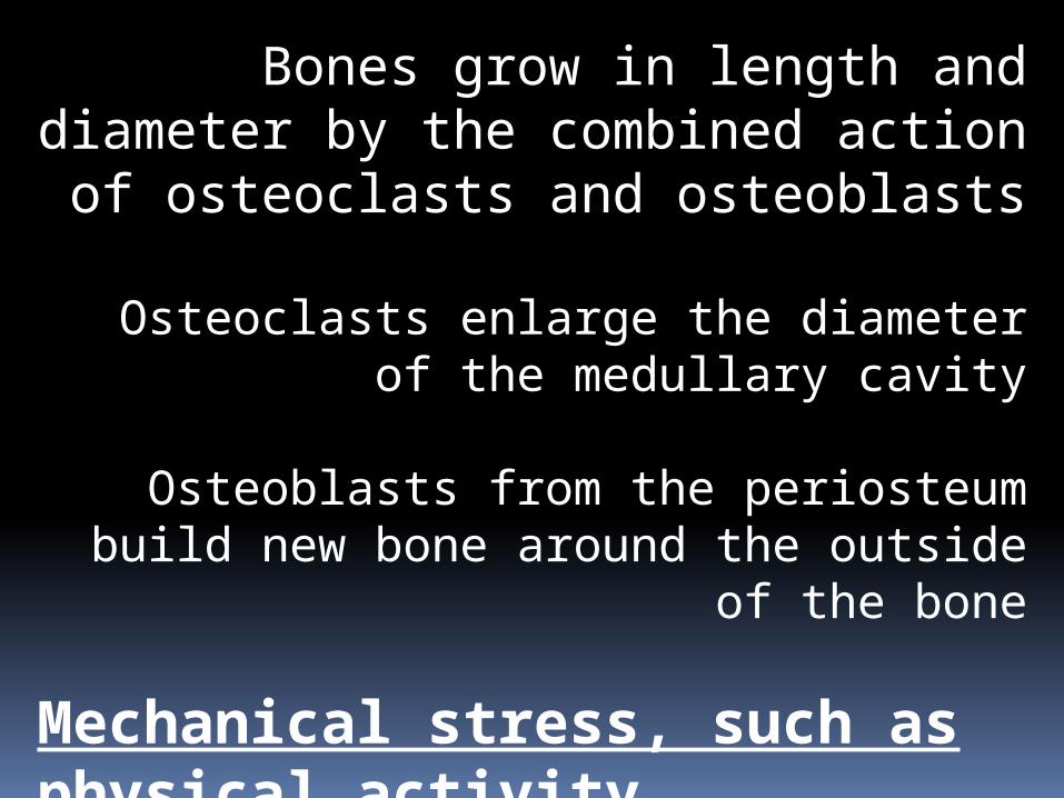
Bones grow in length and diameter by the combined action of osteoclasts and
osteoblasts
Osteoclasts enlarge the diameter of the medullary cavity
Osteoblasts from the periosteum build new bone around the outside of the bone
Mechanical stress, such as physical activity, strengthens bone
Fractures and Repairs
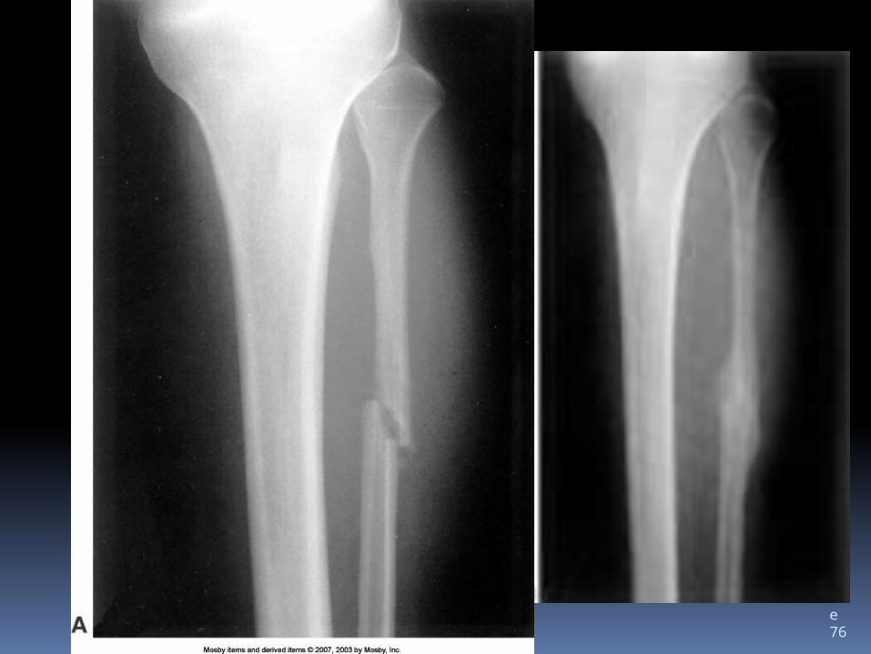
Slide 76
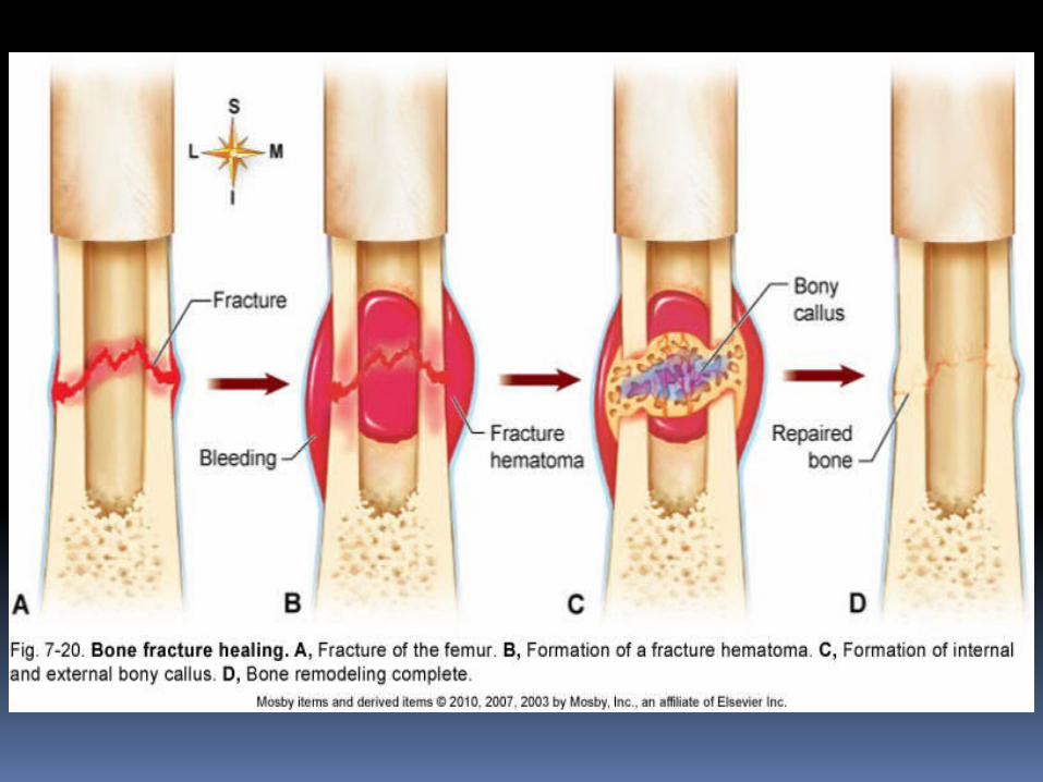
REPAIR OF BONE FRACTURES Fracture: break in the continuity of a bone
Fracture healing Fracture tears and destroys blood vessels
that carry nutrients to osteocytes
Vascular damage initiates repair sequence
Callus: special repair tissue that binds the broken ends of the fracture together
Fracture hematoma: blood clot occurring immediately after the fracture, which is then resorbed and replaced by callus
CYCLE OF LIFE: SKELETAL ISSUES Skeleton fully ossified by mid-20s
Soft tissue may continue to grow; ossifies more slowly
Adults: changes occur from specific conditions Increased density and strength from
exercise Decreased density and strength from
pregnancy, nutritional deficiencies, and illness
Advanced adulthood: apparent degeneration Hard bone matrix replaced by softer
connective tissue Exercise can counteract degeneration