Embed Size (px)
Citation preview
Chapter 6
Ribosomes & Endoplasmic
Reticulum 6.1. Ribosomes Ribosomes are the protein-synthesizing machines of the cell. They translate the
information encoded in messenger RNA (mRNA) into a polypeptide.
Ribosomes are roughly spherical with a diameter of ~20 nm, and can be seen only with
the electron microscope. They can make up 25% of the dry weight of cells (e.g., pancreas
cells) that specialize in protein synthesis. (A single pancreas cell can synthesize 5 million
molecules of protein per minute.) In eukaryotes, ribosomes that synthesize proteins for
use within the cytosol (e.g., enzymes of glycolysis) are suspended in the cytosol.
Ribosomes that synthesize proteins destined for secretion (by exocytosis), the plasma
membrane (e.g., cell surface receptors) and lysosomes are attached to the cytosolic face
of the membranes of the endoplasmic reticulum. As the polypeptide is synthesized, it is
extruded into the interior (lumen) of the endoplasmic reticulum. Then, before these
proteins reach their final destinations, they undergo a series of processing steps in the
Golgi apparatus. Ribosomes that synthesize 13 of the proteins destined for the inner
membrane of mitochondria are found within the mitochondrion itself and are quite
different in structure from the others. The ribosomes of bacteria, eukaryotes, and
mitochondria differ in many details of their structure.
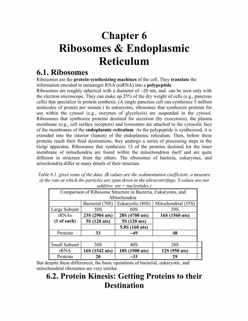
Table 6.1. gives some of the data. (S values are the sedimentation coefficient: a measure
of the rate at which the particles are spun down in the ultracentrifuge. S values are not
additive. nts = nucleotides.)
Comparison of Ribosome Structure in Bacteria, Eukaryotes, and
Mitochondria
Bacterial (70S) Eukaryotic (80S) Mitochondrial (55S)
Large Subunit 50S 60S 39S
23S (2904 nts) 28S (4700 nts) 16S (1560 nts)
5S (120 nts) 5S (120 nts)
rRNAs
(1 of each)
5.8S (160 nts)
Proteins 33 ~49 48
Small Subunit 30S 40S 28S
rRNA 16S (1542 nts) 18S (1900 nts) 12S (950 nts)
Proteins 20 ~33 29
But despite these differences, the basic operations of bacterial, eukaryotic, and
mitochondrial ribosomes are very similar.
6.2. Protein Kinesis: Getting Proteins to their
Destination
Proteins are the major building blocks of life. Eukaryotic cells synthesize proteins for
thousands of different functions. Some examples:
1. To build the components of the cytosol (e.g. microtubules, glycolytic enzymes);
2. To build the receptors and other molecules exposed at the surface of the cell
embedded in the plasma membrane;
3. To supply some of the components of the mitochondria and (in plant cells)
chloroplasts;
4. Proteins secreted from the cell to supply the needs of other cells and tissues (e.g.
collagen to support cells, hormones to signal them).
All proteins are synthesized by ribosomes using the information encoded in molecules of
messenger RNA (mRNA). This process is called translation. The various destinations for
proteins occur in two major sets:
1. One set for those proteins synthesized by ribosomes that remain suspended in the
cytosol, and
2. A second set for proteins synthesized by ribosomes that are attached to the
membranes of the endoplasmic reticulum (ER) forming "rough endoplasmic
reticulum" (RER).
So the first decision that must be made as a ribosome begins to translate a mRNA into a
polypeptide is whether to remain free in the cytosol or to bind to the ER.
6.3. Pathways through the Endoplasmic Reticulum (ER) The decision to enter the ER is dictated by the presence of a signal sequence on the
growing polypeptide.
(a) The Signal Sequence The signal sequence consists of the first portion of the elongating polypeptide chain (so
the signal sequence occurs at the amino terminal of the polypeptide). Typical signal
sequences contain 15 - 30 amino acids. The precise amino acid sequence varies
surprisingly from one protein to the next, but all signal sequences include many
hydrophobic amino acids. The 1999 Nobel Prize in Physiology or Medicine was awarded
on October 11, 1999 to Dr. Günter Blobel for his discovery of the signal sequence and
other intrinsic signals that enable proteins to reach their proper destinations. If a signal
sequence is present, translation ceases after it has been synthesized. The signal sequence
is recognized by and is bound by a signal recognition particle (SRP) The complex of
ribosome with its nascent polypeptide and the SRP binds to a receptor on the surface
(facing the cytosol) of the ER. The SRP leaves and translation recommences. The
growing polypeptide chain is extruded through a pore in the ER membrane and into the
lumen of the ER. The signal sequence is usually clipped off the polypeptide unless the
polypeptide is to be retained as an integral membrane protein. Other proteins, called
molecular chaperones, present in the lumen of the ER, bind the growing polypeptide
chain and assist it to fold into its correct tertiary structure. Sugar residues may be added
to the protein. The process is called glycosylation and often is essential for proper
folding of the final product, a glycoprotein.
(b) Destinations of Proteins Synthesized Within the ER Proteins synthesized within the ER are transported to the Golgi apparatus. Portions of
the ER are pinched off, forming transport vesicles. These carry their load of proteins to
the Golgi apparatus. The membrane of the transport vesicle fuses with the membrane of
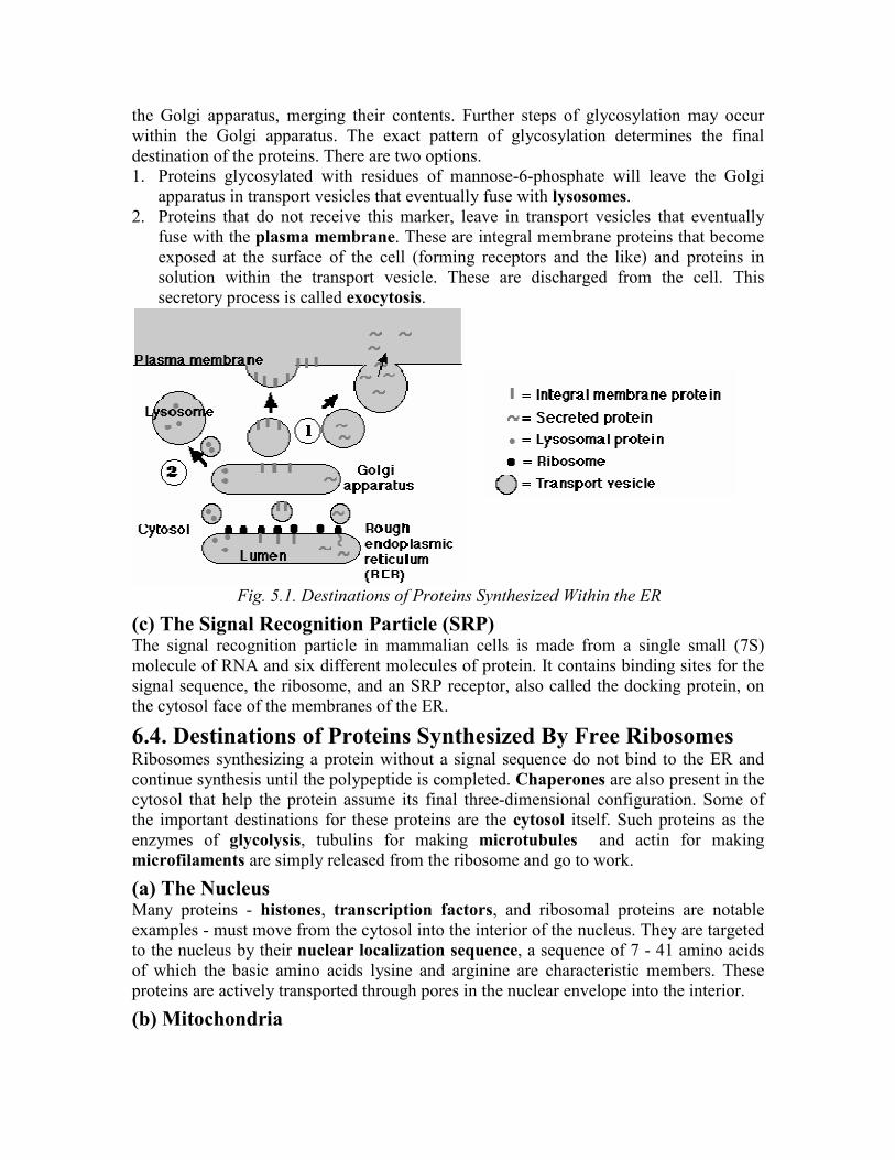
the Golgi apparatus, merging their contents. Further steps of glycosylation may occur
within the Golgi apparatus. The exact pattern of glycosylation determines the final
destination of the proteins. There are two options.
1. Proteins glycosylated with residues of mannose-6-phosphate will leave the Golgi
apparatus in transport vesicles that eventually fuse with lysosomes.
2. Proteins that do not receive this marker, leave in transport vesicles that eventually
fuse with the plasma membrane. These are integral membrane proteins that become
exposed at the surface of the cell (forming receptors and the like) and proteins in
solution within the transport vesicle. These are discharged from the cell. This
secretory process is called exocytosis.
Fig. 5.1. Destinations of Proteins Synthesized Within the ER
(c) The Signal Recognition Particle (SRP) The signal recognition particle in mammalian cells is made from a single small (7S)
molecule of RNA and six different molecules of protein. It contains binding sites for the
signal sequence, the ribosome, and an SRP receptor, also called the docking protein, on
the cytosol face of the membranes of the ER.
6.4. Destinations of Proteins Synthesized By Free Ribosomes Ribosomes synthesizing a protein without a signal sequence do not bind to the ER and
continue synthesis until the polypeptide is completed. Chaperones are also present in the
cytosol that help the protein assume its final three-dimensional configuration. Some of
the important destinations for these proteins are the cytosol itself. Such proteins as the
enzymes of glycolysis, tubulins for making microtubules and actin for making
microfilaments are simply released from the ribosome and go to work.
(a) The Nucleus Many proteins - histones, transcription factors, and ribosomal proteins are notable
examples - must move from the cytosol into the interior of the nucleus. They are targeted
to the nucleus by their nuclear localization sequence, a sequence of 7 - 41 amino acids
of which the basic amino acids lysine and arginine are characteristic members. These
proteins are actively transported through pores in the nuclear envelope into the interior.
(b) Mitochondria
Although the mitochondrion has its own genome and protein synthesizing machinery,
most of the proteins used by mitochondria are encoded by genes in the nucleus of the cell
synthesized in the cytosol, must be imported into the mitochondrion. Proteins destined for
mitochondrion contain a characteristic signal sequence. This is recognized and bound by
a chaperone called mitochondrial stimulation factor (MSF). MSF targets the protein to
a receptor embedded in the outer membrane of the mitochondrion. Other factors and
receptors shepherd proteins through the intermembrane space to the inner mitochondrial
membrane (e.g. some proteins of the electron transport chain) and the matrix.
(c) Chloroplasts Chloroplasts, like mitochondria, have their own genome and their own protein-
synthesizing machinery. But also like mitochondria, most of the proteins used in
chloroplasts are encoded by genes in the nucleus of the cell, are synthesized by ribosomes
in the cytosol, and must then be imported into the chloroplast. Proteins destined for
chloroplasts are recognized by their characteristic transit sequence. Chaperones are also
needed to get them to their final destination: stroma, thylakoid membrane, etc.
(d) Peroxisomes Proteins destined for peroxisomes are synthesized with a Peroxisomal Targeting Signal
(PTS) that binds to a receptor molecule that takes the protein into the peroxisome and
then returns for another load. Two peroxisomal targeting signals have been identified: a
9-amino acid sequence at the N-terminal of the protein and a tripeptide at the C-
terminal. Each has its own receptor to take it to the peroxisome.