Embed Size (px)
Citation preview
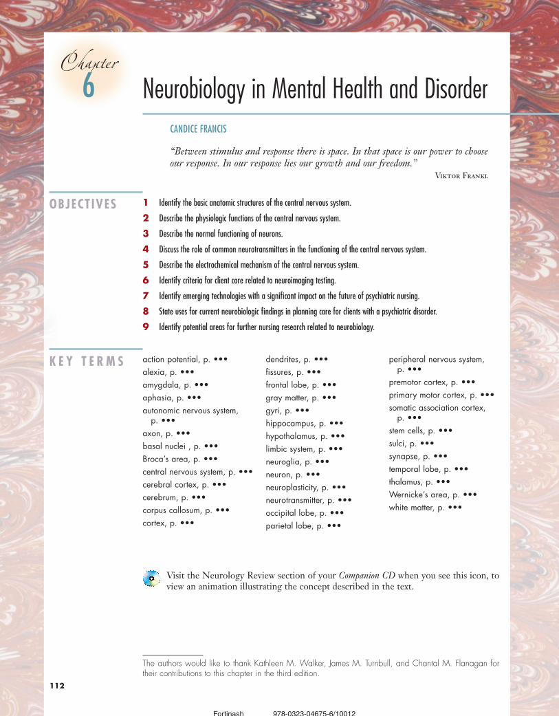
Chapter 6 Neurobiology in Mental Health and Disorder
CANDICE FRANCIS
“Between stimulus and response there is space. In that space is our power to choose our response. In our response lies our growth and our freedom.”
Viktor Frankl
OBJECTIVES 1 Identify the basic anatomic structures of the central nervous system.
2 Describe the physiologic functions of the central nervous system.
3 Describe the normal functioning of neurons.
4 Discuss the role of common neurotransmitters in the functioning of the central nervous system.
5 Describe the electrochemical mechanism of the central nervous system.
6 Identify criteria for client care related to neuroimaging testing.
7 Identify emerging technologies with a signifi cant impact on the future of psychiatric nursing.
8 State uses for current neurobiologic fi ndings in planning care for clients with a psychiatric disorder.
9 Identify potential areas for further nursing research related to neurobiology.
K E Y T E R M S action potential, p. •••alexia, p. •••amygdala, p. •••aphasia, p. •••autonomic nervous system,
p. •••axon, p. •••basal nuclei , p. •••Broca’s area, p. •••central nervous system, p. •••cerebral cortex, p. •••cerebrum, p. •••corpus callosum, p. •••cortex, p. •••
dendrites, p. •••fi ssures, p. •••frontal lobe, p. •••gray matter, p. •••gyri, p. •••hippocampus, p. •••hypothalamus, p. •••limbic system, p. •••neuroglia, p. •••neuron, p. •••neuroplasticity, p. •••neurotransmitter, p. •••occipital lobe, p. •••parietal lobe, p. •••
peripheral nervous system, p. •••
premotor cortex, p. •••primary motor cortex, p. •••somatic association cortex,
p. ••• stem cells, p. •••sulci, p. •••synapse, p. •••temporal lobe, p. ••• thalamus, p. ••• Wernicke’s area, p. •••white matter, p. •••
The authors would like to thank Kathleen M. Walker, James M. Turnbull, and Chantal M. Flanagan for their contributions to this chapter in the third edition.
112
Visit the Neurology Review section of your Companion CD when you see this icon, to view an animation illustrating the concept described in the text.
Fortinash 978-0323-04675-6/10012

113Neurobio logy in Menta l Heal th and Disorder Chapter 6
Neuroanatomy The brain is one of the most important structures in the human body. Although it weighs only 3 to 5 pounds, the brain contains approximately 140 billion cells, making it the most complex and vital of human organs (Gribbin, 2002). The human nervous system is composed of two separate but interconnected anatomic divisions. The fi rst division, the central nervous system (CNS), is com-posed of the spinal cord and brain. The second division, the peripheral nervous system (PNS), contains periph-eral nerves, 12 pairs of cranial nerves that originate just outside of the brain stem, and 31 pairs of spinal nerves arising from the spinal cord. These peripheral nerves transmit sensory (incoming) information toward the CNS and motor (outgoing) information away from the CNS to muscles and glands that are controlled by the CNS.
Although the PNS and certain interactions with the autonomic nervous system (ANS) are is of critical impor-tance to human functioning, the understanding of psychi-atric disorders depends on in-depth understanding of the structure and function of the CNS. For that reason, this chapter focuses on the CNS and how the nurse will use that knowledge to provide care to individuals who have psychiatric and neurologic disorders.
Brain cells are categorized as either neurons or neuro-glia. Neurons generate and conduct electrical signals. Neuroglia provide the mechanical and physiologic sup-port for neurons. White matter is composed of the axons of neurons that are insulated by myelin. White matter makes up the core of major brain structures such as the cerebrum and cerebellum. The gray matter, or cortex, typically covers the surface of these organs. The cortex is the functional area of the brain where neurons communi-cate with each other and where neurotransmitters are concentrated.
CerebrumThe cerebrum is the largest part of the brain and it is divided into two halves called cerebral hemispheres. The cerebral hemispheres contain important functional areas such as the cerebral cortex, basal nuclei, and lim-bic system.
The cerebral hemispheres account for more than 70% of the neurons in the CNS and are responsible for func-tions such as hearing, vision, language, cognitive func-tions, control of muscles, and sensory interpretation. The left hemisphere is dominant in almost 95% of people and mainly controls motor and sensory functions on the right side of the body. The right hemisphere controls functions on the left side of the body. Most right-handed people, and half of left-handed people, have a dominant left hemi-sphere. In rare cases, some people have mixed dominance, with one side dominant for language expression and the other for motor functions such as handwriting.
Effective coordinated human activity requires a com-plex interrelationship and communication within and be-tween the two hemispheres. A large bundle of white mat-
Neuroscientifi c discovery and developments continue to advance at a remarkable pace and contribute to
the evolving understanding of brain function. This re-search provides new strategies for clinicians, clients, and families addressing mental disorders. Many now recog-nize mental disorders as brain-based illnesses. Although some still misunderstand mental disorders and stigmatize those clients, research on brain biology has helped re-shape perceptions of mental illness. To understand treat-ment approaches to psychiatric illness, the psychiatric mental health nurse requires a fi rm foundation in the fundamentals of neurobiology, physiology and genetics, and how these affect treatment of psychiatric disorders. Increased knowledge in this area has changed the way mental health care providers perceive and treat mental disorders. This chapter reviews the fundamentals of neu-robiology and examines concepts and clinical approaches to the treatment of mental illness with regard to our growing knowledge of neuroscience.
UNDERSTANDING NEUROBIOLOGIC FUNCTIONSThe biologic model of psychiatric illness is not a new phenomenon, but the availability of new tools makes it increasingly more sophisticated. Science has advanced beyond merely making educated guesses about how the brain works and has developed scientifi c models that al-low for testing brain-based interventions and developing new effective treatments (Gur, 2002). The best psychiatric mental health care begins with an understanding that the symptoms associated with psychiatric disorders are usu-ally manifested behaviorally. Clients with psychiatric dis-orders frequently behave in ways society considers “not normal.” They often express their disorders through re-sponses such as hearing voices, considering suicide, or wearing a winter coat on a hot summer day. These abnor-mal perceptions, thoughts, and behaviors usually have a neurobiologic basis. Knowledge of normal brain structure and function helps mental health care providers to offer an optimal level of treatment to people with brain-based illnesses. Understanding the structural or neurochemical defects that affect clients with psychiatric disorders helps psychiatric nurses to more effectively assess clients’ re-sponses and plan interventions.
NEUROANATOMY AND NEUROPHYSIOLOGY OF THE HUMAN NERVOUS SYSTEMHuman thoughts, feelings, and actions begin in the cen-tral nervous system. The brain acts as the primary mediator-organ, controlling and determining how peo-ple interact with the world. All human responses are the result of the complex interaction between underlying neuroanatomy and neurophysiology, as well as the ge-netic, environmental, and developmental factors that infl uence those systems.
Fortinash 978-0323-04675-6/10012
114 PART I FOUNDATIONS FOR PSYCHIATRIC MENTAL HEALTH NURSING
ter called the corpus callosum connects the two hemispheres. Sensorimotor information constantly fl ows between the two hemispheres via nerve pathways in the corpus callosum. The corpus callosum has to be intact for full, smooth, and coordinated communication between the hemispheres.
The outermost surface of the cerebral cortex contains corrugated wrinkles with many grooves and indentations. Shallow grooves are called sulci, and the deeper grooves extending deep into the brain are called fi ssures. The raised areas are called gyri. The sulci and gyri dramatically increase the overall surface area of the cerebrum. The ce-rebral cortex is typically composed of only six layers of cells, but it covers an area that, if spread out, equals almost 2.5 square feet. In contrast, the cortex of a chimpanzee would cover only a single sheet of paper, while a rat’s cor-tex occupies an area roughly equal to the size of a postage stamp (Gribbin, 2002). Most discussions on cerebral func-tions focus on the outer layer, the cerebral cortex.
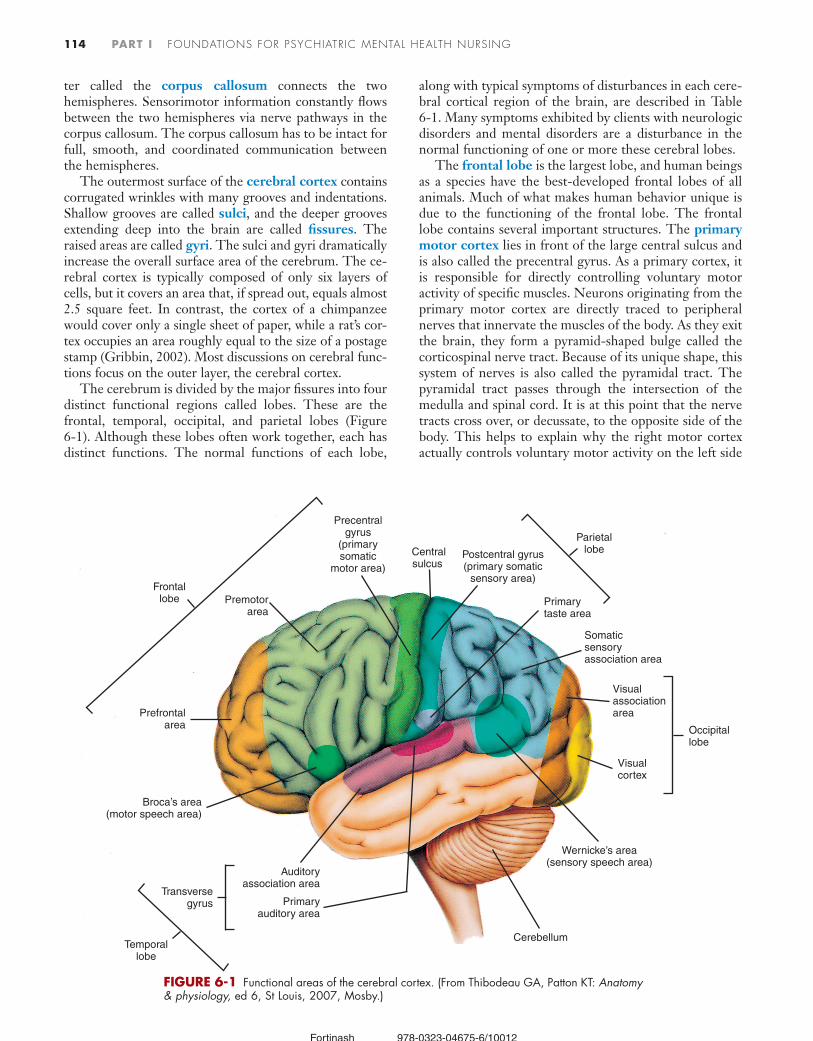
The cerebrum is divided by the major fi ssures into four distinct functional regions called lobes. These are the frontal, temporal, occipital, and parietal lobes (Figure 6-1). Although these lobes often work together, each has distinct functions. The normal functions of each lobe,
along with typical symptoms of disturbances in each cere-bral cortical region of the brain, are described in Table 6-1. Many symptoms exhibited by clients with neurologic disorders and mental disorders are a disturbance in the normal functioning of one or more these cerebral lobes.
The frontal lobe is the largest lobe, and human beings as a species have the best-developed frontal lobes of all animals. Much of what makes human behavior unique is due to the functioning of the frontal lobe. The frontal lobe contains several important structures. The primary motor cortex lies in front of the large central sulcus and is also called the precentral gyrus. As a primary cortex, it is responsible for directly controlling voluntary motor activity of specifi c muscles. Neurons originating from the primary motor cortex are directly traced to peripheral nerves that innervate the muscles of the body. As they exit the brain, they form a pyramid-shaped bulge called the corticospinal nerve tract. Because of its unique shape, this system of nerves is also called the pyramidal tract. The pyramidal tract passes through the intersection of the medulla and spinal cord. It is at this point that the nerve tracts cross over, or decussate, to the opposite side of the body. This helps to explain why the right motor cortex actually controls voluntary motor activity on the left side
Premotorarea
Precentralgyrus
(primarysomatic
motor area)
Centralsulcus
Postcentral gyrus(primary somatic
sensory area)
Primarytaste area
Parietallobe
Frontallobe
Temporallobe
Occipitallobe
Somaticsensoryassociation area
Visualassociationarea
Visualcortex
Wernicke’s area(sensory speech area)
Cerebellum
Transversegyrus
Auditoryassociation area
Primaryauditory area
Broca’s area(motor speech area)
Prefrontalarea
FIGURE 6-1 Functional areas of the cerebral cortex. (From Thibodeau GA, Patton KT: Anatomy & physiology, ed 6, St Louis, 2007, Mosby.)
Fortinash 978-0323-04675-6/10012

115Neurobio logy in Menta l Heal th and Disorder Chapter 6
of the body and the left motor cortex controls motor ac-tivity on the right side of the body.
The frontal lobe also contains two other important structures. The premotor cortex is responsible for the coordinated movement of multiple muscles, and the so-matic association cortex integrates motor commands. Researchers have identifi ed a number of brain regions as association regions. In fact, some estimate that 70% to 75% of all cortical regions are association regions that integrate functions in the primary region. The primary regions are generally involved in analysis, initiation, inter-pretation, and integrative activities. In the case of the frontal lobe, the somatic association cortex is the area of the brain responsible for coordinating learned motor skills. Cognition, memory, and analytic functions are largely functions of a third region of the frontal lobe known as the prefrontal cortex. Damage to this area of the frontal lobe causes changes in personality. Other func-tions of the prefrontal cortex, sometimes described as ex-ecutive functions, include reasoning, planning, prioritiz-ing, sequencing behavior, insight, fl exibility, and judgment (Young and Pigott, 1999). Normal frontal cortical func-tions help suppress and moderate more primitive impulses and actions. The frontal cortex also allows a person to ap-propriately process incoming sensory stimuli, reason, fo-cus on tasks, and respond to social cues. Diffi culty in performing these activities often manifests as symptoms of psychiatric disorders. Two key functions, working memory and behavioral inhibition, have increasingly be-come targets of research interest as scientists explore the
neurobiology of psychiatric disorders (Dubin, 2002). An-other important area usually localized only in the left frontal lobe is Broca’s area, which controls the muscles necessary to speak. Damage to Broca’s area from causes such as accidents or stroke results in the inability to speak (motor aphasia). Speech is a vital part of communication and appropriate social interaction.
The temporal lobe is responsible for some functions of language, memory, and emotion. Wernicke’s area is a specialized area of the temporal lobe responsible for orga-nizing words so they will be recognized and express the correct emotional content. Written speech, verbal speech, and the visual recognition that is critical to communica-tion are all functions of the temporal lobe. Language is one example where two distinct regions, Broca’s area in the frontal lobe and Wernicke’s area in the temporal lobe, work together to facilitate normal communication. Apha-sia, a communication disorder, sometimes has several ori-gins within the brain, most notably Wernicke’s area of the temporal lobe and Broca’s area in the frontal lobe. The auditory association area of the temporal lobe is involved with memory, especially those connected to visual and auditory cues.
The occipital lobe contains the primary visual cortex and is most responsible for visual functioning. Color rec-ognition, the ability to recognize and name objects, and the ability to track moving objects are functions of the occipital lobe. The occipital lobe is sensitive to hypoxia, and trauma to this region of the brain sometimes results in blindness, even if the optic nerves and eyes remain in-
TABLE 6-1
Normal Functions and Symptoms of Dysfunction of the Cerebrum LOBE LOCATION NORMAL FUNCTION SYMPTOMS OF ALTERATIONS IN BRAIN FUNCTIONING
Frontal Anterior, or front area, of brain
Programming and execution of motor functions
Higher thought processes such as planning, ability to abstract, trial-and-error learning, and decision making
Intellectual insight, judgmentExpression of emotion
Changes in affect such as fl atteningAlteration in language productionAlteration in motor functioningImpulsive behaviorImpaired decision makingConcrete thinking
Parietal Posterior to central sulcus Sensory perception: taking in information from environment, organizing it, and com-municating this information to rest of brain
Association areas that allow for such things as accurately following directions on a map, reading a clock, building a bird-house, or dressing oneself
Altered sensory perceptions such as de-creased consciousness of pain sensation
Diffi culty with time concepts such as inability to keep appointment times
Alteration in personal hygieneAlteration in ability to calculate numbersInability to adequately perform common mo-
tor actions of writingMixing up right and leftPoor attention span
Temporal Lies beneath skull on both sides; commonly called the temple
Primarily responsible for hearing and receiv-ing information via ears
Auditory hallucinationsIncreased sexual focusDecreased motivationAlterations in memoryAltered emotional responsesSensory aphasia
Occipital Most posterior of brain lobes—back of head
Primarily responsible for seeing and receiving information via eyes
Visual hallucinations
Fortinash 978-0323-04675-6/10012

116 PART I FOUNDATIONS FOR PSYCHIATRIC MENTAL HEALTH NURSING
tact. Lesions of the occipital lobe can cause visual halluci-nations and other abnormalities of visual functioning, such as alexia, or the inability to read.
The parietal lobe of the brain functions as the primary sensory processing center. The postcentral sensory gyrus area of the parietal lobe, or the somesthetic cortex, inter-prets sensory information. This includes visual, tactile, and auditory information. Posterior to the somesthetic cortex is the somesthetic association area. Again, as an as-sociation area, it is responsible for organizing, integrat-ing, and analyzing sensory information that the primary sensory cortex in the postcentral gyrus will interpret more specifi cally.
Basal Nuclei. The basal nuclei, also known as basal ganglia, are concentrations of cell bodies closely involved with motor functions and association. Basal nuclei are concentrations of gray matter located within the white matter of the cerebrum and midbrain. They have many connections to both the superfi cial cortex above and the deep midbrain structures below. Among the most well known basal nuclei are the caudate lobe, putamen, globus pallidus, and substantia nigra. These basal nuclei trans-late movements such as walking while it is happening, and they also modulate and correct muscle functioning that allows movements to occur in a coordinated manner. The basal nuclei aid in the learning and programming of motor behavior. Activities that are well learned and re-hearsed over the course of a person’s life often become automatic. Complex motor skills involved in walking, eating, or driving become so natural that a person does not have to think consciously to perform them. This helps to explain why some people with dementia retain some of these complex behaviors long after a severe memory or language loss.
Conditions such as Huntington’s disease and Parkin-son’s disease are associated with basal nuclear dysfunction and their inability to effectively communicate with the cerebral cortex (Montoya, 2006). Some medications used to treat psychiatric disorders alter the basal nuclei (Box 6-1). For example, chlorpromazine (Thorazine) and halo-peridol (Haldol) are two older neuroleptic antipsychotic medications that sometimes cause hypertonicity, or dysto-nia, a condition marked by excessive muscle tone.
Limbic System. Instincts, primitive drives, sexual arousal, fear, aggression, and other emotions are part of the functions of the structures deep within the brain called the limbic system or limbic lobe. It is often called a system because researchers believe its functions are a result of the interrelated, closely coordinated actions of its various structures. Table 6-2 and Figure 6-2 identify some of the structural components of the limbic system. Part of the limbic system, the amygdala, is instrumental in emo-tional functioning and in regulating affective responses to events. The amygdala modulates common emotional states such as feelings of anger and aggression, love, and comfort in social settings. The limbic system’s function of emotional regulation is linked with the olfactory pathways that connect to the amygdala. Some suggest that this ex-
plains why certain smells evoke strong emotional re-sponses and memories in some individuals The limbic system holds increasing interest for researchers trying to identify the biologic etiology of bipolar disorder. Some researchers have hypothesized that the rapid misfi ring of neurons in the amygdala is instrumental in the develop-ment of the typical symptoms of bipolar disorder. Re-searchers are also studying the amygdala in an attempt to better understand abnormal fear reactions such as panic and violent-rage behaviors (Carlson, 2001).
The thalamus, a part of the brain collectively referred to as the diencephalon, is another part of the limbic sys-
TABLE 6-2
Structures of the Limbic SystemSTRUCTURE FUNCTION
Amygdala Modulate emotional statesRegulate affective responses to events
Thalamus Relay all sensory information, except smellFilter incoming information regarding emo-
tions, mood, and memory to prevent cortex from becoming overloaded
Hypothalamus Regulate basic human functions such as sleep-rest patterns, body temperature, and physi-cal drives of hunger and sex
Hippocampus Control learning and recall of an event with its associated memory
BOX 6-1
Extrapyramidal Symptoms: Adverse Effects From Antipsychotic Medications
• Acute dystonia. Marked by prolonged, often painful, mus-cle contractions that often occur in the eye (oculogyral cri-sis), tongue (glossospasm), neck (torticollis), and back (ret-rocollis). The nurse assesses a client who complains of a stiff neck, backache or other muscle aches, and pains after receiving antipsychotic medication to either rule out or treat this extrapyramidal symptom. Treatment includes antiparkin-son medication.
• Akathisia. Possibly a result of the blockade effect of these drugs on the neurotransmitter dopamine. Signs include mo-tor restlessness, a subjective sense of anxiety, and an in-ability to lie down or sit still.
• Pseudoparkinsonian symptoms. Marked by decreased mo-tor movements, muscle rigidity, drooling, masklike facies (blunted or fl at facial expression), and shuffl ing gait (walk). Treatment includes antiparkinson medication.
• Tardive dyskinesia. A signifi cant adverst effect of antipsy-chotic drug therapy. Usually an irreversible and late-onset complication, it is characterized by the presence of abnor-mal, stereotyped, rhythmic movements of the limbs and torso, tongue protrusion, and chewing movements. Will af-fect any muscle in the body, including the diaphragm; usu-ally occurs after abrupt termination of the drug, after reduc-tion in dosage, or after long-term, high-dose therapy. Nurses minimize incidence with careful dose management, drug holidays, and administration of antiparkinson drugs.
Fortinash 978-0323-04675-6/10012

117Neurobio logy in Menta l Heal th and Disorder Chapter 6
tem. It is primarily a structure that acts as gateway direct-ing sensory information to the cerebral cortex. All sensory information, except smell, comes from the PNS to the cerebral cortex of the CNS via the thalamus. This critical structure helps to fi lter incoming sensory information and to direct it to specifi c regions of the cortex where it can be interpreted and evaluated more fully. This includes sen-sory information that infl uences emotions, mood, and memory.
The hypothalamus is another functional part of the limbic system that rests deep within the brain and helps regulate some of the most basic human functions includ-ing sleep-rest patterns, body temperature, thirst, and physical drives of hunger and sex. Research indicates that some symptomatic behaviors, such as appetite and sleep problems in the depressed client, the seasonal mood changes of seasonal affective disorder (SAD), and tem-perature regulation problems in clients with schizophre-nia (e.g., wearing winter coats in the summer) are a hypo-thalamic dysregulation. Because of the close physical and physiologic association with the pituitary, hypothalamic activity infl uences hormonal regulatory events attributed to the pituitary as well.
The hippocampus is located deep within the temporal lobe below the thalamus (see Figure 6-2). It has direct connections with the hypothalamus and the amygdala, and it plays a major role in the encoding, consolidation,
and retrieval of memories. Clients with Alzheimer’s dis-ease have damage to the hippocampus, resulting in diffi -culties with short-term memory and learning ability.
NeurophysiologyAmong the billions of cells that make up the human brain, approximately 10% are neurons. The neurons are directly responsible for impulse conduction that allows the brain to initiate signals and process information. Each neuron has thousands, if not hundreds of thousands, of connec-tions to other neurons. The connections, called synapses, allow various areas of the brain to communicate with each other, to interpret sensory information, and to initiate stimuli to activate muscles. This constant brain nerve cell (neuronal) activity accounts for the complex perceptions and behaviors that make us human. The vast numbers of synaptic interconnections makes the brain far more com-plex and sophisticated than any computer.
There are several types of neurons in the brain. Nuclei and other major organelles are typically in a region of the cell known as the cell body or cyton. Two kinds of pro-cesses originate from the cell body region. Dendrites carry electrical impulses toward the cell body, while the axon carries impulses away from the cell body. Axons end at small presynaptic axon terminals. Figure 6-3 illustrates one common type of neuron, called a motor neuron, that stimulates glands and muscle cells.
Occipital lobe
Cerebellum
Fourth ventricleMedulla
Pituitary gland
Amygdala
Septum pellucidum
Frontal lobe
Hypothalamic sulcus
Third ventricle
Cingulate gyrus Parietal lobe Corpus callosum (CORTEX)
Cerebrum
Thalamus
Hippocampus
Cortical sulci
Brain stem
Hippocampal gyrus
LIMBIC SYSTEM:
FIGURE 6-2 The limbic system.
Fortinash 978-0323-04675-6/10012

118 PART I FOUNDATIONS FOR PSYCHIATRIC MENTAL HEALTH NURSING
Nerve Cell Electrical Function: Action PotentialAll neurons are capable of detecting, processing, and con-ducting electrical signals known as action potential. Neurons in the CNS are also capable of generating their own electrical impulses. Cells conduct electricity by con-centrations of ions such as sodium, potassium, and chlo-ride. These ions differ inside and outside of the cell. Be-cause these ions carry an electrical charge, the difference in the distribution of electrically charged ions on the two
sides of the membrane creates an electrical potential, or ability to conduct an electrical current. The neuronal membranes allow selective movement of these ions across the membrane. An action potential occurs as a result of the movement of ions across the cell membrane, tempo-rarily shifting the electrical charge on each side of the nerve cell membrane.
Many kinds of events initiate action potentials given suffi cient stimulus strength. Typically, when an action potential reaches the synaptic terminal, it causes a change in the permeability of the membrane, allowing chemical neurotransmitter substances to be released into the gap or synaptic cleft between adjacent, or neighboring, neurons. Figure 6-4 illustrates a typical synaptic structure. The
Golgi apparatusDendrites
Neuroncellbody(soma)
Mitochondrion
Nucleolus
Nucleus
Nissl bodies
Gemmule
Axon hillock
Axon
Schwann cell
Myelin sheath
Collateral axon
Node of Ranvier
Telodendria Synaptic knobs
FIGURE 6-3 Structural features of neurons: dendrites, cell body, and axon. (From Lewis SM et al: Medical-surgical nursing: assessment and management of clinical problems, ed 7, St Louis, 2007, Mosby.)
���
�
��
�
�
�
�
�
��
�
��
��
�
�
�
��
��
�
��
Tight junction
Gap junctions
Conduction ofaction potential
Electricalcharges
Postsynaptic cell
Presynaptic cell
Tight junction
FIGURE 6-4 Electrical and chemical synapses. A, Electrical synapses involve gap junctions that allow action potentials to move from cell to cell directly by allowing electrical current to fl ow between cells. B, Chemical synapses involve transmitter chemicals (neurotransmitters) that signal postsynaptic cells, possibly inducing an action potential. (From Thibodeau GA, Patton KT: Anatomy & physiology, ed 6, St Louis, 2007, Mosby.)
Receptor molecule
Mitochondrion
Synaptic vesicles
Postsynaptic membraneSynaptic
cleft
Neurotransmittermolecules
Presynaptic cell Postsynaptic cell
A
B
Fortinash 978-0323-04675-6/10012
119Neurobio logy in Menta l Heal th and Disorder Chapter 6
movement of neurotransmitters across cell membranes plays a large role in mental health and mental disorders.
Nerve Cell Chemical Function: Neurotransmitters As the depolarization of neurons reaches the synapse, the action potential is no longer effective in communicating directly with the next neuron in sequence. The space be-tween the two cell membranes in most synapses is about 20 to 30 nanometers (nm). Although this is small, it is too large for most action potentials to cross directly. So com-munication between one neuron and another depends on the release of chemicals known as neurotransmitters by the presynaptic cell and their reception on the postsynap-tic membrane. Neurotransmitter movement, while much slower than action potentials, is effective in sending and regulating signals from one neuron to the next. It is the specifi city of neurotransmitter receptor sites on the post-synaptic membrane that forms the basis of chemical con-trol of all neurologic functions.
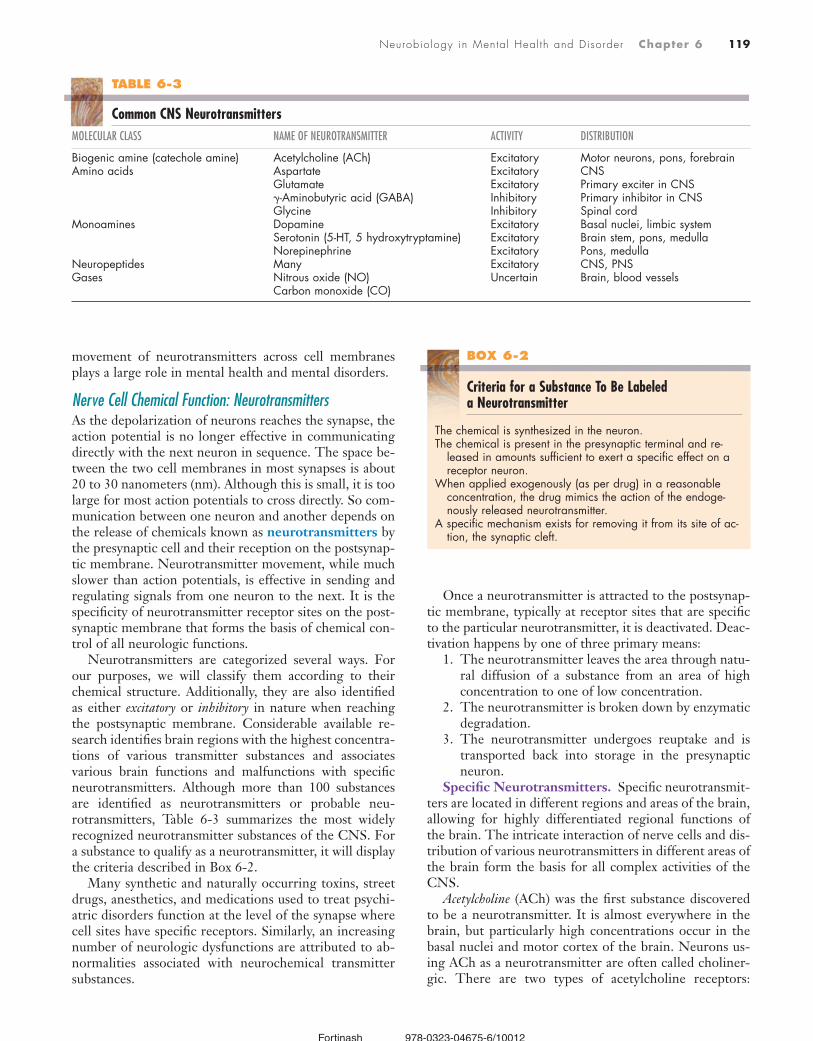
Neurotransmitters are categorized several ways. For our purposes, we will classify them according to their chemical structure. Additionally, they are also identifi ed as either excitatory or inhibitory in nature when reaching the postsynaptic membrane. Considerable available re-search identifi es brain regions with the highest concentra-tions of various transmitter substances and associates various brain functions and malfunctions with specifi c neurotransmitters. Although more than 100 substances are identifi ed as neurotransmitters or probable neu-rotransmitters, Table 6-3 summarizes the most widely recognized neurotransmitter substances of the CNS. For a substance to qualify as a neurotransmitter, it will display the criteria described in Box 6-2.
Many synthetic and naturally occurring toxins, street drugs, anesthetics, and medications used to treat psychi-atric disorders function at the level of the synapse where cell sites have specifi c receptors. Similarly, an increasing number of neurologic dysfunctions are attributed to ab-normalities associated with neurochemical transmitter substances.
Once a neurotransmitter is attracted to the postsynap-tic membrane, typically at receptor sites that are specifi c to the particular neurotransmitter, it is deactivated. Deac-tivation happens by one of three primary means:
1. The neurotransmitter leaves the area through natu-ral diffusion of a substance from an area of high concentration to one of low concentration.
2. The neurotransmitter is broken down by enzymatic degradation.
3. The neurotransmitter undergoes reuptake and is transported back into storage in the presynaptic neuron.
Specifi c Neurotransmitters. Specifi c neurotransmit-ters are located in different regions and areas of the brain, allowing for highly differentiated regional functions of the brain. The intricate interaction of nerve cells and dis-tribution of various neurotransmitters in different areas of the brain form the basis for all complex activities of the CNS.
Acetylcholine (ACh) was the fi rst substance discovered to be a neurotransmitter. It is almost everywhere in the brain, but particularly high concentrations occur in the basal nuclei and motor cortex of the brain. Neurons us-ing ACh as a neurotransmitter are often called choliner-gic. There are two types of acetylcholine receptors:
BOX 6-2
Criteria for a Substance To Be Labeled a Neurotransmitter
The chemical is synthesized in the neuron.The chemical is present in the presynaptic terminal and re-
leased in amounts suffi cient to exert a specifi c effect on a receptor neuron.
When applied exogenously (as per drug) in a reasonable concentration, the drug mimics the action of the endoge-nously released neurotransmitter.
A specifi c mechanism exists for removing it from its site of ac-tion, the synaptic cleft.
TABLE 6-3
Common CNS NeurotransmittersMOLECULAR CLASS NAME OF NEUROTRANSMITTER ACTIVITY DISTRIBUTION
Biogenic amine (catechole amine) Acetylcholine (ACh) Excitatory Motor neurons, pons, forebrainAmino acids Aspartate
Glutamate�-Aminobutyric acid (GABA)Glycine
ExcitatoryExcitatoryInhibitoryInhibitory
CNSPrimary exciter in CNSPrimary inhibitor in CNSSpinal cord
Monoamines DopamineSerotonin (5-HT, 5 hydroxytryptamine) Norepinephrine
ExcitatoryExcitatoryExcitatory
Basal nuclei, limbic systemBrain stem, pons, medullaPons, medulla
Neuropeptides Many Excitatory CNS, PNSGases Nitrous oxide (NO)
Carbon monoxide (CO)Uncertain Brain, blood vessels
Fortinash 978-0323-04675-6/10012

120 PART I FOUNDATIONS FOR PSYCHIATRIC MENTAL HEALTH NURSING
muscarinic and nicotinic. Many drugs, such the older neuroleptic antipsychotics, interact with ACh and its re-ceptor sites to produce anticholinergic side effects, which occur when muscarinic acetylcholine receptors are blocked. Side effects include dry mouth, blurred vision, constipation, and urinary retention. These side effects are troubling to clients and are a common reason why clients stop using their medications and fail to comply with the treatment regimen. In severe cases, muscarinic receptor blockade produces confusion and delirium in clients, especially in older clients. Nicotinic receptors respond positively to nicotine and are common in neuro-muscular synapses as well as in some CNS and PNS re-gions. Nicotine, found in tobacco, binds with the nico-tinic receptor sites and is able to mimic the effects of ACh released in some centers of the brain that are associ-ated with pleasure, making nicotine highly addictive. Exposure to excessive levels of nicotine will sometimes cause paralysis. Nicotine is also an effective insecticide and a common cause of poisoning in children. Good cli-ent teaching and nursing care designed to manage the side effects and adverse effects of drugs are signifi cant aspects of psychiatric mental health nursing.
Glutamate (glutamic acid) is an amino acid and the most widely distributed excitatory neurotransmitter in the brain. Some theorize that excessive glutamate activity is a part of the neurodegenerative process seen in such ill-nesses as schizophrenia and Alzheimer’s disorder (Alexan-der et al., 2002; Goff and Coyle, 2001; Stahl, 2000). �-Aminobutyric acid (GABA), chemically derived from glutamate, is the brain’s principal inhibitory neurotrans-mitter. Nerve cells stimulated by inhibitory neurotrans-mitters such as GABA will be turned off, which slows or stops actions completely in postsynaptic neurons.
Dopamine is a neurotransmitter well localized in the CNS. Dopaminergic neurons occur in several brain re-gions including the substantia nigra, midbrain, and hypo-thalamus. Dopamine-containing cells in the midbrain project to the limbic cortex. Researchers believe that these areas are the parts of the brain that malfunction in schizophrenia.
Norepinephrine or noradrenaline is concentrated in a small area of the brain known as the locus ceruleus. Many studies now indicate that clients suffering from mood disorders, particularly major depression, suffer from a defi cit of norepinephrine. Sympathetic nerves that inner-vate smooth muscles in blood vessels have a heavy con-centration of norepinephrine, which helps to explain its role in elevating blood pressure in the fi ght-or-fl ight re-sponse. When released directly into the bloodstream, norepinephrine acts as a hormone that enhances the effect of locally released norepinephrine at neuromuscular junc-tions. Both norepinephrine and its chemical relative, epi-nephrine, are synthesized from the amino acid tyrosine. Norepinephrine, epinephrine, dopamine, serotonin, and histamine belong to the class of neurotransmitters known as monoamines. Norepinephrine-producing neurons are sometimes referred to as adrenergic.
Serotonin has a pattern of action similar to norepineph-rine and is made from tryptophan, another amino acid. Serotonin production occurs in the brain stem and is also widely dispersed throughout the cerebral cortex and the spinal cord. Serotonin helps to regulate a constant internal environment. Maintaining a normal body temperature, normal eating and sleep-rest patterns, and normal moods is dependent on adequate levels of serotonin. Clinically signifi cant problems occur when clients have low levels of serotonin, and many behavioral symptoms common to depression occur when available serotonin is depleted.
Researchers suspect that two other gases—carbon mon-oxide (CO) and nitric oxide (NO)—function as neurotrans-mitter-like substances. NO and CO are both poisonous, unstable gases found in automobile emissions. Nitric oxide shares few of the characteristics of other neurotransmit-ters. It is not stored in synaptic vesicles. NO actually works in the opposite direction and feeds back toward the pre-synaptic neurons, and has no known specifi c receptor sites. Yet it functions as a chemical messenger in the brain and in peripheral blood vessels. It is involved in blood vessel contraction in the clitoris and penis during sexual arousal. Research suggests that nitric oxide plays a role in the brain’s memory function and is possibly part of the com-plex illness of major depression (McLeod, Lopez-Figueroa, and Lopez-Figueroa, 2001). Research indicates that car-bon monoxide acts in similar ways.
Other larger neuropeptide molecules such as cholecysto-kinin and endorphins, whose functions are still being re-searched, occur at multiple sites in the brain. Researchers believe these molecules play a role in the complex func-tioning of the brain.
Clinical Signifi cance of Neurotransmitters. Exten-sive research has been directed toward developing new drugs that operate at the synaptic level within the brain. Any chemical that mimics, competes, destroys, or pre-vents a neurotransmitter from binding on specifi c recep-tor sites on the postsynaptic membrane alters the effec-tiveness of communication between neurons. Researchers have made countless advances in the treatment of psychi-atric disorders. This is due to an increased understanding of neurotransmitters as well as an understanding of the way neurotransmitters are synthesized and deactivated. A brief discussion of some of the more common brain-related illnesses linked to neurotransmitter dysfunctions follows. Table 6-4 summarizes specifi c disorders and re-lated neurotransmitters.
Depression. Serotonin and its close chemical relatives, dopamine and norepinephrine, are the neurotransmitters most widely involved in various forms of depression. The two major classes of antidepressants—tricyclic and selec-tive serotonin reuptake inhibitors (SSRI) agents—differ primarily in their effects on either norepinephrine or se-rotonin levels. This explains why certain drugs, such as fl uoxetine or paroxetine, that specifi cally target serotonin may not be effective for some clients but work well for others (see the case study presented later in the chapter). The selective serotonin reuptake inhibitor (SSRI) class of
Fortinash 978-0323-04675-6/10012

121Neurobio logy in Menta l Heal th and Disorder Chapter 6
antidepressants inhibits the reuptake of serotonin by the presynaptic secreting cells. This reduces the availability of serotonin for subsequent release. Other antidepressants function as monoamine oxidase inhibitors (MAOIs). Monoamine oxidase (MO) is an enzyme that typically deactivates serotonin and dopamine. Deactivating this enzyme leaves the system unable to turn off the effects of these transmitter substances on postsynaptic neurons. Conversely, an enzyme or drug that acts opposite to MO or is an MAOI reduces the transmission of signals be-tween neurons. Catechol-O-methyl transferase (COMT), an enzyme normally responsible for deactivating norepi-nephrine, is sometimes present in excess in certain syn-apses. Too much of this enzyme prevents adrenergic neurons from effectively communicating with one an-other. Antidepressants that inhibit or reduce COMT levels restore neuronal communication ability. Because norepinephrine is also important in regulating activities such as heart rate and blood pressure, antidepressants operating on the norepinephrine system may have adverse side effects on these functions.
Anxiety. A number of conditions related to anxiety such as panic disorders and extreme phobias are triggered by an overproduction of some excitatory neurotransmit-ters causing a hyperexcitability of the postsynaptic mem-brane. GABA, one of the key inhibitory neurotransmitters in the CNS, normally counteracts the effect of these transmitters. Many antianxiety medications such as diaz-epam (Valium) or alprazolam (Xanax) act by stimulating GABA synthesis, which then modulates the effect of excit-atory neurotransmitters. This produces a calming effect in clients experiencing anxiety.
Schizophrenia. A complex disorder such as schizo-phrenia most likely has multiple contributing factors in-cluding genetic predisposition, prenatal development, and the environment. The direct cause of symptoms mani-fested in schizophrenia is probably a disruption of normal neurotransmitter activity, particularly dopamine. One plausible explanation for schizophrenia is the dopaminer-gic theory, which hypothesizes that the dopamine levels in people with schizophrenia are elevated. Some maintain that at least six other neurotransmitters—glutamate, sero-tonin, norepinephrine, acetylcholine, GABA, and chole-cystokinin—are also involved in schizophrenia. The most
commonly prescribed antipsychotic drugs suppress dopa-mine and similar transmitter substances. Current treat-ment continues to focus primarily on the dopaminergic theory.
Parkinsonism. Most researchers agree that the imme-diate cause of parkinsonism is a defi ciency of dopamine, particularly in the basal nuclei involved in motor coordina-tion. Characteristically, patients suffering from Parkinson’s disease display tremors, a shuffl ing gait, and a progressive lack of motor control. Clients sometimes have a loss of facial motor control, slurring speech, and make facial ex-pressions that are fl at or masklike. Public fi gures such as the late Pope John Paul II, boxer Muhammad Ali, and ac-tor Michael J. Fox displayed several of these symptoms, raising public awareness of the condition. The causes of dopamine defi ciency in Parkinson’s patients appear to be both genetic and environmental. Currently, parkinsonism is treated with L-dopa, a dopamine precursor capable of crossing the blood-brain barrier of the brain. According to the theory, brain cells containing the appropriate enzymes will convert the L-dopa into dopamine.
Alzheimer’s Disease. Alzheimer’s disease is among the leading causes of disability and death of older adults in the United States, and the number of affected persons increases each year. Acetylcholine is the neurotransmit-ter primarily involved in Alzheimer’s disease. Decreased levels of ACh produce many of the behavioral manifesta-tions of the disease, such as memory loss and disorienta-tion. This helps to explain why drugs such as donepezil (Aricept) are useful in the treatment of Alzheimer’s dis-ease. Aricept and other similar drugs inhibit the cholin-esterase enzyme that breaks down ACh thus increasing the amount of available acetylcholine, therefore pro-longing the onset of symptoms of Alzheimer’s disease (Stahl, 2000).
Both Parkinson’s and Alzheimer’s diseases are examples of recognized organic brain disorders. They are included in this discussion because of their relationship to specifi c transmitter substances and because the devastating effects of these degenerative conditions are clearly linked to mood disorders and dementia that psychiatric mental health nurses encounter. The Case Study is an example of a neurodegenerative disorder, multiple sclerosis, that fre-quently has associated symptoms of a mental disorder.
TABLE 6-4
Relationship of Neurotransmitter Dysfunction to Mental Disorders
NEUROTRANSMITTER DYSFUNCTION MENTAL DISORDER
Dopamine Increase SchizophreniaSerotonin Decrease DepressionNorepinephrine Decrease Depression?-Aminobutyric acid
(GABA)Decrease Anxiety disorders
Acetylcholine Decrease Alzheimer’s disease
Shawn, a 43-year-old woman, was diagnosed with multiple sclerosis 5 years ago. She was taking gabapentin (Neurontin) and paroxetine
(Paxil), and muscle pains and migraine headaches made it dif-fi cult for her to sleep. She also complained of feeling depressed and hopeless as a result of the recent exacerbation of debilitat-ing MS symptoms. Shawn reported a 10-pound weight loss in the past 2 months.
CRITICAL THINKING1 What questions will the nurse ask at this time?2 What changes will the nurse suggest to improve Shawn’s
insomnia?
Fortinash 978-0323-04675-6/10012

122 PART I FOUNDATIONS FOR PSYCHIATRIC MENTAL HEALTH NURSING
INTERRELATED SYSTEMSEvidence is now clear that the CNS operates in delicate balance with other body systems. Research demonstrates that the CNS both affects and is affected by the immune system, the endocrine system, and the body’s natural bio-logic rhythms, as well as other systems. The following are some examples of the interactions between body systems and how the disruption of these systems sometimes results in mental, emotional, and behavioral dysfunction and disorder.
PsychoneuroimmunologyPsychoneuroimmunology (PNI) studies the relationship between the neurologic, endocrine, and immune systems and behaviors associated with these systems. Cytokines, chemical messengers between immune cells, signal the brain to produce changes of activity in the endocrine sys-tem as well as the immune system. Research studies focus on the relationship of cytokines and the pathophysiology of medical diseases such as cancer, allergies, and autoim-mune diseases. More recent studies focus on psychiatric disorders such as major depression, schizophrenia, and Alzheimer’s disease (Kronfol and Remick, 2000).
Brain receptor sites for neuropeptides produced by the immune system are associated with changes in emotions and behaviors. Stress causes the discharge of corticotrophin-releasing factors that suppress the immune system. Studies indicate that negative emotions, anxiety, and psychiatric disorders such as schizophrenia and mood disorders are sometimes associated with a decreased functioning of the immune system. Posttraumatic stress syndrome is associ-ated with long-term immunosuppression (Kawamura, Kim, and Asukai, 2001).
NeuroendocrinologyNeuroendocrinology studies the relationship between the nervous system and the endocrine system. A number of hormones, including epinephrine, actually function as neurotransmitter-like substances. This affects chemical communication between many cells, even ones that are distant from the source of the hormone. Several hormon-ally based disorders result in medical conditions that produce psychiatric symptoms, as described next.
Research studies correlate hypothyroidism with de-pressive symptoms and Addison’s disease with depres-sion and fatigue. Other endocrine disorders are linked to autoimmune conditions such as Graves’ disease, which causes excessive thyroid secretion. This sometimes fol-lows an acute infection suggesting an immunologic ori-gin for Graves’ disease. People who suffer from Graves’ disease commonly report symptoms of emotional stress, nervousness, fatigue, weight loss, heat intolerance, and gastrointestinal symptoms. Also, because schizophrenia and other psychiatric disorders occur more frequently during the reproductive period of life when sex hor-mones are most active, this suggests an endocrine-related origin.
ChronobiologyChronobiology is the study of the biologic rhythms of the body, such as the circadian rhythms. These rhythms manifest in metabolic rate, sleep-wakefulness cycles, blood pressure, hormone levels, and body temperature. Re-searchers believe the brain controls these rhythms and their interactions with various endocrine organs. Many psychiatric and medical disorders occur more frequently when sleep patterns and biologic rhythms are disrupted.
Many hypothesize that dreams result from the activa-tion of electrical activity in brain regions that recall recent memories and reinforce long-term memories. One theory maintains that mental disorders are the result of brain circuits that are not activating competently because of abnormal brain wave patterns. When incompetent brain circuits are activated while the individual is awake, clients often report hallucinations and illusions. While sleeping, these incompetent brain circuits appear to produce bi-zarre or illusory dreams (Kavanau, 2000). Psychoactive drugs modify brain waves in psychotic clients, which tem-porarily restore more normal brain circuits. Antidepres-sants increase brain waves and suppress or reduce rapid eye movement (REM) sleep. Electroconvulsive therapy suppresses abnormal brain waves, allowing more normal slow waves to dominate. Additional information on sleep disorders is in Chapter 18.
Sundowning, or Sundowner’s syndrome, is the exacerba-tion, or worsening, of psychotic or depressive symptoms during the afternoon or evening resulting in confusion and disorientation. Some studies connect sundowning with a disturbance of circadian rhythms. Psychiatric and medical conditions such as Alzheimer’s disease also disrupt the cli-ent’s circadian rhythm (Volicer et al., 2001). Decreased exposure to light during the winter months has also shown to produce depressive symptoms in clients suffering from seasonal affective disorder (SAD). PMH nurses need to be aware of these examples of chronobiologic disruptions that produce symptoms of mental illnesses.
EMERGING CONCEPTS IN PSYCHOBIOLOGYGenetic ResearchGenetics is the study of genes and the role they play in the functioning of living organisms. The Human Genome Project that began in 1990 resulted in the identifi cation of all of the genes contained on the 23 pairs of human chro-mosomes. The knowledge of precise locations of genes responsible for every human biologic characteristic, as well as their biochemical structure, has opened up endless possibilities for research into the genetic causes of nearly every human disease or condition. This includes psychiat-ric disorders. However, recognizing that genes only de-termine the potential to develop any normal or abnormal condition signifi cantly complicates the problem of identi-fying genetic causes of specifi c psychiatric disorders. Ex-cellent evidence attributes disorders such as Huntington’s
Fortinash 978-0323-04675-6/10012

123Neurobio logy in Menta l Heal th and Disorder Chapter 6
disease and Parkinsonism to specifi c genes. For example, both diseases are located on chromosome number 4, but evidence for genetic causes of some other specifi c neuro-logic disorders is not so clear.
Because there appear to be familial tendencies in some disorders, researchers are attempting to specify schizo-phrenia genes. According to the most current literature, there are as many as 150 genes on nearly a dozen different chromosomes that contribute to the causes of schizophre-nia, making the situation more complex (Badner and Gershon, 2002; Lewis et al., 2003). Research indicates that schizophrenia results from the interaction of multiple genes rather than a single gene. In theory, defective genes code for incorrect synthesis of neurotransmitters, or their deactivating enzymes, or other factors interfere with the proper transmission of vital chemical agents.
Research has also identifi ed genes that are linked to bipolar disorder and substance dependence (Schindler et al., 2001). This helps to explain why certain psychiatric disorders recur in families and why fi rst-degree relatives of individuals with psychiatric disorders have increased risk for developing the same or similar disorders. The genetic origins of other psychiatric dysfunction and disor-ders, namely attention defi cit hyperactivity disorder, anti-social personality disorder, and violent behaviors, are be-ing explored (Doyle, Roe, and Faraone, 2001; Raine et al., 2000).
Although few researchers believe that a single gene causes a psychiatric illness, genetics clearly plays a signifi -cant role in infl uencing mental health and disorder. The interaction of genes is highly complex, and the link of genes to behavior remains controversial. It appears that many genes infl uence psychiatric illness and the dysfunc-tional behaviors that are symptomatic of those illnesses (Petronis et al., 2003). There is also increased evidence that environmental and developmental conditions in utero contribute to the expression of these genes that subse-quently manifest as abnormal behavior.
Stem Cell TechnologyStem cell technology is perhaps the most controversial and promising technique that will lead to treatments and cures for neurobiologic disease and injury. Stem cells are cells that have the complete genome intact and have not yet differentiated, or developed, into a specifi c cell type. A fertilized egg is totipotent, or has total potential to de-velop into an entire human being. As embryonic cells replicate, some become specialized genes while others are turned off. Adult stem cells have already differenti-ated, or begun to develop to a certain degree. For exam-ple, some adult stem cells have already developed into epithelia rather than connective tissue cells or muscle or nervous tissues. Although there are adult stem cells in a variety of tissues including bone marrow, some connec-tive tissues, and even brain tissue, the ability to success-fully culture them and use them for therapeutic purposes is currently limited.
The reason why stem cell technology is so promising is because undifferentiated stem cells from embryos have the potential to develop into any type of cell, so research-ers are able to control gene expression deliberately. Spe-cialized stem cells can be developed into organs for trans-plants and have fewer complications from tissue rejection. The controversy surrounding stem cell research is an ethical issue, primarily regarding the source of embryos that provide undifferentiated stem cells. Many public fi g-ures have taken seemingly rational and passionate but opposing points of view increasing the intensity of the debate. These points of view will shape public policy and the direction of stem cell research, which will determine matters of life and death. Debate continues for or against the use of embryos or amniotic fl uid as researchers seek cures and treatments for conditions and diseases and remedies that stem cells might provide.
Regardless of the source of stem cells, adult or embry-onic, numerous technical challenges remain, but the po-tential therapeutic applications for this research seem unlimited at the moment. Active research continues on numerous neurobiologic conditions including brain and spinal cord injury, neurogenetic disorders affecting brain development, and degenerative conditions such as amyo-tropic lateral sclerosis (ALS), Parkinson’s disease and Al-zheimer’s disease, among others (Shihabuddin et al., 1999). The implications of stem cell research for the fu-ture of psychiatric nursing are promising as is the poten-tial of stem cell therapy itself.
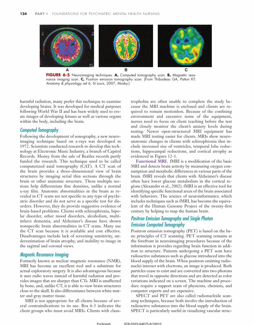
DIAGNOSTIC AND EVALUATION PROCEDURES NeuroimagingBefore modern neuroimaging techniques were available, clinicians had few noninvasive tools to examine the hu-man brain, with the exception of the x-ray. The brain re-mained a mystery. The development of imaging tech-niques since the early 1980s has dramatically changed the understanding of brain structure and function. Brain anatomy and physiology has now been mapped in exqui-site detail, providing valuable information using a variety of techniques. Useful neuroimaging techniques available today include ultrasonography (US), computed tomogra-phy (CT), magnetic resonance imaging (MRI), functional magnetic resonance imaging (fMRI), positron emission tomography (PET), and single photon emission com-puted tomography (SPECT) (Figure 6-5). Unlike older x-ray technology that uses fi lm, these techniques use com-puters to generate images. Table 6-5 identifi es common nursing considerations for clients undergoing neuroimag-ing tests.
UltrasonographyUltrasonography, also known as echoencephalography, uses high-frequency sound waves to form images of brain spaces and masses. Because ultrasonography does not use
Fortinash 978-0323-04675-6/10012
124 PART I FOUNDATIONS FOR PSYCHIATRIC MENTAL HEALTH NURSING
harmful radiation, many prefer this technique to examine developing brains. It was developed for medical purposes following World War II and has been widely used to cre-ate images of developing fetuses as well as various organs within the body, including the brain.
Computed TomographyFollowing the development of sonography, a new neuro-imaging technique based on x-rays was developed in 1972. Scientists conducted research to develop this tech-nology at Electronic Music Industry, a branch of Capitol Records. Money from the sale of Beatles records partly funded the research. This technique used to be called computerized axial tomography (CAT). A CT scan of the brain provides a three-dimensional view of brain structures by imaging serial thin sections through the brain or other anatomic structure. These multiple sec-tions help differentiate fi ne densities, unlike a normal x-ray fi lm. Anatomic abnormalities in the brain as re-vealed in CT scans are not specifi c to any type of psychi-atric disorder and do not serve as a specifi c test for dis-orders. However, they do provide suggestive evidence of brain-based problems. Clients with schizophrenia, bipo-lar disorder, other mood disorders, alcoholism, multi-infarct dementia, and Alzheimer’s disease have shown nonspecifi c brain abnormalities in CT scans. Many use the CT scan because it is available and cost effective. Disadvantages include lack of screening sensitivity, un-derestimation of brain atrophy, and inability to image in the sagittal and coronal views.
Magnetic Resonance ImagingFormerly known as nuclear magnetic resonance (NMR), MRI has become an excellent tool and a substitute for actual exploratory surgery. It is also advantageous because it uses radio waves instead of harmful radiation and pro-vides images that are sharper than CTs. MRI is unaffected by bone, and, unlike CT, it is able to view brain structures close to the skull. It also differentiates between white mat-ter and gray matter tissue.
MRI is not appropriate for all clients because of sev-eral contraindications to its use. Box 6-3 indicates the client groups who must avoid MRIs. Clients with claus-
trophobia are often unable to complete the study be-cause the MRI machine is enclosed and clients are re-quired to remain motionless. Because of the confi ning environment and excessive noise of the equipment, nurses need to focus on client teaching before the test and closely monitor the client’s anxiety levels during testing. Newer open-structured MRI equipment has made MRI testing easier for clients. MRIs show neuro-anatomic changes in clients with schizophrenia that in-clude increased size of ventricles, temporal lobe reduc-tions, hippocampal reductions, and cortical atrophy as evidenced in Figure 12-1.
Functional MRI. fMRI is a modifi cation of the basic MRI and detects brain activity by measuring oxygen con-sumption and metabolic differences in various parts of the brain. fMRI reveals that clients with Alzheimer’s disease often have lower glucose metabolism in the cortical re-gions (Alexander et al., 2002). fMRI is an effective tool for identifying specifi c functional areas of the brain associated with behaviors. The science of neuroinformatics, which includes techniques such as fMRI, has become the equiva-lent of the Human Genome Project of the twenty-fi rst century by helping to map the human brain.
Positron Emission Tomography and Single Photon Emission Computed TomographyPositron emission tomography (PET) is based on the ba-sic principles of CT scanning. PET scanning remains at the forefront in neuroimaging procedures because of the information it provides regarding brain function in addi-tion to structure. Patients undergoing a PET scan have radioactive substances such as glucose introduced into the blood supply of the brain. When positron-emitting radio-nuclei interact with electrons, an image is produced. Both particles cease to exist and are converted into two photons that travel in opposite directions and are detected as color variations indicated on a screen. The machine and proce-dure require a support team of physicists, chemists, and computer experts and are expensive.
SPECT and PET are also called radionucleide scan-ning techniques, because both involve the introduction of radioactive substances into the blood supply of the brain. SPECT is particularly useful in visualizing vascular struc-
FIGURE 6-5 Neuroimaging techniques. A, Computed tomography scan. B, Magnetic reso-nance imaging scan. C, Positron emission tomography scan. (From Thibodeau GA, Patton KT: Anatomy & physiology, ed 6, St Louis, 2007, Mosby.)
A B C
Fortinash 978-0323-04675-6/10012

125Neurobio logy in Menta l Heal th and Disorder Chapter 6
tures in the brain and in diagnosing disorders such as cerebrovascular accidents (CVAs).
These techniques are particularly useful for demon-strating variable levels of brain activity and associated blood fl ow within the brain. SPECT scans have detected abnormalities in the frontal cortex, occipital, and tempo-ral lobes, and parahippocampal gyrus in clients with panic disorders.
TABLE 6-5
Nursing Considerations with Neuroimaging ProceduresTEST GENERAL CONSIDERATIONS COMMON NURSING CARE COMMON CONTRAINDICATIONS
ANATOMIC IMAGINGComputed tomography
(CT)Three-dimensional view of
brain structuresDifferentiate fi ne-density struc-
tures, unlike normal x-ray fi lm
Examination time: 15-30 minClear fl uids meal before test
Explain purpose of test and all procedures.Reassure client that test is safe and that radi-
ation exposure is not a concern.Assess client’s anxiety level and monitor for
symptoms of claustrophobia.Reassure client that hearing monotonous
noise is common.Instruct client to lie still to ensure good
imaging.If using contrast iodine, monitor for gastroin-
testinal upset, fl ushing, and perceptions of excess warmth.
Allergy to iodine (not all CT requires iodine)
Inability to lie completely still
Claustrophobia
Magnetic resonance imaging (MRI)
Separates view of white mat-ter from gray matter tissue
Examination time: 15-60 min
Explain purpose of test and all procedures.Reassure client that test uses magnets, not
radiation; radiation exposure is not a concern.
Assess client’s anxiety level and monitor for symptoms of claustrophobia.
Instruct client to lie still to ensure good imaging.
Instruct client that a clear plastic helmet with antenna will be put over head.
Reassure client that hearing monotonous noise is common.
Inability to lie completely still
ClaustrophobiaPacemakersMetallic implants, plates, or
screwsLife support equipment
needed for clientInfusion pumpsGenerally not used when
client is pregnant
FUNCTIONAL IMAGINGPositron emission
tomography (PET)Two-dimensional view of
brain structuresMeasures physiologic and
chemical functioning such as glucose uptake by cells in brain, as well as infor-mation on anatomic structures
Short half-life isotopes used
Explain purpose of test and all procedures.Inform client that isotopes are radioactive
and discuss concerns.Assess client’s anxiety level and monitor for
symptoms of claustrophobia.Explain that there will be time interval of
about 45 min between injection of isotope and scanning procedure.
Explain that client may be blindfolded and have earplugs to decrease environmental stimulus during testing.
Instruct client to lie still to ensure good imaging.
Make sure client does not fall asleep during procedure—this will affect test results.
Inability to lie completely still
Claustrophobia Severe anxiety levelRecent use of sedating/
tranquilizing medication because these medica-tions alter cellular glu-cose use patterns
Breast-feedingRequires expensive
cyclotron
Single photon emission computed tomography (SPECT)
Two-dimensional view of brain structures
Measures physiologic and chemical functioning such as glucose uptake by cells in brain, as well as infor-mation on anatomic structures
Long half-life isotopes usedNo onsite cyclotron required
As for PET above. Breast-feedingInability to lie completely
stillClaustrophobia
BOX 6-3
Client Group Contraindications for MRI
Individuals with pacemakersIndividuals with metallic objects such as screws, prostheses,
and orthopedic devicesClients on life-support systems
Fortinash 978-0323-04675-6/10012
126 PART I FOUNDATIONS FOR PSYCHIATRIC MENTAL HEALTH NURSING
NEUROBIOLOGY AND PSYCHIATRIC NURSINGPsychiatric mental health nursing provides care to clients with brain-based illnesses. Increasingly, a strong back-ground in neurobiology is part of the standards of practice for psychiatric mental health nursing (American Nurses Association, 2006). By synthesizing the fi ndings of the nursing assessment discussed in Chapter 3 and the nurse’s understanding of psychobiologic issues, effective nursing care will assist clients in achieving wellness.
As we are learning, each structure and each neuro-chemical produced and used by the brain has a specifi c function. The brain is a dynamic, continually changing environment, and researchers are discovering more of the complexities of the brain. In adults, the brain seems less able to repair itself after injury or replace degenerative cells when compared with other parts of the body. This neuroplasticity, or the ability of the brain to change its structure and function, is providing insights into the role of certain brain areas in the development of illness (Mohr and Mohr, 2001). New understanding and application of concepts such as the neuroplastic nature of brain tissue are also leading to new approaches for treating disorders. Until now, many believed that the capacity of the brain to repair itself after injury or to replace degenerative cells was minimal, particularly in adults, but current research reveals brain cell regeneration in several conditions.
Genetics and stem cell research are just two emerging technologies that are opening the door to potential treat-ments for psychiatric illnesses. Much of the stigma at-tached to psychiatric illness was due to a lack of under-standing regarding the biologic basis of these disorders. Therefore, effective client and family teaching is an im-
portant function of the role of the psychiatric mental health nurse as researchers discover new information re-garding the structures and functioning of the CNS. The Client and Family Teaching Guidelines box displays the highlights of effective client teaching regarding the bio-logic basis of psychiatric disorders. Psychiatric mental health nurses will continue to play an important part by directly assisting clients with brain-based disorders and by teaching and informing clients, families, and the general public about advances in neurobiology.
New research fi ndings continue to change the way people with psychiatric disorders are cared for and treated. The Research for Evidence-Based Practice box highlights the importance of critical thinking as psychiatric mental health nurses approach and plan modifi cations of care based on new research fi ndings.
Knowledge of the neurobiologic basis of psychiatric disorders is essential in effective psychiatric mental health nursing practice. Nurses need to include biologic princi-ples in all aspects of nursing care, from assessment to evaluation, to ensure comprehensive and quality nursing. More and more information will be available regarding structure and functioning of the brain, and because of this, the role and function of the psychiatric mental health nurse will continue to change. Staying current with dy-namic development in the fi eld will continue to stimulate and positively challenge the truly professional psychiatric mental health nurse.
CHAPTER SUMMARY ● Current knowledge about the brain and its functions is
continually changing. ● The brain is the most complex and one of the most im-
portant organs in the human body because of its multi-ple functions.
BIOLOGIC BASIS OF PSYCHIATRIC DISORDERS • Determine a mutually acceptable time and location for the
teaching session.• Select an environment that is favorable for learning.• Identify the client’s readiness for learning.• Identify the client’s motivation for learning.• Identify the client’s knowledge about the topic and accu-
racy of that knowledge.• Identify with the client the specifi c content that is requested
and required.• Defi ne a measurable outcome with the client to determine
that learning has occurred.• Defi ne the evaluation method used to determine the effec-
tiveness of teaching.• Use multiple teaching-learning approaches, such as visual
and auditory, based on client’s needs.• Monitor the client’s anxiety level during the teaching
session, as increased anxiety will decrease information processing.
• Identify alternative resources available to the client to in-crease learning potential.
• Identify the process that the client will follow to access sup-port persons if the client requires more reinforcement.
Gross-Isseroff R et al: The suicide brain: a review of postmortem receptor transporter binding studies, Neuroscience and Biobehavior Review 22:653, 1998.
Some research has reported that the brains of individuals who commit suicide are different from the brains of individuals who have died of natural causes. Postmortem examination of the receptor/transport binding sites of the brain tissue of suicide victims and of individuals who have died of natural causes show that the brains of suicide victims have unique, specifi c neurochemical characteristics that make “suicide brains” differ-ent from the brains of individuals who have died of natural causes. Scientists are using such studies to formulate a hypoth-esis of molecular markers that will possibly help defi ne and identify individuals at risk for suicidal behavior. Researchers are developing tests to measure identifi ed markers. When these markers are specifi cally identifi ed and prove to be reliable in their ability to predict suicidal behavior, the test will likely be-come a routine aspect of mental health evaluation.
Fortinash 978-0323-04675-6/10012

127Neurobio logy in Menta l Heal th and Disorder Chapter 6
● Psychiatric disorders are brain-based illnesses with ana-tomic or physiologic components.
● It is imperative for nurses to understand the anatomy and physiology of the brain and other systems that in-teract with the nervous system. Nurses also will become familiar with psychobiologic approaches to treat psy-chiatric disorders.
● One key to understanding treatment strategies for psy-chiatric disorders is to recognize the role that neu-rotransmitter substances play in neural communication.
● Nurses will become familiar with psychobiologic ap-proaches to treat psychiatric disorders.
● Modern neuroimaging techniques help explain structure and function and their relation to brain differences and psychiatric illnesses.
● Emerging fi elds in neuroscience such as genetics and stem cell research will continue to bring advanced tech-nologies that lead to improved care for patients suffer-ing from neurobiologic disorders.
REVIEW QUESTIONS1 Which statement by a family member of a person with
schizophrenia demonstrates effective learning about the disease?1. “The disease was probably caused by problems with
several genes. These genes cause changes in how certain brain chemicals work.”
2. “The disease could be cured if our politicians and laws allowed for more stem cell research. Adult stem cells hold so much promise.”
3. “The disease probably resulted from the mother’s smok-ing during pregnancy. Nicotine is actually a neu-rotransmitter.”
4. “If our family had more money, we could afford the promising psychoneuroimmunologic treatments avail-able in other countries.”
2 Which assessment fi nding best indicates release of norepi-nephrine?1. Pulse rates changes from 70 to 622. Pupil size changes from 8 mm to 3 mm3. Client begins complaining of “intestinal cramping”4. Blood pressure changes from 126/70 to 158/84
3 These clients are scheduled to have magnetic resonance imaging (MRI). For which client(s) should additional as-sessment information be gathered before the diagnostic procedure? You may select more than one answer. A cli-ent with the following conditions:1. A history of wounds from exploding shrapnel during
military service2. A comorbid diagnosis of bleeding peptic ulcers for the
past 3 years3. Current complaints of extreme sensitivity to loud noises4. Reports of allergies to iodine, eggs, and shellfi sh5. A 3-year history of Parkinson’s disease
4 An adult has panic attacks. Which neurotransmitter is most implicated in this problem?1. Norepinephrine2. Acetylcholine
3. Serotonin4. Gamma aminobutyric acid (GABA)
5 A nurse plans the care for an adult with a tumor in the brain’s frontal lobes. Initial interventions should focus on the client’s anticipated problems with the following:1. Motor function and judgment2. Sensory and calculation abilities3. Interpretation of visual stimuli4. Hearing and hygiene
Additional self-study exercises and learning resources are available to you on the Companion CD at the back of the book and on the Evolve website at http://evolve.elsevier.com/Fortinash/.
REFERENCESAlexander GE et al: Longitudinal PET evaluation of cerebral met-
abolic decline in dementia: a potential outcome measure in Al-zheimer’s disease treatment studies, Am J Psychiatry 159:238-245, 2002.
Amen DG: Brain SPECT imaging in psychiatry, Prim Psychiatry 5:83-87, 1998.
American Nurses Association: Scope and standards of psychiatric men-tal health practice, Washington, DC, 2006, American Nurses Publishing.
Badner JA, Gershon ES: Meta-analysis of whole-genome linkage scans of bipolar disorder and schizophrenia, Mol Psychiatry 7:405-411, 2002.
Carlson NR: Physiology of behavior, ed 7, Boston, 2001, Allyn & Bacon.
Doyle AE, Roe CM, Faraone SV: The genetics of attention defi cit hyperactivity disorder, Prim Psychiatry 8:65-71, 2001.
Dubin MW: How the brain works, Williston, Vt, 2002, Blackwell Science.
Goff D, Coyle J: The emerging role of glutamate in the patho-physiology and treatment of schizophrenia, Am J Psychiatry 158:1367-1377, 2001.
Gribbin J: How the brain works: a beginner’s guide to the mind and consciousness, New York, 2002, Doring Kindersley.
Gross-Isseroff R et al: The suicide brain: a review of postmortem receptor transporter binding studies, Neurosci Biobehav Rev 22:653, 1998.
Gur R: Functional imaging is fulfi lling some promises, Am J Psy-chiatry 159:693-694, 2002.
Kavanau J: Sleep, memory, maintenance and mental disorders, J Neuropsychiatry 12:199-208, 2000.
Kawamura N, Kim Y, Asukai N: Suppression of cellular immunity in men with a past history of posttraumatic stress disorder, Am J Psychiatry 158:484-486, 2001.
Keltner NL et al: Psychobiological foundations of psychiatric care, St Louis, 1998, Mosby.
Kronfol Z, Remick, D: Cytokines and the brain implications for clinical psychiatry, Am J Psychiatry 157:683-694, 2000.
Lewis CM et al: Genome scan meta-analysis of schizophrenia and bipolar disorder, part II, Am J Human Genetics 73:34-48, 2003.
McLeod TM, Lopez-Figueroa A, Lopez-Figueroa MO: Nitric ox-ide, stress and depression, Psychopharmacol Bull 35:24-41, 2001.
Mohr WK, Mohr B: Brain, behavior, connections, and implica-tions: psychodynamics no more, Arch Psychiatr Nurs 15:171-181, 2001.
Montoya A et al: Brain mapping and cognitive dysfunction in Huntington’s disease, J Psychiatr Neuroscience 31:21-29, 2006.
Petronis A et al: Monozygotic twins exhibit numerous epigenetic differences: clues to twin discordance? Schizophrenia Bull 29:169-178, 2003.
Raine A et al: Reduced gray matter volume and reduced autonomic activity in antisocial personality disorder, Arch Gen Psychiatry 57:119-129, 2000.
Fortinash 978-0323-04675-6/10012

128 PART I FOUNDATIONS FOR PSYCHIATRIC MENTAL HEALTH NURSING
Rapoport JL: Rhe neurodevelopmental model of schizophrenia: update 2005, Mol Psychiatry 10:614, 2005.
Schindler KM et al: Candidate genes for schizophrenia: further evaluation of KCNN3, Prim Psychiatry 8:51-53, 2001.
Shihabuddin LS et al: Stem cell technology for basic science and clinical applications, Arch Neurol 6:29-32, 1999.
Stahl SM: Essential psychopharmacology: neuroscientifi c basis and practi-cal applications, ed 2, New York, 2000, Cambridge University Press.
Thibodeau G, Patton K: Anatomy and physiology, ed 6, St Louis, 2007, Mosby.
Volicer L et al: Sundowning and circadian rhythms in Alzheimer’s disease, Am J Psychiatry 158:704-711, 2001.
Young GB, Pigott SE: Neurobiological basis of consciousness, Arch Neurol 56:153-157, 1999.
ONLINE RESOURCESAmerican Academy of Sleep Medicine: www.aasmnet.orgAmerican Society of Neuroimaging: www.asnweb.orgCenter for Sleep and Circadian Biology: www.northwestern.edu/cscbEncyclopedia of Psychology: Psychobiology: www.psychology.org/links/Publications/
PsychobiologyInternational Society of Developmental Psychobiology: www.oswego.edu/isdpNational Institute of Mental Health: The Human Brain Project: www.nimh.nih.gov/
neuroinformatics/National Sleep Foundation: www.sleepfoundation.orgSociety for Light Treatment and Biological Rhythms: www.sltbr.org
Fortinash 978-0323-04675-6/10012