Embed Size (px)
Citation preview
105
CHAPTER 6
INNATE IMMUNITY IN C. ELEGANS
Ilka Engelmann and Nathalie Pujol*Centre d’Immunologie de Marseille‑Luminy, Université de la Méditerranée, Marseille, France *Corresponding Author: Nathalie Pujol—Email: [email protected]‑mrs.fr
Abstract: The nematode Caenorhabditis elegans is proving to be a powerful invertebrate model to study host‑pathogen interactions. In common with other invertebrates, C. elegans relies solely on its innate immune system to defend itself against pathogens. Studies of the nematode response to infection with various fungal and bacterial pathogens have revealed that the innate immune system of C. elegans employs evolutionary conserved signalling pathways. They regulate the expression of various effectors molecules, some of which are also conserved. Here, we summarize the current knowledge of the pathways and effector molecules involved in the nematode immune response, with a particular focus on the antifungal immune response of the C. elegans epidermis.
INTRODUCTION
C. elegans is a free‑living soil nematode that feeds on bacteria and is therefore constantly exposed to potential pathogens.1 Like other invertebrates, C. elegans lacks an adaptive immune system. In contrast to many invertebrate species, however, C. elegans does not appear to have specialized immune cells. For example, while Drosophila has macrophage‑like hemocytes, which engulf invading microbes, the only cells in the nematode body cavity, the 6 coelomocytes, do not seem to be capable of phagocytosis but function as scavenger cells with a high endocytic capacity.2
C. elegans possesses three major mechanisms of defences against microbial attacks:1 Avoidance behaviour: It has been demonstrated that worms are able to distinguish between different bacteria. Whereas most bacteria attract C. elegans, some repel the nematode and cause an avoidance behaviour. Such an aversive response can be specifically directed against the pathogenic strains of a bacterial species (reviewed in ref. 3). Olfactory neurons, G protein coupled receptors and the only Toll‑like receptor (TLR) in C. elegans, TOL‑1,
Invertebrate Immunity, edited by Kenneth Söderhäll. ©2010 Landes Bioscience and Springer Science+Business Media.
.noitubirtsiD rof to
N .ecneicsoiB sednaL thgirypoC
1002©

106 INVERTEBRATE IMMUNITY
are involved in triggering the avoidance behaviour to pathogenic Serratia marcescens.4,5 Worms can “remember” odours6 and can even learn to avoid bacteria that are recognized as noxious.7 This discrimination relies in part on pairs of asymmetric chemosensory neurons.8 Their correct development requires a signalling cassette that includes an intracellular TIR‑domain adapter protein (TIR‑1) acting upstream of a p38 MAPK cascade.9 This cassette, which will be described in more detail below, appears also to play a direct behavioural role as it has been found to be involved in the neuroendocrine regulation of serotonin‑dependent aversion to Pseudomonas aeruginosa.10,2 The second axis of protection against pathogen invasion is a strong cuticle, made of collagen and chitin and constituting the exoskeleton of the worm. It acts as a physical barrier that is relatively resistant to puncturing. As a complement, the pharyngeal grinder destroys pathogens that are taken up during feeding. It prevents live pathogens from reaching the intestine and establishing an infection. Indeed, mutants with defective grinder function are more susceptible to infection.11,12,3 The third line of defence involves inducible mechanisms. These will be the main focus of this chapter. C. elegans possesses a complex inducible defence system involving multiple signalling cascades that regulate the production of antimicrobial peptides (AMP) and proteins in a pathogen‑ and tissue‑specific way.
ROUTES OF INFECTION
Most of the known pathogens of C. elegans use two main routes of infection, through the pharynx or the epidermis (Fig. 1). Many Gram‑positive and Gram‑negative bacteria as well as yeast, infect worms upon oral up‑take during feeding and establish an intestinal infection. They must survive the passage through the grinder to reach the intestine, proliferate and establish an infection. In some cases, it has been shown that the pathogen destroys the grinder,13 in others it appears that the infectious particles, such as the spores of Bacillus thuringensis are resistant to the mechanical action of the grinder.14 Almost all characterised intestinal pathogens of C. elegans remain extracellular, apart from Salmonella typhimurium and the microsporidium Nematocida parisii, that have been shown to establish intracellular infection in the intestinal cells.15,16
Some pathogenic bacteria and fungi can adhere to the cuticle and infect the C. elegans epidermis. For example, Microbacterium nematophilum adheres to the anal region of the nematode and induces hindgut swelling17 and Leucobacter chromiireducens is capable of causing lethal uterine infections18 (Fig. 1). Different fungi that are pathogenic for nematodes, including Drechmeria coniospora and species of Haptocillium, produce spores that adhere and then penetrate the cuticle and grow into the epidermis (Figs. 1 and 2).19,20 Although some pathogens, such as certain strains of P. aeruginosa, produce fast‑acting toxins,21 against which C. elegans appears defenceless, in many cases, infection provokes an immune response.
PATHOGEN RECOGNITION
The first step of an inducible defence is the recognition of the pathogen. Conserved structures on pathogens that are not present in the host and thus recognized as foreign, so called microbe‑associated molecular patterns (MAMPs), bind to pattern recognition
.noitubirtsiD rof to
N .ecneicsoiB sednaL thgirypoC
1002©

107INNATE IMMUNITY IN C. ELEGANS
receptors (PRRs) in many organisms.22 PRRs include peptidoglycan recognition proteins (PGRP), Gram negative binding proteins (GNBP), nucleotide‑binding oligomerization domain (NOD) and NACHT domain proteins.23 Genes encoding proteins of these families are absent from the C. elegans genome.
One prominent class of PRRs, in vertebrates the TLRs, can sense outer membrane components of the bacteria, RNA or DNA.22 As mentioned above, the single worm TLR, TOL‑1, is involved in behavioural avoidance of some pathogenic bacteria,4,5 but does not seem to play a role in the resistance to several pathogens,5 nor in the regulation of certain immune effectors.24 One study showed that tol‑1 mutants are more susceptible to S. typhimurium infection,25 but it is unclear whether this is due to an involvement of tol‑1 in a protective immune response or rather due to a defect in cell adherence in the pharynx of the tol‑1 mutant leading to a defect in a physical barrier thus favouring pathogen invasion.
TLRs, as well as a number of other PRR families, in both plants and animals, share a common domain, the leucine rich repeat (LRR) domain. In a recent study, the role in host defences of each of the 14 predicted transmembrane proteins with LRR domains encoded in the C. elegans genome, was assayed. Loss‑of‑function mutants in one gene, fshr‑1, which encodes a glycopeptide hormone receptor homologue, were found to be more susceptible to infection by Gram positive and Gram negative bacteria. It has yet to be determined if FSHR‑1, which is expressed in the intestine, acts as a pathogen receptor or rather functions as a positive modulator of the nematode immune response.26
C‑type lectins are carbohydrate‑binding proteins that can exhibit very narrow ligand specificity. In mammals, a number of C‑type lectins have established roles in innate immunity. For example, Dectin‑1 is highly expressed on macrophages and recognizes beta‑glucan, a component of the fungal cell wall and thereby acts as a PRR.23 C. elegans possesses 278 genes encoding C‑type lectins, but it is currently unclear as to whether any of them function as PRRs or rather as effector molecules (see below).
While there is no clear Dectin‑1 orthologue in C. elegans, there are a number of potential scavenger receptors (SR), another class of protein known to be involved in pathogen recognition in other species.27 Indeed, there are six proteins homologous to CD36 and Croquemort, members of the SR‑B family and one well‑characterised SCARF orthologue CED‑1. Because of its expression in the intestine throughout development, one of these, C03F11.3, was suggested a number of years ago to be potentially involved in the recognition of microbial molecules.28 A study published last year supports such an idea, as CED‑1/SCARF and C03F11.3/CD36 appear to function in host resistance to Candida albicans and Cryptococcus neoformans in C. elegans.29 Whether in the nematode these proteins in fact recognize yeast cell wall beta‑glucans and act as PRRs has not been formally demonstrated. Alternatively, given CED‑1’s known function in recognizing dying cells during programmed cell death, it might instead recognize damaged host material and then induce the unfolded protein response (UPR, see below) in an attempt to contain this damage. So we still do not know whether the worm responds through the detection of specific MAMPs or more generally to the cellular damage and stress caused by the pathogen (so‑called danger theory30) or both. Nevertheless, the finding that C. elegans shows distinct immune responses to different pathogens that infect via the same route and have similar levels of virulence,31,32 clearly supports a model of C. elegans specifically recognizing pathogens.
.noitubirtsiD rof to
N .ecneicsoiB sednaL thgirypoC
1002©

108 INVERTEBRATE IMMUNITY
Figure 1. Pathogens of C. elegans and their route of infection. Most known pathogens of C. elegans are ingested and establish an infection in the intestinal lumen. Certain bacteria produce toxins (*) that can kill the nematode. The fungus D. coniospora and the bacteria M. nematophilum adhere to the cuticle and infect the nematode via the epidermis. Not all known pathogens of C. elegans are shown.
Figure 2. Fungal infection of C. elegans. (A and B) D. coniospora, (C and D) Haptocillium. (A and C) adhesion of the spores to the cuticle after few hours, (B and D) after 2 days fungal hyphae grow out of the worm. Scale bars are 10 mm (A), 100 mm (B) and 50 mm (C and D).
.noitubirtsiD rof to
N .ecneicsoiB sednaL thgirypoC
1002©

109INNATE IMMUNITY IN C. ELEGANS
SIGNALLING PATHWAYS INVOLVED IN THE IMMUNE RESPONSE
Even if the manner in which the immune response in C. elegans is initiated has not been fully elucidated, several signalling cascades have been described that are activated specifically by certain pathogens (Table 1) and lead to the production of effector molecules which have the potential to destroy pathogens.
Table 1. Summary of the major signalling pathways in the C. elegans Immune System (updated from ref. 86)
Pathway Tissue Components Homologues References
p38 MAPK Epidermis GPA‑12, RACK‑1 G protein subunits 82EGL‑8, PLC‑3 Phospholipase C 82NIPI‑3 Tribbles kinase 35
Epidermis and TPA‑1 Protein kinase C 82,87intestine TIR‑1 SARM 24,34,88
NSY‑1, SEK‑1, PMK‑1
MAP kinases 12,35
FSHR‑1 Intestine FSHR‑1 G protein coupled receptor
26
ZIP‑2 Intestine ZIP‑2 b‑zip transcription factor
65
Insulin Nervous system INS‑7 Insulin‑like peptide 76signalling Intestine DAF‑2 Insulin receptor 42
AGE‑1 PI3 kinase 42AKT‑1, AKT‑2 Akt kinase 43DAF‑16 FOXO transcription
factor42
TGF‑b Nervous system DBL‑1 TGF‑b 54,55epidermis SMA‑6 TGF‑breceptor 55
SMA‑3 SMAD protein 55Wnt/Hox Intestine/ BAR‑1 b‑catenin 61
Hindgut EGL‑5 Hox transcription factor
61,64
ERK MAPK
Hindgut LIN‑45, MEK‑2, MPK‑1
ERK MAP kinase 39
EGL‑8 Phospholipase C 89SUR‑2 Mediator component 39
UPR1 Intestine XBP‑1 HSP‑4
X box protein Heat shock protein
50,52
Pharynx CED‑1, C03F11.3 Scavenger receptor 51Autophagy Intestine BEC‑1, LGG‑1 ATG proteins 16
1The recent results of Richardson et al suggest that the primary function of the UPR is to protect against ER stress arising from the increase secretory response. Whether this is the case for Bt toxin50 and for the noncanonical UPR51 remains to be seen.
.noitubirtsiD rof to
N .ecneicsoiB sednaL thgirypoC
1002©

110 INVERTEBRATE IMMUNITY
Mitogen‑Activated Protein Kinase (MAPK) Pathways
Mitogen‑activated protein kinase (MAPK) pathways are considered to be the most ancient signal transduction cascades in immunity, found in both animals and plants. Three MAPK cascades are implicated in C. elegans immunity. A role for the p38 MAPK pathway in C. elegans defence was first revealed in a genetic screen for mutants hypersensitive to infection by Pseudomonas aeruginosa.12 Since then, the p38 MAPK cascade has been shown to protect the worm against other Gram negative and positive bacteria and also fungi and seems to be one of the main signal transduction cascades in the worm’s innate immune response.33‑38
A second MAPK cascade implicated in C. elegans immunity is the extracellular signal‑regulated kinase (ERK) pathway that is involved in the resistance of C. elegans to infection by the Gram positive bacterium M. nematophilum.39 Thirdly, the MAPK kinase protein MEK‑1 of the c‑Jun N‑terminal kinase (JNK) pathway is required for full activation of the p38 MAPK PMK‑1, revealing an interaction between the different MAPK pathways.40
DAF‑2/Insulin‑Like Receptor (ILR) Pathway
The DAF‑2/insulin‑like receptor (ILR) pathway, which involves the Foxo family transcription factor DAF‑16, is also clearly important for the immune response of C. elegans, but its precise role is less clear. Active DAF‑2 retains DAF‑16/FOXO in the cytoplasm. In daf‑2 mutants, DAF‑16 is predominantly in the nucleus. This results in an increase in DAF‑16‑dependent gene expression.41 DAF‑2 is well known to be important for the control of lifespan. In addition to being long‑lived, daf‑2 mutants show increased resistance to infection by several bacteria.42 Genetic evidence suggests, however, that the role of DAF‑2 in immune signalling is distinct from its role in ageing. Downstream of DAF‑2, four known serine threonine kinases, PDK‑1, SGK‑1, AKT‑1 and AKT‑2, regulate lifespan in at least 3 independent pathways. Mutants in these four kinases are long‑lived, but only akt‑1 and akt‑2 mutants are more resistant to infection by P. aeruginosa.43
It has been postulated that increased resistance of daf‑2 mutants may be linked to changes in expression for multiple antimicrobial genes.44 But a direct comparison of the genes transcriptionally regulated by DAF‑16/FOXO and the genes regulated after infection reveals a surprisingly limited overlap. Indeed most of the pathogen‑induced immunity genes downstream of the PMK‑1/p38 pathway are repressed by DAF‑16/FOXO.45,46 Further, in contrast to what is seen upon exposure of C. elegans to several different abiotic stresses, nuclear translocation of DAF‑16/FOXO has never been detected after infection. Additionally, it has been shown that DAF‑16 transcriptionally regulates many genes involved in stress responses.44,47 It is therefore more probable that the DAF‑2/DAF‑16 pathway is part of a general stress response rather than a specific immune response. It should, however, be mentioned that the DAF‑2/DAF‑16 pathway controls multiple aspects of C. elegans physiology. Among other things, the DAF‑2/DAF‑16 pathway appears to influence the worm’s pathogen avoidance behaviour, by an as yet undetermined mechanism.48
The Unfolded Protein Response
In vertebrates, the endoplasmic reticulum (ER) unfolded protein response (UPR) is particularly important for the development and survival of highly secretory cells such
.noitubirtsiD rof to
N .ecneicsoiB sednaL thgirypoC
1002©

111INNATE IMMUNITY IN C. ELEGANS
as plasma cells and exocrine gland acinar cells, which secrete immunoglobulins and digestive enzymes, respectively, as well as in dendritic cells and other antigen presenting cells.49 In C. elegans, the UPR has been shown to be protective against B. thuringiensis. As detailed more fully below, it is activated by the poreforming toxins through the p38 MAPK pathway.50 The UPR is also involved in the immune response to S. typhimurium and it appears that the scavenger receptor CED‑1 is required for the activation of the UPR pathway.51
Very recently, the IRE‑1‑XBP‑1 branch of the UPR was shown to be involved in defences against P. aeruginosa. Abrogation of xbp‑1 blocks part of the UPR and leads to a disruption of ER morphology. This has no major detrimental effect when worms are cultured under normal conditions, but if they are raised on P. aeruginosa, they are unable to complete their development and arrest as larvae. The developmental requirement for XBP‑1 is bypassed in mutants of the p38/PMK‑1 pathway, such that xbp‑1; pmk‑1 double mutants can grow on P. aeruginosa. This led the authors to suggest that the production of antimicrobial proteins and peptides places a stress on the ER, which needs to be balanced by the activation of the UPR. In other words, the UPR may protect the host from the potentially damaging effect of its own innate immune against microbes.52
TGF‑b
A comparison of known targets of the developmentally important transforming growth factor b (TGF‑b)/DBL‑1 pathway53 with those upregulated in adults upon infection with S. marcescens revealed a number of genes in common, including some encoding lectins and lysozymes.54 More recently, TGF‑b has been shown to be necessary for the regulation of AMP expression after a fungal infection (see below, ref. 55).
Autophagy, Apoptosis and Necrosis
A transcriptome analysis comparing the host genes affected by different bacterial infections revealed that among the genes induced by multiple bacteria were ones required for necrotic cell death. One might interpret this as indicating that necrosis could be a protective host defence mechanism. But when necrosis‑defective mutants were tested, they were found to be more resistant to a bacterial infection than wild type.32 Similarly, a recent study has shown that a loss‑of‑function mutation in ced‑3 that encodes a caspase involved in apoptosis, also protects the worm against infection with S. typhimurium.16 This could be consistent with a deliberate triggering of necrotic cell death or apoptosis by pathogenic bacteria, as a strategy to increase their effective virulence.
Conversely, autophagy appears to be protective against the intracellular pathogen S. typhimurium. Thus bec‑1 or lgg‑1 mutants that are autophagy‑defective show an increased susceptibility to infection, with an accumulation of Salmonella containing vacuoles (SCV) in the intestinal cell compared to wild type worms. Interestingly, these autophagy‑defective mutants suppress the enhanced resistance to S. typhimurium infection of daf‑2/Insulin receptor mutants and of a strain overexpressing DAF‑16/FOXO,16 while at the same time increasing normal life span.56 This suggests that increased intestinal epithelial cell autophagic activity may partially underlie the resistance of daf‑2 mutants to intracellular pathogens.16 It will be interesting to establish whether intestinal cell autophagy also contributes to the resistance of C. elegans to extracellular pathogens.
.noitubirtsiD rof to
N .ecneicsoiB sednaL thgirypoC
1002©

112 INVERTEBRATE IMMUNITY
TRANSCRIPTION FACTORS INVOLVED IN THE IMMUNE RESPONSE
The transcription factor NF‑kB links the reception and transmission of an infection signal to the expression of effector proteins in vertebrates and insects. Therefore, its absence from the nematode genome is remarkable and opens the possibility of studying alternative mechanisms of transcriptional regulation potentially conserved in other species. For example, having shown that most of the effectors induced by P. aeruginosa infection in the intestine of C. elegans are under the control of the GATA transcription factor ELT‑2, Tan and colleagues were able to show that ELT‑2 increases host resistance to intestinal infection with bacterial45 and this was subsequently also shown to be the case for intestinal fungal pathogens.57 Another GATA transcription factor ELT‑3 contributes to the proper expression in the epidermis of AMP genes. But it was also shown to be required for the expression in the epidermis of genes important for osmoregulation, not directly related to innate immunity. This led to the suggestion that this GATA TF acts as a more generic transcription factor in the epidermis.58 This latter conclusion is in line with a study published this year showing that ELT‑3 in the epidermis, but also ELT‑2 in the intestine, are essential for tissue‑specific activation of osmosensitive gene expression and promote survival under osmotically stressful conditions.59
Just as the response to infection and osmotic adaptation may be controlled via regulation of common tissue‑specific GATA transcription factors, so too is there a link between innate immunity and temperature adaptation. A mild heat shock has been shown to increase the resistance of C. elegans to infection with Gram positive and Gram negative bacteria. This resistance is independent of the p38 MAPK/PMK‑1 pathway and requires the heat shock factor HSF‑1 and heat shock proteins. The forkhead transcription factor DAF‑16 is positively regulated by heat shock and is required for the induction of HSF‑1 thus linking the heat shock pathway to the DAF‑2/ILR pathway.60
The transcriptional cofactor BAR‑1/b‑catenin and the homeobox gene egl‑5 have been shown to play a role in C. elegans intestinal epithelial immunity and resistance to S. aureus,61 in addition to its established role in cell fate decision during development.62 EGL‑5 is also necessary in the hindgut to induce swelling upon M. nematophilum infection.63,64 Interestingly, the human homologues of EGL‑5, HOXA9 and HOXA10 dampen NFkB‑dependent TLR2 signalling, suggesting a conserved role in innate immune defence.61
Further insights into the complexity of innate immune signalling were obtained in a study that used a gene specifically induced by virulent P. aeruginosa strains called “infection response gene 1” (irg‑1). It was chosen as its expression is independent of the PMK‑1/p38 pathway. Several candidates required for the full induction of irg‑1 were identified from a screen of more than 300 transcription factors, based on RNA interference. Among them, most interest was focused on the bZIP transcription factor zip‑2. It was shown to be required for the induction not only of irg‑1 but of several putative effector genes, in all cases independent of the PMK‑1/p38 pathway and also of FSHR‑1. Certain target genes, such as irg‑3, were demonstrated to be regulated by yet another pathway, involving neither zip‑2, nor p38, nor FSHR‑1, suggesting that at least 4 independent pathways contribute to pathogen resistance in the C. elegans intestine upon P. aeruginosa infection.65
.noitubirtsiD rof to
N .ecneicsoiB sednaL thgirypoC
1002©

113INNATE IMMUNITY IN C. ELEGANS
EFFECTOR MOLECULES INVOLVED IN THE IMMUNE RESPONSE
Antimicrobial Peptides
C. elegans possesses different types of antimicrobial proteins and several classes of AMP. Among them are the mollusc defensin/mycitin‑like peptides (ABF‑1 to ABF‑6). One of them, ABF‑2 has demonstrated antimicrobial activity.66 It is strongly upregulated upon prolonged exposure to S. typhimurium.67 Additionally, there are the neuropeptide‑like proteins (NLPs) and the caenacins (CNC),24,55,58 that are rapidly and strongly induced by fungal infection and that will be discussed in detail below.
Caenopores
Caenopores is the name given to a number of C. elegans proteins that contain the saposin domain, common to mammalian NK‑lysin and granulysin and the protozoan amoebapores.68 Members of this family were first identified more than a decade ago, when two among them, SPP‑1 and SPP‑5, were shown to have a bactericidal function.69 SPP‑5 is constitutively expressed and kills bacteria by permeabilising their membrane. Interestingly, another member of this family, SPP‑3, is expressed both upon starvation and contact with certain bacteria, thus suggesting a potential link between nutrition and immunity.68
Lysozymes
Lysozymes are another class of molecules known to be involved in immune defence in many species. In contrast to arthropods, C. elegans does not have C‑type lysozymes, but possesses a repertoire of 15 genes, falling into 3 classes, two related to protist lysozymes and one specific to invertebrates.70 Certain lysozymes, including lys‑7, are induced upon bacterial challenge and their inactivation has been shown to render worms more susceptible to M. nematophilum and P. aeruginosa.54,71,72 The expression of other lysozymes, mainly from the invertebrate class, has been reported to be repressed upon infection. Although the exact function of these latter genes still remains to be determined, the amplification of the lysozyme family by gene duplication in the nematode is a clear example of evolutionary function diversification.70
Lectins
Lectins are also involved in innate defences in many species and can be involved in pathogen recognition but also in immune effector functions. In C. elegans, there are a very large number of lectin genes, including 11 galectins, in the lec gene class and 265 C‑type lectins in the clec gene class. The expression of some lec and clec genes is up‑regulated by several pathogens; for others their induction appears to be relatively pathogen‑specific. This differential upregulation has led to the suggestion that they might be an element conferring specificity to the immune response of C. elegans.31,32,54,72 In some cases, they have demonstrable role in host defence. Inactivation of some lectins, for example, renders worms more susceptible to M. nematophilum.72 Unfortunately, there is currently little
.noitubirtsiD rof to
N .ecneicsoiB sednaL thgirypoC
1002©

114 INVERTEBRATE IMMUNITY
direct functional information about most of the large number of lectins. One exception is the glycolipid‑binding galectin LEC‑8 that has been shown recently to play a role in host defence against B. thuringiensis infection by competitively inhibiting the binding of the toxin Cry5B to its host glycolipid receptor.73
Reactive Oxygen Species
In addition to its arsenal of antimicrobial proteins, C. elegans also has the capacity to produce bactericidal reactive oxygen species (ROS) in response to exposure to pathogens. This has been best characterised in the case of infection with the Gram‑positive pathogen Enterococcus faecalis, which provokes ROS production via the action of the dual oxidase BLI‑3. ROS are relatively unspecific in their capacity to kill cells. They affect invading micro‑organisms, but can also damage host tissues. As a result, increased levels of ROS triggers a protective stress response in the host. This involves up‑regulation of the superoxide dismutase SOD‑3 and the catalase CTL‑2, which sequential detoxify the ROS. Both enzymes are targets of DAF‑16. Indeed, their combined action is part of the mechanism underlying the increased resistance to infection of daf‑2 mutants. Consistent with a protective role for oxidative stress, the addition of compounds that scavenge ROS increase the sensitivity of C. elegans to infection with E. faecalis.74,75
MODULATION OF THE IMMUNE RESPONSE BY THE NERVOUS SYSTEM
ROS can act in a relatively unspecific manner over a distance to affect cells not in direct contact with a pathogen. Several recent papers attempt to provide evidence for more refined noncell autonomous mechanisms involved in controlling the response of C. elegans to infection. These examples involve the nervous system. Kawli and Tan demonstrated that the release of dense core vesicles (DCVs) from neurons suppresses the intestinal immune response of C. elegans to P. aeruginosa and that this neuronal control mechanisms is mediated in the intestine by the DAF‑2/ILR pathway. The insulin‑like peptide INS‑7 has been proposed to provide the link between DCV release in neurons and the DAF‑2/ILR pathway in the intestine.76 In a second study, Stryer et al reported that NPR‑1, a G‑protein‑coupled receptor related to mammalian neuropeptide Y receptors, functions to suppress innate immunity to P. aeruginosa, by acting upstream of the p38 MAPK signalling cascade.77 It should be noted, however, that the reported changes in gene expression seen in the npr‑1 mutant are minimal compared to those seen upon infection by P. aeruginosa. Further, the results of Stryer et al have been contradicted by a more recent study showing that the difference in susceptibility in the npr‑1 mutant strain is due to its well‑characterised behaviour of clumping, a behaviour which is linked to sensing oxygen concentration.78 In a final example, as mentioned above and described more fully below, the expression of cnc AMP genes appears to involve regulation by neuronally‑derived TGF‑b.55
IMMUNE RESPONSE TO PORE‑FORMING TOXINS
Some bacteria, such as B. thuringiensis, are able to produce multiple toxins that target host cells. These pore‑forming toxins (PFTs) make holes in membranes and alone can
.noitubirtsiD rof to
N .ecneicsoiB sednaL thgirypoC
1002©

115INNATE IMMUNITY IN C. ELEGANS
cause the death of C. elegans. Not surprisingly, the nematode has evolved mechanisms to protect itself from the nefarious effects of PFTs. It has been demonstrated that the toxin directly binds glycolipids and that the major mechanism for PFT resistance in C. elegans entails a loss of glycolipid carbohydrates.79 Moreover, in the case of the B. thuringensis Cry5B toxin, this response involves the PMK‑1/p38 and cJunN‑terminal kinase‑like pathways. The mechanism has an inbuilt component of amplification, since these kinases are also transcriptionally upregulated by Cry5B.38 Activation of the p38 MAPK pathway by Cry5B activates the IRE‑1 UPR pathway. IRE‑1 induces an alternative splicing of the transcription factor xbp‑1. The resultant infection‑specific transcript then drives the expression of a number of target genes which protect against the effects of the PFT.50 There is some cross‑talk between the mechanisms involved in the response to PFTs and those required to tolerate conditions of low oxygen. Indeed, activation of the hypoxia pathway also increases resistance against PFTs. Resistance to hypoxia also involves the UPR and in common with the response to PFTs, it is mediated by the transcription factor HIF‑1.80
EPIDERMAL IMMUNE RESPONSE TO THE FUNGUS DRECHMERIA CONIOSPORA
Most of the bacterial or fungal pathogens described in the previous sections infect the worm through the intestinal lumen, which is primarily programmed for destroying microbes as part of normal feeding and digestion. In some instances, this can blur the distinction between an immune response and the consequence of a change of diet. Other pathogens infect worms via the cuticle. Among them, Drechmeria coniospora is a natural fungal pathogen of nematodes, including C. elegans (M.A Felix, personal communication). D. coniospora pierces the worm’s cuticle and its hyphae proliferate first in the epidermis, then throughout the organism. This provokes a complex transcriptional response involving, among others, the upregulation of AMP genes.24,58 These include members of two phylogenetically‑related families, the nlp and cnc genes, which are present in clusters in the genome. Phylogenetic analysis shows that these AMP genes, arose through recent duplication and diversification, have been under selective pressure during evolution and are thus likely to be important in nature for the survival of C. elegans.58
As this fungal infection involves breaching the cuticle and epidermis, the question of whether C. elegans epidermis responds to physical injury was addressed. Needle pricking or laser wounding not only provokes an up‑regulation of AMP genes, but also triggers a cellular wound‑healing and scarring mechanism.35 These two processes appears to be independent, but are normally kept in check by a common negative regulator, the nematode Death‑associated protein kinase (DAPK).81 While infection and injury induces the expression of both nlp and cnc family genes, the molecular mechanisms that regulate each class appear to be strikingly different.
Cell‑Autonomous Regulation of nlp Gene Expression
Through direct genetic screens, proteomics and a candidate gene approach, two signalling pathways required for the regulation of nlp gene expression in the epidermis have been described. One is specific for infection, the second is also activated by wounding. Both pathways converge on a protein kinase C, TPA‑1, which, in turn, acts
.noitubirtsiD rof to
N .ecneicsoiB sednaL thgirypoC
1002©
116 INVERTEBRATE IMMUNITY
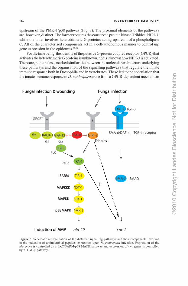
upstream of the PMK‑1/p38 pathway (Fig. 3). The proximal elements of the pathways are, however, distinct. The former requires the conserved protein kinase Tribbles, NIPI‑3, while the latter involves heterotrimeric G proteins acting upstream of a phospholipase C. All of the characterised components act in a cell‑autonomous manner to control nlp gene expression in the epidermis.35,82
For the time being, the identity of the putative G‑protein coupled receptor (GPCR) that activates the heterotrimeric G proteins is unknown, nor is it known how NIPI‑3 is activated. There are, nonetheless, marked similarities between the molecular architecture underlying these pathways and the organisation of the signalling pathways that regulate the innate immune response both in Drosophila and in vertebrates. These led to the speculation that the innate immune response to D. coniospora arose from a GPCR‑dependent mechanism
Figure 3. Schematic representation of the different signalling pathways and their components involved in the induction of antimicrobial peptides expression upon D. coniospora infection. Expression of the nlp genes is controlled by a PKC/SARM/p38 MAPK pathway and expression of cnc genes is controlled by a TGF‑b pathway.
.noitubirtsiD rof to
N .ecneicsoiB sednaL thgirypoC
1002©

117INNATE IMMUNITY IN C. ELEGANS
used to detect cellular damage, which was subsequently ameliorated by the addition of a pathogen‑specific detection mechanism.
Paracrine Regulation of cnc Gene Expression
Surprisingly, the cnc gene family is regulated in an entirely different fashion, as it is largely independent of the PMK‑1/p38 pathway. Induction of the cnc genes after infection requires the ligand DBL‑1/TGF‑b, produced by certain neuronal cells, which acts via its normal receptor SMA‑6/DAF‑4 expressed on epidermal cells (Fig. 3). The resultant signal is transduced by a noncanonical TGF‑b pathway that does not involve all three SMAD proteins, hitherto considered to be indispensable for TGF‑b signalling in C. elegans. Artificially altering the level of expression of the TGF‑b modulated the strength of cnc induction after infection but did not affect the basal level of cnc gene expression, suggesting that infection triggers the conversion of an inactive precursor into an active TGF‑b. This is reminiscent of the proteolytic activation of Spaetzle required for triggering the Toll pathway during the immune response in Drosophila. As the canonical TGF‑b pathway undoubtedly existed before the appearance of the cnc genes, this is another clear example of the co‑option and adaptation of a pre‑existing signalling pathway for use by the innate immune system.55
CONCLUSION
In the last 10 years, our knowledge of the way C. elegans defends itself against microbes has greatly expanded. The worm clearly shows specific immune responses to pathogens and their toxins. The signalling processes and effector molecules involved in this response have been elucidated to some extent. Interestingly, the main signalling pathways involved in the C. elegans immune response are conserved in other species, in part because they also play important developmental roles.
On the other hand, much remains to be learnt about the way the nematode recognizes pathogens and the receptors involved in this process, as well as the temporal and spatial dynamics of the downstream signalling processes. For the latter, techniques to visualise proteins and transcripts at the single molecule level within living cells must be improved.
It is important to note how deeply the defence mechanisms are embedded in the physiology of the organism. We described how important are several aspects of behaviour, digestion or stress resistance to defence. Other studies have shown connections with reproduction, where sterile mutants are more resistant to bacterial infection in a DAF‑16/FOXO dependent manner.83 The same is true for osmotic stress since several osmotic mutants are more resistant to fungal infection and some immune effectors genes are induced upon osmotic stress although apparently regulated by a dedicated pathway.58,84 Lastly, lipid synthesis has been shown to be required for the basal activity of the PMK‑1/p38 pathway that influences resistance against P. aeruginosa.71 Unbiased forward genetics screens combined with global functional genomic approaches will help to unravel the complex and intricate biology that underlies successful host defences in C. elegans.
Another powerful approach to understand more fully how the innate immune system works is to take advantage of the fact that pathogens can specifically interfere with the defence mechanisms of the host. For the moment there are only a few examples in
.noitubirtsiD rof to
N .ecneicsoiB sednaL thgirypoC
1002©
118 INVERTEBRATE IMMUNITY
C. elegans, such as the down‑regulation of several intestinal immune effectors through activation of DAF‑2 by P. aeruginosa which requires bacterial virulence factors controlled by the signalling molecule GacA.85 The study of these interactions will also teach us more about innate immune defence and the virulence strategies that pathogens have developed to escape host immunity.
ACKNOWLEDGEMENT
We thank J. Ewbank for insightful comments on the manuscript. Work in our laboratory is supported by institutional grants from INSERM and the CNRS and grants from the ANR and FRM. IE is supported by a Marie Curie fellowship.
REFERENCES
1. Barrière A, Felix MA. Isolation of C. elegans and related nematodes. The C. elegans Research Community: WormBook; 2006.
2. Fares H, Greenwald I. Genetic analysis of endocytosis in Caenorhabditis elegans: coelomocyte uptake defective mutants. Genetics 2001; 159(1):133‑45.
3. Schulenburg H, Ewbank JJ. The genetics of pathogen avoidance in Caenorhabditis elegans. Mol Microbiol 2007; 66(3):563‑70.
4. Pradel E, Zhang Y, Pujol N et al. Detection and avoidance of a natural product from the pathogenic bacterium Serratia marcescens by Caenorhabditis elegans. Proc Natl Acad Sci USA 104(7):2295‑300.
5. Pujol N, Link EM, Liu LX et al. A reverse genetic analysis of components of the Toll signalling pathway in Caenorhabditis elegans. Curr Biol 2001; 11(11):809‑21.
6. Remy JJ, Hobert O. An interneuronal chemoreceptor required for olfactory imprinting in C. elegans. Science (New York, NY) 2005; 309(5735):787‑90.
7. Zhang Y, Lu H, Bargmann CI. Pathogenic bacteria induce aversive olfactory learning in Caenorhabditis elegans. Nature 2005; 438(7065):179‑84.
8. Wes PD, Bargmann CI. C. elegans odour discrimination requires asymmetric diversity in olfactory neurons. Nature 2001; 410(6829):698‑701.
9. Chuang CF, Bargmann CI. A Toll‑interleukin 1 repeat protein at the synapse specifies asymmetric odorant receptor expression via ASK1 MAPKKK signaling. Genes Dev 2005; 19(2):270‑81.
10. Shivers RP, Kooistra T, Chu SW et al. Tissue‑specific activities of an immune signaling module regulate physiological responses to pathogenic and nutritional bacteria in C. elegans. Cell Host Microbe 2009; 6(4):321‑30.
11. Labrousse A, Chauvet S, Couillault C et al. Caenorhabditis elegans is a model host for Salmonella typhimurium. Curr Biol 2000; 10(23):1543‑5.
12. Kim DH, Feinbaum R, Alloing G et al. A conserved p38 MAP kinase pathway in Caenorhabditis elegans innate immunity. Science (New York, NY) 2002; 297(5581):623‑6.
13. Kurz CL, Chauvet S, Andres E et al. Virulence factors of the human opportunistic pathogen Serratia marcescens identified by in vivo screening. EMBO J 2003; 22(7):1451‑60.
14. Borgonie G, Claeys M, Leyns F et al. Effect of nematicidal Bacillus thuringiensis strains on free‑living nematodes. 1. Light microscopic observations, species and biological stage specificity and identification of resistant mutants of Caenorhabditis elegans. Fundam appl Nematol 1996; 19(4):391‑8.
15. Troemel ER, Felix MA, Whiteman NK et al. Microsporidia are natural intracellular parasites of the nematode Caenorhabditis elegans. PLoS Biol 2008; 6(12):2736‑52.
16. Jia K, Thomas C, Akbar M et al. Autophagy genes protect against Salmonella typhimurium infection and mediate insulin signaling‑regulated pathogen resistance. Proc Natl Acad Sci USA 2009; 106(34):14564‑9.
17. Hodgkin J, Kuwabara PE, Corneliussen B. A novel bacterial pathogen, Microbacterium nematophilum, induces morphological change in the nematode C. elegans. Curr Biol 2000; 10(24):1615‑8.
18. Muir RE, Tan MW. Leucobacter chromiireducens subsp. solipictus Exerts Virulence on Caenorhabditis elegans, Characterization of a Novel Host‑Pathogen Interaction. Appl Environ Microbiol 2008.
.noitubirtsiD rof to
N .ecneicsoiB sednaL thgirypoC
1002©
119INNATE IMMUNITY IN C. ELEGANS
19. Barron GL. Nematophagous destroying fungi. Topics in Mycobiology [serial on the Internet] 1977; 1.20. Jansson HB. Adhesion of conidia of Drechmeria coniospora to Caenorhabditis elegans wild type and
mutants. J Nematol 1994; 26:430‑5.21. Gallagher LA, Manoil C. Pseudomonas aeruginosa PAO1 kills Caenorhabditis elegans by cyanide poisoning.
J Bacteriol 2001; 183(21):6207‑14.22. Akira S, Uematsu S, Takeuchi O. Pathogen recognition and innate immunity. Cell 2006;
124(4):783‑801.23. Palm NW, Medzhitov R. Pattern recognition receptors and control of adaptive immunity. Immunol Rev
2009; 227(1):221‑33.24. Couillault C, Pujol N, Reboul J et al. TLR‑independent control of innate immunity in Caenorhabditis
elegans by the TIR domain adaptor protein TIR‑1, an ortholog of human SARM. Nature immunology 2004; 5:488‑94.
25. Tenor JL, Aballay A. A conserved Toll‑like receptor is required for Caenorhabditis elegans innate immunity. EMBO Rep 2008; 9(1):103‑9.
26. Powell JR, Kim DH, Ausubel FM. The G protein‑coupled receptor FSHR‑1 is required for the Caenorhabditis elegans innate immune response. Proc Natl Acad Sci USA 2009; 106(8):2782‑7.
27. Gordon S. Pattern recognition receptors. Doubling up for the innate immune response. Cell 2002; 111(7):927‑30.
28. Nicholas HR, Hodgkin J. Responses to infection and possible recognition strategies in the innate immune system of Caenorhabditis elegans. Mol Immunol 2004; 41(5):479‑93.
29. Means TK, Mylonakis E, Tampakakis E et al. Evolutionarily conserved recognition and innate immunity to fungal pathogens by the scavenger receptors SCARF1 and CD36. J Exp Med 2009.
30. Matzinger P. The danger model: a renewed sense of self. Science (New York, NY) 2002; 296(5566):301‑5.
31. Schulenburg H, Hoeppner MP, Weiner J 3rd et al. Specificity of the innate immune system and diversity of C‑type lectin domain (CTLD) proteins in the nematode Caenorhabditis elegans. Immunobiology 2008; 213(3‑4):237‑50.
32. Wong D, Bazopoulou D, Pujol N et al. Genome‑wide investigation reveals pathogen‑specific and shared signatures in the response of Caenorhabditis elegans to infection. Genome Biol 2007; 8(9):R194.
33. Sifri CD, Begun J, Ausubel FM et al. Caenorhabditis elegans as a model host for Staphylococcus aureus pathogenesis. Infect Immun 2003; 71(4):2208‑17.
34. Liberati NT, Fitzgerald KA, Kim DH et al. Requirement for a conserved Toll/interleukin‑1 resistance domain protein in the Caenorhabditis elegans immune response. Proc Natl Acad Sci USA 2004; 101(17):6593‑8.
35. Pujol N, Cypowyj S, Ziegler K et al. Distinct innate immune responses to infection and wounding in the C. elegans epidermis. Curr Biol 2008; 18(7):481‑9.
36. Begun J, Gaiani JM, Rohde H et al. Staphylococcal biofilm exopolysaccharide protects against Caenorhabditis elegans immune defenses. PLoS Pathog 2007; 3(4):e57.
37. Aballay A, Drenkard E, Hilbun LR et al. Caenorhabditis elegans innate immune response triggered by Salmonella enterica requires intact LPS and is mediated by a MAPK signaling pathway. Curr Biol 2003; 13(1):47‑52.
38. Huffman DL, Abrami L, Sasik R et al. Mitogen‑activated protein kinase pathways defend against bacterial pore‑forming toxins. Proc Natl Acad Sci USA 2004; 101(30):10995‑1000.
39. Nicholas HR, Hodgkin J. The ERK MAP kinase cascade mediates tail swelling and a protective response to rectal infection in C. elegans. Curr Biol 2004; 14(14):1256‑61.
40. Kim DH, Liberati NT, Mizuno T et al. Integration of Caenorhabditis elegans MAPK pathways mediating immunity and stress resistance by MEK‑1 MAPK kinase and VHP‑1 MAPK phosphatase. Proc Natl Acad Sci USA 2004; 101(30):10990‑4.
41. Lin K, Hsin H, Libina N et al. Regulation of the Caenorhabditis elegans longevity protein DAF‑16 by insulin/IGF‑1 and germline signaling. Nat Genet 2001; 28(2):139‑45.
42. Garsin DA, Villanueva JM, Begun J et al. Long‑lived C. elegans daf‑2 mutants are resistant to bacterial pathogens. Science (New York, NY) 2003; 300(5627):1921.
43. Evans EA, Chen WC, Tan MW. The DAF‑2 Insulin‑like signaling pathway independently regulates aging and immunity in C. elegans. Aging Cell 2008; 7(6):879‑93.
44. Murphy CT, McCarroll SA, Bargmann CI et al. Genes that act downstream of DAF‑16 to influence the lifespan of Caenorhabditis elegans. Nature 2003; 424(6946):277‑83.
45. Shapira M, Hamlin BJ, Rong J et al. A conserved role for a GATA transcription factor in regulating epithelial innate immune responses. Proc Natl Acad Sci USA 2006; 103(38):14086‑91.
46. Troemel ER, Chu SW, Reinke V et al. p38 MAPK regulates expression of immune response genes and contributes to longevity in C. elegans. PLoS Genetics 2006; 2(11):e183.
.noitubirtsiD rof to
N .ecneicsoiB sednaL thgirypoC
1002©

120 INVERTEBRATE IMMUNITY
47. Alper S, McBride SJ, Lackford B et al. Specificity and complexity of the Caenorhabditis elegans innate immune response. Mol Cell Biol 2007; 27(15):5544‑53.
48. Hasshoff M, Bohnisch C, Tonn D et al. The role of Caenorhabditis elegans insulin‑like signaling in the behavioral avoidance of pathogenic Bacillus thuringiensis. FASEB J 2007; 21(8):1801‑12.
49. Todd DJ, Lee AH, Glimcher LH. The endoplasmic reticulum stress response in immunity and autoimmunity. Nat Rev Immunol 2008; 8(9):663‑74.
50. Bischof LJ, Kao CY, Los FC et al. Activation of the unfolded protein response is required for defenses against bacterial pore‑forming toxin in vivo. PLoS Pathog 2008; 4(10):e1000176.
51. Haskins KA, Russell JF, Gaddis N et al. Unfolded protein response genes regulated by CED‑1 are required for Caenorhabditis elegans innate immunity. Dev Cell 2008; 15(1):87‑97.
52. Richardson CE, Kooistra T, Kim DH. An essential role for XBP‑1 in host protection against immune activation in C. elegans. Nature 2010; 463(6784):1092‑5.
53. Mochii M, Yoshida S, Morita K et al. Identification of transforming growth factor‑beta‑ regulated genes in Caenorhabditis elegans by differential hybridization of arrayed cDNAs. Proc Natl Acad Sci USA 1999; 96(26):15020‑5.
54. Mallo GV, Kurz CL, Couillault C et al. Inducible antibacterial defense system in C. elegans. Curr Biol 2002; 12(14):1209‑14.
55. Zugasti O, Ewbank JJ. Neuroimmune regulation of antimicrobial peptide expression by a noncanonical TGF‑beta signaling pathway in Caenorhabditis elegans epidermis. Nature immunology 2009; 10(3):249‑56.
56. Hashimoto Y, Ookuma S, Nishida E. Lifespan extension by suppression of autophagy genes in Caenorhabditis elegans. Genes Cells 2009; 14(6):717‑26.
57. Kerry S, Tekippe M, Gaddis NC et al. GATA transcription factor required for immunity to bacterial and fungal pathogens. PLoS ONE 2006; 1:e77.
58. Pujol N, Zugasti O, Wong D et al. Anti‑fungal innate immunity in C. elegans is enhanced by evolutionary diversification of antimicrobial peptides. PLoS Pathog 2008; 4(7):e1000105.
59. Rohlfing AK, Miteva Y, Hannenhalli S et al. Genetic and physiological activation of osmosensitive gene expression mimics transcriptional signatures of pathogen infection in C. elegans. PLoS One 2010; 5(2):e9010.
60. Singh V, Aballay A. Heat‑shock transcription factor (HSF)‑1 pathway required for Caenorhabditis elegans immunity. Proc Natl Acad Sci USA 2006; 103(35):13092‑7.
61. Irazoqui JE, Ng A, Xavier RJ et al. Role for beta‑catenin and HOX transcription factors in Caenorhabditis elegans and mammalian host epithelial‑pathogen interactions. Proc Natl Acad Sci USA 2008; 105(45):17469‑74.
62. Chisholm A. Control of cell fate in the tail region of C. elegans by the gene egl‑5. Development 1991; 111(4):921‑32.
63. Nicholas HR, Hodgkin J. The C. elegans Hox gene egl‑5 is required for correct development of the hermaphrodite hindgut and for the response to rectal infection by Microbacterium nematophilum. Dev Biol 2009; 329(1):16‑24.
64. Gravato‑Nobre MJ, Nicholas HR, Nijland R et al. Multiple genes affect sensitivity of Caenorhabditis elegans to the bacterial pathogen Microbacterium nematophilum. Genetics 2005; 171(3):1033‑45.
65. Estes KA, Dunbar TL, Powell JR et al. bZIP transcription factor zip‑2 mediates an early response to Pseudomonas aeruginosa infection in Caenorhabditis elegans. Proc Natl Acad Sci USA 2010; 107(5):2153‑8.
66. Kato Y, Aizawa T, Hoshino H et al. abf‑1 and abf‑2, ASABF‑type antimicrobial peptide genes in Caenorhabditis elegans. Biochem J 2002; 361(Pt 2):221‑30.
67. Alegado RA, Tan MW. Resistance to antimicrobial peptides contributes to persistence of Salmonella typhimurium in the C. elegans intestine. Cell Microbiol 2008; 10(6):1259‑73.
68. Roeder T, Stanisak M, Gelhaus C et al. Caenopores are antimicrobial peptides in the nematode Caenorhabditis elegans instrumental in nutrition and immunity. Developmental and comparative immunology 2009.
69. Banyai L, Patthy L. Amoebapore homologs of Caenorhabditis elegans. Biochim Biophys Acta 1998; 1429(1):259‑64.
70. Schulenburg H, Boehnisch C. Diversification and adaptive sequence evolution of Caenorhabditis lysozymes (Nematoda: Rhabditidae). BMC Evol Biol 2008; 8:114.
71. Nandakumar M, Tan MW. Gamma‑linolenic and stearidonic acids are required for basal immunity in Caenorhabditis elegans through their effects on p38 MAP kinase activity. PLoS Genet 2008; 4(11):e1000273.
72. O’Rourke D, Baban D, Demidova M et al. Genomic clusters, putative pathogen recognition molecules and antimicrobial genes are induced by infection of C. elegans with M. nematophilum. Genome Res 2006; 16(8):1005‑16.
.noitubirtsiD rof to
N .ecneicsoiB sednaL thgirypoC
1002©

121INNATE IMMUNITY IN C. ELEGANS
73. Ideo H, Fukushima K, Gengyo‑Ando K et al. A Caenorhabditis elegans glycolipid‑binding galectin functions in host defense against bacterial infection. J Biol Chem 2009; 284(39):26493‑501.
74. Chavez V, Mohri‑Shiomi A, Maadani A et al. Oxidative stress enzymes are required for DAF‑16‑mediated immunity due to generation of reactive oxygen species by Caenorhabditis elegans. Genetics 2007; 176(3):1567‑77.
75. Chavez V, Mohri‑Shiomi A, Garsin DA. Ce‑Duox1/BLI‑3 generates reactive oxygen species as a protective innate immune mechanism in Caenorhabditis elegans. Infect Immun 2009; 77(11):4983‑9.
76. Kawli T, Tan MW. Neuroendocrine signals modulate the innate immunity of Caenorhabditis elegans through insulin signaling. Nature immunology 2008; 9(12):1415‑24.
77. Styer KL, Singh V, Macosko E et al. Innate immunity in Caenorhabditis elegans is regulated by neurons expressing NPR‑1/GPCR. Science (New York, NY) 2008; 322(5900):460‑4.
78. Reddy KC, Andersen EC, Kruglyak L et al. A polymorphism in npr‑1 is a behavioral determinant of pathogen susceptibility in C. elegans. Science (New York, NY) 2009; 323(5912):382‑4.
79. Griffitts JS, Haslam SM, Yang T et al. Glycolipids as receptors for Bacillus thuringiensis crystal toxin. Science (New York, NY.) 2005; 307(5711):922‑5.
80. Bellier A, Chen CS, Kao CY et al. Hypoxia and the hypoxic response pathway protect against pore‑forming toxins in C. elegans. PLoS Pathog 2009; 5(12):e1000689.
81. Tong A, Lynn G, Ngo V et al. Negative regulation of Caenorhabditis elegans epidermal damage responses by death‑associated protein kinase. Proc Natl Acad Sci USA 2009; 106(5):1457‑61.
82. Ziegler K, Kurz CL, Cypowyj S et al. Antifungal innate immunity in C. elegans: PKCdelta links G protein signaling and a conserved p38 MAPK cascade. Cell Host Microbe 2009; 5(4):341‑52.
83. Miyata S, Begun J, Troemel ER et al. DAF‑16‑dependent suppression of immunity during reproduction in Caenorhabditis elegans. Genetics 2008; 178(2):903‑18.
84. Lee KZ, Kniazeva M, Han M et al. The fatty acid synthase fasn‑1 acts upstream of WNK and Ste20/GCK‑VI kinases to modulate antimicrobial peptide expression in C. elegans epidermis. Virulence 2010; 1(3).
85. Evans EA, Kawli T, Tan MW. Pseudomonas aeruginosa suppresses host immunity by activating the DAF‑2 insulin‑like signaling pathway in Caenorhabditis elegans. PLoS Pathog 2008; 4(10):e1000175.
86. Partridge FA, Gravato‑Nobre MJ, Hodgkin J. Signal transduction pathways that function in both development and innate immunity. Dev Dyn 2010.
87. Ren M, Feng H, Fu Y et al. Protein kinase D (DKF‑2), a diacylglycerol effector, is an essential regulator of C. elegans innate immunity. Immunity 2009; 30(4):521‑32.
88. Kurz CL, Shapira M, Chen K et al. Caenorhabditis elegans pgp‑5 is involved in resistance to bacterial infection and heavy metal and its regulation requires TIR‑1 and a p38 map kinase cascade. Biochem Biophys Res Commun 2007; 363(2):438‑43.
89. Yook K, Hodgkin J. Mos1 Mutagenesis Reveals a Diversity of Mechanisms Affecting Response of Caenorhabditis elegans to the Bacterial Pathogen Microbacterium nematophilum. Genetics 2007; 175(2):681‑97.
.noitubirtsiD rof to
N .ecneicsoiB sednaL thgirypoC
1002©