Embed Size (px)
Citation preview

Chapter 5 - Subphylum Kinetoplasta
Trypanosomes and their kin

Taxonomy of Protozoans from Chapter 5
Phylum Euglenozoa
Subphylum Kinetoplasta (formerly an Order)
Class Trypanosomatidea
Order Trypanosomatida
Family Trypanosomatidae
Genera: Trypanosoma, Leishmania
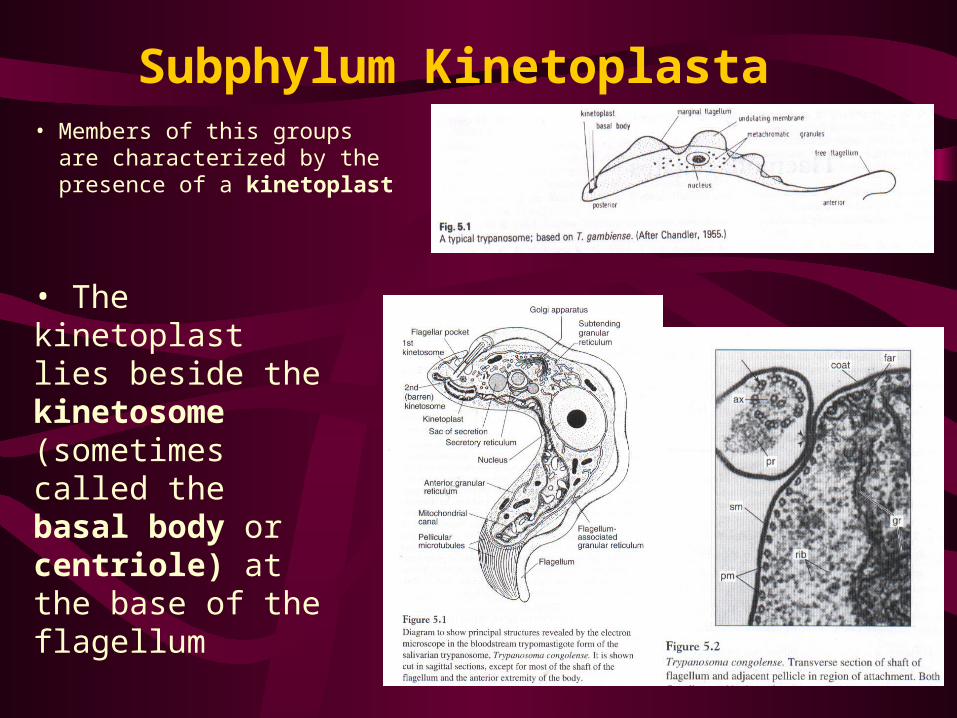
Subphylum Kinetoplasta • Members of this groups are
characterized by the presence of a kinetoplast
• The kinetoplast lies beside the kinetosome (sometimes called the basal body or centriole) at the base of the flagellum

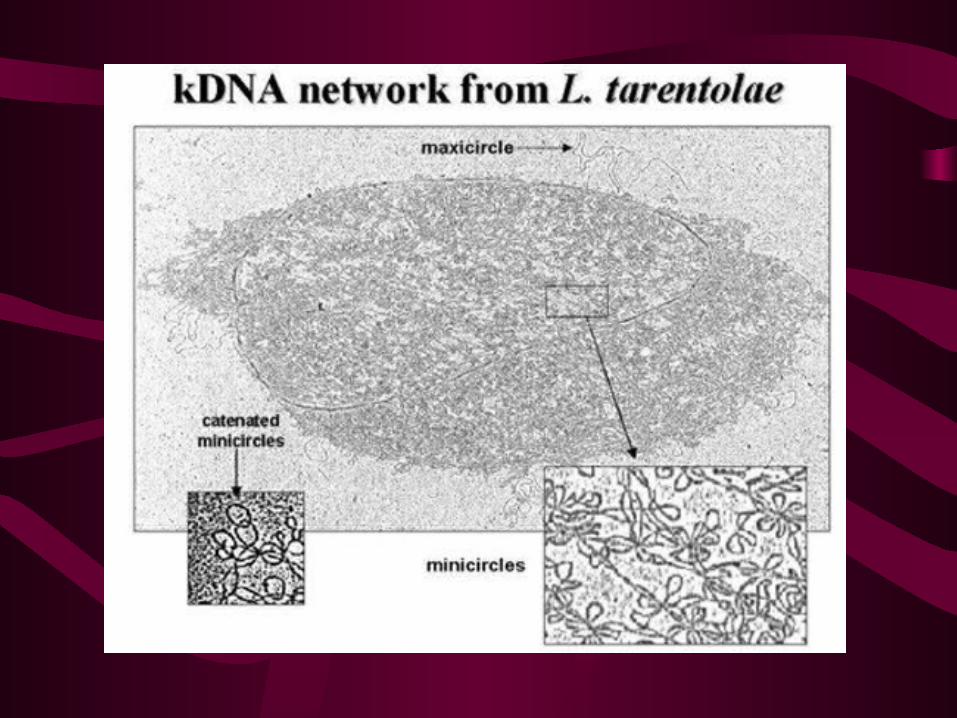
The Kinetoplast-Mitochondrion
• Leishmania and other trypanosomatids have a single mitochondrion that contains a large mass of mitochondrial DNA known as kinetoplast DNA. • The kDNA consists of a giant network of catenated minicircles and maxicircles. There are approximately 10,000 minicircles and 50 maxicircles per network. • This is one of the most unusual DNA structures in nature.

Family Trypanosomatidae
• Typically referred to as the hemoflagellates
• Single nucleus; elongate with a protruding flagellum or more rounded with a nonprotruding flagellum
• 8 body types (we will study 4 major ones)– They vary in size, body shape, location of the flagellum,
location of the kinetoplast, and other structures

Types of Trypanosomatids1. Promastigote - elongated form with
antenuclear kinetoplast; flagellum arising near it and emerging from the anterior end of body; e.g. Leptomonas
2. Epimastigote - elongated form with a juxtanuclear kinetoplast; flagellum arising near it and emerging from the side of the body to run along short undulating membrane; e.g. Blastocrithidia and some Trypanosoma

Types of Trypanosomatids con’t3. Trypomastigote - the “true”
trypanosome type; postnuclear
kinetosplast; flagellum arising
near it to run along a long
undulating membrane
4. Amastigote - rounded or oval forms devoid of external flagellum; e.g. Leishmania

Genus Trypanosoma • Life cycle usually includes both an invertebrate and a vertebrate host
(=heteroxenous life cycle)
• Usually found in the blood and tissue fluids of vertebrate hosts; but, some of the more popular pathogenic forms (e.g. T. cruzi) occur intracellularly
• They typically occur in the digestive tracts of invertebrates, especially arthropods
• Divided into 2 main groups or “sections”:– Salivaria - develop in the anterior part of the digestive tract of insect vector
(=anterior station); infect new hosts as a consequence of insect biting
– Stercoraria - develop in the hindgut of insect vector (=posterior station); leave insects with the feces and infect new hosts through the skin or mucous membranes or by lesions made by vector bites

Section Salivaria, Type Example: Trypanosoma brucei
• Widely distributed throughout Africa especially between latitude 15oN and 25o S; strongly dictated by the distribution of the insect vectors (e.g. tsetse flies of the genus Glossina)
• T. brucei brucei is primarily a parasite of native ruminants such as antelopes; also occurs in a variety of livestock (e.g. sheep, goats, horses, mules, donkeys, and camels)• It is extremely pathogenic to these animals and causes the disease known as nagana; humans are not susceptible
• The vectors for T. brucei are tsetse flies

Section Salivaria con’t
T. b. gambiense and T. b. rhodesiense cause African Sleeping Sickness
• T. b. gambiense is endemic to west and central portions of Africa
• It causes a chronic form of sleeping sickness
• Vectors include: Glossina palpalis and G. tachinoides
• T. b. rhodesiense occurs in central and east central Africa
• It causes a more acute form of sleeping sickness
• Vectors are the same species of tsetse flies used by T. brucei:
G. morsitans, G. pallidipes and G. swynnertoni)

Generalized Life Cycle
• The life cycle of T. b. brucei is highly polymorphic in its vertebrate host

Generalized Life Cycle1. Infected tsetse fly takes a blood meal, injecting metacyclic trypomastigotes
into its host
2. Metacyclic trypomastigotes transform into slender trypomastigotes,
3. These forms multiply and spread rapidly within the host, eventually migrating to the cerebralspinal fluid

8. Epimastigotes undergo several generations of asexual reproduction in the salivary glands and eventually transform into metacyclic trypomastigotes; the infective stage in vertebrates hosts
Generalized Life Cycle cont.
4. and 5. Trypomastigotes are ingested by the tsetse flies when they take another blood meal from an infected mammalian host
6. These transform into procyclic trypomastigotes in the insect’s midgut; here they undergo asexual reproduction7. Procyclic forms migrate forward to the foregut and transform into epimastigotes

Pathogenesis
• T. brucei primarily invade the blood, lymph, and spinal fluid
• The course of T. brucei infections depend on the susceptibility of the animals involved
• Horses, mules, and even dogs are acute sufferers and can die within 2 weeks of infection
• Symptoms of the infection include: anemia, edema and fever
• Cattle suffer the same kinds of symptoms but often live a few extra months
• Pigs can recover from the infection

Pathogenesis cont.• In humans, T. b. gambiense causes the Gambian or slow, chronic
form of sleeping sickness• It primarily invades the blood, nervous, and lymphatic system; may
also be an invasion of the dermis with the production of chancre soars• 1-2 week incubation period followed by fever, chills, headache, and
loss of appetite• Later invasion and enlargement of the spleen, liver, and lymph nodes• Invasion of nervous system and organs leads to weakness, anemia,
apathy, headache, disturbed vision, etc.• Coma, emaciation and death complete the course of the disease,
which may last for several years
Pathogenesis cont.
• T. b. rhodesiense causes the Rhodesian or quick, acute form of human sleeping sickness; usually results in death within a year
• Rhodesian trypanosomiasis rarely, if ever, causes the symptoms normally associated with sleeping sickness
• Compared with Gambian sleeping sickness, there is less invasion of the CNS, but the lymph nodes become swollen and congested
• The effects on the cardiac system are more severe
Immunology• Infections are often highly variable for the life of the organism,
attributable to the ability of these parasites to change the chemical composition of their surface coat (glycocalyx)
• They produce a parade of successive variant antigenic types (VATs) in the vertebrate host
• There is a period of remission during which the host produces antibodies that are capable of destroying the parasite
• However, the parasite counters this by producing a new surface glycoprotein for which the host has not yet developed antibodies
• Once this is accomplished the parasite can begin to multiply rapidly
Immunology cont.
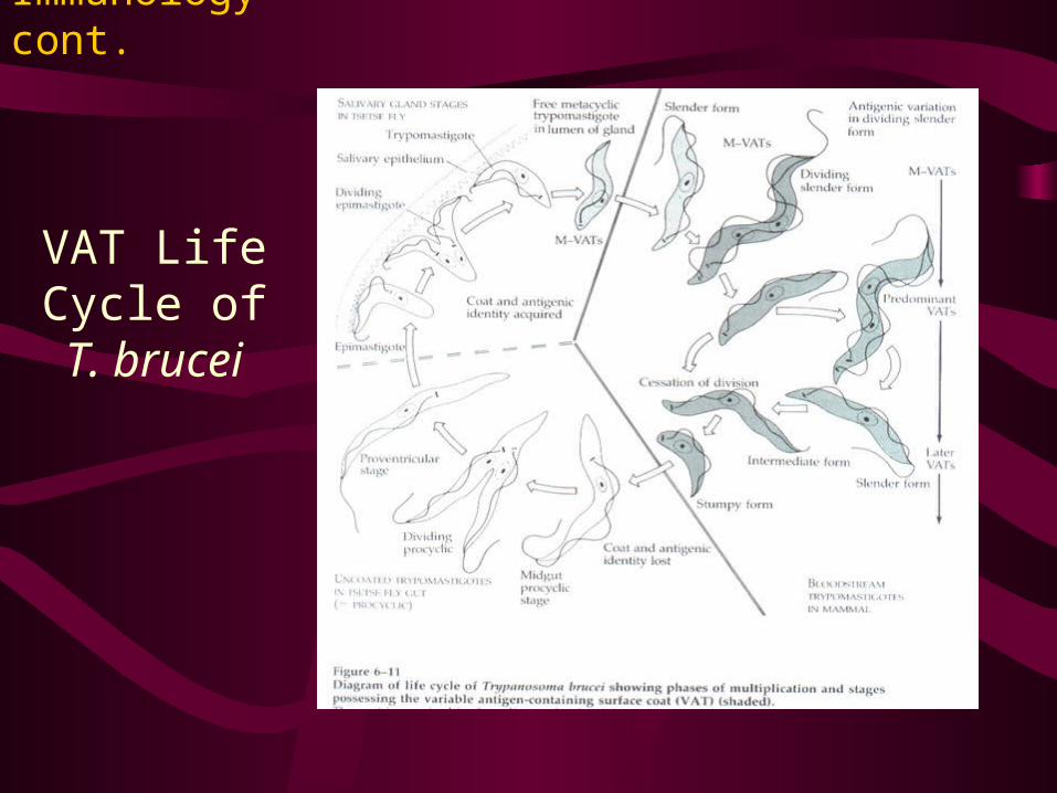
VAT Life Cycle of T.
brucei

Immunology cont.
• The clinical course of the infection is characterized by an increase in trypanosome numbers, followed by a crash or sudden decrease in the population which is repeated over a number of cycles.
• Again, remission coincides with an increase in host protective antibody against specific surface expressed antigens on the trypanosome, located in the surface coat or glycocalyx. The cyclic nature appears to occur due to an almost endless change in the Variant Antigen Type (VAT).
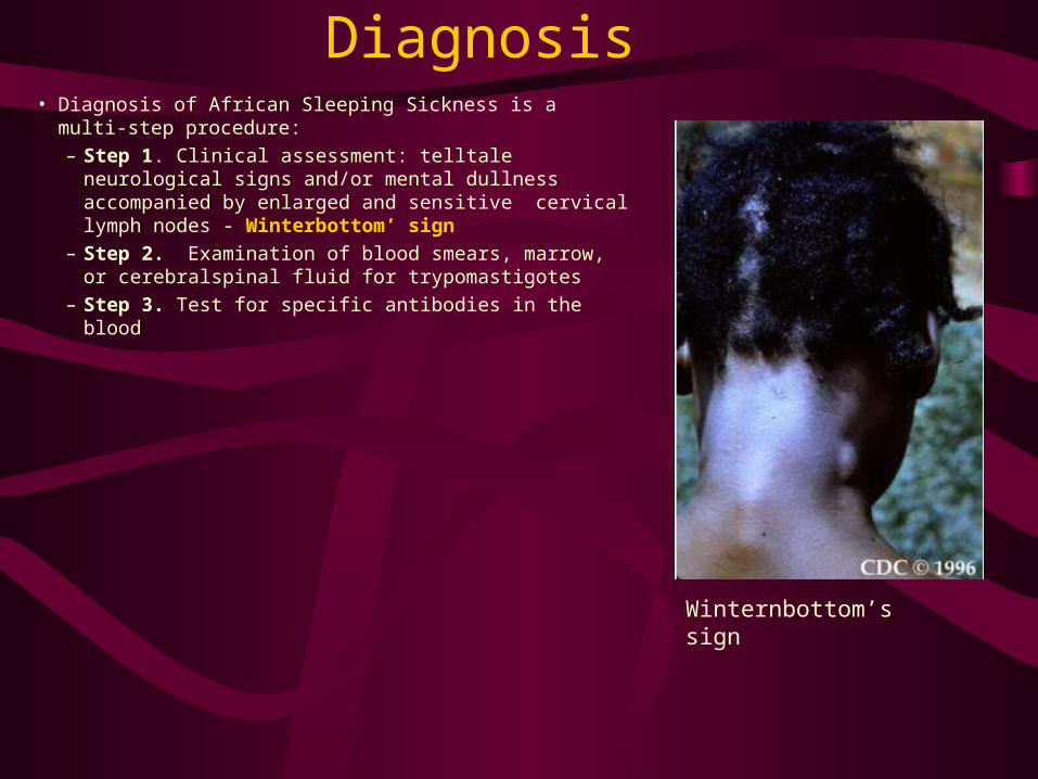
Diagnosis• Diagnosis of African Sleeping Sickness is a multi-step
procedure:
– Step 1. Clinical assessment: telltale neurological signs and/or mental dullness accompanied by enlarged and sensitive cervical lymph nodes - Winterbottom’ sign
– Step 2. Examination of blood smears, marrow, or cerebralspinal fluid for trypomastigotes
– Step 3. Test for specific antibodies in the blood
Winternbottom’s sign

Treatment
• Trypan blue and trypan red
• Suramin sodium
• Melarsoprol
• Difluoromethylornithine

Section Stercoraria, Type Example: Trypanosoma cruzi
• T. cruzi is the causative agent of Chagas’ disease
• T. cruzi infections are prevalent throughout South America and Latin America, affecting an estimated 12-19 million people

Life Cycle• Reduvid bug takes a blood meal and releases trypomastigotes in its feces near the site of the bite wound.
Trypomastigotes enter the host through the wound or through intact mucosal membranes, such as the conjunctiva .
• Inside the host, the trypomastigotes invade cells, where they differentiate into intracellular amastigotes
• Amastigotes undergo asexual reproduction and differentiate into trypomastigotes, and then are released into the circulation as trypomastigotes
• Trypomastigotes infect cells from a variety of tissues and transform into intracellular amastigotes in new infection sites.

Life Cycle cont.
• The “kissing” bug becomes infected by feeding on human or animal blood that contains circulating parasites .
• The ingested trypomastigotes transform into epimastigotes in the vector’s midgut• The parasites multiply and differentiate in the midgut and differentiate into infective metacyclic trypomastigotes in the hindgut .

Pathology• Upon introduction into the human, the parasites invade
cells of the subcutaneous tissue at the site of the infection, causing an acute inflammatory reaction or
swelling often called a chagoma • When they enter through the conjunctiva of the eye, they
cause a unilateral edema of the eyelid and conjunctivitis, a syndrome known as Romana’s sign
• In acute cases, about 1-3 weeks after infection, fever, headache, malaise and prostration may develop• Chronic symptoms include: dysfunction of the peripheral and CNS, myocardial damage, and gastrointestinal problems
Romana’s sign

Diagnosis
• Examination of fresh blood within the first month or two following infection may reveal T. cruzi trypomastigotes
• Direct agglutination tests with the IgM serum fraction • Xenodiagnosis

Genus Leishmania: Life Cycle• Upon biting a host, the sand fly
(G. Phlebotomus) vector, ingests amastigotes from infected host
• These migrate to the hindgut and transform to promastigotes, which undergo asexual reproduction
Phlebotomus sp.

Genus Leishmania: Life Cycle cont.
• Promastigotes migrate forward to the esophagus and are inoculated into a new host when vector bites another victim • Inoculated promastigotes are ingested by macrophages and then undergo transformation to become amastigotes• Following asexual reproduction, infected host cells rupture and a large number of amastigotes are released• These are engulfed by other phagocytic cells, spreading the infection
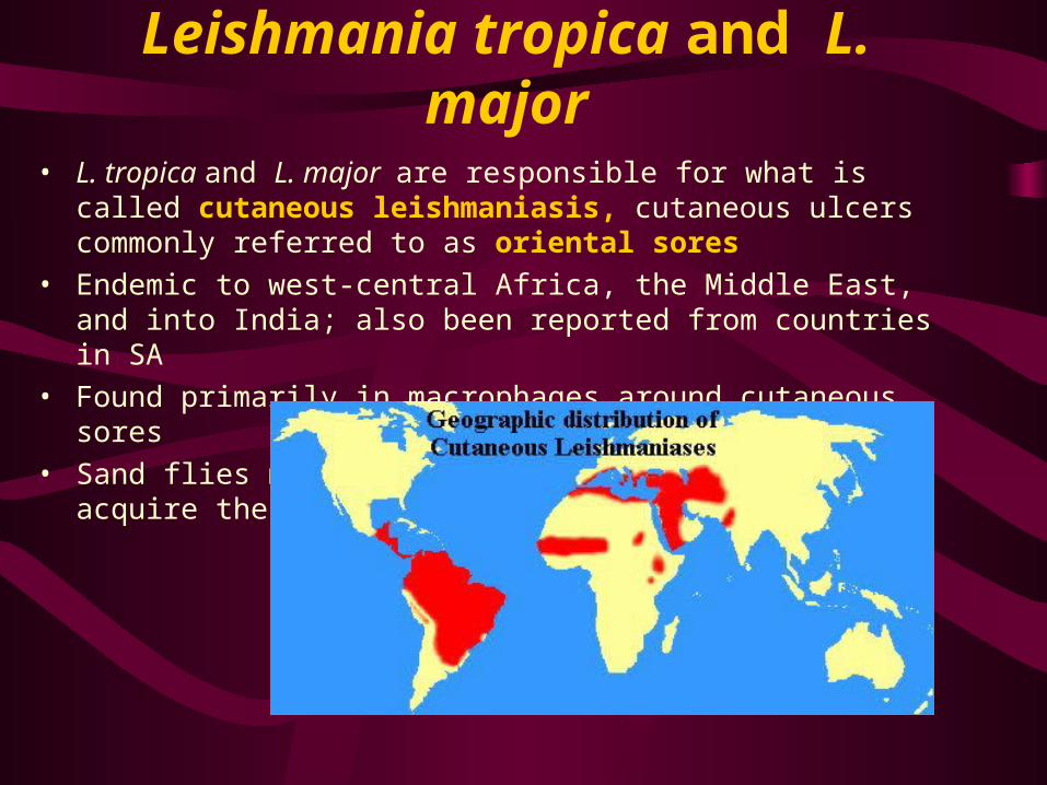
Leishmania tropica and L. major
• L. tropica and L. major are responsible for what is called cutaneous leishmaniasis, cutaneous ulcers commonly referred to as oriental sores
• Endemic to west-central Africa, the Middle East, and into India; also been reported from countries in SA
• Found primarily in macrophages around cutaneous sores
• Sand flies must feed at these sites in order to acquire the infective amastigotes
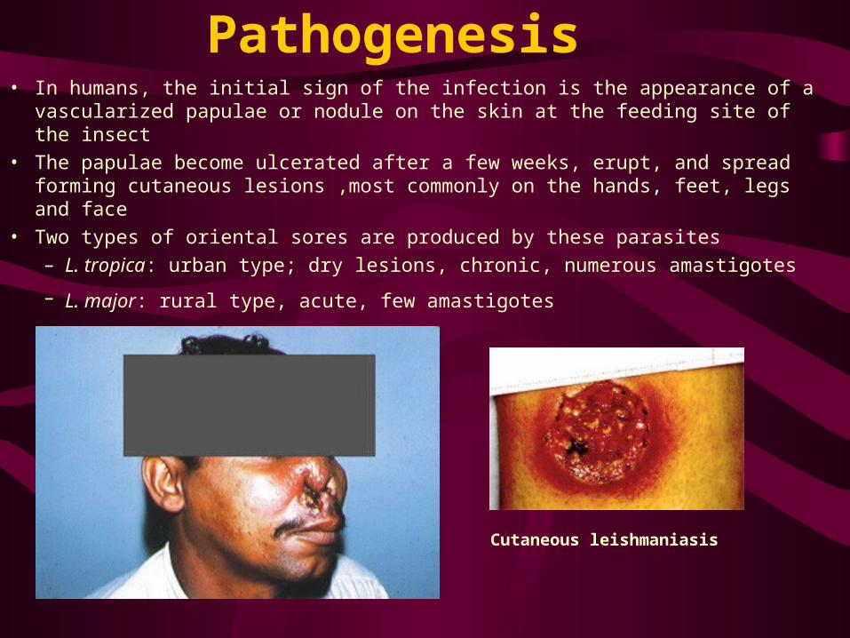
Pathogenesis• In humans, the initial sign of the infection is the appearance of a vascularized
papulae or nodule on the skin at the feeding site of the insect
• The papulae become ulcerated after a few weeks, erupt, and spread forming cutaneous lesions ,most commonly on the hands, feet, legs and face
• Two types of oriental sores are produced by these parasites
– L. tropica: urban type; dry lesions, chronic, numerous amastigotes
– L. major: rural type, acute, few amastigotes
Cutaneous leishmaniasis
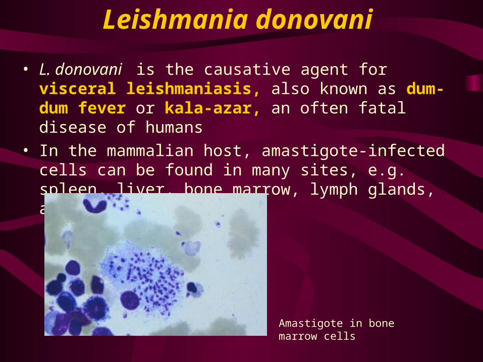
Leishmania donovani
• L. donovani is the causative agent for visceral leishmaniasis, also known as dum-dum fever or kala-azar, an often fatal disease of humans
• In the mammalian host, amastigote-infected cells can be found in many sites, e.g. spleen, liver, bone marrow, lymph glands, and intestinal mucosa
Amastigote in bone marrow cells
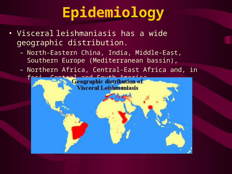
Epidemiology• Visceral leishmaniasis has a wide geographic distribution.
– North-Eastern China, India, Middle-East, Southern Europe (Mediterranean bassin),
– Northern Africa, Central-East Africa and, in foci, Central and South America

Pathogenesis• The disease typically starts off slowly with fever, chills, and
general anemia
• Since leishmaniasis is primarily a disease of the reticulo-endothelial system, replacement of infected cells produces hyperplasia and consequent enlargement of the visceral organs associated with the system (e.g., spleen and liver)
• A concomitant decrease in RBC and WBC production results in anemia and leukopenia, facilitating secondary bacterial infection
• In areas of India, a post-kala-azar dermal leishmanoid may develop in which numerous parasite-laden nodules appear in the skin
Post-kala-azar dermal leishmaniasis
Symptoms of visceral leishmaniasis