Embed Size (px)
Citation preview

Chapter 5
Ground Rules of
Metabolism Sections 6-10

5.6 Cofactors in Metabolic Pathways
• Most enzymes require cofactors
• Energy in ATP drives many endergonic reactions

Table 5-1 p86
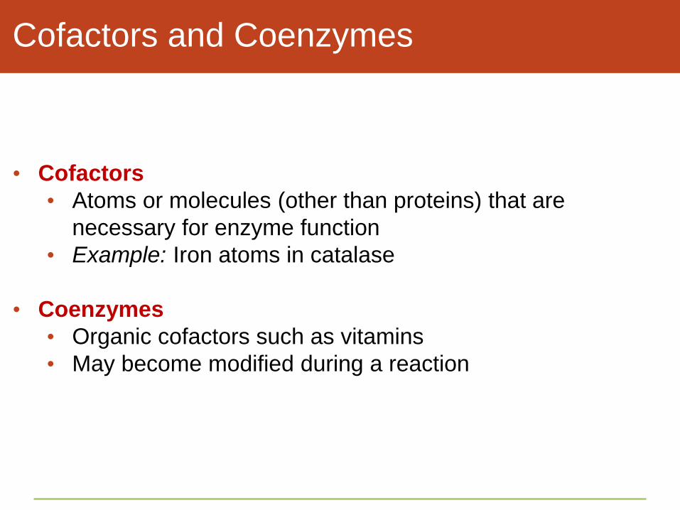
Cofactors and Coenzymes
• Cofactors
• Atoms or molecules (other than proteins) that are
necessary for enzyme function
• Example: Iron atoms in catalase
• Coenzymes
• Organic cofactors such as vitamins
• May become modified during a reaction

Catalase and Cofactors
• Catalase is an antioxidant that neutralizes free radicals
(atoms or molecules with unpaired electrons that attack
biological molecules)
• Catalase has four hemes (small organic compound with an
iron atom at its center)
• Catalase works by holding a substrate molecule close to one
of its iron atoms (cofactors)
• Iron pulls on the substrate’s electrons, bringing on the
transition state

Heme

ATP—A Special Coenzyme
• ATP (adenosine triphosphate)
• A nucleotide with three phosphate groups
• Transfers a phosphate group and energy to other
molecules
• Phosphorylation
• A phosphate-group transfer
• ADP binds phosphate in an endergonic reaction to
replenish ATP (ATP/ADP cycle)

Figure 5-18 p87
adenine
three phosphate
groups
ribose
adenine
ribose
AMP ADP
energy in energy out
ADP + phosphate
ATP
A
B
C

Coupled Reactions

Take-Home Message:
How do cofactors work?
• Cofactors associate with enzymes and assist their function.
• Metal ions stabilize the structure of many enzymes. They also
participate in some enzymatic reactions by donating or
accepting electrons
• Many coenzymes carry chemical groups, atoms, or electrons
from one reaction to another
• The formation of ATP from ADP is an endergonic reaction;
ADP forms again when a phosphate group is transferred from
ATP to another molecule – energy from such transfers drives
cellular work

5.7 A Closer Look at Cell Membranes
• A membrane is a continuous, selectively permeable barrier
• A cell membrane is organized as a lipid bilayer with many
proteins embedded in it and attached to its surfaces

Membrane Lipids
• Phospholipid molecules in the plasma membrane have two
parts
• Hydrophilic heads interact with water molecules
• Hydrophobic tails interact with each other, forming a
barrier to hydrophilic molecules

The Fluid Mosaic Model
• Fluid mosaic model
• Describes the organization of cell membranes
• Phospholipids drift and move like a fluid
• The bilayer is a mosaic mixture of phospholipids, steroids,
proteins, and other molecules

Cell Membrane Organization
one layer
of lipids
one layer
of lipids

Membrane Proteins
• Cell membrane function begins with the many proteins
associated with the lipid bilayer
• Peripheral membrane proteins temporarily attach to the lipid
bilayer’s surfaces by interactions with lipids or other proteins
• Integral membrane proteins permanently attach to a bilayer

Types of Membrane Proteins
• Each type of protein in a membrane has a special function
• Adhesion proteins
• Recognition proteins
• Receptor proteins
• Enzymes
• Transport proteins (active and passive)

Types of Membrane Proteins
B Recognition proteins
such as this MHC
molecule tag a cell as
belonging to one’s own
body.
c Receptor proteins such
as this B cell receptor bind
substances outside the
cell. B cell receptors help
the body eliminate toxins
and infectious agents.
D Transport proteins
bind to molecules on
one side of the
membrane, and
release them on the
other side. This one
transports glucose.
E This transport
protein, an ATP
synthase, makes
ATP when
hydrogen ions flow
through its interior.
Extracellular
Fluid
Lipid
bilayer
Cytoplasm

ANIMATED FIGURE: Cell membranes
To play movie you must be in Slide Show Mode
PC Users: Please wait for content to load, then click to play
Mac Users: CLICK HERE

Table 5-2 p88

Take-Home Message:
What is a cell membrane?
• The structural foundation of all cell membranes is the lipid
bilayer
• Adhesion proteins, recognition proteins, transport proteins,
receptors, and enzymes embedded in or associated with the
lipid bilayer impart functionality to a cell membrane

ANIMATION: Lipid bilayer organization
To play movie you must be in Slide Show Mode
PC Users: Please wait for content to load, then click to play
Mac Users: CLICK HERE

5.8 Diffusion and Membranes
• Ions and molecules tend to move spontaneously from regions
of higher to lower concentration
• Water diffuses across cell membranes by osmosis

Diffusion
• Diffusion
• The net movement of molecules down a concentration
gradient
• Moves substances into, through, and out of cells
• A substance diffuses in a direction set by its own
concentration gradient, not by the gradients of other
solutes around it

Diffusion

The Rate of Diffusion
• Rate of diffusion depends on five factors
• Size
• Temperature
• Steepness of the concentration gradient
• Charge
• Pressure

Concentration Gradients
• Concentration
• The number of molecules (or ions) of substance per unit
volume of fluid
• Concentration gradient
• The difference in concentration between two adjacent
regions
• Molecules move from a region of higher concentration to
one of lower concentration
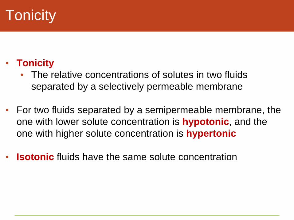
Tonicity
• Tonicity
• The relative concentrations of solutes in two fluids
separated by a selectively permeable membrane
• For two fluids separated by a semipermeable membrane, the
one with lower solute concentration is hypotonic, and the
one with higher solute concentration is hypertonic
• Isotonic fluids have the same solute concentration

Osmosis
• Osmosis
• The movement of water down its concentration gradient –
through a selectively permeable membrane from a region
of lower solute concentration to a region of higher solute
concentration
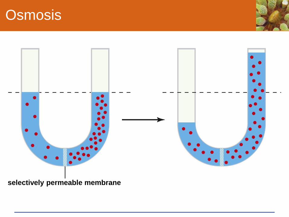
Osmosis
selectively permeable membrane

Membrane Permeability
• Selective permeability
• The ability of a cell membrane to control which substances
and how much of them enter or leave the cell
• Allows the cell to maintain a difference between its internal
environment and extracellular fluid
• Supplies the cell with nutrients, removes wastes, and
maintains volume and pH

Selective Permeability of Lipid Bilayers
gases glucose and
other polar
molecules;
ions
lipid
bilayer water

Effects of Fluid Pressure
• Hydrostatic pressure (turgor)
• The pressure exerted by a volume of fluid against a
surrounding structure (membrane, tube, or cell wall) which
resists volume change
• Osmotic pressure
• The amount of hydrostatic pressure that can stop water
from diffusing into cytoplasmic fluid or other hypertonic
solutions

Effects of Fluid Pressure

Take-Home Message: What influences the
movement of ions and molecules?
• Molecules or ions tend to diffuse into an adjoining region of
fluid in which they are not as concentrated
• he steepness of a concentration gradient as well as
temperature, molecular size, charge, and pressure affect the
rate of diffusion
• Osmosis is a net diffusion of water between two fluids that
differ in water concentration and are separated by a
selectively permeable membrane
• Fluid pressure that a solution exerts against a membrane or
wall influences the osmotic movement of water

5.9 Membrane Transport Mechanisms
• Many types of molecules and ions can cross a lipid bilayer
only with the help of transport proteins

How Substances Cross Membranes
• Gases and nonpolar molecules diffuse freely across a lipid
bilayer
• Ions and large polar molecules require other mechanisms to
cross the cell membrane
• Passive transport
• Active transport
• Endocytosis and exocytosis

Passive Transport
• Passive transport (facilitated diffusion)
• Requires no energy input
• A passive transport protein allows a specific solute (such
as glucose) to follow its concentration gradient across a
membrane
• A gated passive transporter changes shape when a
specific molecule binds to it

Passive Transport of Glucose

Active Transport
• Active transport
• Requires energy input (usually ATP)
• Moves a solute against its concentration gradient, to the
concentrated side of the membrane
• Calcium pumps
• Active transporters move calcium ions across muscle cell
membranes into the sarcoplasmic reticulum

Active Transport: Calcium Pump
Extracellular Fluid
Cytoplasm

Active Transport: Calcium Pump

Active Transport: Calcium Pump

ANIMATED FIGURE: Active transport
To play movie you must be in Slide Show Mode
PC Users: Please wait for content to load, then click to play
Mac Users: CLICK HERE

Cotransport
• Cotransporter
• An active transport protein that moves two substances
across a membrane at the same time
• Example: The sodium-potassium pump moves Na+ out of
the cell and K+ into the cell

Cotransport: Sodium-Potassium Pump
Extracellular Fluid
Cytoplasm
ADP

Take-Home Message: How do
molecules or ions cross a cell membrane?
• Transport proteins help specific molecules or ions to cross
cell membranes
• In passive transport, a solute binds to a protein that releases
it on the opposite side of the membrane; he movement is
driven by a concentration gradient
• In active transport, a transport protein pumps a solute across
a membrane, against its concentration gradient; the
movement is driven by an energy input, such as ATP

ANIMATED FIGURE: Passive transport
To play movie you must be in Slide Show Mode
PC Users: Please wait for content to load, then click to play
Mac Users: CLICK HERE

5.10 Membrane Trafficking
• By processes of endocytosis and exocytosis, cells take in and
expel particles that are too big for transport proteins, as well
as substances in bulk
• Requires formation and movement of vesicles formed from
membranes, involving motor proteins and ATP

Exocytosis and Endocytosis
• Exocytosis
• The fusion of a vesicle with the cell membrane, releasing
its contents to the surroundings
• Endocytosis
• The formation of a vesicle from cell membrane, enclosing
materials near the cell surface and bringing them into the
cell

F Some vesicles and their contents are delivered to lysosomes.
lysosome
B The pits sink inward and become endocytic vesicles.
C Vesicle contents are sorted.
Exocytosis
D Many of the sorted molecules cycle to the plasma membrane.
E Some vesicles are routed to the nuclear envelope or ER membrane. Others fuse with Golgi bodies.
Golgi
Endocytosis
A Molecules get concentrated inside coated pits at the plasma membrane.
coated pit
Stepped Art
Figure 5-27 p94
ANIMATED FIGURE: Membrane cycling
To play movie you must be in Slide Show Mode
PC Users: Please wait for content to load, then click to play
Mac Users: CLICK HERE

Figure 5-28a p94
plasma
membrane

Figure 5-28b p94
aggregates of
lipoproteins

Three Pathways of Endocytosis
• Receptor-mediated endocytosis • Specific molecules bind to surface receptors, which are
then enclosed in an endocytic vesicle
• Phagocytosis • Larger target particles such as microbes or cellular debris
are engulfed by pseudopods which merge as a vesicle, which fuses with a lysosome in the cell
• Pinocytosis • A less selective endocytic pathway that brings materials
in bulk into the cell
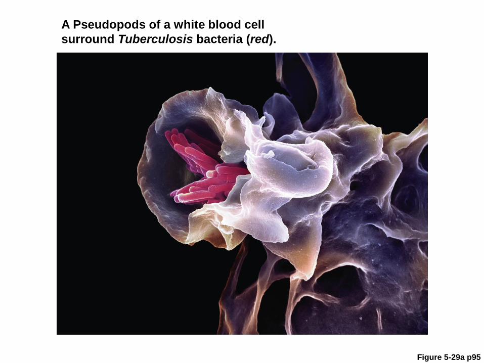
Figure 5-29a p95
A Pseudopods of a white blood cell
surround Tuberculosis bacteria (red).

ANIMATED FIGURE: Phagocytosis
To play movie you must be in Slide Show Mode
PC Users: Please wait for content to load, then click to play
Mac Users: CLICK HERE

Phagocytosis
B Endocytic vesicle
forms.
c Lysosome fuses with
vesicle; enzymes digest
pathogen.
D Cell uses the
digested material
or expels it.

Membrane Cycling
• Exocytosis and endocytosis continually replace and withdraw
patches of the plasma membrane
• New membrane proteins and lipids are made in the ER,
modified in Golgi bodies, and form vesicles that fuse with
plasma membrane

Forming New Plasma Membrane

Take-Home Message: How do cells take in
large particles and bulk substances?
• Exocytosis and endocytosis move materials in bulk across
plasma membranes
• In exocytosis, a cytoplasmic vesicle fuses with the plasma
membrane and releases its contents to the outside of the cell
• In endocytosis, a patch of plasma membrane sinks inward
and forms a vesicle in the cytoplasm
• Phagocytosis is an endocytic pathway by which cells engulf
particles such as microorganisms