Embed Size (px)
Citation preview

11/1/2011 1
DISORDERS AND CONDITIONS OF
THE EYE
Dr Ibraheem Bashayreh, RN, PhD

11/1/2011 2
Eyes• Anatomy of Eye
– Housed in a cone of fatty tissueEyeball– Three layers– External fibrous layer – Middle vascular layer – Inner layer of nerve tissue

11/1/2011 3
Anatomy of the Eye

11/1/2011 4
External Fibrous Layer• Sclera
– “white of eye’• Protective & supportive outer layer
• Cornea– Dense fibrous connective tissue
• Must be transparent to allow light

11/1/2011 5
Middle Vascular Layer• Heavily pigmented• Blood vessels

11/1/2011 6
Inner Layer• Retina
– Continuous with optical nerve in rear– Ora serrata in front– Two parts
• Outer part-pigmented-attached to choroid layer• Inner part is nerve tissue
11/1/2011 7
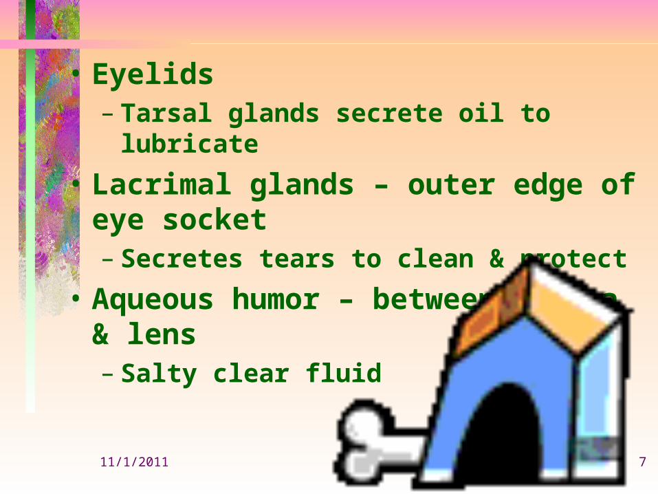
• Eyelids– Tarsal glands secrete oil to lubricate
• Lacrimal glands – outer edge of eye socket– Secretes tears to clean & protect
• Aqueous humor – between cornea & lens– Salty clear fluid
11/1/2011 8
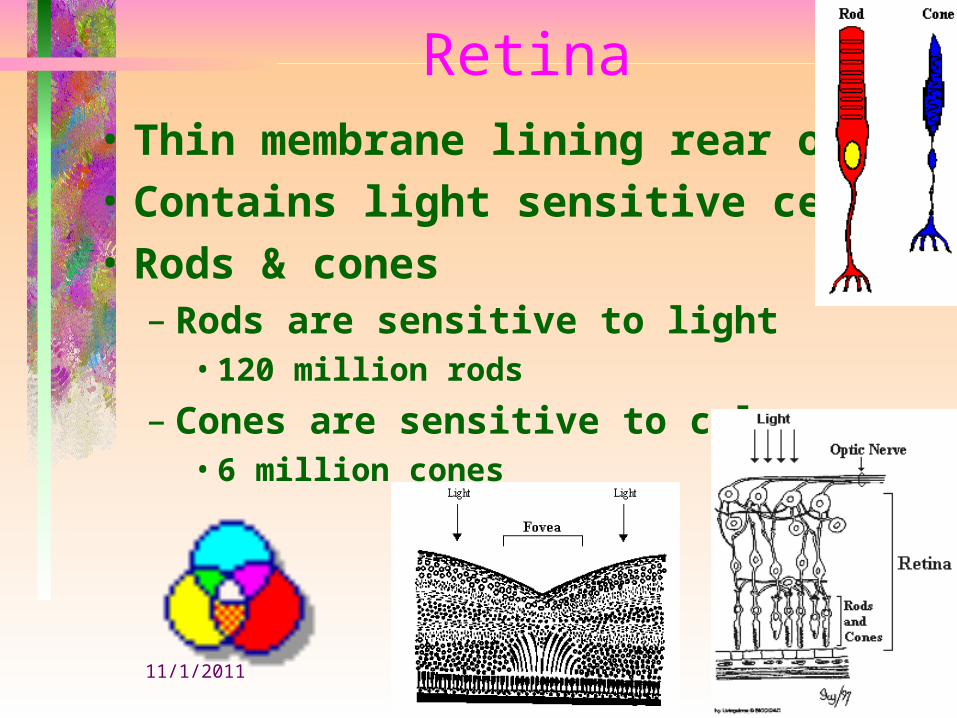
Retina• Thin membrane lining rear of eye• Contains light sensitive cells• Rods & cones
– Rods are sensitive to light• 120 million rods
– Cones are sensitive to colors• 6 million cones

11/1/2011 9
EYE DISORDERS• REFRACTIVE ERRORS• MUSCULAR DISORDERS• DISORDERS OF THE EYELID• DISORDERS OF THE GLOBE
OF THE EYE

11/1/2011 10
• REFRACTIVE ERRORS• HYPEROPIA• MYOPIA• ASTIGMATISM• PRESBYOPIA

11/1/2011 11
HYPEROPIA (FAR SIGHTEDNESS)
• MECHANISM– * object focuses behind the retina
* able to see only far objects• ETIOLOGY
– * genetic link
11/1/2011 12
• SYMPTOMS AND SIGNS– * blurred vision
* squinting* eye rubbing* headaches
• DIAGNOSIS– * Snellen visual acuity test– * ophthalmoscope
• TREATMENT– * Convex lens
11/1/2011 13
MYOPIA (NEAR SIGHTEDNESS)
• MECHANISM– * object focuses in front of the retina
* able to see only close objects• ETIOLOGY
– * genetic link• SYMPTOMS AND SIGNS
– * blurred vision* squinting* eye rubbing* headaches
11/1/2011 14
• DIAGNOSIS– * Snellen visual acuity test
* opthalmoscope• TREATMENT
– * concave lens* radical keratotomy - shallow incision in the cornea causing it to flatten in desired area (could have significant complications)

11/1/2011 15
ASTIGMATISM• MECHANISM
– * Abnormal shaped cornea (egg shape instead of spherical)* object is partially clear & other blurred
• ETIOLOGY– * genetic link
11/1/2011 16
• SYMPTOMS AND SIGNS– * blurred vision
* squinting* eye rubbing* headaches
• DIAGNOSIS– * Snellen visual acuity test
* opthalmoscope• TREATMENT
– * artificial lens transplant* radial keratotomy
11/1/2011 17
PRESBYOPIA• MECHANISM
– * Rigidity of the lens (old age)* unable to focus
• ETIOLOGY– * genetic link
• SYMPTOMS AND SIGNS– * blurred vision
* squinting* eye rubbing* headaches

11/1/2011 18
• DIAGNOSIS– * Snellen visual acuity test
* opthalmoscope• TREATMENT
– * lens transplant
11/1/2011 19
MUSCULAR DISORDERS
• NYSTAGMUS• STRABISMUS (CROSS EYED)
11/1/2011 20
NYSTAGMUS• MECHANISM* repetitive involuntary movements of one or both eyes• ETIOLOGY* Congenital * Brain tumors* CV lesions * Ear lesions* Alcohol/drug abuse

11/1/2011 21
• SYMPTOMS AND SIGNS– * Eye Movements
*Horizontal, vertical, circular, or combination* blurred vision
• DIAGNOSIS* viewing of the eyes - involuntary
movement* complete neurological tests
• TREATMENT* Treat the underlying condition* Congenital stays for life

11/1/2011 22
STRABISMUS (CROSS EYED)• MECHANISM
– * Failure of eyes to look in the same direction at the same time* Weakness of muscles of one eye(superior oblique, interior oblique, lateral)
• ETIOLOGY– in childhood: associated with amblyopia (decreased vision
in one eye)(reversible after 7 years of age)in adults: Usually caused by disease: i.e. diabetes, high blood pressure, brain trauma

11/1/2011 23
• SYMPTOMS AND SIGNS– * TYPES:
1. Esotropia (convergent-cross eye of one eye) 2. Exotropia (divergent- one eye turns outward) 3. Diplopia (adults strabismus) 4. Congenital (no strabismus exists)
11/1/2011 24
• DIAGNOSIS– * complete ophthalmic examination
* Diagnose underlying disease• TREATMENT
– * Treat early* Corrective glasses* orthoptic training* surgery to restore eye muscle balance* treat underlying disorder
11/1/2011 25
DISORDERS OF THE EYE LID
• HORDEOLUM (STYE)• CHALAZION (MEIBOMIAN CYST)• BLEPHARITIS• ENTROPION• ECTROPON• CONJUNCTIVITIS (PINK EYE)
11/1/2011 26
HORDEOLUM (STYE)• MECHANISM
– * Inflammatory infection of the hair follicle of the eye lid• ETIOLOGY
– * staphylococcal infection* usually associated with Blepharitis
• SYMPTOMS AND SIGNS– * occurs on the outside
* Pain/swelling/redness/pus* patient feels something in the eye
11/1/2011 27
• DIAGNOSIS– * Visual exam
* culture if needed• TREATMENT
– * Hot compress to alleviate pain* Topical or systemic antibiotics
11/1/2011 28
CHALAZION (MEIBOMIAN CYST)
• MECHANISM– * Collection of fluid or soft mass cyst
• ETIOLOGY– * Blockage of meibomian gland
• SYMPTOMS AND SIGNS– * Pea size cyst
* painless slow swelling of the inner part of eye lid* Could become infected
11/1/2011 29
• DIAGNOSIS– * Visual Examination
• TREATMENT– * small ones usually disappear spontaneously
after a month or two* large ones usually need surgical removal

11/1/2011 30
BLEPHARITIS• MECHANISM
– * Inflammation of the margins of the eye lids• ETIOLOGY
– * Ulcerative: staphy infection* nonulcerative: allergies, smoke, dust, chemicals, seborrhea, stye, chalazions
• SYMPTOMS AND SIGNS– * Persistent redness & crusting on eyelids
* itching / burning sensation* feeling something in the eye* Ulcers can cause eye lashes to fall out* Scales can get into eye causing conjunctivitis
11/1/2011 31
• DIAGNOSIS– * visual examination
* Culture (confirm staphy infection)• TREATMENT
– * Salt & water cleansing for 2 weeks* If unsuccessful - local antibiotics or sulfonamide
11/1/2011 32
ENTROPION• MECHANISM
– * Inversion of eye lid into eye• ETIOLOGY
– * aging (course fibrous tissue)• SYMPTOMS AND SIGNS
– * Foreign body sensation* Tearing / itching / redness* Continuous rubbing causes conjunctivitis or corneal ulcers* Decreased visual acuity if not corrected

11/1/2011 33
• DIAGNOSIS– * visual examination
• TREATMENT– * clean up on its own
* if not, minor surgery
11/1/2011 34
ECTROPON• MECHANISM
– * Outurned eye lids• ETIOLOGY
– * elderly (weakness of eye lid muscles)• SYMPTOMS AND SIGNS
– * dryness of the exposed part of the eye* tears run down the cheeks* if not treated can cause ulcers and permanent damage to cornea

11/1/2011 35
• DIAGNOSIS– * visual examination
• TREATMENT– * minor surgery if doesn’t disappear
11/1/2011 36
BLEPHAROPTOSIS (PTOSIS)• MECHANISM
– * weakness of eye muscle that raises eyelid (superior rectus, superior oblique)
• ETIOLOGY– * familial – * trauma
* diabetes mellitus – * muscular dystrophy
* myasthenia gravis – * brain tumors
11/1/2011 37
• SYMPTOMS AND SIGNS– * “drooping eye”
* Blocks vision• DIAGNOSIS
– * ophthalmic examination* blood work to rule out underlying disease
• TREATMENT– * Surgery (strengthen muscles)
* eye glasses with raised eyelid support* treat underlying disease

11/1/2011 38
CONJUNCTIVITIS (PINK EYE)• MECHANISM
– * inflammation of the conjunctiva• ETIOLOGY
– * Viral / bacterial* irritants (allergies, chemicals, UV light)
• SYMPTOMS AND SIGNS– * Redness / swelling / itching
* tearing when exposed to light * pus if infectious* “contagious” with contaminated hands, washcloths
11/1/2011 39
DIAGNOSIS
• Ophthalmic examination• Culture discharge
TREATMENT• Warm compress 3-4 times daily (10-15 min.)• If bacterial (antibiotics)• If viral- self limiting
11/1/2011 40
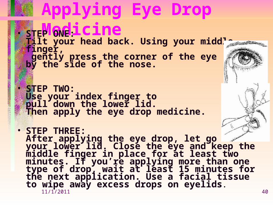
Applying Eye Drop Medicine• STEP ONE:
Tilt your head back. Using your middle finger, gently press the corner of the eye by the side of the nose.
• STEP TWO:Use your index finger to pull down the lower lid.Then apply the eye drop medicine.
• STEP THREE:After applying the eye drop, let go of your lower lid. Close the eye and keep the middle finger in place for at least two minutes. If you’re applying more than one type of drop, wait at least 15 minutes for the next application. Use a facial tissue to wipe away excess drops on eyelids.
11/1/2011 41
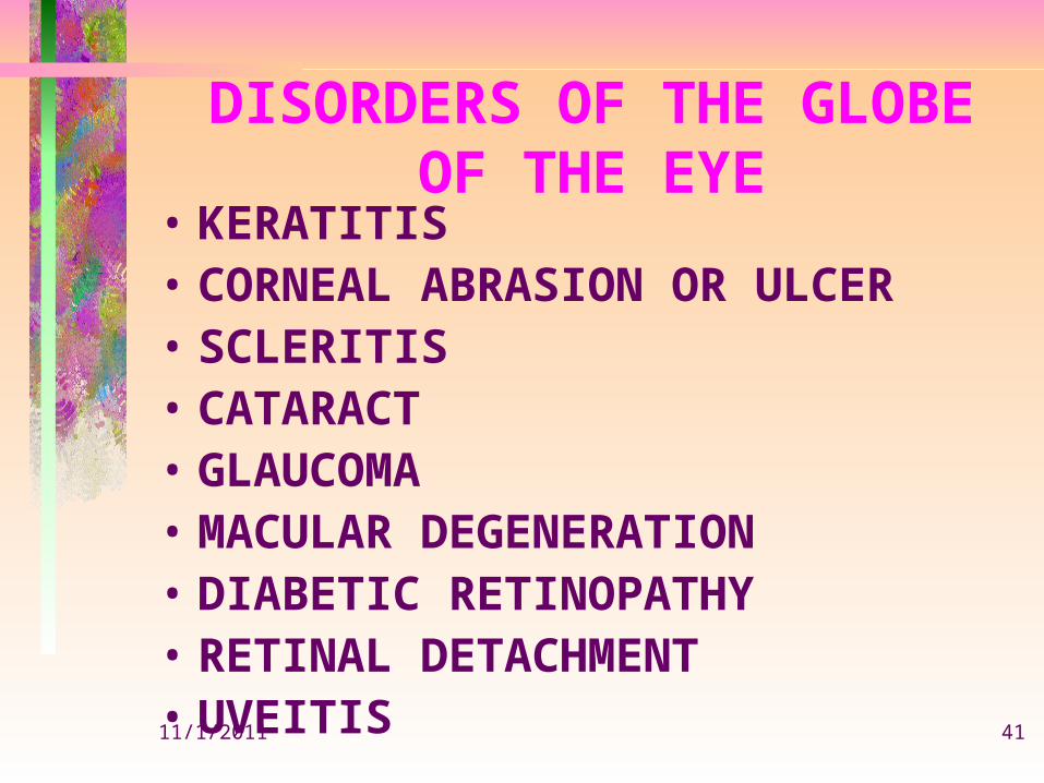
DISORDERS OF THE GLOBE OF THE EYE
• KERATITIS• CORNEAL ABRASION OR ULCER• SCLERITIS• CATARACT• GLAUCOMA• MACULAR DEGENERATION• DIABETIC RETINOPATHY• RETINAL DETACHMENT• UVEITIS

11/1/2011 42
KERATITIS
• MECHANISM– * inflammation and ulceration of the cornea
• ETIOLOGY– * herpes simplex virus (cold sores)
* other bacteria & fungi* trauma* dry air or intense light (welding)
11/1/2011 43
• SYMPTOMS AND SIGNS– * pain or numbness of the cornea
* decreased visual acuity* irritation
– * tearing* photophobia * mild conjunctivitis

11/1/2011 44
• DIAGNOSIS– * examination of cornea using slit lamp
* medical history* previous upper respiratory tract infection
• TREATMENT– * eye patch to protect from photophobia
11/1/2011 45
CORNEAL ABRASION OR ULCER
• ETIOLOGY– * foreign bodies
* trauma (fingernail, contact lenses)• SYMPTOMS AND SIGNS
– * pain / redness & tearing* something constantly in eye* vision impairment
11/1/2011 46
• DIAGNOSIS– * visual examination
* fluorescien (stain)• TREATMENT
– * remove foreign bodies* eye wear for protection & promote hearing* eye dressing to reduce movement

11/1/2011 47
SCLERITIS• MECHANISM
– * Inflammation of sclera• ETIOLOGY
– * rheumatoid arthritis* digestive disorders (Crohn’s)
• SYMPTOMS AND SIGNS– * Dull pain – * Intense redness
* loss of vision (posterior sclera inflammation)* if untreated can lead to perforation or loss of eye

11/1/2011 48
• DIAGNOSIS– * ophthalmic examination
* Blood work to uncover underlying cause• TREATMENT
– * MILD: eye drops (antibiotics)* SEVERE: immunosupressive drugs* PERFORATION: surgery

11/1/2011 49
CATARACT• MECHANISM
– * Gradual deterioration of lens• ETIOLOGY
– * familial – * old age
* congenital – * trauma
* drug toxicity (high level of steroids)* diabetes mellitus

11/1/2011 50
• SYMPTOMS AND SIGNS– * Cloudy / white opaque area of the lens
* reduce visual acuity* Blurring of vision* photosensitivity
• DIAGNOSIS– * Visual examination
* pen light of slit lamp confers the presence of a cataract• TREATMENT
– * Intracapsular phacoemulsification(involves breakage of cataract then aspiration)* Extracapsular phacoemulsification: (artificial lens replacement)

11/1/2011 51
GLAUCOMA
•Chronic Open-Angle Glaucoma– MECHANISM
• * Increased intraocular pressure due to a malfunction in eyes aqueous humor drainage system - can lead to optic nerve damage
– ETIOLOGY• * trauma
* overuse of steriods
11/1/2011 52
• SYMPTOMS AND SIGNS– * Gradual loss of peripheral vision.
* If untreated - eventually complete vision loss• DIAGNOSIS
– * ophthalmic examination* tonometry (pressure measure)
• TREATMENT– * Medication that helps decrease aqueous humor
production or opens drainage system* laser to open drainage* surgery (bypass)

11/1/2011 53
•Acute Angle-Closure Glaucoma– MECHANISM
• * complete blockage of aqueous humor drainage system
– ETIOLOGY• * trauma

11/1/2011 54
• SYMPTOMS AND SIGNS– * Blurred vision – * severe eye pain
* redness of the eye – * nausea & vomiting
* photophobia (sees “halo” around light)* hazy cornea (elevated pressure)* if untreated --> blindness
• DIAGNOSIS– * goniolens (special lens to view the opening)
• TREATMENT– * LASER IRIDOTOMY (creation of a hole in the iris between the
anterior and posterior chamber)* medications to reduce pressure

11/1/2011 55
MACULAR DEGENERATION• MECHANISM
– (The area next to optic disc that defines fine details at the center of visual field = macula)* not enough blood supply to area (disappearance of central vision due to deterioration of pigment layer of retina)
• ETIOLOGY– * age – * atherosclerosis
* hemorrhage
11/1/2011 56
• SYMPTOMS AND SIGNS– * Fine detailed vision is impaired
* Sharp vision deterioration (reading)* peripheral vision is not affected* loss of central vision
• DIAGNOSIS– * Ophthalmoscopy
* fluorescein angiography* patient history

11/1/2011 57
• TREATMENT– * no known cure
* laser photocoagulation* increase zinc in diet* strong magnifying glasses
11/1/2011 58
DIABETIC RETINOPATHY• MECHANISM
– * constriction of ocular blood vessels & leakage of blood into retina (microaneurysms, neovascularization = new blood vessels)* leakage of blood into vitreous humor* scar tissue
• ETIOLOGY– diabetics with uncontrolled glucose levels

11/1/2011 59
• SYMPTOMS AND SIGNS– * impaired sharp vision
* blurred vision* could lead to permanent blindness
• DIAGNOSIS– * Ophthalmoscopy
• TREATMENT– * Laser photocoagulation
* vitrectomy
11/1/2011 60
RETINAL DETACHMENT• MECHANISM
– * elevation & detachment of the retina from the choriod (partial or complete)
• ETIOLOGY– * Near sightedness (myopia)
* trauma• SYMPTOMS AND SIGNS
– * visual floaters – * light flashes
* dark/opaque shadow extending form periphery inward from lower field to upper* If central retina is involved, could lead to blindness
11/1/2011 61
• DIAGNOSIS– * Ophthalmoscopy
• TREATMENT– * Photocoagulation (laser)
* cryotherapy
11/1/2011 62
UVEITIS• MECHANISM
– * Inflammation of uveal tract (iris, ciliary body, choriod)
• ETIOLOGY– * autoimmune
* infections (syphilis, tuberculosis, toxoplasmosis, histoplasmosis)* unknown etiology
• SYMPTOMS AND SIGNS– * unilateral / bilateral
* pain – * photophobia
* blurred vision – * redness
* pupillary constriction

11/1/2011 63
• DIAGNOSIS– * complete visual examination
* skin test for TB, toxoplasmosis, histoplasmosis* blood test
• TREATMENT– * treat underlying disease if known
* cycloplegics and steroids
11/1/2011 64
EXOPHTHALMOS• MECHANISM
– * Edema of the soft tissue that lines bony orbit of eye• ETIOLOGY
– * hyperthyroidism (bilateral)* hemorrhage or inflammation (unilateral)
• SYMPTOMS AND SIGNS– * protrusion of eye balls
* dizziness – * double vision
* restricted eye movement* seriously blurred vision
11/1/2011 65
• DIAGNOSIS– * ophthalmic examination
* blood work – * x ray / CT
* echography• TREATMENT
– * treat underlying disorder (thyroid)* surgery* steroids (control edema)