Embed Size (px)
Citation preview

Chapter 5
CELL MEMBRANE
Structure and Function
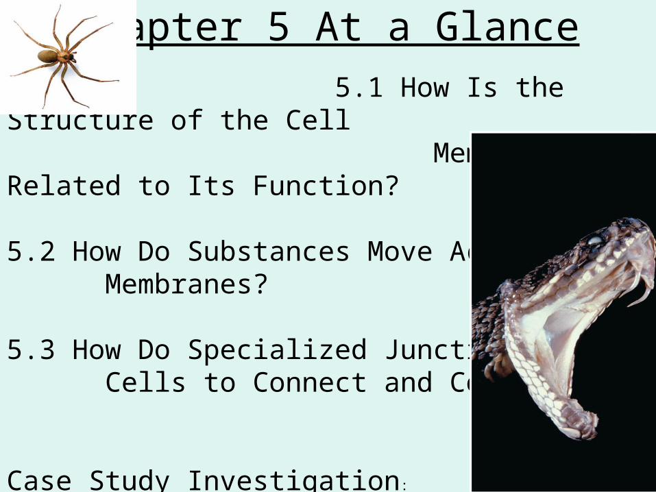
Chapter 5 At a Glance
5.1 How Is the Structure of the Cell Membrane Related to Its Function?
5.2 How Do Substances Move Across Membranes?
5.3 How Do Specialized Junctions Allow Cells to Connect and Communicate?
Case Study Investigation:
Y:\Biology\D Cell Membrane and Transport\Transport CSI Reading.pdf

5.1 How is the Structure of the Cell Membrane Related to its Function?
All cell membranes have a similar basic structureProteins suspended in a double layer of phospholipids
Responsible for:1.selectively exchanging
substances 2.Communicating with
environment3.Controlling biochemical
reactions4.Forming connections
btwn cells
Responsible for:1.Isolating cells contents

Crucial functions of the plasma cell membrane: isolates cell’s contents from external environment regulates exchange of essential substances allows communication between cells creates attachments within and btwn cells regulates biochemical reactions
extremely thin–10,000 membranes still thinner than txtbk page
membranes change in response to their surroundings
5.1 How is the Structure of the Cell Membrane Related to its Function?

cholesterol
interstitial fluid (outside)
cytosol (fluid inside cell)
enzyme cytoskeletonrecognitionprotein
binding site
connectionprotein
phospholipid
receptorprotein
glycoprotein
phospholipidbilayer
pore
transportprotein
carbohydrate
proteinextra-cellularmatrix
Cell Membrane = Fluid Mosaic Model named in 1972 by S.J. Singer and G.L. Nicolson proteins and other structures move within the membrane
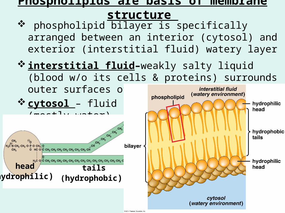
Phospholipids are basis of membrane structure phospholipid bilayer is specifically arranged between an
interior (cytosol) and exterior (interstitial fluid) watery layer
interstitial fluid–weakly salty liquid (blood w/o its cells & proteins) surrounds outer surfaces of animal cell membranes
cytosol – fluid portion of cytoplasm (mostly water)
head(hydrophilic)
tails(hydrophobic)
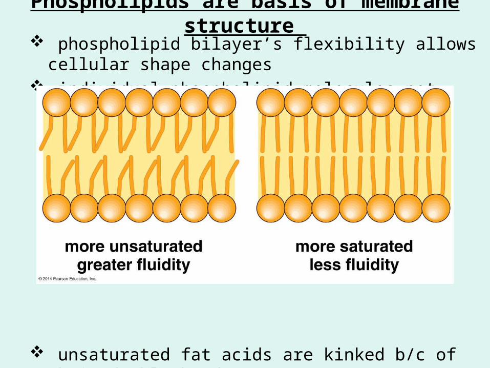
Phospholipids are basis of membrane structure phospholipid bilayer’s flexibility allows cellular shape changes individual phospholipid molecules not bonded to each other
unsaturated fat acids are kinked b/c of their double bond(s) all reasons why membrane is fluid

membranes more fluid at high temps (more movement) and less fluid at low temps (less movement) cell membranes of organisms living in low temps tend to be more unsaturated to help maintain fluidity
cholesterol stabilizes membranes – makes it less fluid at high temps and less solid at lower temps; becomes more selective
Phospholipids structure = selectively permeable

Phospholipids structure = selectively permeable
water soluble (polar) substances cannot easily cross phospholipid bilayers (i.e. salts, amino acids, sugars)
small molecules (i.e. H2O, O2, CO2) can slip in
larger lipid soluble (nonpolar) substances (i.e. estrogen, testosterone) can pass through

Variety of Proteins Form a Mosaic
variety of proteins are embedded within or attached to phospholipid bilayer many have carbohydrates attached to their outer surface
(glycoproteins)
5 major categories of membrane proteins enzymes receptor proteins recognition proteins connection proteins transport proteins

Membrane Proteins
1. Enzymes - proteins that promote chemical reactions that synthesize or break apart molecules (i.e. enzymes that line the small intestine that digest carbs & proteins)
2. Receptor proteins – trigger cellular responses (i.e. hormones that bind, immune response)
3. Recognition proteins – glycoproteins that serve as identification tags on cell surface (i.e. self vs non-self)
Membrane Proteins
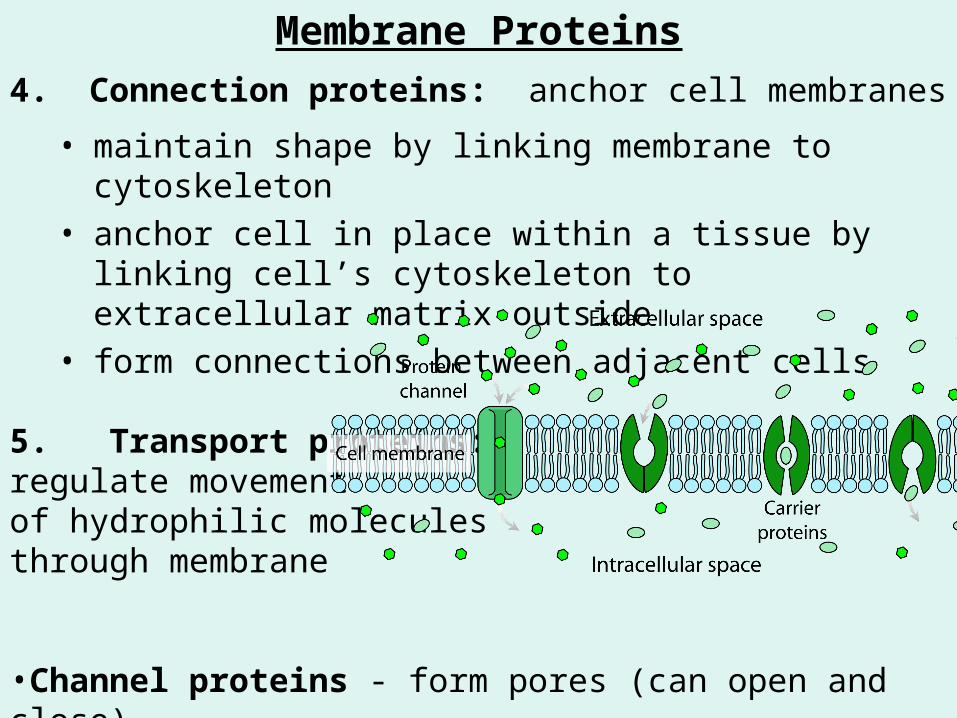
4. Connection proteins: anchor cell membranes
• maintain shape by linking membrane to cytoskeleton• anchor cell in place within a tissue by linking cell’s
cytoskeleton to extracellular matrix outside• form connections between adjacent cells
5. Transport proteins: regulate movement of hydrophilic molecules through membrane
•Channel proteins - form pores (can open and close)•Carrier proteins – bind to and carry substances through membrane

5.2 How Do Substances Move Across Membrane some substance can move across the membrane by diffusing
through phospholipid bilayer through specialized proteins solute: substance that can be dissolved in a solvent (i.e. sugar,
salt, etc) solvent: fluid capable of dissolving a solute (i.e. H2O,
gasoline, alcohol)• Water is a universal solvent

concentration: defines the amount of solute in a given amount of solvent
gradient: physical difference in temperature, pressure, charge or concentration of a solute in a fluid - btwn 2 adjoining regions of space
concentration gradient: differences in solute concentrations across a membrane

Molecules in Fluids Diffuse in Response to Gradients atoms, molecules, & ions are in constant random motion
diffusion: net movement of solutes from regions of high concentration to regions of low concentration (down a gradient)• gradients cause molecule movement• the greater the gradient the faster the rate of diffusion• the higher the temperature the faster the rate of diffusion• movement continues until molecules are evenly dispersed (unless disrupted)
Hot Water Cold Water

A drop of dye isplaced in water
Dye moleculesdiffuse into the water;water molecules diffuseinto the dye
Both dye moleculesand water molecules areevenly dispersed
water molecule
dye molecules

Movement Through Membranes Occurs by Passive or Active Transport
cells w/o gradients are dead so….. membrane proteins must use energy to create and maintain
gradients in order to carry out crucial biochemical process selective permeable membrane creates a barrier that helps maintain
gradients
PASSIVE TRANSPORT diffusion of substancesacross cell membranes DOWN concentration gradients•simple diffusion•facilidated diffusion•osmosis
ACTIVE TRANSPORT requires energy to movesubstances usually AGAINST concentration gradients
•endocytosis•exocytosis

Active Transport
Passive T
ransp
ort
Passive Transport: Simple & Facilitated Diffusion
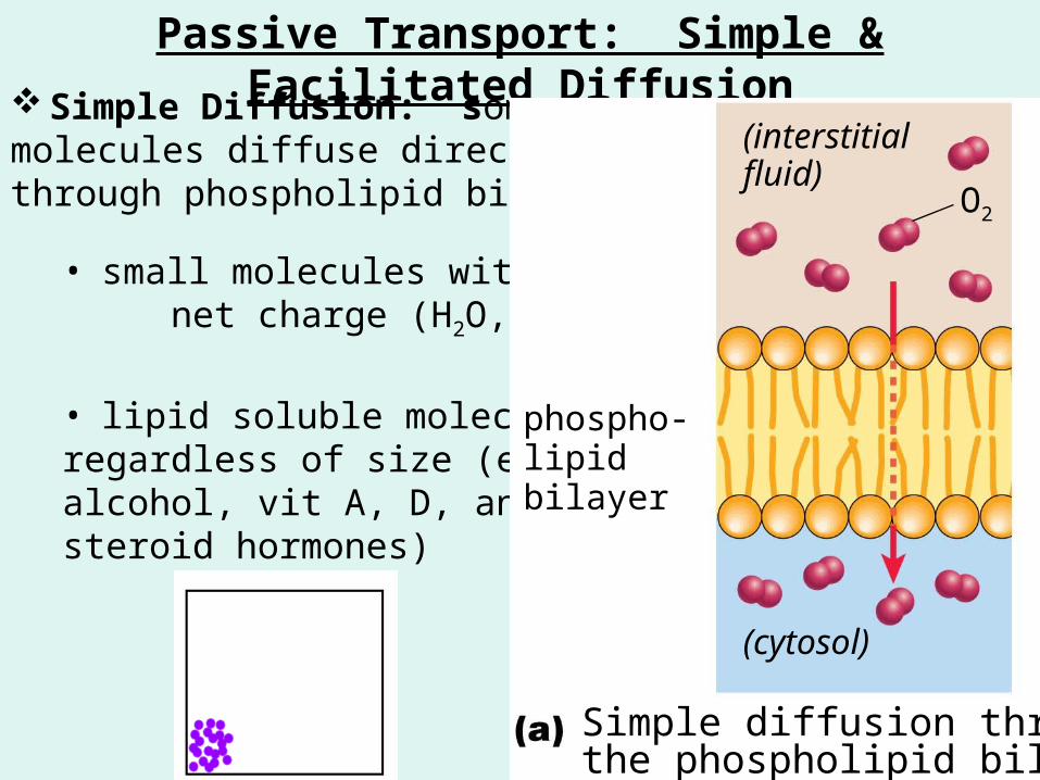
Simple Diffusion: some molecules diffuse directly through phospholipid bilayers
• small molecules with no net charge (H2O, O2, CO2)
• lipid soluble molecules regardless of size (ethyl alcohol, vit A, D, and E, steroid hormones)
phospho-lipidbilayer
(interstitialfluid)
O2
(cytosol)
Simple diffusion throughthe phospholipid bilayer

Facilitated Diffusion: transport proteins help larger polar molecules & ions (sugars, K+, Na+, Cl-, Ca 2+) to cross membranes• carrier proteins: loosely bind to specific ions/molecules (i.e.
sugars & small proteins), change shape & transfer the bound particles across membrane
• channel proteins: form pores through cell membranes
carrierprotein
Facilitated diffusion through carrier proteins
Facilitated diffusion through channel proteins
channelprotein
Ion channels very selective(charge/size)
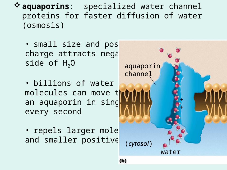
aquaporins: specialized water channel proteins for faster diffusion of water (osmosis)
• small size and positive charge attracts negative side of H2O
• billions of watermolecules can move throughan aquaporin in single fileevery second
• repels larger moleculesand smaller positive ions
(cytosol)water
aquaporinchannel

Osmosis is the Diffusion of Water Osmosis: diffusion of water across a selectively permeable
membrane in response to concentration gradient, pressure, or temp.
• H2O moves from regions of high concentration to regions of low concentration across a membrane
• dissolved substances reduces concentration of free H2O molecules (& purity) in a solution• osmotic strength:tendency to attract H2O across a membrane; greaterthe solute conc., the greaterthe o.s.
High water
concentration
Low water
concentration

Isotonic, Hypertonic, Hypotonic SolutionsNo net flowof water
Water flows out;the sac shrinks
Water flows in;the sac expands
A sac in anisotonic solution
A sac in ahypertonic solution
A sac in ahypotonic solution
iso = same equalconcentrations of
solute and solvent
hyper = more more solute higher osmotic strengthH2O moves out of less concentrated sln
hypo = less less solute lower osmotic
strength H2O moves into more
concentrated slnhttp://www.youtube.com/watch?v=_slUL3kMZlU http://www.youtube.com/watch?v=SSS3EtKAzYc Egg Experiment

Osmosis Plays an Important Role in Lives of Cells water uptake by roots of plants
absorption of dietary water from intestine reabsorption of water in kidneys
organisms that live in fresh water must use energy to counteract osmosis b/c cells are hypertonic to surrounding water
Turgor Plasmolysis
pressure: when water
when water leaves cell
flows into a causing cell
cell and to shrink inflates the away
from cell; pressure cell wallon the cell wall (droopy
plants)


When water is plentiful, it fills the centralvacuole, pushes the cytoplasm against thecell wall, and helps maintain the cell’s shape
cytoplasm
Turgor pressure provides support
central vacuole
Water pressure supportsthe leaves of thisimpatiens plant
When water is scarce, the central vacuoleshrinks and the cell wall is unsupported
Loss of turgor pressure causes the plant to wilt
Deprived of the supportfrom water, the plant wilts
plasma membranecell wall

Active Transport, Endocytosis, Exocytosis
energy-expending cellular activities are crucial to sustaining life• maintaining concentration gradients• acquiring food• excreting wastes • cell to cell communication
active transport: membrane proteins use energy to move molecules/ions across a membrane AGAINST their concentration gradient (low concentration to a high concentration)

Active Transporthttp://www.youtube.com/watch?v=yz7EHJFDEJs
ATP & specific molecule/ion Energy comes from breaking high energy bond of 3rd phosphate
active transport proteins = pumps
Energy from ATPchanges the shapeof the transport proteinand moves the ionacross the membrane
bindingsite
The proteinreleases the ionand the remnantsof ATP (ADP and P)and closes
The transportprotein binds bothATP and Ca2
ATPbindingsite
ATPATP
Ca2 (cytosol)
ADP
(interstitial fluid)
P

Endocytosis Allows Cells to Engulf Particles or Fluids
When cells need materials thatare too large to pass through membrane they use…
Endocytosis: form of active transport that allows cells to engulf particles or fluids
1. Pinocytosis:“cell drinking” moves liquids into cells
vesicle containinginterstitialfluid(cytosol)
(interstitial fluid)
cytosol
interstitial fluid
Pinocytosis
TEM of pinocytosis
A dimple forms in the plasma membrane, whichdeepens and surrounds the interstitial fluid. Themembrane encloses the interstitial fluid, forming a vesicle.
http://www.youtube.com/watch?v=InG6xF9D4EM

2. Receptor-mediated endocytosis: moves specific molecules into cells (packets of protein and cholesterol)
Receptor proteins for specificmolecules or complexes ofmolecules are localized at coatedpit sites.
(cytosol)
(interstitial fluid)
(cytosol)
(interstitial fluid)
coated pit
nutrient molecule
coated vesicle
coated pitproteincoating plasma membrane
extracellular particlesbound to receptors
Receptor-mediated endocytosis
TEM of receptor-mediated endocytosis
coated vesicle
The receptor bind themolecules and the membranedimples inward.
The coated pit region of themembrane encloses the receptor-bound molecules. A vesicle (“coated vesicle”)containing the bound moleculesis released into the cytosol.
receptor
http://www.youtube.com/watch?v=knJQzBmqOZw

3. Phagocytosis: “cell eating”moves large particles (or whole organisms) into cells
(cytosol)
(interstitial fluid)
pseudopodsfood particle
Phagocytosis
food vacuole
An Amoeba engulfs aParamecium
A white blood cell engulfs adisease-causing fungal cell
The plasma membrane extends pseudopods toward an extracellularparticle (for example, food). The ends of the pseudopods fuse,encircling the particle. A vesicle called a food vacuole is formedcontaining the engulfed particle.
http://www.youtube.com/watch?v=a1xPpsxvhVA

Exocytosis Moves Material Out of the Cell
energy is used to dispose of undigested particles of waste or secrete substances (i.e. hormones) into interstitial fluid
(cytosol)
(interstitial fluid)plasma membrane
secreted material
plasmamembrane
Material is enclosed in a vesicle that fuses with theplasma membrane, allowingits contents to diffuse out
vesicle
http://www.youtube.com/watch?v=dPKvHrD1eS4

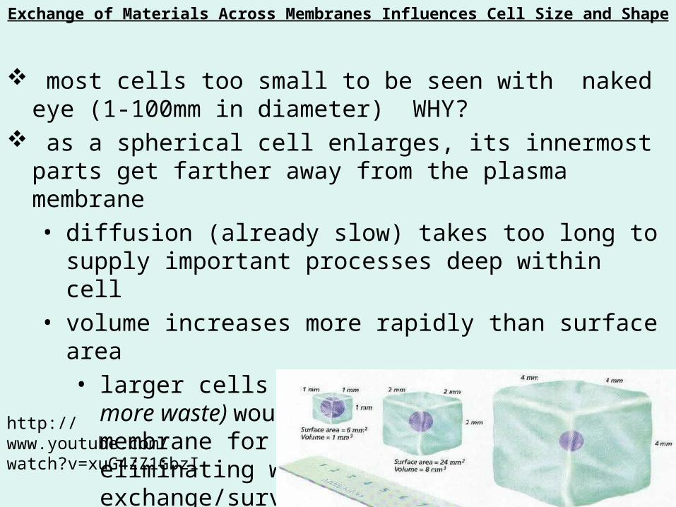
Exchange of Materials Across Membranes Influences Cell Size and Shape
most cells too small to be seen with naked eye (1-100mm in diameter) WHY?
as a spherical cell enlarges, its innermost parts get farther away from the plasma membrane• diffusion (already slow) takes too long to supply important
processes deep within cell• volume increases more rapidly than surface area
• larger cells (require more nutrients & create more waste) would have a smaller area of membrane for acquiring nutrients and eliminating wastes not enough to exchange/survive
http://www.youtube.com/watch?v=xuG4ZZ1GbzI

Case Study many rattlesnake & spider venoms contain phospholipases - break
down membrane phospholipids causing cells to rupture and die phospholipases attack membranes of capillary cells causing blood
vessels to rupture & release blood into surrounding tissue • causes anemia (inadequate # of oxygen-carrying RBC)• attack muscle cell membranes
antivenin – contains specialized proteins that bind and neutralize snake venom proteins
no antivenin for brown recluse bites (treatments only)
How does the role of phospholipases in snake venom differ from its role in the snake’s digestive tract?

Study…. Reread each section; reread your notes
Reorganize your notes; re-write; make charts, tables, lists
Ch. 5 Vocab
Read Summary of Key Concepts (pg. 91)
Complete Thinking Through the Concepts 1 – 6 (pg 91-92)
Be able to answer Review Questions 1-8 (pg. 92)
Concept check questions
Use Mastering Biology to help study