Embed Size (px)
DESCRIPTION
Chapter 47. Animal Development. Overview: A Body-Building Plan for Animals. It is difficult to imagine that each of us began life as a single cell, a zygote A human embryo at about 6–8 weeks after conception shows development of distinctive features. LE 47-1. 1 mm. - PowerPoint PPT Presentation
Citation preview
Copyright © 2005 Pearson Education, Inc. publishing as Benjamin Cummings
PowerPoint Lectures for Biology, Seventh Edition
Neil Campbell and Jane Reece
Lectures by Chris Romero
Chapter 47Chapter 47
Animal Development

Copyright © 2005 Pearson Education, Inc. publishing as Benjamin Cummings
Overview: A Body-Building Plan for Animals
• It is difficult to imagine that each of us began life as a single cell, a zygote
• A human embryo at about 6–8 weeks after conception shows development of distinctive features

LE 47-1
1 mm

Copyright © 2005 Pearson Education, Inc. publishing as Benjamin Cummings
• The question of how a zygote becomes an animal has been asked for centuries
• As recently as the 18th century, the prevailing theory was called preformation
• Preformation is the idea that the egg or sperm contains a miniature infant, or “homunculus,” which becomes larger during development

Copyright © 2005 Pearson Education, Inc. publishing as Benjamin Cummings
Copyright © 2005 Pearson Education, Inc. publishing as Benjamin Cummings
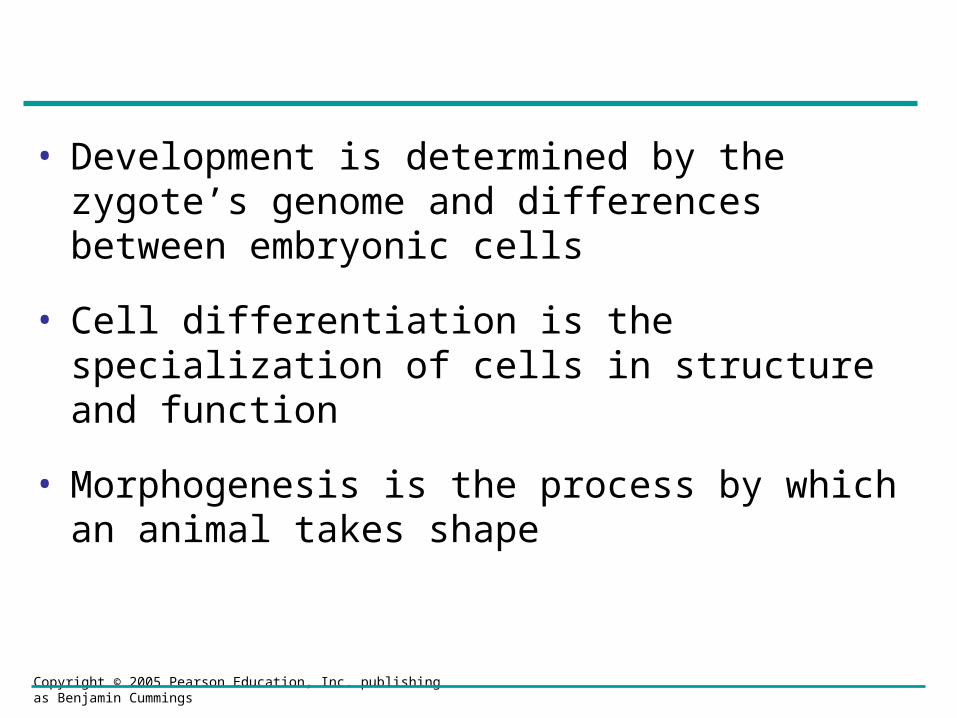
• Development is determined by the zygote’s genome and differences between embryonic cells
• Cell differentiation is the specialization of cells in structure and function
• Morphogenesis is the process by which an animal takes shape

Copyright © 2005 Pearson Education, Inc. publishing as Benjamin Cummings
Concept 47.1: After fertilization, embryonic development proceeds through cleavage, gastrulation, and organogenesis
• Important events regulating development occur during fertilization and the three stages that build the animal’s body

Copyright © 2005 Pearson Education, Inc. publishing as Benjamin Cummings
Fertilization
• Fertilization brings the haploid nuclei of sperm and egg together, forming a diploid zygote
• The sperm’s contact with the egg’s surface initiates metabolic reactions in the egg that trigger the onset of embryonic development

Copyright © 2005 Pearson Education, Inc. publishing as Benjamin Cummings
The Acrosomal Reaction
• The acrosomal reaction is triggered when the sperm meets the egg
• This reaction releases hydrolytic enzymes that digest material surrounding the egg

LE 47-3
Sperm-bindingreceptors
Jelly coat
Acrosome
Actin
Spermhead
Basal body(centriole)
Sperm plasmamembrane
SpermnucleusContact
Acrosomalreaction
Acrosomalprocess
Contact and fusionof sperm and eggmembranes Entry of sperm
nucleus
Cortical reaction
Fertilizationenvelope
Egg plasmamembrane
Vitelline layer
Hydrolytic enzymes
Corticalgranule
Fused plasmamembranes
Perivitellinespace
Cortical granulemembrane
EGG CYTOPLASM

Copyright © 2005 Pearson Education, Inc. publishing as Benjamin Cummings
• Gamete contact and/or fusion depolarizes the egg cell membrane and sets up a fast block to polyspermy

Copyright © 2005 Pearson Education, Inc. publishing as Benjamin Cummings
The Cortical Reaction
• Fusion of egg and sperm also initiates the cortical reaction
• This reaction induces a rise in Ca2+ that stimulates cortical granules to release their contents outside the egg
• These changes cause formation of a fertilization envelope that functions as a slow block to polyspermy

LE 47-4
1 sec beforefertilization
Point ofspermentry
10 sec afterfertilization
Spreading waveof calcium ions
20 sec 30 sec
500 µm

Copyright © 2005 Pearson Education, Inc. publishing as Benjamin Cummings
Activation of the Egg
• The sharp rise in Ca2+ in the egg’s cytosol increases the rates of cellular respiration and protein synthesis by the egg cell
• With these rapid changes in metabolism, the egg is said to be activated
• In a sea urchin, a model organism, many events occur in the activated egg

LE 47-5
Binding of sperm to egg
Acrosomal reaction: plasma membranedepolarization (fast block to polyspermy)
Increased intracellular calcium level
Cortical reaction begins (slow block to polyspermy)
Formation of fertilization envelope complete
Increased intracellular pH
Fusion of egg and sperm nuclei complete
Increased protein synthesis
Onset of DNA synthesis
First cell division
1
Se
co
nd
s
2
3
68
10
4
20
30
501
2
40
34
10
5
20
3040
9060
Min
ute
s

Copyright © 2005 Pearson Education, Inc. publishing as Benjamin Cummings
Fertilization in Mammals
• In mammalian fertilization, the cortical reaction modifies the zona pellucida as a slow block to polyspermy

LE 47-6
Folliclecell
Acrosomalvesicle
Egg plasmamembrane
Zonapellucida Sperm
nucleus
Corticalganules
Spermbasalbody
EGG CYTOPLASM
Copyright © 2005 Pearson Education, Inc. publishing as Benjamin Cummings
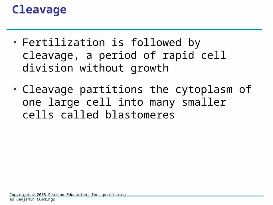
Cleavage
• Fertilization is followed by cleavage, a period of rapid cell division without growth
• Cleavage partitions the cytoplasm of one large cell into many smaller cells called blastomeres

LE 47-7
Fertilized egg Four-cell stage Morula Blastula

Copyright © 2005 Pearson Education, Inc. publishing as Benjamin Cummings
• The eggs and zygotes of many animals, except mammals, have a definite polarity
• The polarity is defined by distribution of yolk, with the vegetal pole having the most yolk
• The development of body axes in frogs is influenced by the egg’s polarity

LE 47-8
Anterior
Right
Animal pole
Graycrescent
DorsalVentral
Left
Posterior
Body axes Establishing the axes
Futuredorsalside oftadpole
Point ofspermentry
Firstcleavage
Vegetalhemisphere Vegetal pole
Point of sperm entry
Animalhemisphere

Copyright © 2005 Pearson Education, Inc. publishing as Benjamin Cummings
• Cleavage planes usually follow a pattern that is relative to the zygote’s animal and vegetal poles

LE 47-9
Zygote
2-cellstageforming
8-cellstage
4-cellstageforming
Animal pole Blasto-coel
Blastula(crosssection)
Vegetal poleBlastula (at least 128 cells)
0.25 mm
Eight-cell stage (viewedfrom the animal pole)
0.25 mm

Copyright © 2005 Pearson Education, Inc. publishing as Benjamin Cummings
• Meroblastic cleavage, incomplete division of the egg, occurs in species with yolk-rich eggs, such as reptiles and birds

LE 47-10
Blastocoel
Fertilized egg
BLASTODERM
HypoblastEpiblastYOLK MASS
Cutaway view ofthe blastoderm
Blastoderm
Four-cell stage
Zygote
Disk ofcytoplasm
Copyright © 2005 Pearson Education, Inc. publishing as Benjamin Cummings
• Holoblastic cleavage, complete division of the egg, occurs in species whose eggs have little or moderate amounts of yolk, such as sea urchins and frogs

Copyright © 2005 Pearson Education, Inc. publishing as Benjamin Cummings
Gastrulation
• Gastrulation rearranges the cells of a blastula into a three-layered embryo, called a gastrula, which has a primitive gut
Copyright © 2005 Pearson Education, Inc. publishing as Benjamin Cummings
• The three layers produced by gastrulation are called embryonic germ layers
– The ectoderm forms the outer layer
– The endoderm lines the digestive tract
– The mesoderm partly fills the space between the endoderm and ectoderm
Video: Sea Urchin Embryonic Development

LE 47-11
Animalpole
Blastopore
Filopodiapullingarchenterontip
Archenteron
Mesenchymecells
Blastocoel
Future ectoderm
Vegetalpole
Key
Future mesoderm
Future endoderm
Vegetalplate
Blastocoel
Mesenchymecells
Archenteron
Blastocoel
Mesenchume(mesodermforms futureskeleton)
50 µm
Mouth
Ectoderm
Blastopore
Digestive tube (endoderm)
Anus (from blastopore)

Copyright © 2005 Pearson Education, Inc. publishing as Benjamin Cummings
• The mechanics of gastrulation in a frog are more complicated than in a sea urchin

LE 47-12
Future ectoderm
Key
Future mesoderm
Future endoderm
Archenteron
Blastocoelremnant
Ectoderm
MesodermEndoderm
Yolk plugYolk plugGastrula
Blastocoelshrinking
Blastocoel
Dorsal tip of blastopore
CROSS SECTION
Animal pole
Dorsal lipof blastopore
Vegetal pole Blastula
SURFACE VIEW

Copyright © 2005 Pearson Education, Inc. publishing as Benjamin Cummings
• Gastrulation in the chick and frog is similar, with cells moving from the embryo’s surface to an interior location
• During gastrulation, some epiblast cells move toward the blastoderm’s midline and then detach and move inward toward the yolk

LE 47-13
Futureectoderm
Epiblast
Migratingcells(mesoderm)
YOLK
HypoblastEndoderm
Primitivestreak

Copyright © 2005 Pearson Education, Inc. publishing as Benjamin Cummings
Organogenesis
• During organogenesis, various regions of the germ layers develop into rudimentary organs
Copyright © 2005 Pearson Education, Inc. publishing as Benjamin Cummings
• Early in vertebrate organogenesis, the notochord forms from mesoderm, and the neural plate forms from ectoderm
Video: Frog Embryo Development

LE 47-14a
Neural folds
Neuralplate
LM1 mm
Neuralfold
Notochord
Archenteron
Neural plate formation
Endoderm
Mesoderm
Ectoderm

Copyright © 2005 Pearson Education, Inc. publishing as Benjamin Cummings
• The neural plate soon curves inward, forming the neural tube

LE 47-14b
Neuralfold
Neural plate
Neural tube
Formation of the neural tube
Neural crest
Outer layerof ectoderm
Neural crest

Copyright © 2005 Pearson Education, Inc. publishing as Benjamin Cummings
• Mesoderm lateral to the notochord forms blocks called somites
• Lateral to the somites, the mesoderm splits to form the coelom

LE 47-14c
1 mm
Notochord
Archenteron(digestive cavity)
Neural tube
Neural crest
Eye Somites Tail bud
SEM
Coelom Somite
Somites

Copyright © 2005 Pearson Education, Inc. publishing as Benjamin Cummings
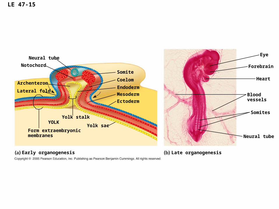
• Organogenesis in the chick is quite similar to that in the frog
LE 47-15
Notochord
ArchenteronEndoderm
Mesoderm
Ectoderm
Neural tube
Eye
Coelom
Somite
Somites
Neural tube
Lateral fold
Yolk stalkYOLK
Form extraembryonicmembranes
Yolk sac
Early organogenesis
Forebrain
Heart
Bloodvessels
Late organogenesis

Copyright © 2005 Pearson Education, Inc. publishing as Benjamin Cummings
• Many structures are derived from the three embryonic germ layers during organogenesis

Copyright © 2005 Pearson Education, Inc. publishing as Benjamin Cummings

Copyright © 2005 Pearson Education, Inc. publishing as Benjamin Cummings
Developmental Adaptations of Amniotes
• Embryos of birds, other reptiles, and mammals develop in a fluid-filled sac in a shell or the uterus
• Organisms with these adaptations are called amniotes
• In these organisms, the three germ layers also give rise to the four membranes that surround the embryo
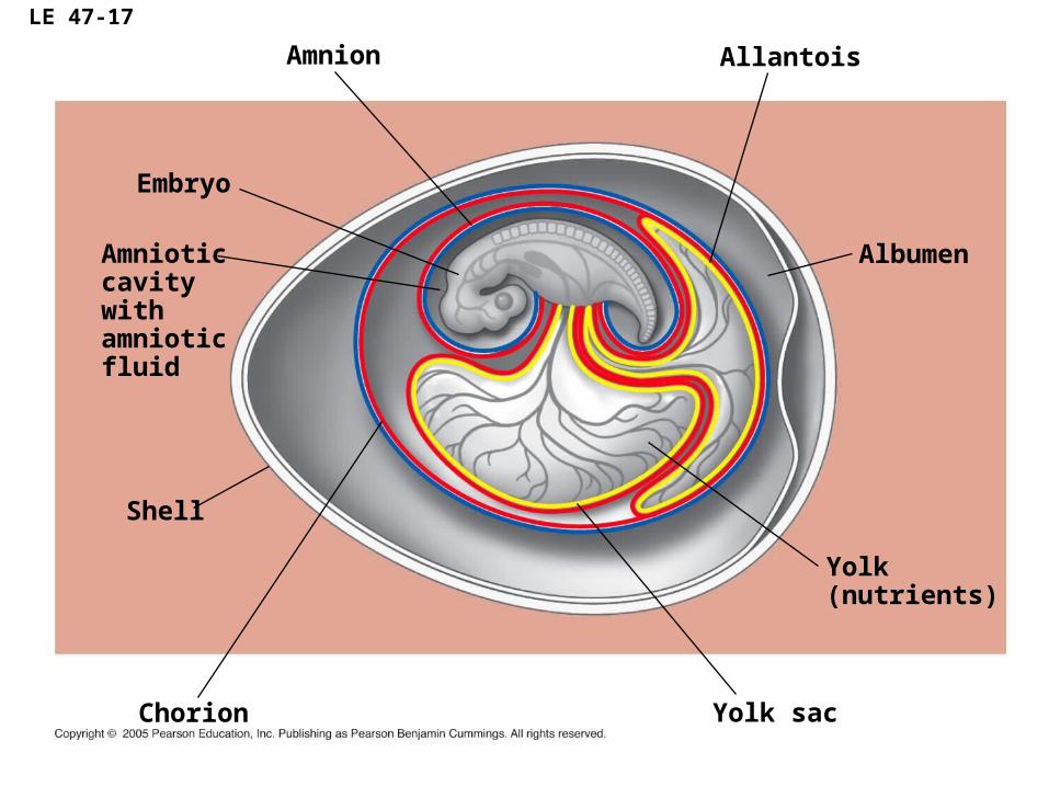
LE 47-17
Embryo
Amnioticcavitywithamnioticfluid
AllantoisAmnion
Albumen
Yolk(nutrients)
Yolk sacChorion
Shell

Copyright © 2005 Pearson Education, Inc. publishing as Benjamin Cummings
Mammalian Development
• The eggs of placental mammals
– Are small and store few nutrients
– Exhibit holoblastic cleavage
– Show no obvious polarity
• Gastrulation and organogenesis resemble the processes in birds and other reptiles
• Early cleavage is relatively slow in humans and other mammals

Copyright © 2005 Pearson Education, Inc. publishing as Benjamin Cummings
• At completion of cleavage, the blastocyst forms
• The trophoblast, the outer epithelium of the blastocyst, initiates implantation in the uterus, and the blastocyst forms a flat disk of cells
• As implantation is completed, gastrulation begins
• The extraembryonic membranes begin to form
• By the end of gastrulation, the embryonic germ layers have formed

LE 47-18a
Blastocystreaches uterus.
Endometrium(uterine lining)
Maternalbloodvessel
Blastocystimplants.
Inner cell mass
Trophoblast
Blastocoel
Hypoblast
Trophoblast
Epiblast
Expandingregion oftrophoblast

LE 47-18b
Hypoblast
Chorion (fromtrophoblast
Epiblast
Amnioticcavity
Amnion
Yolk sac (fromhypoblast)
Extraembryonic mesoderm cells(from epiblast)
Extraembryonicmembranes startto form andgastrulationbegins.
Amnion
Chorion
Endoderm
Mesoderm
Ectoderm
Yolk sac
Extraembryonicmesoderm
Gastrulation has produced a three-layered embryo with fourextraembryonic membranes.
Allantois
Expandingregion oftrophoblast
Copyright © 2005 Pearson Education, Inc. publishing as Benjamin Cummings
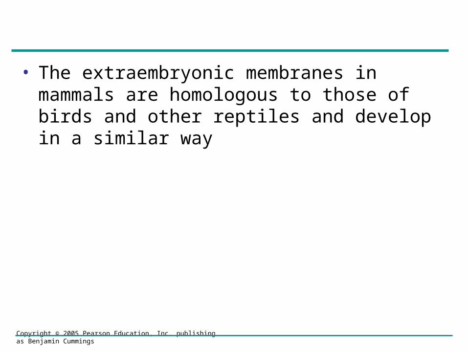
• The extraembryonic membranes in mammals are homologous to those of birds and other reptiles and develop in a similar way
Copyright © 2005 Pearson Education, Inc. publishing as Benjamin Cummings
Concept 47.2: Morphogenesis in animals involves specific changes in cell shape, position, and adhesion
• Morphogenesis is a major aspect of development in plants and animals
• But only in animals does it involve the movement of cells

Copyright © 2005 Pearson Education, Inc. publishing as Benjamin Cummings
The Cytoskeleton, Cell Motility, and Convergent Extension
• Changes in cell shape usually involve reorganization of the cytoskeleton
• Microtubules and microfilaments affect formation of the neural tube

LE 47-19Ectoderm
Neuralplate
Copyright © 2005 Pearson Education, Inc. publishing as Benjamin Cummings
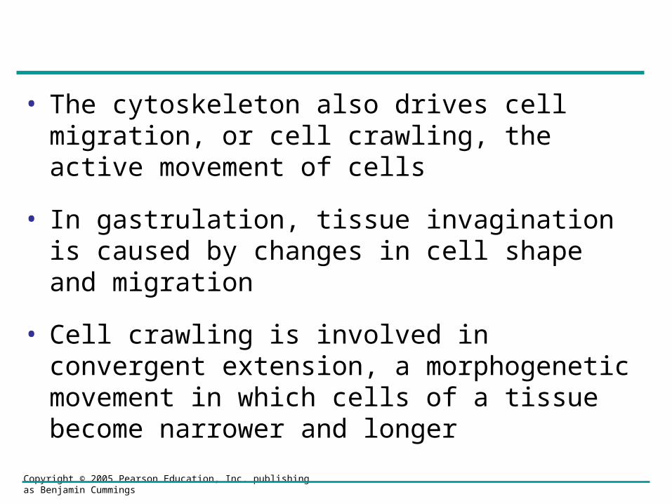
• The cytoskeleton also drives cell migration, or cell crawling, the active movement of cells
• In gastrulation, tissue invagination is caused by changes in cell shape and migration
• Cell crawling is involved in convergent extension, a morphogenetic movement in which cells of a tissue become narrower and longer

LE 47-20
ConvergenceExtension

Copyright © 2005 Pearson Education, Inc. publishing as Benjamin Cummings
Roles of the Extracellular Matrix and Cell Adhesion Molecules
• Fibers of the extracellular matrix may function as tracks, directing migrating cells along routes
• Several kinds of glycoproteins, including fibronectin, promote cell migration by providing molecular anchorage for moving cells
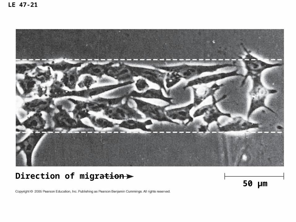
LE 47-21
Direction of migration50 µm

Copyright © 2005 Pearson Education, Inc. publishing as Benjamin Cummings
• Cell adhesion molecules contribute to cell migration and stable tissue structure
• One class of cell-to-cell adhesion molecule is the cadherins, which are important in formation of the frog blastula

LE 47-22
Control embryo
Experimental embryo
Copyright © 2005 Pearson Education, Inc. publishing as Benjamin Cummings
Concept 47.3: The developmental fate of cells depends on their history and on inductive signals
• Coupled with morphogenetic changes, development requires timely differentiation of cells at specific locations
• Two general principles underlie differentiation:
– During early cleavage divisions, embryonic cells must become different from one another
– After cell asymmetries are set up, interactions among embryonic cells influence their fate, usually causing changes in gene expression

Copyright © 2005 Pearson Education, Inc. publishing as Benjamin Cummings
Fate Mapping
• Fate maps are general territorial diagrams of embryonic development
• Classic studies using frogs indicated that cell lineage in germ layers is traceable to blastula cells

LE 47-23a
Fate map of a frog embryo
Epidermis Centralnervoussystem
Blastula
Epidermis
Neural tube stage(transverse section)
Endoderm
Mesoderm
Notochord

Copyright © 2005 Pearson Education, Inc. publishing as Benjamin Cummings
• Techniques in later studies marked an individual blastomere during cleavage and followed it through development

LE 47-23b
Cell lineage analysis in a tunicate
Copyright © 2005 Pearson Education, Inc. publishing as Benjamin Cummings
Establishing Cellular Asymmetries
• To understand how embryonic cells acquire their fates, think about how basic axes of the embryo are established

Copyright © 2005 Pearson Education, Inc. publishing as Benjamin Cummings
The Axes of the Basic Body Plan
• In nonamniotic vertebrates, basic instructions for establishing the body axes are set down early, during oogenesis or fertilization
• In amniotes, local environmental differences play the major role in establishing initial differences between cells and, later, the body axes

Copyright © 2005 Pearson Education, Inc. publishing as Benjamin Cummings
Restriction of Cellular Potency
• In many species that have cytoplasmic determinants, only the zygote is totipotent
• That is, only the zygote can develop into all the cell types in the adult
Copyright © 2005 Pearson Education, Inc. publishing as Benjamin Cummings
• Unevenly distributed cytoplasmic determinants in the egg cell help establish the body axes
• These determinants set up differences in blastomeres resulting from cleavage

LE 47-24
Left (control): Fertilized salamander eggs were allowed to divide normally, resulting in the gray crescent being evenly divided between the two blastomeres.
Right (experimental): Fertilized eggs were constricted by a thread so that the first cleavage plane restricted the gray crescent to one blastomere.
Gray crescent
Gray crescent
Normal Bellypiece
Normal
The two blastomeres were then separated and allowed to develop.

Copyright © 2005 Pearson Education, Inc. publishing as Benjamin Cummings
• As embryonic development proceeds, potency of cells becomes more limited

Copyright © 2005 Pearson Education, Inc. publishing as Benjamin Cummings
Cell Fate Determination and Pattern Formation by Inductive Signals
• After embryonic cell division creates cells that differ from each other, the cells begin to influence each other’s fates by induction
Copyright © 2005 Pearson Education, Inc. publishing as Benjamin Cummings
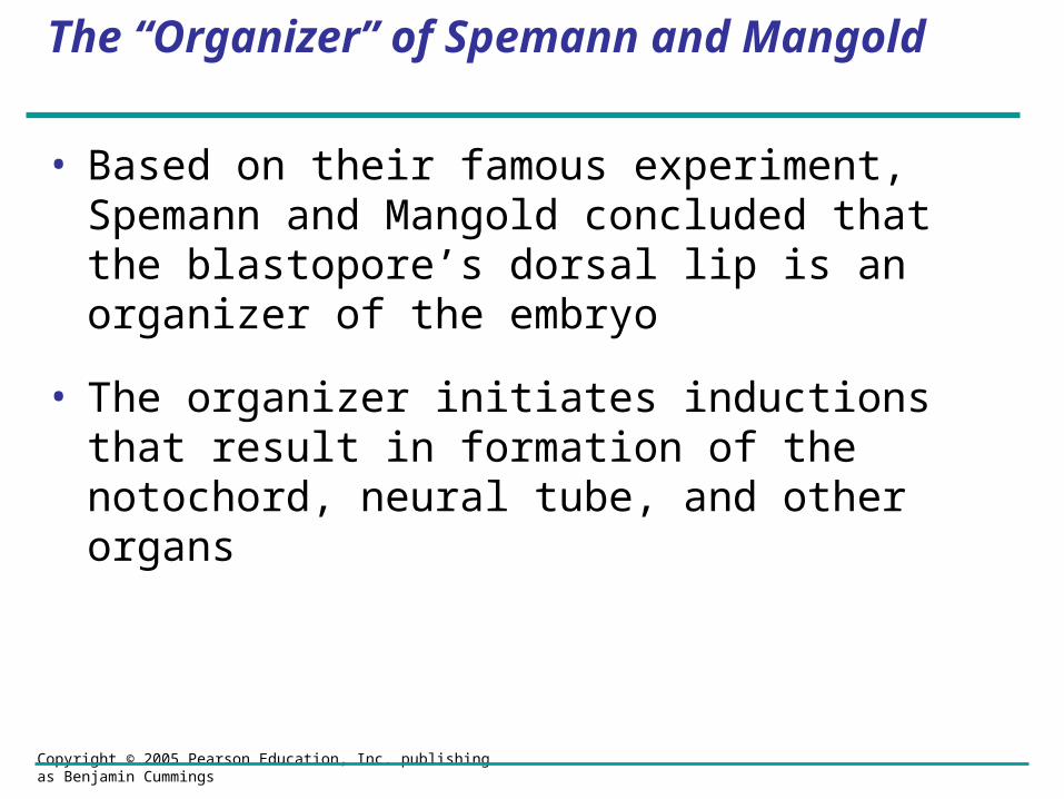
The “Organizer” of Spemann and Mangold
• Based on their famous experiment, Spemann and Mangold concluded that the blastopore’s dorsal lip is an organizer of the embryo
• The organizer initiates inductions that result in formation of the notochord, neural tube, and other organs

LE 47-25a
Nonpigmented gastrula(recipient embryo)
Pigmented gastrula(donor embryo)
Dorsal lip ofblastopore

LE 47-25b
Secondary (induced) embryo
Primarystructures:
Secondarystructures:
Neural tube
Notochord
Notochord (pigmented cells)
Neural tube (mostly nonpigmented cells)
Primary embryo

Copyright © 2005 Pearson Education, Inc. publishing as Benjamin Cummings
Formation of the Vertebrate Limb
• Inductive signals play a major role in pattern formation, development of spatial organization

Copyright © 2005 Pearson Education, Inc. publishing as Benjamin Cummings
• The molecular cues that control pattern formation are called positional information
• This information tells a cell where it is with respect to the body axes
• It determines how the cell and its descendents respond to future molecular signals

Copyright © 2005 Pearson Education, Inc. publishing as Benjamin Cummings
• The wings and legs of chicks, like all vertebrate limbs, begin as bumps of tissue called limb buds
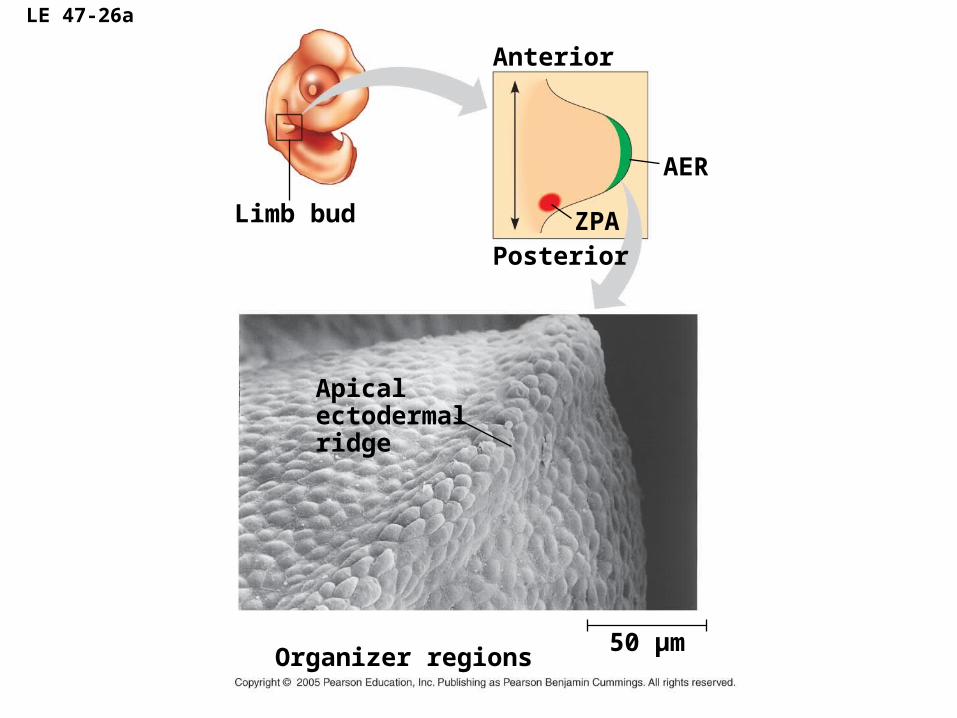
LE 47-26a
Anterior
Organizer regions
Limb bud
PosteriorZPA
AER
50 µm
Apicalectodermalridge

Copyright © 2005 Pearson Education, Inc. publishing as Benjamin Cummings
• The embryonic cells in a limb bud respond to positional information indicating location along three axes

LE 47-26b
Digits
AnteriorVentral
Distal
Posterior
Proximal
Dorsal
Wing of chick embryo
Copyright © 2005 Pearson Education, Inc. publishing as Benjamin Cummings
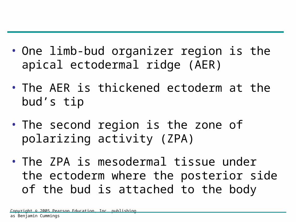
• One limb-bud organizer region is the apical ectodermal ridge (AER)
• The AER is thickened ectoderm at the bud’s tip
• The second region is the zone of polarizing activity (ZPA)
• The ZPA is mesodermal tissue under the ectoderm where the posterior side of the bud is attached to the body
Copyright © 2005 Pearson Education, Inc. publishing as Benjamin Cummings
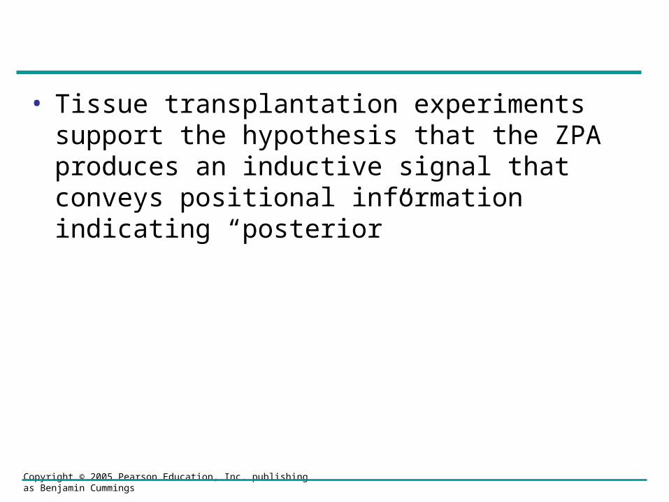
• Tissue transplantation experiments support the hypothesis that the ZPA produces an inductive signal that conveys positional information indicating “posterior”
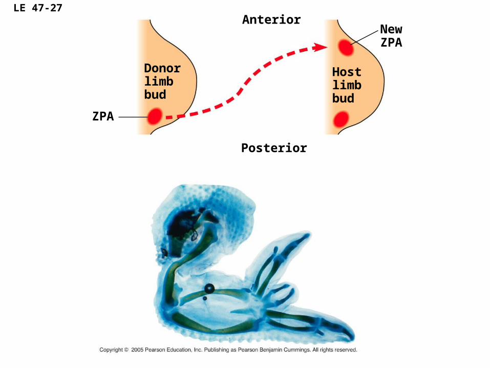
LE 47-27Anterior
Posterior
New ZPA
Hostlimbbud
ZPA
Donorlimbbud
Copyright © 2005 Pearson Education, Inc. publishing as Benjamin Cummings
• Signal molecules produced by inducing cells influence gene expression in cells receiving them
• Signal molecules lead to differentiation and the development of particular structures