Embed Size (px)
Citation preview
Chapter 44Chapter 44
Osteoporosis Associated with Pregnancy
Copyright © 2013 Elsevier Inc. All rights reserved.
Copyright © 2013 Elsevier Inc. All rights reserved.
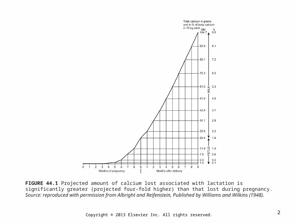
FIGURE 44.1 Projected amount of calcium lost associated with lactation is significantly greater (projected four-fold higher) than that lost during pregnancy. Source: reproduced with permission from Albright and Reifenstein, Published by Williams and Wilkins (1948).
2
Copyright © 2013 Elsevier Inc. All rights reserved.
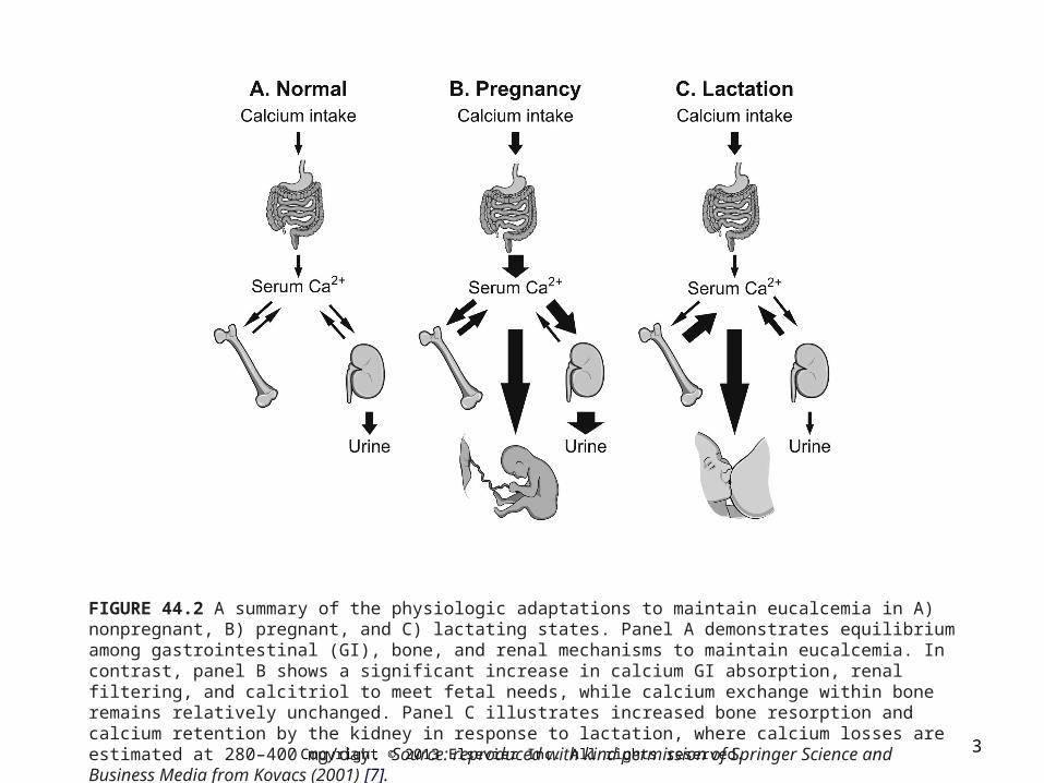
FIGURE 44.2 A summary of the physiologic adaptations to maintain eucalcemia in A) nonpregnant, B) pregnant, and C) lactating states. Panel A demonstrates equilibrium among gastrointestinal (GI), bone, and renal mechanisms to maintain eucalcemia. In contrast, panel B shows a significant increase in calcium GI absorption, renal filtering, and calcitriol to meet fetal needs, while calcium exchange within bone remains relatively unchanged. Panel C illustrates increased bone resorption and calcium retention by the kidney in response to lactation, where calcium losses are estimated at 280–400 mg/day. Source: reproduced with kind permission of Springer Science and Business Media from Kovacs (2001) [7].
3
Copyright © 2013 Elsevier Inc. All rights reserved.
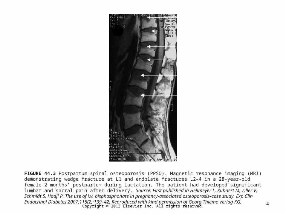
FIGURE 44.3 Postpartum spinal osteoporosis (PPSO). Magnetic resonance imaging (MRI) demonstrating wedge fracture at L1 and endplate fractures L2–4 in a 28-year-old female 2 months’ postpartum during lactation. The patient had developed significant lumbar and sacral pain after delivery. Source: First published in Hellmeyer L, Kuhnert M, Ziller V, Schmidt S, Hadji P. The use of i.v. bisphosphonate in pregnancy-associated osteoporosis–case study. Exp Clin Endocrinol Diabetes 2007;115(2):139–42. Reproduced with kind permission of Georg Thieme Verlag KG. 4
Copyright © 2013 Elsevier Inc. All rights reserved.
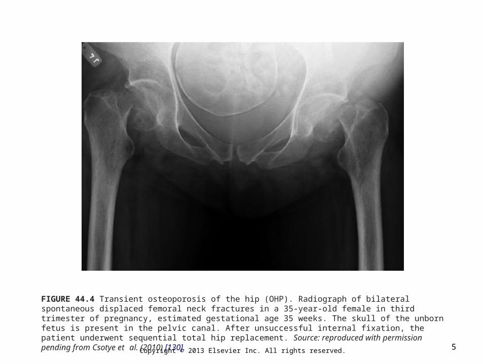
FIGURE 44.4 Transient osteoporosis of the hip (OHP). Radiograph of bilateral spontaneous displaced femoral neck fractures in a 35-year-old female in third trimester of pregnancy, estimated gestational age 35 weeks. The skull of the unborn fetus is present in the pelvic canal. After unsuccessful internal fixation, the patient underwent sequential total hip replacement. Source: reproduced with permission pending from Csotye et al. (2010) [130].
5