Embed Size (px)
Citation preview
43
CHAPTER-4 PLANT PROFILE AND EVALUATION OF ANTIEPILEPTIC
ACTIVITY OF ACALYPHA FRUTICOSA
S. No Name of the Sub-Title Page No
4.1 Taxonomy 45-45
4.2 Distribution 45-45
4.3 Description 45-46
4.4 Chemical constituents 46-46
4.5 Traditional uses 47-47
4.6
4.7
Previous Investigations
Reason for selection
47-51
51-51
4.8 Materials and methods 51-55
4.9 Results 55-83
4.10 Discussion 84-88
44
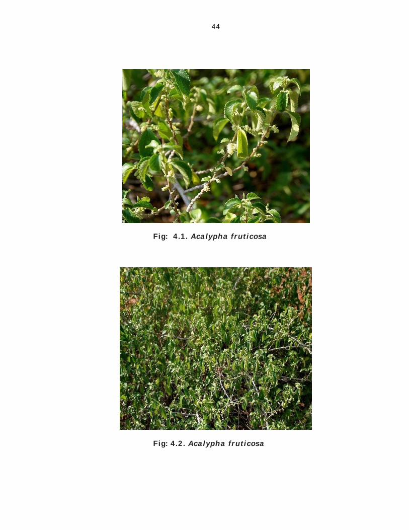
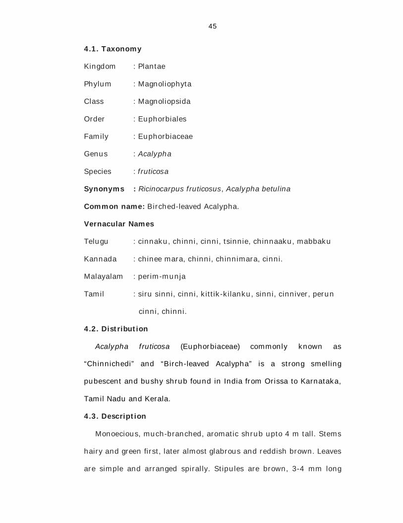
Fig: 4.1. Acalypha fruticosa
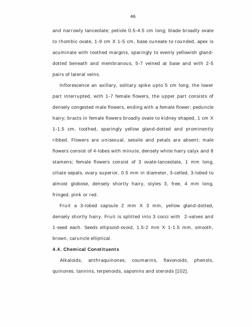
Fig: 4.2. Acalypha fruticosa
45
4.1. Taxonomy
Kingdom : Plantae
Phylum : Magnoliophyta
Class : Magnoliopsida
Order : Euphorbiales
Family : Euphorbiaceae
Genus : Acalypha
Species : fruticosa
Synonyms : Ricinocarpus fruticosus, Acalypha betulina
Common name: Birched-leaved Acalypha.
Vernacular Names
Telugu : cinnaku, chinni, cinni, tsinnie, chinnaaku, mabbaku
Kannada : chinee mara, chinni, chinnimara, cinni.
Malayalam : perim-munja
Tamil : siru sinni, cinni, kittik-kilanku, sinni, cinniver, perun
cinni, chinni.
4.2. Distribution
Acalypha fruticosa (Euphorbiaceae) commonly known as
“Chinnichedi” and “Birch-leaved Acalypha” is a strong smelling
pubescent and bushy shrub found in India from Orissa to Karnataka,
Tamil Nadu and Kerala.
4.3. Description
Monoecious, much-branched, aromatic shrub upto 4 m tall. Stems
hairy and green first, later almost glabrous and reddish brown. Leaves
are simple and arranged spirally. Stipules are brown, 3-4 mm long
46
and narrowly lanceolate; petiole 0.5-4.5 cm long; blade broadly ovate
to rhombic ovate, 1-9 cm X 1-5 cm, base cuneate to rounded, apex is
acuminate with toothed margins, sparingly to evenly yellowish gland-
dotted beneath and membranous, 5-7 veined at base and with 2-5
pairs of lateral veins.
Inflorescence an axillary, solitary spike upto 5 cm long, the lower
part interrupted, with 1-7 female flowers, the upper part consists of
densely congested male flowers, ending with a female flower; peduncle
hairy; bracts in female flowers broadly ovate to kidney shaped, 1 cm X
1-1.5 cm, toothed, sparingly yellow gland-dotted and prominently
ribbed. Flowers are unisexual, sessile and petals are absent; male
flowers consist of 4-lobes with minute, densely white hairy calyx and 8
stamens; female flowers consist of 3 ovate-lanceolate, 1 mm long,
ciliate sepals, ovary superior, 0.5 mm in diameter, 3-celled, 3-lobed to
almost globose, densely shortly hairy, styles 3, free, 4 mm long,
fringed, pink or red.
Fruit a 3-lobed capsule 2 mm X 3 mm, yellow gland-dotted,
densely shortly hairy. Fruit is splitted into 3 cocci with 2-valves and
1-seed each. Seeds ellipsoid-ovoid, 1.5-2 mm X 1-1.5 mm, smooth,
brown, caruncle elliptical.
4.4. Chemical Constituents
Alkaloids, anthraquinones, coumarins, flavonoids, phenols,
quinones, tannins, terpenoids, saponins and steroids [102].
47
4.5. Traditional uses
In traditional system of medicine the young twigs and leaves of the
plant are prescribed in the treatment of dyspepsia, colic, diarrhoea
and in cholera. The Paliyar and Irula tribes in Western Ghats of South
India use leaves and roots of Acalypha fruticosa to treat stomachache,
skin diseases, wounds and poisonous bites [103]. In Siddha Materia
Medica it is used to treat poisonous bites. Medicinally the leaves are
used as stomachic in dyspepsia, attenuant, alterative and vulnerary
[104]. In Yemen, leaf and stem have been used to treat skin diseases,
malaria and wounds [105].
In Tanzania, a leaf decoction is drunk to treat epilepsy. Leaf
maceration is used to treat eye infections. A leaf infusion is used to
treat stomach problems and swellings of the body. Leaf sap is used as
nose drops to treat cough and chest problems. Leaf paste is applied to
scabies and sores. Stems ground in water are applied to wounds of
animals. The Suiei hunter-gatherers of northern Kenya used root
decoction to treat convulsions, colds, fever and swellings of the
scrotum. They used root infusion to treat whooping cough. They
chewed stem and root to relieve toothache. In southern Africa a root
decoction is taken to treat snakebites, fever and ulcers of venereal
origin. Ground fresh leaves mixed with water are rubbed in and
inhaled as a sedative [21].
4.6. Previous Investigations
• Mathad et al., evaluated the antidiarrhoeal activity of methanol
and aqueous extracts of Acalypha fruticosa leaves by using
48
castor oil-induced diarrhoea model in rats. They reported that
methanol and aqueous extracts decreased propulsion of the
charcoal meal (p<0.001) through the gastrointestinal tract at the
oral dose of 200mg/kg as compared with control group [106].
• Gupta et al., reported the antioxidant and anti-inflammatory
activities of the methanol extract of the leaves of Acalypha
fruticosa by DPPH radical scavenging assay and in vitro lipid
perioxdation induced by Fe2+-ascorbate system in the rat liver
homogenate, carrageenan induced paw oedema and cotton
pellet induced granuloma in rats. They reported that the extract
showed marked free radical scavenging activities in DPPH
radical scavenging assay, in vitro lipid perioxidation induced by
Fe2+-ascorbate system in the rat liver homogenate and
significant anti-inflammatory activity in carrageenan-induced
paw oedema in rats [107].
• Duraipandiyan and coworkers reported the antimicrobial
activity of the hexane and methanol extracts of Acalypha
fruticosa against some bacterial strains. They reported that the
methanol extract showed significant activity against tested
organisms when compared to hexane extract [108].
• Alshawsh and coworkers reported the antimalarial activity of
the methanol and aqueous extracts of Acalypha fruticosa leaves
against Plasmodium falciparum. They found that the extract
have significant antiplasmodial activity with IC50 values less
than 4µg/ml [109].
49
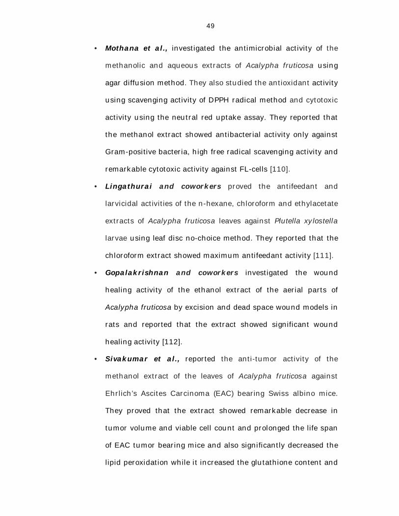
• Mothana et al., investigated the antimicrobial activity of the
methanolic and aqueous extracts of Acalypha fruticosa using
agar diffusion method. They also studied the antioxidant activity
using scavenging activity of DPPH radical method and cytotoxic
activity using the neutral red uptake assay. They reported that
the methanol extract showed antibacterial activity only against
Gram-positive bacteria, high free radical scavenging activity and
remarkable cytotoxic activity against FL-cells [110].
• Lingathurai and coworkers proved the antifeedant and
larvicidal activities of the n-hexane, chloroform and ethylacetate
extracts of Acalypha fruticosa leaves against Plutella xylostella
larvae using leaf disc no-choice method. They reported that the
chloroform extract showed maximum antifeedant activity [111].
• Gopalakrishnan and coworkers investigated the wound
healing activity of the ethanol extract of the aerial parts of
Acalypha fruticosa by excision and dead space wound models in
rats and reported that the extract showed significant wound
healing activity [112].
• Sivakumar et al., reported the anti-tumor activity of the
methanol extract of the leaves of Acalypha fruticosa against
Ehrlich’s Ascites Carcinoma (EAC) bearing Swiss albino mice.
They proved that the extract showed remarkable decrease in
tumor volume and viable cell count and prolonged the life span
of EAC tumor bearing mice and also significantly decreased the
lipid peroxidation while it increased the glutathione content and
50
superoxide dismutase level as compared to that of EAC control
group [113].
• Ireri and coworkers reported the insecticidal activity of the
methanol and ethyl acetate extracts of aerial parts of Acalypha
fruticosa against sandflies (Phlebotomus duboiscqi). They
reported that the extracts were found to be insecticidal to adult
sandflies [114].
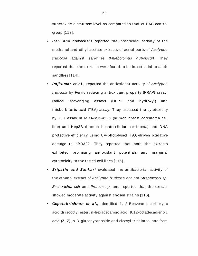
• Rajkumar et al., reported the antioxidant activity of Acalypha
fruticosa by Ferric reducing antioxidant property (FRAP) assay,
radical scavenging assays (DPPH and hydroxyl) and
thiobarbituric acid (TBA) assay. They assessed the cytotoxicity
by XTT assay in MDA-MB-435S (human breast carcinoma cell
line) and Hep3B (human hepatocellular carcinoma) and DNA
protective efficiency using UV-photolysed H2O2-driven oxidative
damage to pBR322. They reported that both the extracts
exhibited promising antioxidant potentials and marginal
cytotoxicity to the tested cell lines [115].
• Sripathi and Sankari evaluated the antibacterial activity of
the ethanol extract of Acalypha fruticosa against Streptococci sp,
Escherichia coli and Proteus sp. and reported that the extract
showed moderate activity against chosen strains [116].
• Gopalakrishnan et al., identified 1, 2-Benzene dicarboxylic
acid di isooctyl ester, n-hexadecanoic acid, 9,12-octadecadienoic
acid (Z, Z), α-D-glucopyranoside and eicosyl trichlorosilane from
51
the aerial parts of Acalypha fruticosa by GC-MS analysis of the
ethanol extract [102].
• Deepa et al., isolated β–caryophyllene, α-humulene,
isocaryophyllene, caryophyllene oxide and tans-phytol from the
essential oil of Acalypha fruticosa leaves [117].
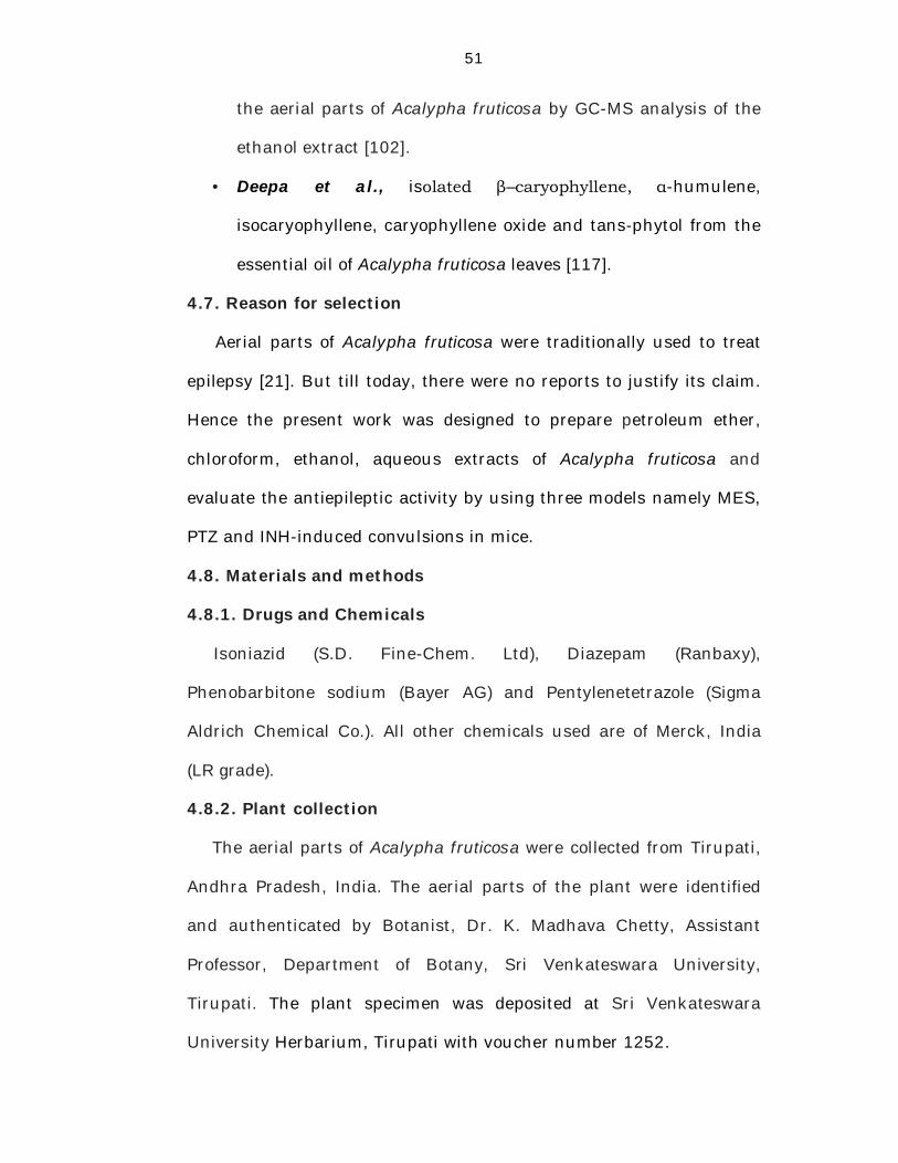
4.7. Reason for selection
Aerial parts of Acalypha fruticosa were traditionally used to treat
epilepsy [21]. But till today, there were no reports to justify its claim.
Hence the present work was designed to prepare petroleum ether,
chloroform, ethanol, aqueous extracts of Acalypha fruticosa and
evaluate the antiepileptic activity by using three models namely MES,
PTZ and INH-induced convulsions in mice.
4.8. Materials and methods
4.8.1. Drugs and Chemicals
Isoniazid (S.D. Fine-Chem. Ltd), Diazepam (Ranbaxy),
Phenobarbitone sodium (Bayer AG) and Pentylenetetrazole (Sigma
Aldrich Chemical Co.). All other chemicals used are of Merck, India
(LR grade).
4.8.2. Plant collection
The aerial parts of Acalypha fruticosa were collected from Tirupati,
Andhra Pradesh, India. The aerial parts of the plant were identified
and authenticated by Botanist, Dr. K. Madhava Chetty, Assistant
Professor, Department of Botany, Sri Venkateswara University,
Tirupati. The plant specimen was deposited at Sri Venkateswara
University Herbarium, Tirupati with voucher number 1252.
52
4.8.3. Preparation of the extracts
The fresh aerial parts of Acalypha fruticosa were collected, shade
dried and was made in to coarse powder. Then petroleum ether,
chloroform, ethanol and aqueous extracts were prepared by following
maceration method [118].
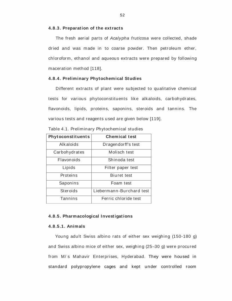
4.8.4. Preliminary Phytochemical Studies
Different extracts of plant were subjected to qualitative chemical
tests for various phytoconstituents like alkaloids, carbohydrates,
flavonoids, lipids, proteins, saponins, steroids and tannins. The
various tests and reagents used are given below [119].
Table 4.1. Preliminary Phytochemical studies
Phytoconstituents Chemical test
Alkaloids Dragendorff’s test
Carbohydrates Molisch test
Flavonoids Shinoda test
Lipids Filter paper test
Proteins Biuret test
Saponins Foam test
Steroids Liebermann-Burchard test
Tannins Ferric chloride test
4.8.5. Pharmacological Investigations
4.8.5.1. Animals
Young adult Swiss albino rats of either sex weighing (150-180 g)
and Swiss albino mice of either sex, weighing (25–30 g) were procured
from M/s Mahavir Enterprises, Hyderabad. They were housed in
standard polypropylene cages and kept under controlled room
53
temperature (24±20C; relative humidity 60-70%) in a 12h light – dark
cycle. The rats and mice were given a standard laboratory diet and
water ad libitum. The animals were acclimatizated before the study.
The experimental protocol was approved by the Institutional Animals
Ethics Committee (IAEC) of Talla Padmavathi College of Pharmacy,
Warangal, Andhra Pradesh (CPCSEA no. 1505/PO/a/11/CPCSEA).
4.8.5.2. Acute toxicity studies
Acute toxicity study was performed for the extracts to ascertain
safe dose by acute oral toxic class method of Organization of
Economic Co-operation and Development, as per 420 guidelines
(OECD). Young adult Swiss albino rats and Swiss albino mice of either
sex were used for the study. Each extract of plant was tested in both
the species upto a dose of 2000 mg/kg, body weight [120].
4.8.5.3. Evaluation of Anti-epileptic activity
4.8.5.3.1. Maximum Electroshock (MES) in mice
Five groups of six Swiss albino mice (25–30 g) of either sex were
used. Mice belonging to Group I were treated with the vehicle, Group
II, III and IV were treated with different doses (30, 100 and 300
mg/kg, p.o.) of Acalypha fruticosa respectively. Mice belonging to
Group V received diazepam (standard) at the dose of 3 mg/kg, p.o.
The test was started one hour after oral treatment with the extract or
the vehicle or the standard. Tonic hind limb extensions were induced
by an apparatus with corneal electrodes. The intensity of the stimulus
was dependent on the apparatus, eg: 45 mA, 50Hz for 0.2 sec has
54
been used. Percentage of inhibition of convulsions relative to control
was calculated [40].
Control - Treated Percentage of Inhibition = -------------------------- X 100 Control
Same treatment schedule was followed for chloroform, ethanol and
aqueous extracts.
4.8.5.3.2. Pentylenetetrazole (PTZ)-induced convulsions
Mice of either sex were randomly allotted to five different groups of
six mice each. Mice belonging to Group I received the vehicle, Group
II, III and IV received Acalypha fruticosa at the doses of 30, 100 and
300 mg/kg, p.o. respectively. Mice belonging to Group V received
phenobarbitone sodium (standard) at the dose of 40 mg/kg, i.p. Mice
belonging to Group I were administered with pentylenetetrazole (PTZ)
(75 mg/kg, i.p.) one hour after vehicle. Mice belonging to Group V
received PTZ, 15 min after phenobarbitone sodium (40 mg/kg, i.p.).
Mice belonging to Group II, III and IV mice received different doses of
plant extracts, p.o. one hour before PTZ. Onset time as well as
duration of convulsions were recorded [85].
Same treatment schedule was followed for chloroform, ethanol and
aqueous extracts.
4.8.5.3.3. Isoniazid (INH)-induced convulsions
Five Groups of six Swiss albino mice (25–30 g) of either sex were
used. Mice belonging to Group I were treated with the vehicle, Group
II, III and IV were treated with different extracts of Acalypha fruticosa
55
at the doses of 30, 100 and 300 mg/kg, p.o. Mice belonging to Group
V received the standard drug, diazepam at the dose of 4 mg/kg, i.p.
One hour after the administration of vehicle or different extracts of
Acalypha fruticosa, isoniazid at a dose of 300mg/kg, s.c. was
administered to mice belonging to Group I, II, III, IV and 15 min after
administration of diazepam to mice belonging to Group V. The mice
were placed in isolated perplex chamber and the latency of
convulsions was recorded [121].
Same treatment schedule was followed for chloroform, ethanol and
aqueous extracts.
4.8.6. Statistical analysis:
The data was analyzed using one-way analysis of variance
(ANOVA), followed by Dunnett’s test and p<0.05 was considered as
statistically significant. The data was expressed as mean ± Standard
deviation (SD).
4.9. Results
4.9.1. Percentage yield of different extracts of Acalypha fruticosa
After extraction with different solvents by maceration method, the
percentage yield was calculated. The percentage yield obtained for
petroleum ether extract-12.74, chloroform extract-14.55, ethanol
extract-31.99 and aqueous extract-9.96.
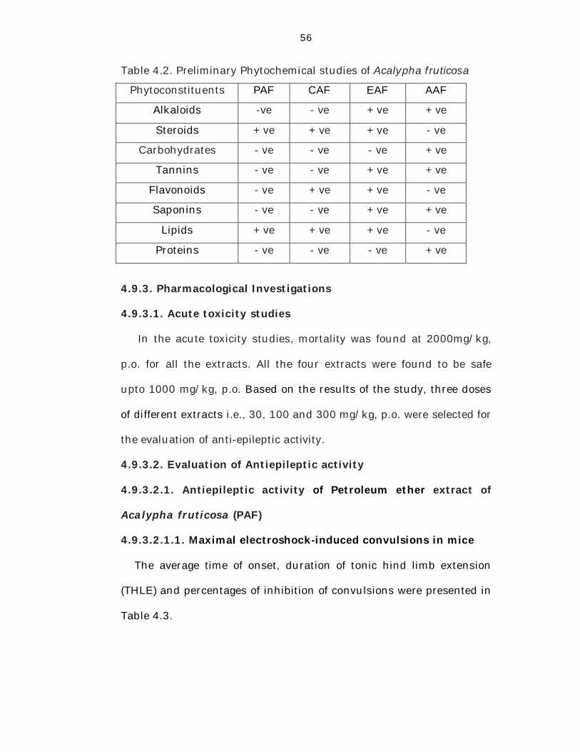
4.9.2. Preliminary Phytochemical Studies
Preliminary phytochemical studies gave positive tests for alkaloids,
steroids, carbohydrates, tannins, flavanoids, saponins, lipids and
proteins in various extracts of the plants.
56
Table 4.2. Preliminary Phytochemical studies of Acalypha fruticosa
Phytoconstituents PAF CAF EAF AAF
Alkaloids -ve - ve + ve + ve
Steroids + ve + ve + ve - ve
Carbohydrates - ve - ve - ve + ve
Tannins - ve - ve + ve + ve
Flavonoids - ve + ve + ve - ve
Saponins - ve - ve + ve + ve
Lipids + ve + ve + ve - ve
Proteins - ve - ve - ve + ve
4.9.3. Pharmacological Investigations
4.9.3.1. Acute toxicity studies
In the acute toxicity studies, mortality was found at 2000mg/kg,
p.o. for all the extracts. All the four extracts were found to be safe
upto 1000 mg/kg, p.o. Based on the results of the study, three doses
of different extracts i.e., 30, 100 and 300 mg/kg, p.o. were selected for
the evaluation of anti-epileptic activity.
4.9.3.2. Evaluation of Antiepileptic activity
4.9.3.2.1. Antiepileptic activity of Petroleum ether extract of
Acalypha fruticosa (PAF)
4.9.3.2.1.1. Maximal electroshock-induced convulsions in mice
The average time of onset, duration of tonic hind limb extension
(THLE) and percentages of inhibition of convulsions were presented in
Table 4.3.
57
Effect on onset time of convulsions
The onset time of THLE in control group animals was found to be
1.34±0.04 sec. PAF treated mice showed the onset time as 1.89±0.05,
2.25±0.05 and 3.04±0.08 sec (p<0.01) respectively at the doses of 30,
100 and 300 mg/kg, p.o. Animals treated with diazepam (3 mg/kg,
p.o.) showed onset time as 2.48±0.05 sec (p<0.01).
Effect on duration of convulsions
The duration of THLE in control group animals was 118.91±0.22
sec. Albino mice pretreated with PAF at the doses of 30, 100 and 300
mg/kg, p.o. showed the duration of 67.33±0.08, 58.66±0.06 and
44.79±0.14 sec (p<0.01) respectively. The standard group animals
(diazepam 3 mg/kg, p.o.) showed 49.36±0.06 sec (p<0.01).
It has been found that the time of onset of THLE in control group
animals was very less when compared to that of animals which
received extract and standard. Duration of THLE in control group
animals was greater when compared to that of animals which received
extract and standard. Albino mice pretreated with PAF at the doses of
30, 100 and 300 mg/kg were provided significant protection from
convulsions induced by electroshock method.
Percentage inhibition of convulsions
The percentage of inhibition achieved in mice pretreated with PAF
at the doses of 30, 100 and 300 mg/kg were 43.38%, 50.67% and
62.33% (p<0.01) respectively when compared to control group
animals. Animals treated with PAF exhibited significant antiepileptic
58
activity and more percentage of inhibition of convulsions at the dose of
300 mg/kg when compared to diazepam treated animals (58.49%).
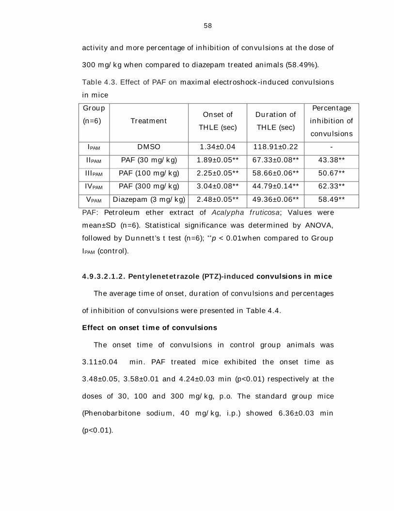
Table 4.3. Effect of PAF on maximal electroshock-induced convulsions
in mice
Group
(n=6)=
6)
Treatment Onset of
THLE (sec)
Duration of
THLE (sec)
Percentage
inhibition of
convulsions
IPAM DMSO 1.34±0.04 118.91±0.22 -
IIPAM PAF (30 mg/kg) 1.89±0.05** 67.33±0.08** 43.38**
IIIPAM PAF (100 mg/kg) 2.25±0.05** 58.66±0.06** 50.67**
IVPAM PAF (300 mg/kg) 3.04±0.08** 44.79±0.14** 62.33**
VPAM Diazepam (3 mg/kg) 2.48±0.05** 49.36±0.06** 58.49**
PAF: Petroleum ether extract of Acalypha fruticosa; Values were
mean±SD (n=6). Statistical significance was determined by ANOVA,
followed by Dunnett’s t test (n=6); **p < 0.01when compared to Group
IPAM (control).
4.9.3.2.1.2. Pentylenetetrazole (PTZ)-induced convulsions in mice
The average time of onset, duration of convulsions and percentages
of inhibition of convulsions were presented in Table 4.4.
Effect on onset time of convulsions
The onset time of convulsions in control group animals was
3.11±0.04 min. PAF treated mice exhibited the onset time as
3.48±0.05, 3.58±0.01 and 4.24±0.03 min (p<0.01) respectively at the
doses of 30, 100 and 300 mg/kg, p.o. The standard group mice
(Phenobarbitone sodium, 40 mg/kg, i.p.) showed 6.36±0.03 min
(p<0.01).
59
Effect on duration of convulsions
The duration of convulsions in control group animals was
22.14±0.05 min. Animals pretreated with PAF at the doses of 30, 100
and 300 mg/kg, p.o. exhibited the duration as 10.43±0.08, 8.59±0.22
and 7.42±0.10 min (p<0.01) respectively. The standard group mice
(Phenobarbitone sodium 40 mg/kg, i.p.) showed 11.13±0.05 min
(p<0.01).
The time of onset of convulsions in control group animals was very
less when compared to the animals treated with extract and standard.
Duration of convulsions in control group animals was greater when
compared to the extract and standard treated animals. It has been
found that animals pretreated with PAF were significantly protected
from convulsions induced by PTZ in a dose-dependent manner.
Percentage inhibition of convulsions
The percentage of inhibition achieved in PAF treated mice were
52.89%, 61.20% and 66.49% (p<0.01) respectively at the doses of 30,
100 and 300 mg/kg when compared to control group animals.
Animals which were treated with PAF exhibited significant
antiepileptic activity and more percentage inhibition of convulsions
when compared to Phenobarbitone sodium treated mice (49.73%,
p<0.01).
60
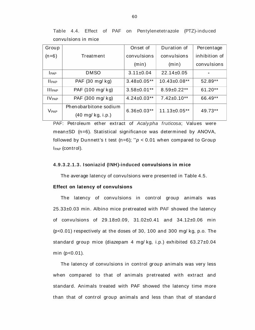
Table 4.4. Effect of PAF on Pentylenetetrazole (PTZ)-induced
convulsions in mice
Group
(n=6)=
6)
Treatment
Onset of
convulsions
(min)
Duration of
convulsions
(min)
Percentage
inhibition of
convulsions
IPAP DMSO 3.11±0.04 22.14±0.05 --
IIPAP PAF (30 mg/kg) 3.48±0.05** 10.43±0.08** 52.89**
IIIPAP PAF (100 mg/kg) 3.58±0.01** 8.59±0.22** 61.20**
IVPAP PAF (300 mg/kg) 4.24±0.03** 7.42±0.10** 66.49**
VPAP Phenobarbitone sodium
(40 mg/kg, i.p.) 6.36±0.03** 11.13±0.05** 49.73**
PAF: Petroleum ether extract of Acalypha fruticosa; Values were
mean±SD (n=6). Statistical significance was determined by ANOVA,
followed by Dunnett’s t test (n=6); **p < 0.01 when compared to Group
IPAP (control).
4.9.3.2.1.3. Isoniazid (INH)-induced convulsions in mice
The average latency of convulsions were presented in Table 4.5.
Effect on latency of convulsions
The latency of convulsions in control group animals was
25.33±0.03 min. Albino mice pretreated with PAF showed the latency
of convulsions of 29.18±0.09, 31.02±0.41 and 34.12±0.06 min
(p<0.01) respectively at the doses of 30, 100 and 300 mg/kg, p.o. The
standard group mice (diazepam 4 mg/kg, i.p.) exhibited 63.27±0.04
min (p<0.01).
The latency of convulsions in control group animals was very less
when compared to that of animals pretreated with extract and
standard. Animals treated with PAF showed the latency time more
than that of control group animals and less than that of standard
61
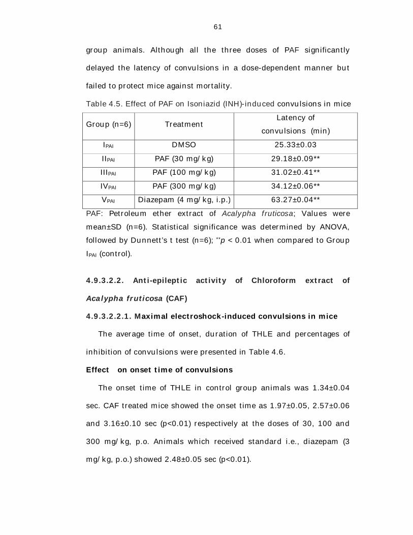
group animals. Although all the three doses of PAF significantly
delayed the latency of convulsions in a dose-dependent manner but
failed to protect mice against mortality.
Table 4.5. Effect of PAF on Isoniazid (INH)-induced convulsions in mice
Group (n=6) Treatment Latency of
convulsions (min)
IPAI DMSO 25.33±0.03
IIPAI PAF (30 mg/kg) 29.18±0.09**
IIIPAI PAF (100 mg/kg) 31.02±0.41**
IVPAI PAF (300 mg/kg) 34.12±0.06**
VPAI Diazepam (4 mg/kg, i.p.) 63.27±0.04**
PAF: Petroleum ether extract of Acalypha fruticosa; Values were
mean±SD (n=6). Statistical significance was determined by ANOVA,
followed by Dunnett’s t test (n=6); **p < 0.01 when compared to Group
IPAI (control).
4.9.3.2.2. Anti-epileptic activity of Chloroform extract of
Acalypha fruticosa (CAF)
4.9.3.2.2.1. Maximal electroshock-induced convulsions in mice
The average time of onset, duration of THLE and percentages of
inhibition of convulsions were presented in Table 4.6.
Effect on onset time of convulsions
The onset time of THLE in control group animals was 1.34±0.04
sec. CAF treated mice showed the onset time as 1.97±0.05, 2.57±0.06
and 3.16±0.10 sec (p<0.01) respectively at the doses of 30, 100 and
300 mg/kg, p.o. Animals which received standard i.e., diazepam (3
mg/kg, p.o.) showed 2.48±0.05 sec (p<0.01).
62
Effect on duration of convulsions
The duration of THLE in control group animals was 118.91±0.22
sec. Mice pretreated with CAF at the doses of 30, 100 and 300 mg/kg,
p.o. showed the duration of 62.07±0.13, 51.96±0.11 and 40.04±0.07
sec (p<0.01) respectively. The standard group mice (diazepam 3
mg/kg, p.o.) showed 49.36±0.06 sec (p<0.01).
The time of onset of THLE in control group animals was very less
when compared to the extract and standard group animals. Duration
of THLE in control group animals was greater when compared to the
extract and standard group animals. Albino mice pretreated with CAF
at doses 30, 100 and 300 mg/kg provided significant protection from
convulsions induced by electroshock method.
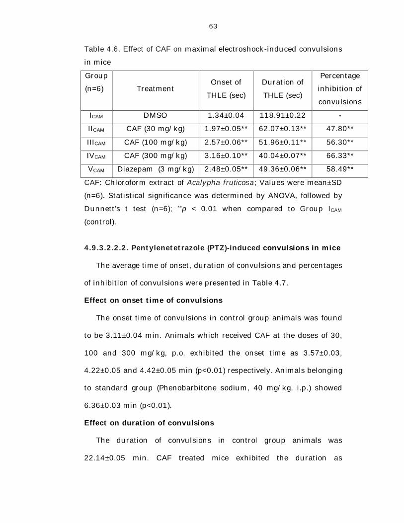
Percentage inhibition of convulsions
The percentage inhibition achieved in CAF treated animals were
47.80% (30 mg/kg), 56.30% (100 mg/kg) and 66.33% (300 mg/kg)
(p<0.01) respectively when compared to control group animals.
Animals pretreated with CAF exhibited significant antiepileptic activity
and more percentage inhibition of convulsions at the dose of 300
mg/kg when compared to diazepam treated animals (58.49%, p<0.01).
63
Table 4.6. Effect of CAF on maximal electroshock-induced convulsions
in mice
Group
(n=6)=
6)
Treatment Onset of
THLE (sec)
Duration of
THLE (sec)
Percentage
inhibition of
convulsions
ICAM DMSO 1.34±0.04 118.91±0.22 ---
IICAM CAF (30 mg/kg) 1.97±0.05** 62.07±0.13** 47.80**
IIICAM CAF (100 mg/kg) 2.57±0.06** 51.96±0.11** 56.30**
IVCAM CAF (300 mg/kg) 3.16±0.10** 40.04±0.07** 66.33**
VCAM Diazepam (3 mg/kg) 2.48±0.05** 49.36±0.06** 58.49**
CAF: Chloroform extract of Acalypha fruticosa; Values were mean±SD
(n=6). Statistical significance was determined by ANOVA, followed by
Dunnett’s t test (n=6); **p < 0.01 when compared to Group ICAM
(control).
4.9.3.2.2.2. Pentylenetetrazole (PTZ)-induced convulsions in mice
The average time of onset, duration of convulsions and percentages
of inhibition of convulsions were presented in Table 4.7.
Effect on onset time of convulsions
The onset time of convulsions in control group animals was found
to be 3.11±0.04 min. Animals which received CAF at the doses of 30,
100 and 300 mg/kg, p.o. exhibited the onset time as 3.57±0.03,
4.22±0.05 and 4.42±0.05 min (p<0.01) respectively. Animals belonging
to standard group (Phenobarbitone sodium, 40 mg/kg, i.p.) showed
6.36±0.03 min (p<0.01).
Effect on duration of convulsions
The duration of convulsions in control group animals was
22.14±0.05 min. CAF treated mice exhibited the duration as
64
10.01±0.35, 8.02±0.22 and 7.04±0.24 min (p<0.01) respectively at the
doses of 30, 100 and 300 mg/kg, p.o. The standard group mice
(Phenobarbitone sodium 40 mg/kg, i.p.) showed 11.13±0.05 min
(p<0.01).
The time of onset of convulsions in control group animals was very
less when compared to the extract and standard treated animals.
Duration of convulsions in control group animals was greater when
compared to the extract and standard treated animals. All the three
doses of CAF afforded significant protection in dose-dependent
manner against convulsions induced by PTZ.
Percentage inhibition of convulsions
The percentage of inhibition achieved in CAF treated mice were
54.79%, 63.78% and 68.20% (p<0.01) respectively at the doses of 30,
100 and 300 mg/kg when compared to control group animals.
Animals pretreated with CAF exhibited significant anti-epileptic
activity and more percentage of inhibition of convulsions when
compared to Phenobarbitone sodium treated animals (49.73%,
p<0.01).
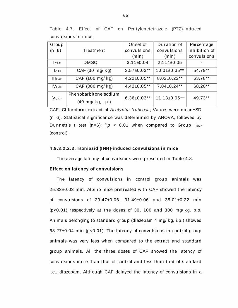
65
Table 4.7. Effect of CAF on Pentylenetetrazole (PTZ)-induced
convulsions in mice
Group (n=6)=
6) Treatment
Onset of convulsions
(min)
Duration of convulsions
(min)
Percentage inhibition of convulsions
ICAP D DMSO MSO 3.11±0.04 22.14±0.05 ---
IICAP CAF (30 mg/kg) 3.57±0.03** 10.01±0.35** 54.79**
IIICAP CAF (100 mg/kg) 4.22±0.05** 8.02±0.22** 63.78**
IVCAP CAF (300 mg/kg) 4.42±0.05** 7.04±0.24** 68.20**
VCAP Phenobarbitone sodium
(40 mg/kg, i.p.) 6.36±0.03** 11.13±0.05** 49.73**
CAF: Chloroform extract of Acalypha fruticosa; Values were mean±SD
(n=6). Statistical significance was determined by ANOVA, followed by
Dunnett’s t test (n=6); **p < 0.01 when compared to Group ICAP
(control).
4.9.3.2.2.3. Isoniazid (INH)-induced convulsions in mice
The average latency of convulsions were presented in Table 4.8.
Effect on latency of convulsions
The latency of convulsions in control group animals was
25.33±0.03 min. Albino mice pretreated with CAF showed the latency
of convulsions of 29.47±0.06, 31.49±0.06 and 35.01±0.22 min
(p<0.01) respectively at the doses of 30, 100 and 300 mg/kg, p.o.
Animals belonging to standard group (diazepam 4 mg/kg, i.p.) showed
63.27±0.04 min (p<0.01). The latency of convulsions in control group
animals was very less when compared to the extract and standard
group animals. All the three doses of CAF showed the latency of
convulsions more than that of control and less than that of standard
i.e., diazepam. Although CAF delayed the latency of convulsions in a
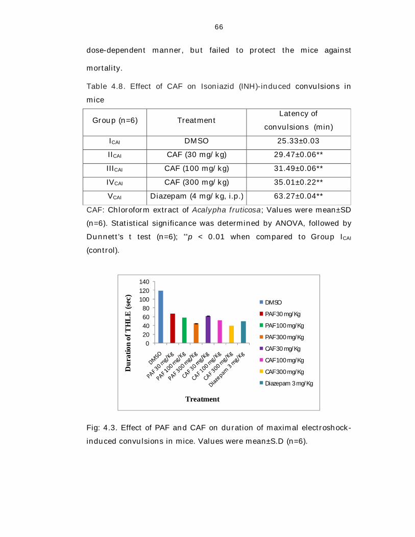
66
dose-dependent manner, but failed to protect the mice against
mortality.
Table 4.8. Effect of CAF on Isoniazid (INH)-induced convulsions in
mice
Group (n=6) Treatment Latency of
convulsions (min)
ICAI DMSO 25.33±0.03
IICAI CAF (30 mg/kg) 29.47±0.06**
IIICAI CAF (100 mg/kg) 31.49±0.06**
IVCAI CAF (300 mg/kg) 35.01±0.22**
VCAI Diazepam (4 mg/kg, i.p.) 63.27±0.04**
CAF: Chloroform extract of Acalypha fruticosa; Values were mean±SD
(n=6). Statistical significance was determined by ANOVA, followed by
Dunnett’s t test (n=6); **p < 0.01 when compared to Group ICAI
(control).
020406080
100120140
Dur
atio
n of
TH
LE
(sec
)
Treatment
DMSO
PAF 30 mg/Kg
PAF 100 mg/Kg
PAF 300 mg/Kg
CAF 30 mg/Kg
CAF 100 mg/Kg
CAF 300 mg/Kg
Diazepam 3 mg/Kg
Fig: 4.3. Effect of PAF and CAF on duration of maximal electroshock-
induced convulsions in mice. Values were mean±S.D (n=6).
67
010203040506070
Perc
enta
ge in
hibi
tion
of
conv
ulsio
ns
Treatment
PAF 30 mg/Kg
PAF 100 mg/Kg
PAF 300 mg/Kg
CAF 30 mg/Kg
CAF 100 mg/Kg
CAF 300 mg/Kg
Diazepam 3 mg/Kg
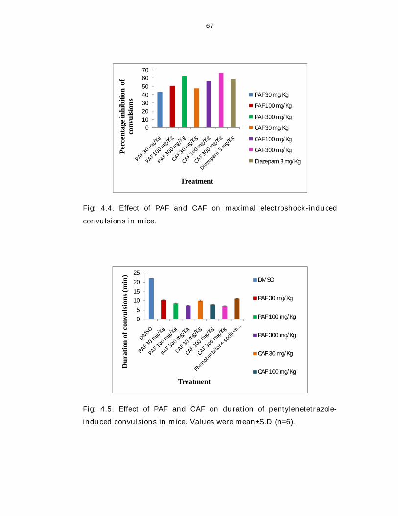
Fig: 4.4. Effect of PAF and CAF on maximal electroshock-induced
convulsions in mice.
05
10152025
Dur
atio
n of
con
vulsi
ons (
min
)
Treatment
DMSO
PAF 30 mg/Kg
PAF 100 mg/Kg
PAF 300 mg/Kg
CAF 30 mg/Kg
CAF 100 mg/Kg
Fig: 4.5. Effect of PAF and CAF on duration of pentylenetetrazole-
induced convulsions in mice. Values were mean±S.D (n=6).
68
01020304050607080
Perc
enta
ge in
hibi
tion
of
conv
ulsio
ns
Treatment
PAF 30 mg/Kg
PAF 100 mg/Kg
PAF 300 mg/Kg
CAF 30 mg/Kg
CAF 100 mg/Kg
CAF 300 mg/Kg
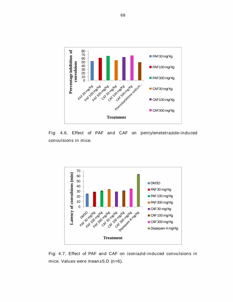
Fig: 4.6. Effect of PAF and CAF on pentylenetetrazole-induced
convulsions in mice.
010203040506070
Late
ncy
of c
onvu
lsion
s (m
in)
Treatment
DMSO
PAF 30 mg/Kg
PAF 100 mg/Kg
PAF 300 mg/Kg
CAF 30 mg/Kg
CAF 100 mg/Kg
CAF 300 mg/Kg
Diazepam 4 mg/Kg
Fig: 4.7. Effect of PAF and CAF on isoniazid-induced convulsions in
mice. Values were mean±S.D (n=6).
69
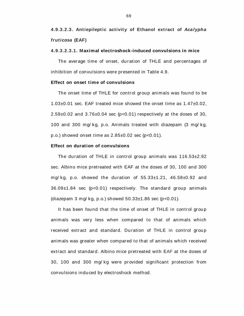
4.9.3.2.3. Antiepileptic activity of Ethanol extract of Acalypha
fruticosa (EAF)
4.9.3.2.3.1. Maximal electroshock-induced convulsions in mice
The average time of onset, duration of THLE and percentages of
inhibition of convulsions were presented in Table 4.9.
Effect on onset time of convulsions
The onset time of THLE for control group animals was found to be
1.03±0.01 sec. EAF treated mice showed the onset time as 1.47±0.02,
2.59±0.02 and 3.76±0.04 sec (p<0.01) respectively at the doses of 30,
100 and 300 mg/kg, p.o. Animals treated with diazepam (3 mg/kg,
p.o.) showed onset time as 2.85±0.02 sec (p<0.01).
Effect on duration of convulsions
The duration of THLE in control group animals was 116.53±2.92
sec. Albino mice pretreated with EAF at the doses of 30, 100 and 300
mg/kg, p.o. showed the duration of 55.33±1.21, 46.58±0.92 and
36.09±1.84 sec (p<0.01) respectively. The standard group animals
(diazepam 3 mg/kg, p.o.) showed 50.33±1.86 sec (p<0.01).
It has been found that the time of onset of THLE in control group
animals was very less when compared to that of animals which
received extract and standard. Duration of THLE in control group
animals was greater when compared to that of animals which received
extract and standard. Albino mice pretreated with EAF at the doses of
30, 100 and 300 mg/kg were provided significant protection from
convulsions induced by electroshock method.
70
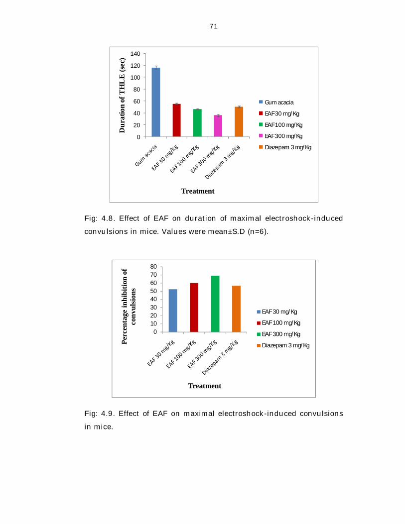
Percentage inhibition of convulsions
The percentage inhibition achieved in mice pretreated with EAF at
the doses of 30, 100 and 300 mg/kg were 52.52%, 60.03% and
69.03% (p<0.01) respectively when compared to control group
animals. Animals treated with EAF exhibited significant antiepileptic
activity and more percentage of inhibition of convulsions at the doses
of both 100 and 300 mg/kg when compared to diazepam treated
animals (56.81%).
Table 4.9. Effect of EAF on maximal electroshock-induced convulsions
in mice
Group
(n=6)=
6)
Treatment Onset of
THLE (sec)
Duration of
THLE (sec)
Percentage
inhibition of
convulsions
IEAM Gum acacia 1.03±0.01 116.53±2.92 -
IIEAM EAF (30 mg/kg) 1.47±0.02** 55.33±1.21** 52.52**
IIIEAM EAF (100 mg/kg) 2.59±0.02** 46.58±0.92** 60.03**
IVEAM EAF (300 mg/kg) 3.76±0.04** 36.09±1. 84** 69.03**
VEAM Diazepam (3 mg/kg) 2.85±0.02** 50.33±1.86** 56.81**
EAF: Ethanol extract of Acalypha fruticosa; Values were mean±SD
(n=6). Statistical significance was determined by ANOVA, followed by
Dunnett’s t test (n=6); **p < 0.01 when compared to Group IEAM
(control).
71
0
20
40
60
80
100
120
140
Dur
atio
n of
TH
LE
(sec
)
Treatment
Gum acacia
EAF 30 mg/Kg
EAF 100 mg/Kg
EAF 300 mg/Kg
Diazepam 3 mg/Kg
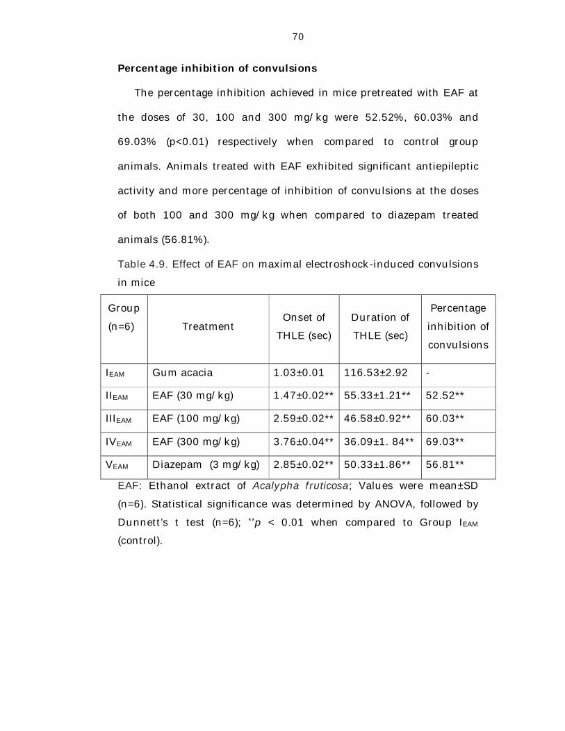
Fig: 4.8. Effect of EAF on duration of maximal electroshock-induced
convulsions in mice. Values were mean±S.D (n=6).
01020304050607080
Perc
enta
ge in
hibi
tion
of
conv
ulsi
ons
Treatment
EAF 30 mg/Kg
EAF 100 mg/Kg
EAF 300 mg/Kg
Diazepam 3 mg/Kg
Fig: 4.9. Effect of EAF on maximal electroshock-induced convulsions
in mice.
72
4.9.3.2.3.2. Pentylenetetrazole (PTZ)-induced convulsions in mice
The average time of onset, duration of convulsions and percentages
of inhibition of convulsions were presented in Table 4.10.
Effect on onset time of convulsions
The onset time of convulsions in control group animals was
3.43±0.04 min. EAF treated mice exhibited the onset time as
4.38±0.05, 4.54±0.02 and 5.14±0.03 min (p<0.01) respectively at the
doses of 30, 100 and 300 mg/kg, p.o. The standard group mice
(Phenobarbitone sodium, 40 mg/kg, i.p.) showed 6.46 ±0.02 min
(p<0.01).
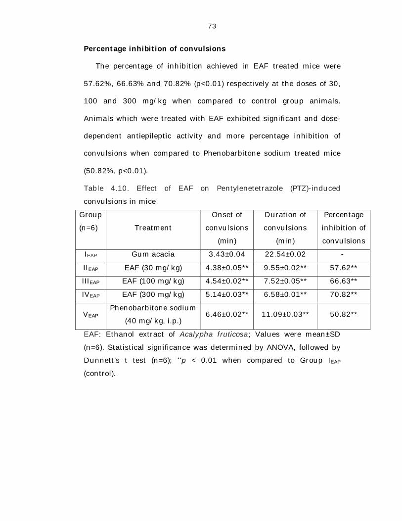
Effect on duration of convulsions
The duration of convulsions in control group animals was
22.54±0.02 min. Animals pretreated with EAF at the doses of 30, 100
and 300 mg/kg, p.o. exhibited the duration as 9.55±0.02, 7.52±0.05
and 6.58±0.01 min (p<0.01) respectively. The standard group mice
(Phenobarbitone sodium 40 mg/kg, i.p.) showed 11.09±0.03 min
(p<0.01).
The time of onset of convulsions in control group animals was very
less when compared to the animals treated with extract and standard.
Duration of convulsions in control group animals was greater when
compared to the extract and standard treated animals. It has been
found that animals pretreated with EAF were significantly protected
from convulsions induced by PTZ in a dose-dependent manner.
73
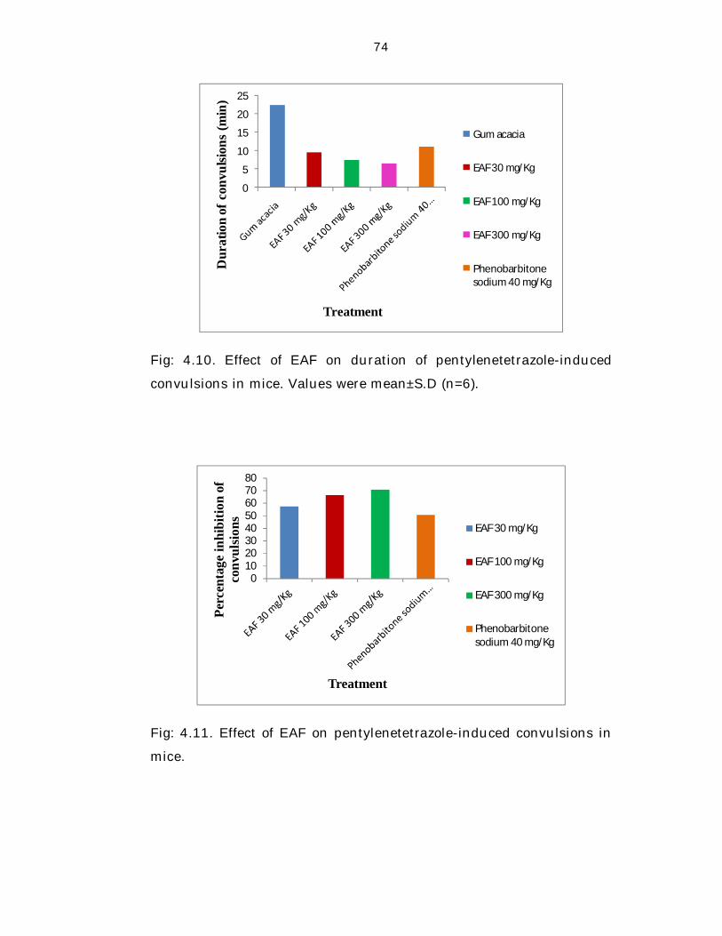
Percentage inhibition of convulsions
The percentage of inhibition achieved in EAF treated mice were
57.62%, 66.63% and 70.82% (p<0.01) respectively at the doses of 30,
100 and 300 mg/kg when compared to control group animals.
Animals which were treated with EAF exhibited significant and dose-
dependent antiepileptic activity and more percentage inhibition of
convulsions when compared to Phenobarbitone sodium treated mice
(50.82%, p<0.01).
Table 4.10. Effect of EAF on Pentylenetetrazole (PTZ)-induced
convulsions in mice
Group
(n=6)=
6)
Treatment
Onset of
convulsions
(min)
Duration of
convulsions
(min)
Percentage
inhibition of
convulsions
IEAP Gum acacia 3.43±0.04 22.54±0.02 --
IIEAP EAF (30 mg/kg) 4.38±0.05** 9.55±0.02** 57.62**
IIIEAP EAF (100 mg/kg) 4.54±0.02** 7.52±0.05** 66.63**
IVEAP EAF (300 mg/kg) 5.14±0.03** 6.58±0.01** 70.82**
VEAP Phenobarbitone sodium
(40 mg/kg, i.p.) 6.46±0.02** 11.09±0.03** 50.82**
EAF: Ethanol extract of Acalypha fruticosa; Values were mean±SD
(n=6). Statistical significance was determined by ANOVA, followed by
Dunnett’s t test (n=6); **p < 0.01 when compared to Group IEAP
(control).
74
0
5
10
15
20
25
Dur
atio
n of
con
vulsi
ons
(min
)
Treatment
Gum acacia
EAF 30 mg/Kg
EAF 100 mg/Kg
EAF 300 mg/Kg
Phenobarbitone sodium 40 mg/Kg
Fig: 4.10. Effect of EAF on duration of pentylenetetrazole-induced
convulsions in mice. Values were mean±S.D (n=6).
01020304050607080
Perc
enta
ge in
hibi
tion
of
conv
ulsio
ns
Treatment
EAF 30 mg/Kg
EAF 100 mg/Kg
EAF 300 mg/Kg
Phenobarbitone sodium 40 mg/Kg
Fig: 4.11. Effect of EAF on pentylenetetrazole-induced convulsions in
mice.
75
4.9.3.2.3.3. Isoniazid (INH)-induced convulsions in mice
The average latency of convulsions were presented in Table 4.11.
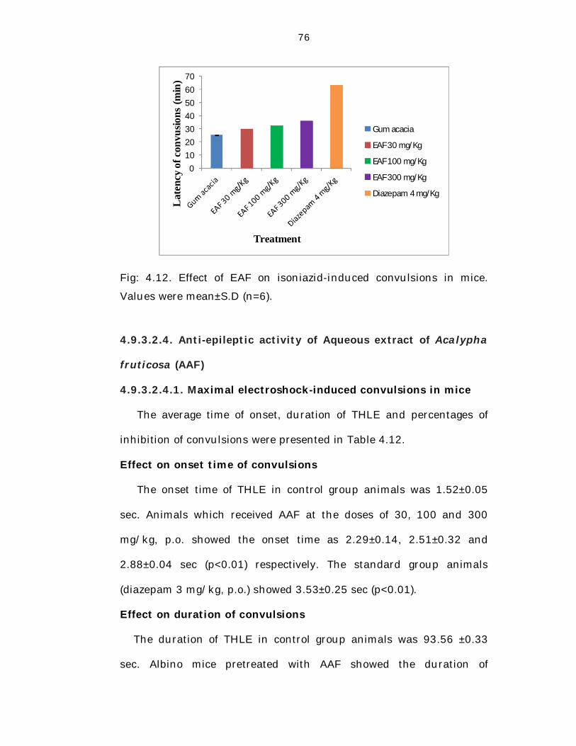
Effect on latency of convulsions
The latency of convulsions in control group animals was
25.15±0.28 min. Albino mice pretreated with EAF showed the latency
of convulsions of 30.08±0.05, 32.52±0.06 and 36.06±0.04 min
(p<0.01) respectively at the doses of 30, 100 and 300 mg/kg, p.o. The
standard group mice (diazepam 4 mg/kg, i.p.) showed 63.27±0.04 min
(p<0.01).
The latency of convulsions in control group animals was very less
when compared to that of animals pretreated with extract and
standard. EAF treated animals showed the latency time more than
that of control group animals and less than that of standard group
animals. Although all the three doses of EAF significantly delayed the
latency of convulsions in a dose-dependent manner but failed to
protect mice against mortality.
Table 4.11. Effect of EAF on Isoniazid (INH)-induced convulsions in
mice
Group (n=6) Treatment Latency of
convulsions (min)
IEAI Gum acacia 25.15±0.28
IIEAI EAF (30 mg/kg) 30.08±0.05**
IIIEAI EAF (100 mg/kg) 32.52±0.06**
IVEAI EAF (300 mg/kg) 36.06±0.04**
VEAI Diazepam (4 mg/kg, i.p.) 63.27±0.04**
EAF: Ethanol extract of Acalypha fruticosa; Values were mean±SD
(n=6). Statistical significance was determined by ANOVA, followed by
Dunnett’s t test (n=6); **p < 0.01 when compared to Group IEAI
(control).
76
010203040506070
Lat
ency
of c
onvu
sions
(min
)
Treatment
Gum acacia
EAF 30 mg/Kg
EAF 100 mg/Kg
EAF 300 mg/Kg
Diazepam 4 mg/Kg
Fig: 4.12. Effect of EAF on isoniazid-induced convulsions in mice.
Values were mean±S.D (n=6).
4.9.3.2.4. Anti-epileptic activity of Aqueous extract of Acalypha
fruticosa (AAF)
4.9.3.2.4.1. Maximal electroshock-induced convulsions in mice
The average time of onset, duration of THLE and percentages of
inhibition of convulsions were presented in Table 4.12.
Effect on onset time of convulsions
The onset time of THLE in control group animals was 1.52±0.05
sec. Animals which received AAF at the doses of 30, 100 and 300
mg/kg, p.o. showed the onset time as 2.29±0.14, 2.51±0.32 and
2.88±0.04 sec (p<0.01) respectively. The standard group animals
(diazepam 3 mg/kg, p.o.) showed 3.53±0.25 sec (p<0.01).
Effect on duration of convulsions
The duration of THLE in control group animals was 93.56 ±0.33
sec. Albino mice pretreated with AAF showed the duration of
77
60.28±0.40, 51.59±0.35 and 42.22±0.22 sec (p<0.01) respectively at
the doses of 30, 100 and 300 mg/kg, p.o. The standard group mice
(diazepam 3 mg/kg, p.o.) showed 39.59±0.35 sec (p<0.01).
The time of onset of THLE in control group animals was very less
when compared to the extract and standard treated animals. Duration
of THLE in control group animals was greater when compared to the
extract and standard treated animals. AAF significantly protected the
mice from convulsions induced by electroshock method in a dose-
dependent manner.
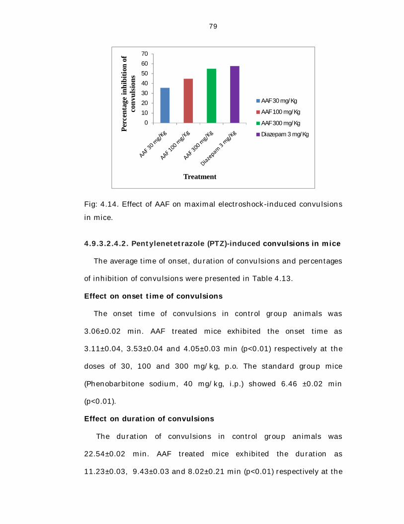
Percentage inhibition of convulsions
The percentage inhibition achieved in AAF treated mice were
35.57% (30 mg/kg), 44.85% (100 mg/kg) and 54.87% (300 mg/kg)
(p<0.01) respectively when compared to control group animals.
Animals pretreated with AAF exhibited significant antiepileptic activity
and less percentage of inhibition of convulsions when compared to
diazepam treated animals (57.69%, p<0.01).
78
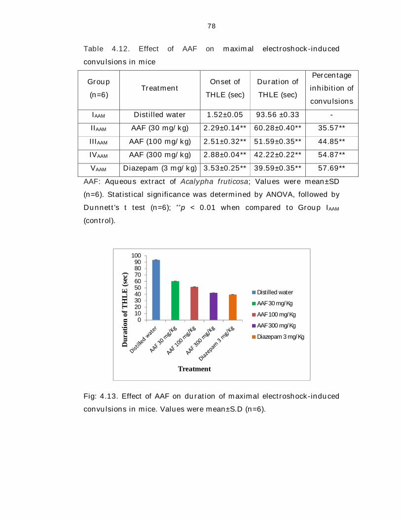
Table 4.12. Effect of AAF on maximal electroshock-induced
convulsions in mice
Group
(n=6) Treatment
Onset of
THLE (sec)
Duration of
THLE (sec)
Percentage
inhibition of
convulsions
IAAM Distilled water 1.52±0.05 93.56 ±0.33 -
IIAAM AAF (30 mg/kg) 2.29±0.14** 60.28±0.40** 35.57**
IIIAAM AAF (100 mg/kg) 2.51±0.32** 51.59±0.35** 44.85**
IVAAM AAF (300 mg/kg) 2.88±0.04** 42.22±0.22** 54.87**
VAAM Diazepam (3 mg/kg) 3.53±0.25** 39.59±0.35** 57.69**
AAF: Aqueous extract of Acalypha fruticosa; Values were mean±SD
(n=6). Statistical significance was determined by ANOVA, followed by
Dunnett’s t test (n=6); **p < 0.01 when compared to Group IAAM
(control).
0102030405060708090
100
Dur
atio
n of
TH
LE (s
ec)
Treatment
Distilled water
AAF 30 mg/Kg
AAF 100 mg/Kg
AAF 300 mg/Kg
Diazepam 3 mg/Kg
Fig: 4.13. Effect of AAF on duration of maximal electroshock-induced
convulsions in mice. Values were mean±S.D (n=6).
79
0
10
20
30
40
50
60
70
Perc
enta
ge in
hibi
tion
of
conv
ulsi
ons
Treatment
AAF 30 mg/Kg
AAF 100 mg/Kg
AAF 300 mg/Kg
Diazepam 3 mg/Kg
Fig: 4.14. Effect of AAF on maximal electroshock-induced convulsions
in mice.
4.9.3.2.4.2. Pentylenetetrazole (PTZ)-induced convulsions in mice
The average time of onset, duration of convulsions and percentages
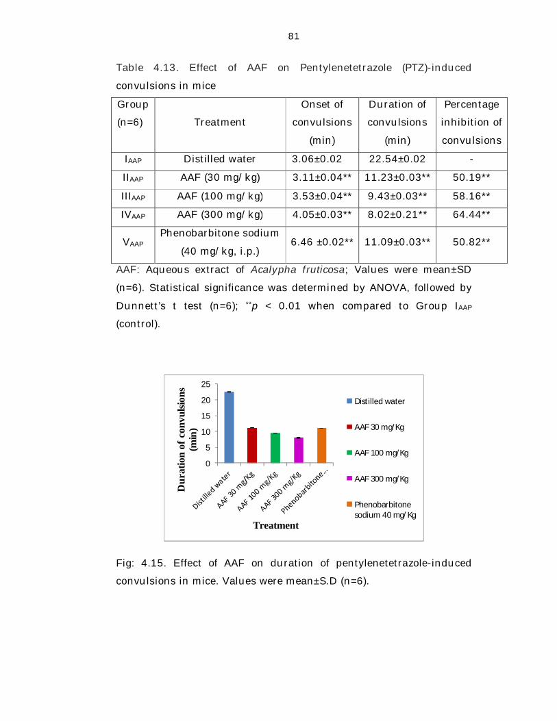
of inhibition of convulsions were presented in Table 4.13.
Effect on onset time of convulsions
The onset time of convulsions in control group animals was
3.06±0.02 min. AAF treated mice exhibited the onset time as
3.11±0.04, 3.53±0.04 and 4.05±0.03 min (p<0.01) respectively at the
doses of 30, 100 and 300 mg/kg, p.o. The standard group mice
(Phenobarbitone sodium, 40 mg/kg, i.p.) showed 6.46 ±0.02 min
(p<0.01).
Effect on duration of convulsions
The duration of convulsions in control group animals was
22.54±0.02 min. AAF treated mice exhibited the duration as
11.23±0.03, 9.43±0.03 and 8.02±0.21 min (p<0.01) respectively at the
80
doses of 30, 100 and 300 mg/kg, p.o. Albino mice pretreated with
Phenobarbitone sodium, 40 mg/kg, i.p. showed 11.09±0.03 min
(p<0.01).
The time of onset of convulsions in control group animals was very
less when compared to the extract and standard group animals.
Duration of convulsions in control group animals was greater when
compared to the extract and standard group animals. Albino mice
pretreated with AAF at the doses of 30, 100 and 300 mg/kg were
provided significant protection from convulsions induced by PTZ.
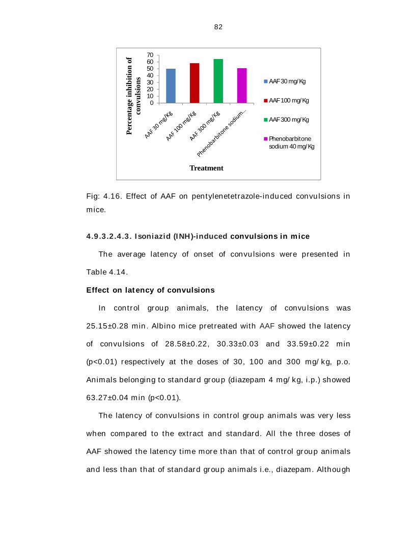
Percentage inhibition of convulsions
The percentage inhibition achieved in mice which received AAF
were 50.19% (30 mg/kg), 58.16% (100 mg/kg) and 64.44% (300
mg/kg) (p<0.01) respectively when compared to control group
animals. AAF treated mice exhibited significant antiepileptic activity
and more percentage of inhibition of convulsions at both 100 and 300
mg/kg when compared to Phenobarbitone sodium 40 mg/kg, i.p.
(50.82%, p<0.01).
81
Table 4.13. Effect of AAF on Pentylenetetrazole (PTZ)-induced
convulsions in mice
Group
(n=6)=
6)
Treatment
Onset of
convulsions
(min)
Duration of
convulsions
(min)
Percentage
inhibition of
convulsions
IAAP Distilled water 3.06±0.020. 22.54±0.02 -
IIAAP AAF (30 mg/kg) 3.11±0.04** 11.23±0.03** 50.19**
IIIAAP AAF (100 mg/kg) 3.53±0.04** 9.43±0.03** 58.16**
IVAAP AAF (300 mg/kg) 4.05±0.03** 8.02±0.21** 64.44**
VAAP Phenobarbitone sodium
(40 mg/kg, i.p.) 6.46 ±0.02** 11.09±0.03** 50.82**
AAF: Aqueous extract of Acalypha fruticosa; Values were mean±SD
(n=6). Statistical significance was determined by ANOVA, followed by
Dunnett’s t test (n=6); **p < 0.01 when compared to Group IAAP
(control).
0
5
10
15
20
25
Dur
atio
n of
con
vulsi
ons
(min
)
Treatment
Distilled water
AAF 30 mg/Kg
AAF 100 mg/Kg
AAF 300 mg/Kg
Phenobarbitonesodium 40 mg/Kg
Fig: 4.15. Effect of AAF on duration of pentylenetetrazole-induced
convulsions in mice. Values were mean±S.D (n=6).
82
010203040506070
Perc
enta
ge in
hibi
tion
of
conv
ulsio
ns
Treatment
AAF 30 mg/Kg
AAF 100 mg/Kg
AAF 300 mg/Kg
Phenobarbitone sodium 40 mg/Kg
Fig: 4.16. Effect of AAF on pentylenetetrazole-induced convulsions in
mice.
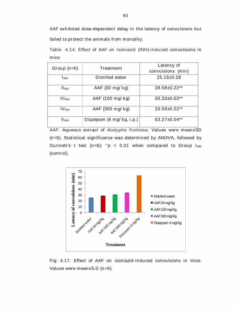
4.9.3.2.4.3. Isoniazid (INH)-induced convulsions in mice
The average latency of onset of convulsions were presented in
Table 4.14.
Effect on latency of convulsions
In control group animals, the latency of convulsions was
25.15±0.28 min. Albino mice pretreated with AAF showed the latency
of convulsions of 28.58±0.22, 30.33±0.03 and 33.59±0.22 min
(p<0.01) respectively at the doses of 30, 100 and 300 mg/kg, p.o.
Animals belonging to standard group (diazepam 4 mg/kg, i.p.) showed
63.27±0.04 min (p<0.01).
The latency of convulsions in control group animals was very less
when compared to the extract and standard. All the three doses of
AAF showed the latency time more than that of control group animals
and less than that of standard group animals i.e., diazepam. Although
83
AAF exhibited dose-dependent delay in the latency of convulsions but
failed to protect the animals from mortality.
Table 4.14. Effect of AAF on Isoniazid (INH)-induced convulsions in
mice
Group (n=6) Treatment Latency of
convulsions (min) IAAI Distilled water 25.15±0.28
IIAAI AAF (30 mg/kg) 28.58±0.22**
IIIAAI AAF (100 mg/kg) 30.33±0.03**
IVAAI AAF (300 mg/kg) 33.59±0.22**
VAAI Diazepam (4 mg/kg, i.p.) 63.27±0.04**
AAF: Aqueous extract of Acalypha fruticosa; Values were mean±SD
(n=6). Statistical significance was determined by ANOVA, followed by
Dunnett’s t test (n=6); **p < 0.01 when compared to Group IAAI
(control).
0
10
20
30
40
50
60
70
Lat
ency
of c
onvu
lsion
s (m
in)
Treatment
Distilled water
AAF 30 mg/Kg
AAF 100 mg/Kg
AAF 300 mg/Kg
Diazepam 4 mg/Kg
Fig: 4.17. Effect of AAF on isoniazid-induced convulsions in mice.
Values were mean±S.D (n=6).
84
4.10. Discussion
Epilepsy is one of the major neurological disorders characterized
by sporadic episodes of abnormal behavior, convulsive seizures,
sensory disturbance and loss of consciousness or all of these
symptoms resulting from a brain dysfunction or an abnormal
discharge of cerebral neurons [35].
Higher prevalence, cultural and social stigma, lack of awareness
and non-availability of proper diagnostic and treatment facilities are
some of the major reasons for the increasing number of people with
epilepsy in the developing countries. All the currently available AEDs
are associated with side-effects, long-term toxicity, teratogenic effects
and about 40% patients are refractory to therapeutic intervention and
thus its effective and safe therapy still remains a challenge [44-46].
Hence there is a mere need to search for AEDs from alternative
sources i.e., exploitation of medicinal plants.
Medicinal plants in traditional medicine can become an invaluable
source for search of new antiepileptic compounds. Literature survey
revealed that many plants like Aegle marmelos, Asparagus racemosus,
Carissa edulis, Cyperus articulatus, Delphinium denudatum, Hibiscus
rosa and Jasminum grandflorum acclaim to possess antiepileptic
activity [57, 58, 96, 122-123].
Acalypha fruticosa is a shrub belonging to the family of
Euphorbiaceae. Aerial parts of Acalypha fruticosa were traditionally
used to treat epilepsy [21]. But till today there were no reports to
85
justify its claim. Hence the present work was designed to evaluate the
antiepileptic activity of Acalypha fruticosa.
In the present study, four different extracts were prepared by using
solvents of increasing polarity like petroleum ether, chloroform,
ethanol and water. Non-polar solvents have low dielectric constants
and dissolve non-polar solutes with similar internal pressures through
induced dipole interactions. Petroleum ether is a non-polar solvent
which solubilises non-polar compounds like steroids and
triterpenoids. Chloroform extracts non-polar to intermediately polar
compounds such as steroids, triterpenoids, flavonoids. Ethanol
dissolves most of the secondary metabolites such as steroids,
triterpenoids, flavonoids, tannins, saponins and enhance their release
from cellular matrix/cell surface. The polar components like
polysaccharides, phenols, aldehydes, ketones, amines, saponins and
other oxygen containing compounds dissolve in water due to
formation of hydrogen bonding [124]. Thus petroleum ether,
chloroform, ethanol and aqueous extracts of Acalypha fruticosa were
prepared to predict which phytoconstituents are responsible for the
remarkable antiepileptic activity.
In preliminary phytochemical studies, the chief phytoconstituents
present in the different extracts of Acalypha fruticosa were alkaloids,
steroids, carbohydrates, tannins, flavanoids, saponins, lipids and
proteins.
86
In acute toxicity studies, petroleum ether, chloroform, ethanol and
aqueous extracts of Acalypha fruticosa were found to be safe upto
1000 mg/kg, (p.o.) body weight. Hence three doses i.e., 30, 100 and
300 mg/kg, p.o. were selected for all the extracts to evaluate
antiepileptic activity.
To screen the antiepileptic activity, most extensively studied, well
established and simple animal seizure models viz. maximal
electroshock, pentylenetetrazole and isoniazid-induced seizures in
mice were selected. Another advantage of using the above models is
the pharmacological profiles were comparable to the human condition
[33-34].
The MES test identifies compounds/extracts which prevent seizure
spread [29-30]. In this model, all the extracts of Acalypha fruticosa
significantly and dose dependently increased the onset time of THLE
and decreased the duration of THLE. But ethanol extract exhibited
maximum significant antiepileptic activity at 300 mg/kg (69.03%). The
order of antiepileptic activity for various extracts in MES model was
ethanol>chloroform>petroleum ether>aqueous (chloroform extract-
66.33%, petroleum ether extract-62.33% and aqueous extract-
54.87%). All the extracts of Acalypha fruticosa might prevent the
seizure spread and contribute to the activity. But the maximum
antiepileptic activity of the ethanol extract may be due to the presence
of phytoconstituents such as tannins and flavonoids.
PTZ test identifies compounds/extracts which primarily raise
seizure threshold [29-30]. In this model, all the extracts of Acalypha
87
fruticosa significantly increased the onset time of convulsions and
decreased the duration of convulsions in a dose-dependent manner.
But ethanol extract showed maximum antiepileptic activity at 300
mg/kg (70.82%) followed by chloroform extract (68.20%), petroleum
ether extract (66.49%) and aqueous extract (64.44%). In this model,
seizure threshold may be raised by all the extracts of Acalypha
fruticosa but the potent activity of ethanol extract may be because of
phytoconstituents like tannins and flavonoids present in the extract.
Isoniazid is an antitubercular drug which was shown to lower the
content of brain GABA in humans to approximately the same extent in
rats and mice [32]. In this model, all the four extracts of aerial parts of
Acalypha fruticosa delayed the latency of convulsions but could not
protect the mice from mortality. At 300 mg/kg dose, ethanol extract
possessed maximum delay in latency of convulsions. In this model,
the extracts act as GABA agonists but unable to increase GABA levels
in brain.
All the extracts exhibited significant antiepileptic activity in all
three tested models. The order of activity is AAF<PAF<CAF<EAF. The
observed antiepileptic activity of AAF in all models could be due to the
presence of tannins. Presence of steroids may attribute to the potent
antiepileptic activity of PAF than AAF because steroids are involved in
neuromodulatory effects [125]. Antiepileptic activity of CAF may be
due to the presence of steroids and flavonoids because flavonoids and
sterols have been involved in central inhibitory and neuromodulatory
effects [126]. Maximum activity of EAF may be because of combined
88
effects of steroids, tannins and flavonoids. Studies carried out till date
suggests that flavonoids inhibit voltage gated sodium channels,
activate Ca+ activated K+ channels, stimulate GABAergic inhibition,
interact with opioid receptors, inhibit NMDA receptors and exhibit
antioxidant actions via modulation of nitric oxide and xanthine
oxidase pathways and by leukocytic immobilization, one or more of
these mechanisms are involved in suppression of epileptic seizures
[127].
Overall the present study demonstrated that the ethanol extract of
aerial parts of Acalypha fruticosa exhibited maximum antiepileptic
activity in all tested models. The order of activity of the extracts were
ethanol> chloroform>petroleum ether>aqueous. Ethanol extract may
act at seizure focus, prevent spread of the seizure and suppresses
THLE induced by MES. In PTZ model, the extract might raised the
seizure threshold or act as GABA agonist and enhanced GABAergic
neurotransmission by increasing GABA levels in brain by facilitating
the opening of GABA-activated chloride channels at GABAA receptors.
The ethanol extract increased the latency of convulsions in INH model
but could not protect the mice from mortality. So, it may act as GABA
agonist but unable to stimulate GABA synthesis. Further, our results
indicated that the beneficial effect of ethanol extract of Acalypha
fruticosa in epilepsy is due to the presence of steroids, tannins and
flavonoids. Present study justify the traditional use of Acalypha
fruticosa in folk medicine.