Embed Size (px)
Citation preview

Chapter 4Chapter 4Neonatal Assessment and ResuscitationNeonatal Assessment and Resuscitation

Labor and Delivery Terms
• Departments: L & D, OB, Post Partum, Antepartum, NICU, Nursery
• APGAR: A measurement of the newborn's response to birth and life outside the womb. The ratings, APGAR, are based on Appearance (color), Pulse (heartbeat), Grimace (reflex), Activity (muscle tone), and Respiration (breathing). The scores, which are taken at 1 and 5 minutes following birth, range from 10 to 1, with 10 being the highest and 1 being the lowest.

Labor and Delivery Terms
• Breech: When the fetus is positioned head up to be born buttocks first or with one or both feet first.
• Cephalopelvic Disproportion(CPD):The baby is too large to safely pass through the mother's pelvis.
• Cervidil: A medication used to ripen the cervix before induction.
• Cesarean: An incision through the abdominal and uterine walls for extraction of the fetus; it may be vertical or more commonly, horizontal. Also called abdominal delivery; commonly called C-Section.

Labor and Delivery Terms
• Colostrum: This is a thin, white fluid discharged from the breasts in the early stage of milk production, and usually noticeable during the last couple weeks of pregnancy
• Complete Breech: The baby's buttocks are presenting at the cervix, but the legs are folded “Indian style,” making vaginal delivery difficult or impossible.
• Contraction: The regular tightening of the uterus, working to push the baby down the birth canal.
• Crowned/Crowning: When the baby's head has passed through the birth canal and the top or “crown” stays visible at the vaginal opening.

Labor and Delivery Terms
• Dilation: The extent to which the cervix has opened in preparation for childbirth. It is measured in centimeters, with full dilation being 10 centimeters.
• Effacement: This refers to the thinning of the cervix in preparation for birth and is expressed in percentages. You will be 100% effaced when you begin pushing.
• Epidural: A common method of anesthesia used during labor. It is inserted through a catheter which is threaded through a needle, into the dura space near the spinal cord.
• Episiotomy: An incision made to the perineum to widen the vaginal opening for delivery.

Labor and Delivery Terms
• Fontanelle: Soft spots between the unfused sections of the baby's skull. These allow the baby's head to compress slightly during passage through the birth canal
• Forceps: Tong shaped instrument that may be used to help guide the baby's head out of the birth canal during deliver
• Frank Breech: The baby's buttocks are presenting at the cervix and the baby's legs are extended straight up to the baby's head.
• Induced Labor: Labor is started or accelerated through intervention, such as placing prostaglandin gel on the cervix, using an IV drip of the hormone oxytocin (Pitocin), or by rupturing the membranes.

Labor and Delivery Terms
• Lightening: When the baby drops in preparation for delivery (Engagement).
• Meconium: This is the greenish substance that builds up in the bowels of a growing fetus and is normally discharged shortly after birth
• Nubain: Synthetic narcotic pain reliever commonly used in labor and delivery.
• Oxytocin: Hormone secreted by the pituitary gland that stimulates contractions and the milk-eject reflex. Pitocin is the synthetic form of this hormone.
• Perineum: The muscle and tissue between the vagina and the rectum.

Labor and Delivery Terms
• Phenergan: A sedative administered that also controls nausea and vomiting.
• Placenta Previa: When the placenta partially or completely covers the cervix.
• Prostaglandin Cream: Medication used to ripen the cervix before induction.
• Ruptured Membranes: Usually refers to the breaking of the fluid filled sac surrounding the baby. The fluid may come as a gush of water or as a slow leak. Slow leaks are sometimes mistaken as incontinence.
• Speculum: An instrument used to open the vagina slightly wider so that the cervix can be seen more easily.

Labor and Delivery Terms• Timing Contractions: Contractions are measured from the
beginning of one contraction until the beginning of the next contraction.
• Tocolysis: Inhibition of contractions used to suppress premature labor with medications, Magnesium Sulfate, Terbutaline, Ritodrine
• Transverse: Baby's body length is horizontal in the uterus. If the baby cannot be moved, it will have to be delivered by cesarean .
• Vacuum Extractor: Instrument that attaches to the baby's head and helps guide it out of the birth canal during delivery.
• Betamethasone: glucocorticoid drug which greatly accelerates fetal lung maturity, but takes one to two days to work.

Labor and Delivery Terms
• Chorioamnionitis : an inflammation of the fetal membranes (amnion and chorion) due to a bacterial infection.
• Prelabor Rupture of Membranes (PROM) or Premature Rupture of Membranes as it is sometimes known, is a condition that occurs in pregnancy when there is rupture of the membrane of the amniotic sac and chorion more than one hour before the onset of labor
• Umbilical cord prolapse happens when the umbilical cord precedes the fetus' exit from the uterus. It is an obstetric emergency during pregnancy or labor that imminently endangers the life of the fetus. Cord prolapse is rare
Labor and Delivery Terms
• Para – the number of births (alive or not) with a viable infant > 22 weeks and at least 500 grams
• Gravida – the number of pregnancies• Pre-eclampsia (toxemia)-Usually occurs at > 24 weeks
gestation, Acute hypertension, edema, renal impairment (proteinuria), sudden weight gain, Occurs in 7% of pregnancies, More common in low socioeconomic groups
• Eclampsia; Toxemia, seizures, coma, convulsions, hemolysis, renal failure, 5% of women with pre-eclampsia develop eclampsia, 15% die from complications; Associated with high fetal mortality due to premature delivery
• Dystocia: Difficult birth

Neonatal Resuscitation• Newly born – infant at time of birth• Newborn – within first few hours of birth• Neonate – within first 30 days of delivery• Pre-term – less than 37 weeks of gestation• Term – 38 to 42 weeks of gestation• Post-term (post-date) – greater than 42 weeks
of gestation• http://vimeo.com/31423498

General Pathophysiology and Assessment
• Approximately 10% of newborns require assistance to begin breathing
• Extensive resuscitation needed in less than 1% of newborns
• Rate of complication increases as the newborn weight and gestational age decrease
• 80% of 30,000 babies born each year weighing less than 3 lbs. (1,500 grams) require resuscitation

Factors that indicate high risk delivery• Factors can be divided into maternal and fetal.
– Maternal factors include age (younger than age 15, older than age 35)– weight (pre-pregnancy weight under 100 lb or obesity)– height (under five feet)– history of complications during previous pregnancies (including stillbirth, fetal loss, preterm labor
and/or delivery, small-for-gestational age baby, large baby, pre-eclampsia or eclampsia)– more than five previous pregnancies– bleeding during the third trimester– abnormalities of the reproductive tract– uterine fibroids– Hypertension– Rh incompatability– gestational diabetes– infections of the vagina and/or cervix– kidney infection– Fever– acute surgical emergency (appendicitis, gallbladder disease, bowel obstruction)– post-term pregnancy– pre-existing chronic illness (such as asthma, autoimmune disease, cancer, sickle cell anemia,
tuberculosis, herpes, AIDS, heart disease, kidney disease, Crohn's disease, ulcerative colitis, diabetes)
Factors that indicate high risk delivery
• Fetal factors include • exposure to infection (especially herpes simplex, viral hepatitis, mumps, rubella,
varicella, syphilis, toxoplasmosis, and infections caused by coxsackievirus)• exposure to damaging medications (especially phenytoin, folic acid antagonists,
lithium, streptomycin, tetracycline, thalidomide, and warfarin)• exposure to addictive substances (cigarette smoking, alcohol intake, and illicit or
abused drugs)• A pregnancy is also considered high-risk when prenatal tests indicate that the
baby has a serious health problem (for example, a heart defect). In such cases, the mother will need special tests, and possibly medication, to carry the baby safely through to delivery. Furthermore, certain maternal or fetal problems may prompt a physician to deliver a baby early, or to choose a surgical delivery (cesarean section) rather than a vaginal delivery.

The Birth Process • At the onset of true labor, the contractions will feel like a tightening of
abdominal muscles, a dull ache or a pressure on the lower pelvis or back.
• At about this time the baby drops down into the pelvis; this is known as engagement because the baby has settled into its position for birth. Another term aptly used is lightening because the baby's new position means the mother has now more space to breathe and digest food.
• With time the contractions start to come at a more regular pace and the pain intensifies. The sensation is like a belt tightening around your back which spreads round underneath the baby. As the labor progresses the contractions last longer and occur at decreasing intervals.
The Birth Process • Bloody Show: A plug of mucous which seals the top of the
vagina acts as a barrier against infection from invading the uterus. Sometimes this plug dislodges itself before the contractions commence and you will notice a 'show' i.e. thick vaginal discharge mixed with blood in the patient’s underwear.
• This however is no indication that you are into proper labor; it may be several days before the action commences. In other instances, the contractions begin well before the expulsion of the mucous plug.
The Birth Process • Water Break: In about 20% of women, the pressure of the
baby's head can puncture the amniotic bag causing the fluids to leak out before labor. Often it occurs in later part of labor. There is no mistaking a rupture; the leakage is clear and watery from the vagina and you can lose as much as 2 pints of fluid. The water can gush out or come in a slow trickle.
• The baby is in risk of infection and chance of umbilical cord's descent into the birth canal. Water breaking can pose a risk in the form of infection or the oxygen supply being affected.

The Birth Process
• other symptoms; nausea, diarrhea and backache. Sometimes tightening the whole day before labor begins; this is indicative of the cervix ripening and shortening at a gradual pace or your baby could be probably lying with its back to your back. In the case of the latter the tightening occurs as the baby rotates round to assume the right position before making its entry into the world.
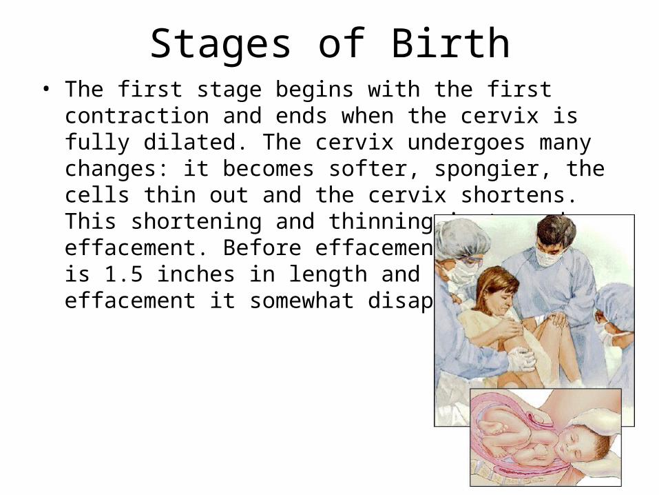
Stages of Birth• The first stage begins with the first contraction and ends
when the cervix is fully dilated. The cervix undergoes many changes: it becomes softer, spongier, the cells thin out and the cervix shortens. This shortening and thinning is termed effacement. Before effacement the cervix is 1.5 inches in length and after effacement it somewhat disappears.


Stages of Birth• Force from the contractions combined with the pressure from
baby's head slowly compels the now 'thin' cervix opening to widen. This is termed as dilation, which basically means the widening of the opening. Full dilation is at 10cm, about the width of a hand. When the cervix is beginning to dilate most mothers will feel the contractions in the back. You will be conscious of this ache but you can still go about with your normal activities. If you do not experience this, the mucous plug will soon dislodge instead.

Stages of Birth• The rate of dilation varies; tends to be slower in first time
mothers. • On average it will take about 9 hours for the cervix to dilate
2.5 cm (the latent phase). The time varies; some women take longer, some with lesser time.
• Next comes the active phase which causes the cervix to dilate from 3 - 10 cm; this phase lasts between 2 - 4 hours. Again the time varies. The pain is intense and the urge to bear down and push the baby out starts now.
• On average one hour is taken up for every centimeter that the cervix dilates

Stages of Birth• The second stage begins at full dilation and ends when the
baby makes an entry into the world.• Uterine contractions are now even more pronounced; they
help in the dilation and force the baby down and out. The contractions now occur every 2-3 minutes, lasting between 1 to 1.5 minutes.
• As the baby makes its descent, the mother works hard on the pushing. In a typical, normal situation, first the head descends followed by the rest of the body through the vagina.

Stages of Birth• It takes a first baby close to an hour to make its way
down the birth canal, through the vagina to the vulva
• The appearance of the baby's head at the mouth of the vagina is called crowning.
• http://www.youtube.com/watch?v=vpeggjIiE9k
• Following the crowning, the doctor may make a small cut into the area between the vagina and the rectum; this is called episiotomy.

Episiotomy

Stages of Birth• In the final stage, the placenta dislodges from the uterus
and is expelled. • After the baby is delivered, the uterus sheds the placenta • Contractions continue even after baby is born. The uterus
begins to get smaller and its walls thicker. This reduces the surface the placenta was attached. The placenta then separates and is pushed down and out of the vagina. Blood clots form immediately at the site of separation preventing any excessive bleeding. Bleeding is also controlled by the uterus contracting and closing the blood vessels that previously supported the placenta.

Antepartum Risk Factors
• Multiple gestation• Pregnant patient <16 or
>35 years of age• Post-term >42 weeks• Preeclampsia, HTN, DM• Polyhydraminos • Premature rupture of
amniotic sac (PROM)
• Fetal malformation (CDH, Gastroscesis…)
• Inadequate prenatal care
• History of prenatal morbidity or mortality
• Maternal use of drugs or alcohol
• Fetal anemia• Oligohydraminos

Risks of Multiple gestation• Preterm labor and birth
About half of twins and nearly all higher-order multiples are premature (born before 37 weeks). The higher the number of fetuses in the pregnancy, the greater the risk for early birth.
• pregnancy-induced hypertensionWomen with multiple fetuses are more than three times as likely to develop high blood pressure of pregnancy. This condition often develops earlier and is more severe than pregnancy with one baby. It can also increase the chance of placental abruption (early detachment of the placenta).

Risks of Multiple gestation• Anemia is more than twice as common in
multiple pregnancies as in a single birth. birth defects
• Multiple birth babies have about twice the risk of congenital (present at birth) abnormalities including neural tube defects (such as spina bifida), gastrointestinal, and heart abnormalities.

Risks of Multiple gestation• A phenomenon called the vanishing twin syndrome in
which more than one fetus is diagnosed, but vanishes (or is miscarried), usually in the first trimester, is more likely in multiple pregnancies. This may or may not be accompanied by bleeding. The risk of pregnancy loss is increased in later trimesters as well.
• twin-to-twin transfusion syndromeTwin-to-twin syndrome is a condition of the placenta that develops only with identical twins that share a placenta. Blood vessels connect within the placenta and divert blood from one fetus to the other. It occurs in about 15 percent of twins with a shared placenta.
Age Related Complications• >35 years more likely to have a multiple
pregnancy. The chance of having twins increases with age. The use of assisted reproductive technologies — such as in vitro fertilization — also can play a role.
• You're more likely to develop gestational diabetes. This type of diabetes occurs only during pregnancy, and it's more common as women get older. Tight control of blood sugar through diet, physical activity and other lifestyle measures is essential.

Age Related Complications• You're more likely to develop high blood
pressure during pregnancy. Some studies suggest that high blood pressure that develops during pregnancy — before 20 weeks (chronic hypertension), after 20 weeks (gestational hypertension) or after 20 weeks and accompanied by protein in the urine (preeclampsia) — might be more common in older women.
• The risk of chromosome abnormalities is higher. Babies born to older mothers have a higher risk of certain chromosome problems, such as Down syndrome.

Preeclampsia, HTN, DM
• Preeclampsia is a condition that occurs only during pregnancy. Diagnoses is made by the combination of high blood pressure and protein in the urine, occurring after week 20 of pregnancy. Preeclampsia may also be called toxemia; who is at risk:– A first-time mom– Previous experience with gestational hypertension or
preeclampsia– Women whose sisters and mothers had preeclampsia– Women carrying multiple babies; women younger than 20 years
and older than age 40– Women who had high blood pressure or kidney disease prior to
pregnancy– Women who are obese or have a BMI of 30 or greater
Preeclampsia
• Mild preeclampsia: high blood pressure, water retention, and protein in the urine.
• Severe preeclampsia/leading to eclampsia: headaches, blurred vision, inability to tolerate bright light, fatigue, nausea/vomiting, urinating small amounts, pain in the upper right abdomen, shortness of breath, and tendency to bruise easily. Seizures and associated symptoms
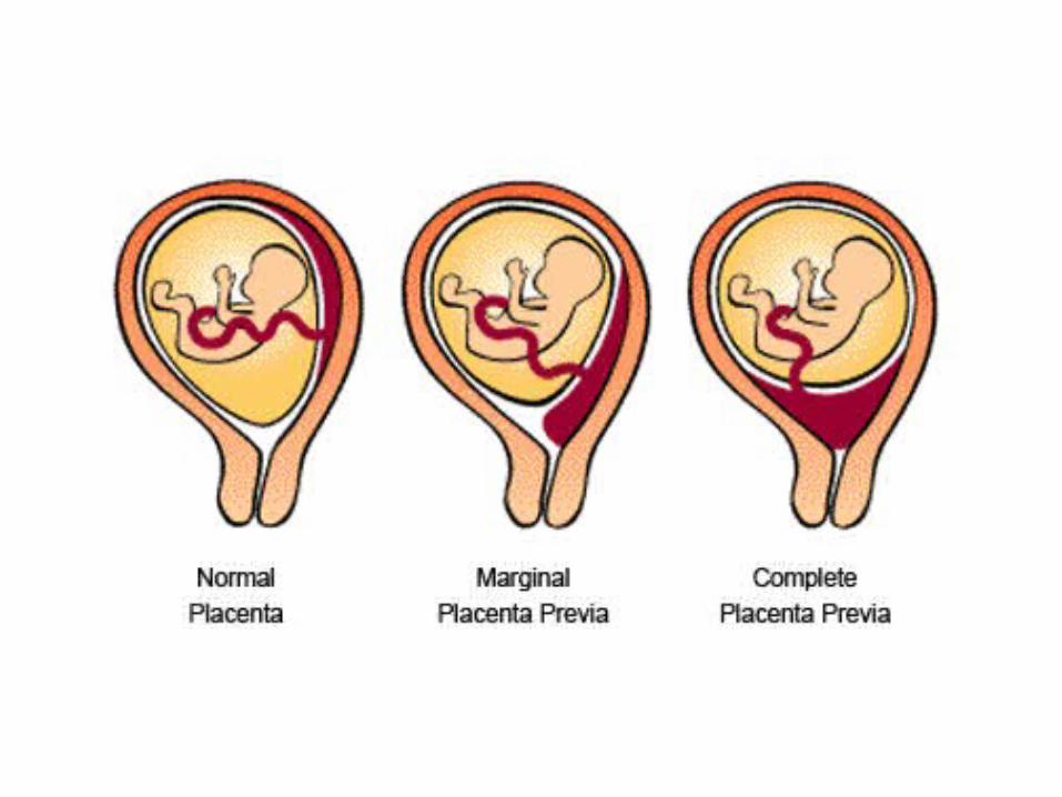
Placenta Previa• Placenta Previa is a condition where the placenta lies low in
the uterus and partially or completely covers the cervix. The placenta may separate from the uterine wall as the cervix begins to dilate (open) during labor.
• Requires C-section Delivery• Signs and symptoms of placenta previa vary, but the most
common symptom is painless bleeding during the third trimester. Other reasons to suspect placenta previa would be:– Premature contractions – Baby is breech, or in transverse position – Uterus measures larger than it should according to gestational
age

HELLP Syndrome
• HELLP Syndrome is a series of symptoms that make up a syndrome
• HELLP syndrome is thought to be a variant of preeclampsia, but it may be an entity all on its own. There are still many questions about the serious condition of HELLP syndrome. The cause is still unclear to many doctors and often HELLP syndrome is misdiagnosed. It is believed that HELLP syndrome affects about 0.2 to 0.6 percent of all pregnancies.

HELLP Syndrome
• What is HELLP Syndrome?• The name HELLP stands for:• H- hemolysis ( breakdown of red blood cells)• EL- elevated liver enzymes (liver function)• LP- low platelets counts (platelets help the
blood clot)

HELLP Syndrome• The most common symptoms of HELLP syndrome include:
– Headaches– Nausea and vomiting that continue to get worse (this may also feel like a
serious case of the flu.)– Upper right abdominal pain or tenderness– Fatigue or malaise
• A woman with HELLP may experience other symptoms that often can be attributed to other things such as normal pregnancy concerns or other pregnancy conditions. These symptoms may include:– Visual disturbances– High blood pressure– Protein in urine– Edema (swelling)– Severe headaches– Bleeding

HELLP Syndrome• Hemolysis -Red blood bells• Abnormal peripheral smear• Lacatate dehydrogenase >600 U/L• Bilirubin > 1.2 mg/dl• Elevated liver Enzyme levels• Serum aspartate amniotransferase >70 U/L• Lacatate dehydrogenase >600 U/L• Low Platelets• Platelet count • How is HELLP Syndrome Treated?• The treatment of HELLP Syndrome is primarily based on the gestation of the
pregnancy, but delivery of the baby is the best way to stop this condition from causing any serious complications for mom and baby. Most symptoms and side effects of HELLP will subside within 2-3 days of delivery.
• http://www.youtube.com/watch?v=LRWLB2T96MQ
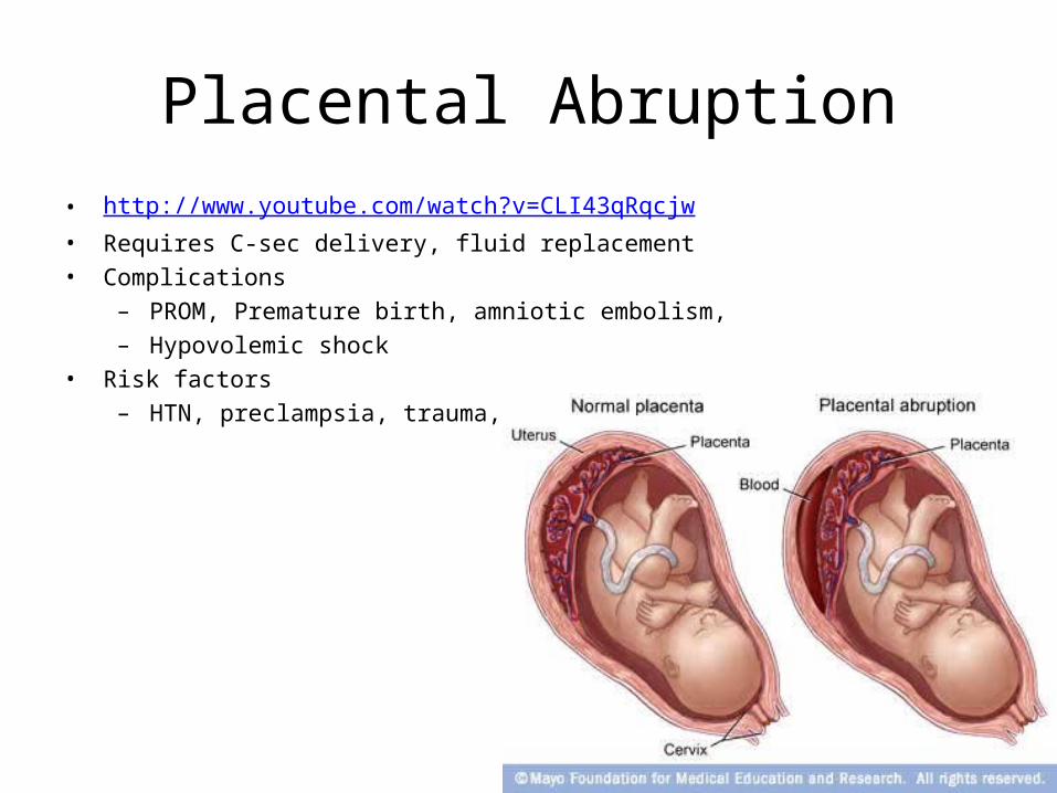
Placental Abruption• http://www.youtube.com/watch?v=CLI43qRqcjw• Requires C-sec delivery, fluid replacement• Complications
– PROM, Premature birth, amniotic embolism, – Hypovolemic shock
• Risk factors– HTN, preclampsia, trauma, smoking

Fetal Alcohol Spectrum Disorders (FASD);Fetal Alcohol Syndrome (FAS)
• Drinking alcohol during pregnancy can result in a number of different physical, neurological and mental effects that range in severity. These effects fall under the term “Fetal Alcohol Spectrum Disorders (FASD)”, which encompasses all the problems that result from prenatal alcohol exposure. The most known of these effects is Fetal Alcohol Syndrome (FAS) and Fetal Alcohol Effects (FAE). Fetal Alcohol Effects can also be separated into two different categories: Alcohol-Related Neurodevelopmental Disorder (ARND) and Alcohol-Related Birth Defects (ARBD).

FAS• The effects of FAS include: mental retardation,
malformations of the skeletal system and major organ systems (specifically the heart and brain), growth deficiencies, central nervous system problems, poor motor skills, mortality, and problems with learning, memory, social interaction, attention span, problem solving, speech and/or hearing.
• There are also facial features that are characteristic of babies with FAS. These features include: small eyes, short or upturned nose, flat cheeks, and thin lips. These features fade as the child grows up, but the child is left with a lifetime of difficulties trying to cope with other effects.

• http://www.youtube.com/watch?v=o-xGBjpGLdI

Other Antepartum risk factors• Ectopic pregnancy• Fetal growth restriction• HIV/AIDS• Listeria• Placenta Accreta• RH Factor • Tipped Uterus• UTI• Yeast Infection• Fetal position
• Chicken Pox• Cytomegalovirus (CMV)
infection• Gestational Diabetes• Group B Strep Infection• Intrauterine Growth
Retardation (IUGR)• STD’s/STI’s• Toxoplasmosis• Hyperemesis Gravidarum

Intrapartum Risk Factors
• Premature labor• PROM >24 hours• Abnormal presentation• Prolapsed cord• Chorioamnionitis
• Meconium-stained amniotic fluid
• Use of narcotics within 4 hours of delivery
• Prolonged labor• Precipitous delivery• Bleeding• Placenta previa

Pain Control during child birth
• Two options, natural child birth (deep breathing, laboring in water, massage…) or pain controlled with opiates
• Opiates do not interfere with the ability to push during delivery, does not numb the pain like a epidural, can reduce anxiety
• Side effects of opiates: nausea, vomiting, sedation, loss of protective airway reflexes

Fetal Transition• Rapid process that allows baby to breathe• Fetal lung is collapsed and filled with fluid• Reduction in pulmonary resistance
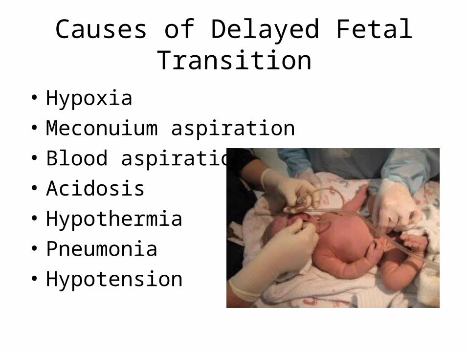
Causes of Delayed Fetal Transition
• Hypoxia• Meconuium aspiration• Blood aspiration• Acidosis• Hypothermia• Pneumonia• Hypotension

Newborn Resuscitation
Basic Assessment•Gestational age•Amniotic fluid•Respiratory effort•Muscle tone•Warmth and stimulation•Oxygenation and ventilation•Circulation•Volume expanders•Cardiotonic medications


Four Phases of Care• Preparation (read charts, setup
equipment, ensure functionality of equipment)
• Stabilization (NRP guidelines)• Assessment (NRP guidelines)• Resuscitation (NRP guidelines)

Preparation• Trained/skilled personnel• Equipment• Perinatal history
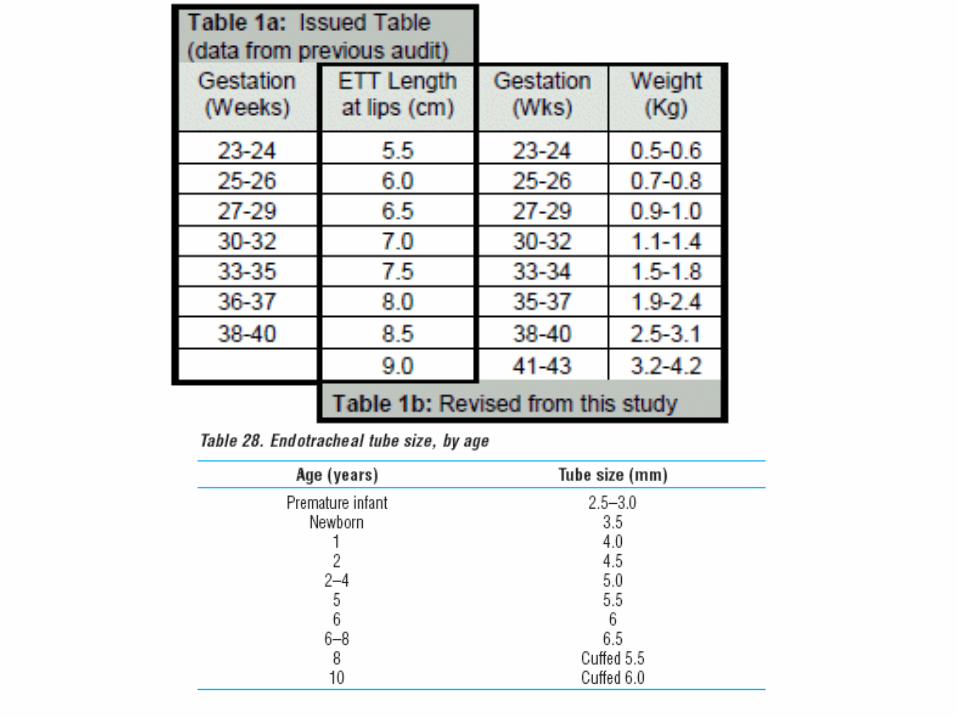
ETT sizes 2.0, 2.5, 3.0 and 3.5 cuffless
ETT holders/neobar or Tape

Equipment at DeliveryBlade sizes 00, 0, 1
Meconium aspirator
Cloth tape
Various size bulb syringes
5, 8, 10 F suction cathetersVacuum at -60 to -80

Radiant warmer with warm blankets, ensure temperature is working

Types of Resuscitators

Prepare the room
• If it is a C-section, you will be in sterile dress (be sure not to contaminate the nurse or doctor), have transport isolate/ventilator outside delivery room
• Insure suction tubing is working

Arrival of the Newborn
• Key questions– Mother’s age– Length of pregnancy (due date)– Presence and frequency of contractions– Presence of or absence of fetal movement– Any pregnancy complications (DM, HTN, fever)– Rupture of membranes
• When?• Color? (clear, meconium, blood)
– Any medications that have been taken
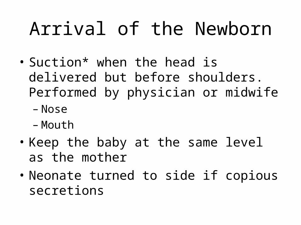
Arrival of the Newborn
• Suction* when the head is delivered but before shoulders. Performed by physician or midwife– Nose– Mouth
• Keep the baby at the same level as the mother• Neonate turned to side if copious secretions

Stabilization• Position• Warm/dry• Head position

SuctioningClear Amniotic Fluid
• Recommendation that suctioning immediately following birth including with a bulb syringe should only be done in babies who have obvious obstruction to spontaneous breathing or require PPV
2010 American Heart Association Guidelines for Cardiopulmonary Resuscitation and Emergency Cardiovascular Care

SuctioningClear Amniotic Fluid
• Suctioning the nasopharynx can cause bradycardia
• Suctioning the trachea in intubated babies – Decreases pulmonary compliance– Decreases oxygenation– Reduces cerebral blood flow
• If secretions are present, suctioning must be performed.
2010 American Heart Association Guidelines for Cardiopulmonary Resuscitation and Emergency Cardiovascular Care

Meconium
• Vigorous or not • If not, suction

Clamp and Cut Cord

Special Consideration
• Polycythemia (excessive red blood cell count) – Delay in clamping the cord– Placing the infant below the placenta
• Do not milk the cord– Destroy or distort RBCs

Special Consideration
Severe prematurity:

Initial Assessment
• Respiratory rate (Cry)• Respiratory effort (Cry)• Pulse rate• Oxygenation
– Color – SpO2

Assess Neonate
• Nearly 90% of newborns are vigorous term babies• Ensure thermoregulation
– Dry– Warm– Place on mother’s chest (skin to skin)
• Suction only if necessary• Assess ventilation (cry)• Asses heart rate• Assess oxygenation (color and SpO2)

Apgar Score
• Determines need and effectiveness of resuscitation
• Performed 1 minute and 5 minutes after birth• If 5 minute Apgar is less than 7, reassess every
5 minutes for 20 minutes

Need for Resuscitation
• Approximately 10% of newborns require additional assistance– 1% requires major resuscitation
• Resuscitation– Intervene Reassess Intervene Reassess– 30 second intervals

Asphyxia
• Hypoxia + Hypercapnia + Acidosis• May lead to irreversible brain damage• The necessity to resuscitate is related to the
degree of asphyxia

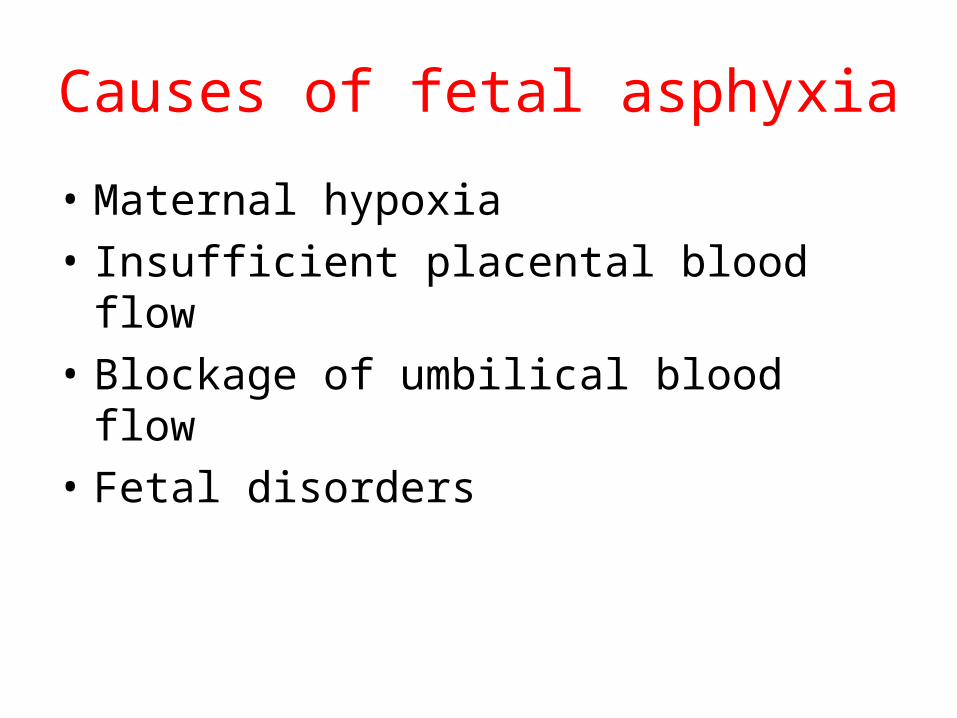
Causes of fetal asphyxia
• Maternal hypoxia• Insufficient placental blood flow• Blockage of umbilical blood flow• Fetal disorders

Primary vs. Secondary Apnea
• Primary– Initial asphyxia– Signs
• Initial period of rapid breathing• Respiratory movements cease• Heart rate and bp drop• Neuromuscular tone diminishes


Secondary Apnea
• If no resuscitation and apnea continues• Signs
– Deep gasping respirations– Heart rate continues to decrease– Blood pressure begins to fall– Infant flaccid– PERFORM PPV


• Primary–Stimulation and
oxygen will usually induce respirations
• Secondary– Infant
unresponsive to stimulation – must be resuscitated


Initial Steps of Resuscitation
• Routine Care – If YES to the following questions– Term gestation?– Amniotic fluid clear?– Breathing or crying?– Good muscle tone?
• Dry• Provide warmth (skin-to-skin)• Cover• Assess color, breathing, acivity
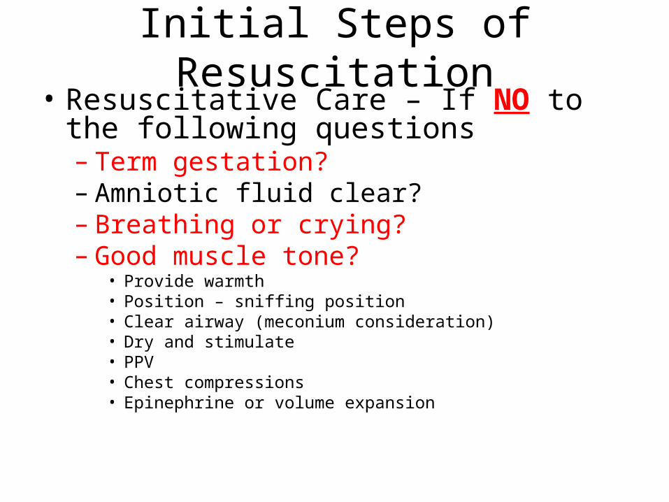
Initial Steps of Resuscitation• Resuscitative Care – If NO to the following
questions– Term gestation?– Amniotic fluid clear?– Breathing or crying?– Good muscle tone?
• Provide warmth• Position – sniffing position• Clear airway (meconium consideration)• Dry and stimulate• PPV• Chest compressions• Epinephrine or volume expansion

Stimulate

Initial Steps (Golden Minute)
• Approximately 60 seconds to complete, reevaluate, and ventilate if necessary– Provide warmth– Clear airway– Dry– Stimulate– Position - sniffing

Initial Steps (Golden Minute)• Decision to proceed beyond initial steps is
based on evaluation of: – Respirations
• Apnea• Gasping• Labored breathing
– Heart rate• Less than 100 bpm• Auscultation of precordial pulse• Palpation of umbilical pulse

Assessment After PPV or Supplemental Oxygenation
• Evaluate– Heart rate– Respirations– Oxygenation
• Most sensitive indicator of successful response is an increase in heart rate
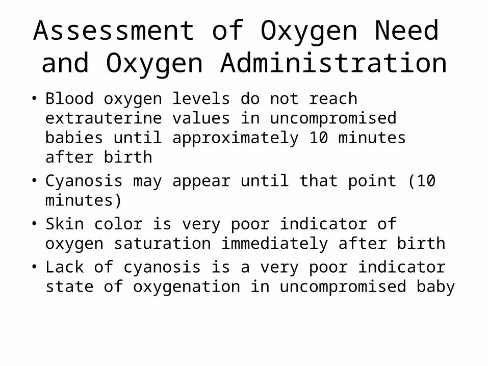
Assessment of Oxygen Need and Oxygen Administration
• Blood oxygen levels do not reach extrauterine values in uncompromised babies until approximately 10 minutes after birth
• Cyanosis may appear until that point (10 minutes)• Skin color is very poor indicator of oxygen saturation
immediately after birth• Lack of cyanosis is a very poor indicator state of
oxygenation in uncompromised baby


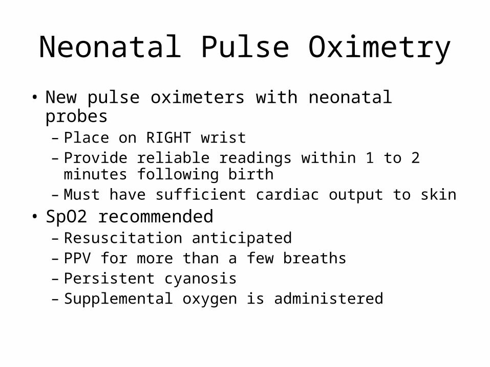
Neonatal Pulse Oximetry
• New pulse oximeters with neonatal probes– Place on RIGHT wrist– Provide reliable readings within 1 to 2 minutes
following birth– Must have sufficient cardiac output to skin
• SpO2 recommended– Resuscitation anticipated– PPV for more than a few breaths– Persistent cyanosis– Supplemental oxygen is administered

Neonatal Pulse Oximetry
• Probe location– Right upper extremity
• Medial surface of the palm• Wrist
• Attach probe to baby prior to device– More rapid acquisition of signal
PPV and Supplemental Oxygen
• 100% oxygen administration is not recommended
• Titrate oxygen to SpO2 range• Initiate resuscitation with air if blended
oxygen is not available– If bradycardia persists (HR <60 bpm) after 90
seconds, increase oxygen to 100% until HR > 100 bpm

Targeted SpO2 After Birth
• 1 minute 60 to 65%• 2 minutes 65 to 70%• 3 minutes 70 to 75%• 4 minutes 75 to 80%• 5 minutes 80 to 85%• 10 minutes 85 to 95%

Newborn Intervention Triggers
• Secretions = suction• Apnea or gasping respirations = PPV• Labored breathing or low SpO2 = oxygen or
CPAP• HR< 100 bpm = PPV• HR< 60 = Chest compressions and PPV• Persistent HR< 60 = epinephrine

Evaluate Respiration, HR, Oxygenation
• Breathing adequate (rate and effort)– No apnea– No gasping– No labored breathing
• HR >100 bpm• SpO2 in normal range
• Observe and suction only to keep airway clear
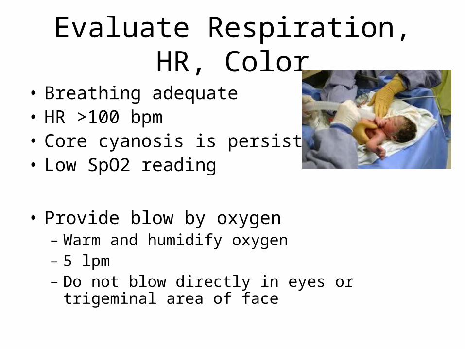
Evaluate Respiration, HR, Color
• Breathing adequate• HR >100 bpm• Core cyanosis is persistent• Low SpO2 reading
• Provide blow by oxygen– Warm and humidify oxygen– 5 lpm– Do not blow directly in eyes or trigeminal area of face

Evaluate Respiration, HR, Color
• Breathing adequate• HR >100 bpm• Acrocyanosis with normal SpO2
• No intervention• If acrocyanosis with poor SpO2 provide blow-
by O2

Evaluate Respiration, HR, SpO2• Breathing inadequate
– Gasping or apnea • HR >100 bpm • Good pink or normal SpO2
• Positive pressure ventilation– Infant size (240 ml)– 5 to 8 ml/kg VT– Disable pop-off (30 to 40 cmH20)– 40 to 60 ventilations/minute– Peak inspiratory pressure 25 cmH2O in full-term

CPAP
• Breathing spontaneously but labored• HR> 100 bpm• SpO2 normal or low
• Research lacking – only studied in preterm babies


Evaluate Respiration, HR, Color
• Breathing adequate• HR <100 bpm • SpO2 normal
• Positive pressure ventilation– Infant size (240 ml)– 5 to 8 ml/kg VT– Disable pop-off (30 to 40 cmH20)– 40 to 60 ventilations/minute– Peak inspiratory pressure 25 mmHg in full-term

Evaluate Respiration, HR, Color• Breathing adequate• HR < 60 bpm• SpO2 not adequate
• PPV• Chest compressions
– Depth 1/3 of anteroposterior diameter of chest– Two thumbs over sternum with hands encircling chest– 3 compressions to one ventilation– Compression rate 120/minute
• 90 compressions and 30 ventilations in one minute
• After 30 seconds of compressions and ventilation – consider epinephrine

Persistent Bradycardia
• Usually due to – Inadequate lung inflation– Profound hypoxemia
• Primary emergency intervention– Adequate ventilation
• HR remains < 60 bpm with 100% oxygen• Consider epinephrine

Epinephrine Administration
• Intravenous (UVC) route is recommended only– 0.01 to 0.03 mg/kg – 1:10,000 dilution
• If ET route is used– 0.05 to 0.1 mg/kg– 1:10,000 dilution
http://www.youtube.com/watch?v=JjBJONanCYU

Volume Expansion
• Blood loss known or suspected– Pale skin– Poor perfusion– Weak pulse– HR not responding to other interventions
• Isotonic crystalloid– 10 mL/kg
• Avoid rapid infusion in premature infants


Oral Airways
• Rarely used for neonates
• Use tongue depressor to insert airway
May be used for Pierre Robin Syndrome or Choanal Atresia

Respiratory Distress or Inadequacy
• HR < 100 bpm = hypoxia• Periodic breathing (20 second or longer period
of apnea)• Intercostal retractions• Nasal flaring• Grunting

Meconium Stained Amniotic Fluid (MSAF)
• 10 to 15% of deliveries• High risk of morbidity• Passage may occur before or during delivery• More common in post-term infants and
neonates small for the gestational age• Fetus normally does not pass stool prior to
brith

Meconium Stained Amniotic Fluid
• Complications if aspirated – Meconium Aspiration Syndrome (MAS)– Atelectasis– Persistent pulmonary hypertension– Pneumonitis– Pneumothorax
Meconium Stained Amniotic Fluid
• Determine if fluid is thin and green or thick and particulate
• If baby is crying vigorously – use standard resuscitation criteria
• If baby is depressed – DO NOT dry or stimulate– Intubate trachea– Attach a meconium aspirator– Apply suction to endotracheal tube– Dry and stimulate– Continue with standard resuscitation

Apnea• Common in infants delivered before 32 weeks of
gestation
• Risk factors– Prematurity– Infection– Prolonged or difficult labor and delivery– Drug exposure– CNS abnormalities– Seizures – Metabolic disorders– Gastroesophageal reflux

Apnea
• Pathophysiology– Prematurity due to underdeveloped CNS– Gastroesophageal reflux can trigger a vagal
response– Drug-induced from CNS depression
• Bradycardia is key assessment finding


Premature and Low Birth Weight Infants
• Delivered before 37th week of gestation
• Less than 5.5 lbs or 2,500 grams
• Premature labor– Genetic factors– Infection– Cervical incompetence– Abruption– Multiple gestations (twins, triplets)– Previous premature delivery– Drug use– Trauma

Premature and Low Birth Weight Infants
• Low birth weight– Chronic maternal HTN– Smoking– Placental anomalies– Chromosomal abnormalities
• Born <24 weeks and less than 1 lb – poor chance of survival

Premature and Low Birth Weight Infants
• Physical appearance– Skin is thin and translucent– No cartilage in the outer ear– Small breast nodule size– Fine thin hair – Lack of creases in soles of feet

Premature and Low Birth Weight Infants
• High risk for respiratory distress and hypothermia– Surfactant deficiency– Thermoregulation is imperative
• Use minimum pressure with PPV• Brain injury may result from hypoxemia, rapid
change in blood pressure• Retinopathy from abnormal vascular development of
retina – May be worsened by long term oxygen administration

Hypoglycemia
• BGL <40 mg/dL• May not be symptomatic until BGL reaches 20
mg/dL• Fetus received glycogen stores from mother in
utero– Liver– Heart– Lung– Skeletal muscle
Hypoglycemia
• Glycogen stores sufficient for 8 to 12 hours after birth
• Disorders related to – Poor glycogen storage
• Small birth weight• Prematurity postmaturity
– Increased glucose use• Infant of DM mother• Large for gestational age• Hypoxia• Hypothermia• Sepsis

Hypoglycemia• Symptoms
– Cyanosis– Apnea– Irritability– Poor sucking or feeding– Hypothermia– Lethargy– Tremors– Twitching or seizures– Coma– Tachycardia– Tachypnea– Vomiting

Hypoglycemia
• Check BGL – heel stick• Establish good airway, ventilation,
oxygenation, and circulation• D10W -10% dextrose
– 2 mL/kg IV if BGL <40 mg/dL– IV infusion of D10W – 60 mL/kg