Embed Size (px)
Citation preview
Chapter 4Molecular Detection of Past Pathogens
Michel Drancourt(*ü ) and Didier Raoult
Abstract Detection and characterisation of DNA is the most widely used approach for the study of past pathogens. This approach can be applied to various specimens, including environmental, vector and animal reservoir specimens as well as human corpses. Experimental data indicated that host-associated microbial DNA can survive for 20,000 years, and bacterial DNA preserved in permafrost specimens has been dated up to 1 million years. Current protocols targeted one pathogen at a time and universal 16S rDNA-based detection of bacteria have yielded ambiguous results. There is no universal detection of ancient virus so far. Major human patho-gens, e.g. Mycobacterium tuberculosis, Mycobacterium leprae, Yersinia pestis, Rickettsia prowazekii, Bartonella spp. and Spanish influenza virus have been detected in suitable human specimens. Ancient M. tuberculosis and Y. pestis organ-isms have been genotyped, whereas the entire RNA genome of Spanish influenza virus was reconstituted for extensive studies. Metagenomic approaches based on high throughput pyrosequencing may help further resolve forthcoming issues. Interpretation of experimental data has to be based upon strict rules due to potential contamination of specimens.
4.1 Introduction
As a discipline, palaeomicrobiology (Zink et al. 2002) began in 1993 with the molecular detection of Mycobacterium tuberculosis DNA in an ancient human skeleton (Spigelman and Lemma 1993). This finding served to illustrate the impor-tance of molecular biology techniques in the quest for pathogens, and microbes at large, in ancient specimens recovered from various human tissues, as well as from environmental samples of potential vectors and reservoirs of past pathogens
Michel DrancourtUnité des Rickettsies, CNRS UMR 6020, Faculté de Médecine, Université de laMéditerranée, 27 Boulevard Jean Moulin, 13385 Marseille Cedex 5, FranceE-mail: [email protected]
D. Raoult and M. Drancourt (eds.), Paleomicrobiology: Past Human Infections. 55© Springer-Verlag Berlin Heidelberg 2008
56 M. Drancourt, D. Raoult
(Drancourt et al. 2005). Indeed, with the exception of some enteric parasites (Bouchet et al. 2003) and rare human viruses, all past pathogens and microbes have been detected and studied thanks to the detection and characterisation of nucleic acids. Experimental data have now demonstrated that bacterial DNA can be detected in 20,000-year-old host specimens, and in up to several thousand-year-old environmental specimens preserved in permafrost (Willerslev et al. 2005). Likewise, Spanish influenza virus RNA has been extensively studied after its recovery from both formalin-preserved human lung tissue (Reid et al. 2000; Taubenberger et al. 1997) and permafrost-preserved human tissues (Reid et al. 2000).
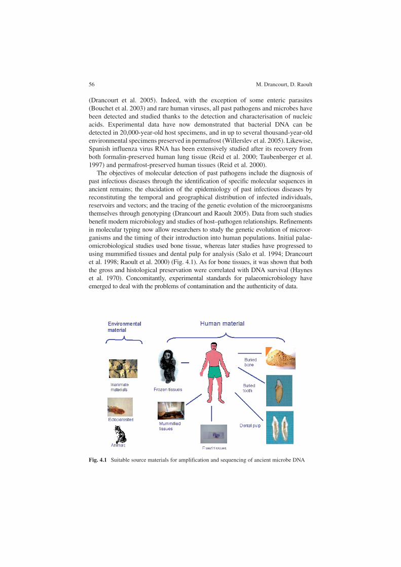
The objectives of molecular detection of past pathogens include the diagnosis of past infectious diseases through the identification of specific molecular sequences in ancient remains; the elucidation of the epidemiology of past infectious diseases by reconstituting the temporal and geographical distribution of infected individuals, reservoirs and vectors; and the tracing of the genetic evolution of the microorganisms themselves through genotyping (Drancourt and Raoult 2005). Data from such studies benefit modern microbiology and studies of host–pathogen relationships. Refinements in molecular typing now allow researchers to study the genetic evolution of microor-ganisms and the timing of their introduction into human populations. Initial palae-omicrobiological studies used bone tissue, whereas later studies have progressed to using mummified tissues and dental pulp for analysis (Salo et al. 1994; Drancourt et al. 1998; Raoult et al. 2000) (Fig. 4.1). As for bone tissues, it was shown that both the gross and histological preservation were correlated with DNA survival (Haynes et al. 1970). Concomitantly, experimental standards for palaeomicrobiology have emerged to deal with the problems of contamination and the authenticity of data.
Fig. 4.1 Suitable source materials for amplification and sequencing of ancient microbe DNA
4 Molecular Detection of Past Pathogens 57
4.2 Protocols for the Molecular Detection of Past Pathogens
Detection and identification of pathogens in ancient human and environmental specimens relies mostly upon the molecular detection of specific nucleic acid sequences. A few studies have focussed on viral RNA for the detection of Spanish influenza virus (Reid et al. 2000; Taubenberger et al. 1997; Reid et al. 1999) but the vast majority of studies have targeted ancient bacterial and parasite DNA. While experi-mental protocols for DNA extraction and its amplification by polymerase chain reaction (PCR) have been empirical, a few systematic studies of experimental parameters now provide clear experimental guidelines for optimal DNA extraction and amplification from bone tissues (Rohland and Hofreiter 2007).
4.2.1 Ancient DNA Characteristics
Empirical observations made over the last 20 years indicated that ancient DNA has adverse characteristics when compared to modern DNA. The amino-acid racemisation ratio was shown to predict the preservation of ancient DNA (Poinar et al. 1996). Ancient DNA is broken into pieces of < 500 bp (Lindahl 1993); consequently, PCR cannot be used to amplify large fragments in ancient specimens. In the case of ancient mammal DNA, this limitation has been circumvented by pre-treatment of the ancient DNA with reconstructive polymerisation (Golenberg et al. 1996) or enzymatic repair by the combined activities of DNA polymerase I and T4 DNA ligase (Pusch et al. 1998; Di et al. 2002). However, nothing has been published regarding the repair of ancient microbial DNA.
A second feature of ancient DNA is chemical modification, comprising both oxidisation and hydrolysis resulting in deamination of nucleotides (Hoss et al. 1996; Hofreiter et al. 2001). Such modifications have been implicated in cases of poor yields from PCR. It has been recently demonstrated that not all DNA polymerases amplify ancient DNA extracted from cave bear bone with the same efficiency (Rohland and Hofreiter 2007).
Third, numerous studies have demonstrated the presence of poorly characterised PCR inhibitors in ancient specimen extracts (Hoss et al. 1996; Hanni et al. 1995). The precise nature of these inhibitors, once correlated to the presence of a brown coloration of extracts (Hanni et al. 1995), is not known. Two strategies have been proposed to circumvent the presence of inhibitors: dilution of extracted specimens and the addition of bovine serum albumin (BSA). The effectiveness of both solutions has recently been demonstrated (Rohland and Hofreiter 2007).
4.2.2 Nucleic Acid Extraction
Since the initial demonstration that DNA can survive in mummified human tissues (Pääbo 1985), nucleic acid extraction from various types of specimens has been
58 M. Drancourt, D. Raoult
achieved. Extraction can be achieved from conjunctive tissues that have been either frozen (Reid et al. 1999; Cano et al. 2000; Rhodes et al. 1998), mummified (Salo et al. 1994; Fornaciari et al. 2003) buried (Reid et al. 1999) (Table 4.1) or fixed (Taubenberger et al. 2005) . Extraction from bone tissues requires extensive decal-cification using EDTA and mechanical grinding prior to DNA extraction. The same holds true for entire teeth. We proposed the use of dental pulp as a suitable specimen for the molecular detection of blood-borne organisms (Drancourt et al. 1998). Several protocols for the extraction of DNA from ancient tissues have been proposed, but the comparative performance of these various protocols has been evaluated only recently (Rohland and Hofreiter 2007).
4.2.3 Amplification, Cloning and Sequencing
All studies dealing with ancient microbial DNA use a PCR amplification step before nucleotide sequencing. Various PCR protocols have been developed, includ-ing one-step conventional PCR in most studies, nested and hemi-nested PCR and, rarely, real-time PCR. The addition of either BSA or a related protein in the PCR cocktail had been advocated in order to prevent PCR inhibition (Rohland and Hofreiter 2007). This empirical observation has recently been verified (Rohland and Hofreiter 2007). The exact nature of the PCR inhibitors in ancient specimens has not been elucidated, and the proposed correlation of the brown colour of the extraction product with PCR inhibition (Hanni et al. 1995) has not been confirmed (Drancourt et al. 1998). In most studies, PCR-amplified fragments are cloned before being sequenced. So far, the conventional Sanger sequencing method has been applied using capillary automatic sequencers.
Table 4.1 Adverse characteristics of ancient microbial DNA limiting PCR-based detection of past pathogens and proposed solutions. PCR Polymerase chain reaction, BSA bovine serum albumin
CharacteristicConsequence for PCR-based detection Proposed solutions
Fragmentation < 500 bp Amplification of small frag-ments only
Select PCR primers in order to amplify a fragment ≤ 300 bp
DNA enzymatic repair using DNA polymerase I/T4 DNA ligasea
Chemical alterations Poor PCR yield Select appropriate Taq DNA polymerase
PCR inhibitors Lack of PCR amplification Run dilutions of extracted DNAAdd BSA to PCR mix
a This technique has been published only for ancient eukaryotic DNA
4 Molecular Detection of Past Pathogens 59
4.3 Contamination of Ancient Specimens
Micro-organisms from the burial site can contaminate specimens before labora-tory analyses, whereas laboratory micro-organisms and their DNA can contami-nate specimens during laboratory analyses. Some PCR mix reagents, including PCR primers, polymerases and water used to complement reaction volumes, have been shown to be contaminated by bacterial DNA. In the detection of past bacte-ria, the contamination threat is particularly great when using a universal approach such as 16S rDNA-based PCR (Gilbert et al. 2004; Zink et al. 2000; Cano et al. 2000). Specific molecular targets carry a smaller risk. The specificity of detection has been shown by analysis of environmental samples in parallel with buried specimens (Papagrigorakis et al. 2006). The use of naturally protected specimens, such as dental pulp, might also limit the risk of external contamination (Drancourt et al. 1998).
4.4 Strategies to Obtain Reliable Data
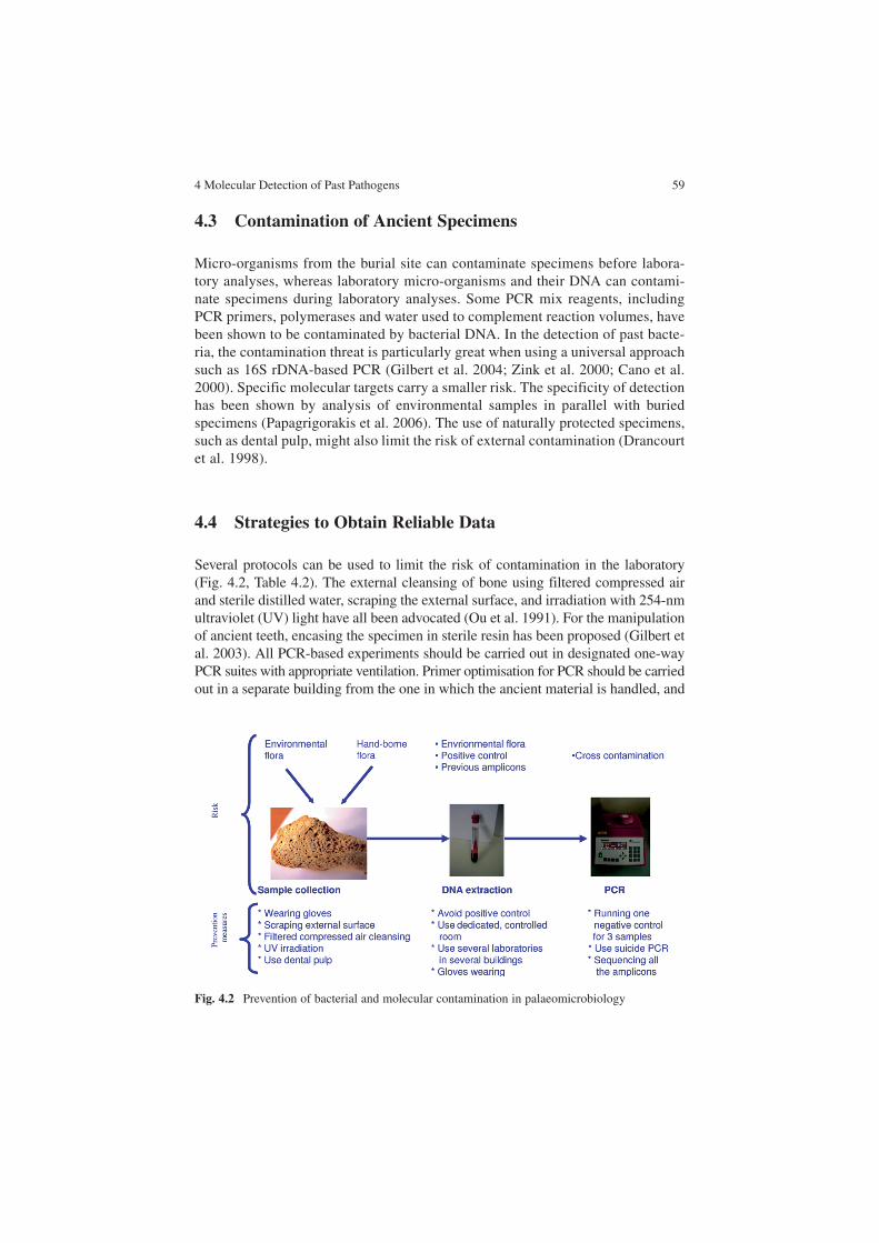
Several protocols can be used to limit the risk of contamination in the laboratory (Fig. 4.2, Table 4.2). The external cleansing of bone using filtered compressed air and sterile distilled water, scraping the external surface, and irradiation with 254-nm ultraviolet (UV) light have all been advocated (Ou et al. 1991). For the manipulation of ancient teeth, encasing the specimen in sterile resin has been proposed (Gilbert et al. 2003). All PCR-based experiments should be carried out in designated one-way PCR suites with appropriate ventilation. Primer optimisation for PCR should be carried out in a separate building from the one in which the ancient material is handled, and
Fig. 4.2 Prevention of bacterial and molecular contamination in palaeomicrobiology
60 M. Drancourt, D. Raoult
PCR and post-PCR experiments should be performed in a separate room using disposable equipment and freshly prepared reagents that have been irradiated with UV light. It has been also advocated that ancient DNA experiments be performed without using a positive control. Alternatively, mock positive controls and DNA from different, related species can be used. Furthermore, we developed “suicide PCR” reactions, which target a new genomic region by using a new PCR primer pair in every new experiment, to prevent vertical contamination from previous ampli-fications (Raoult et al. 2000). The introduction of numerous negative controls helps monitor any carry-over source of contamination. Material collected from unaffected individuals are also of value; for example, lesion-free bones collected from fossilised Canis and Equus species have been used as controls for the molecular detection of M. tuberculosis DNA in extinct bison (Rothschild et al. 2001).
As pathogens are not ubiquitous organisms, the first sequence achieved in a laboratory is reliable if the pathogen and its DNA have never been manipulated in that laboratory. Therefore, standardisation of PCR protocols must be carried out in a laboratory different from the one where the ancient DNA is handled. Likewise, DNA from ancient specimens must be extracted in a laboratory where the targeted pathogens have never been manipulated. We optimised this approach by performing these different experimental steps in laboratories located in different campus build-ings (Drancourt et al. 2004). Also, we designed suicide PCR in order to prevent intra-laboratory contamination resulting from previous experiments (Raoult et al. 2000). Suicide PCR avoids use of positive controls and uses a new PCR primer pair targeting a different genomic region for every new experiment (Raoult et al. 2000). Alternatively, PCR targetting a hypervariable genomic region could be used in order to demonstrate the presence of an original sequence of the pathogen in the ancient specimen.
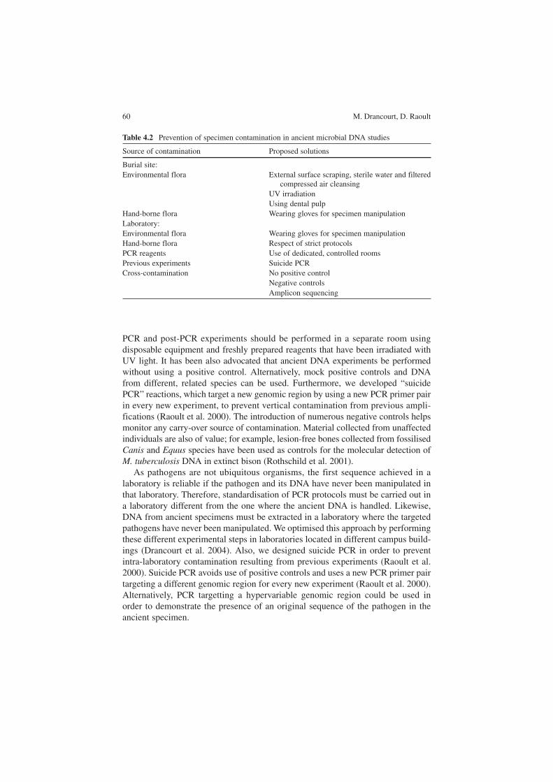
Table 4.2 Prevention of specimen contamination in ancient microbial DNA studies
Source of contamination Proposed solutions
Burial site:Environmental flora External surface scraping, sterile water and filtered
compressed air cleansingUV irradiationUsing dental pulp
Hand-borne flora Wearing gloves for specimen manipulationLaboratory:Environmental flora Wearing gloves for specimen manipulationHand-borne flora Respect of strict protocolsPCR reagents Use of dedicated, controlled roomsPrevious experiments Suicide PCRCross-contamination No positive control
Negative controlsAmplicon sequencing
4 Molecular Detection of Past Pathogens 61
4.5 Interpretation of Data
Strict adherence to the rules for the prevention of contamination is a first step towards ensuring the authenticity of ancient microbial isolates. Absence of any amplicon in negative controls is strictly required. The recovery of an original sequence indicates that laboratory contamination has not occurred and is good evidence for authenticity. The original sequence must be shown in several clones. Chemical modifications of ancient DNA can result in “jumping PCR” – template switching during PCR and C→T and G→A substitutions. The sequencing of multiple clones derived from more than one independent amplification has been advocated to reduce the risk of obtaining incorrect DNA sequences (Hoss et al. 1996; Spencer and Howe 2004). However, there is no evidence of “spontaneous” mutation in ancient DNA (Serre et al. 2004).
Phylogenetic analyses of the gene sequence from the ancient microorganism can confirm its antiquity; for example, phylogenetic analyses of a Bacillus sp. that was once claimed to be 250 million years old showed that it was in fact a modern contaminant (Vreeland et al. 2000; Nickle et al. 2002). The reproducibility of results using different specimens collected from the same individual is another a criterion. Also, the demonstration of two unrelated sequences that identify the same pathogen in the same specimen further increases the specificity of the identification.
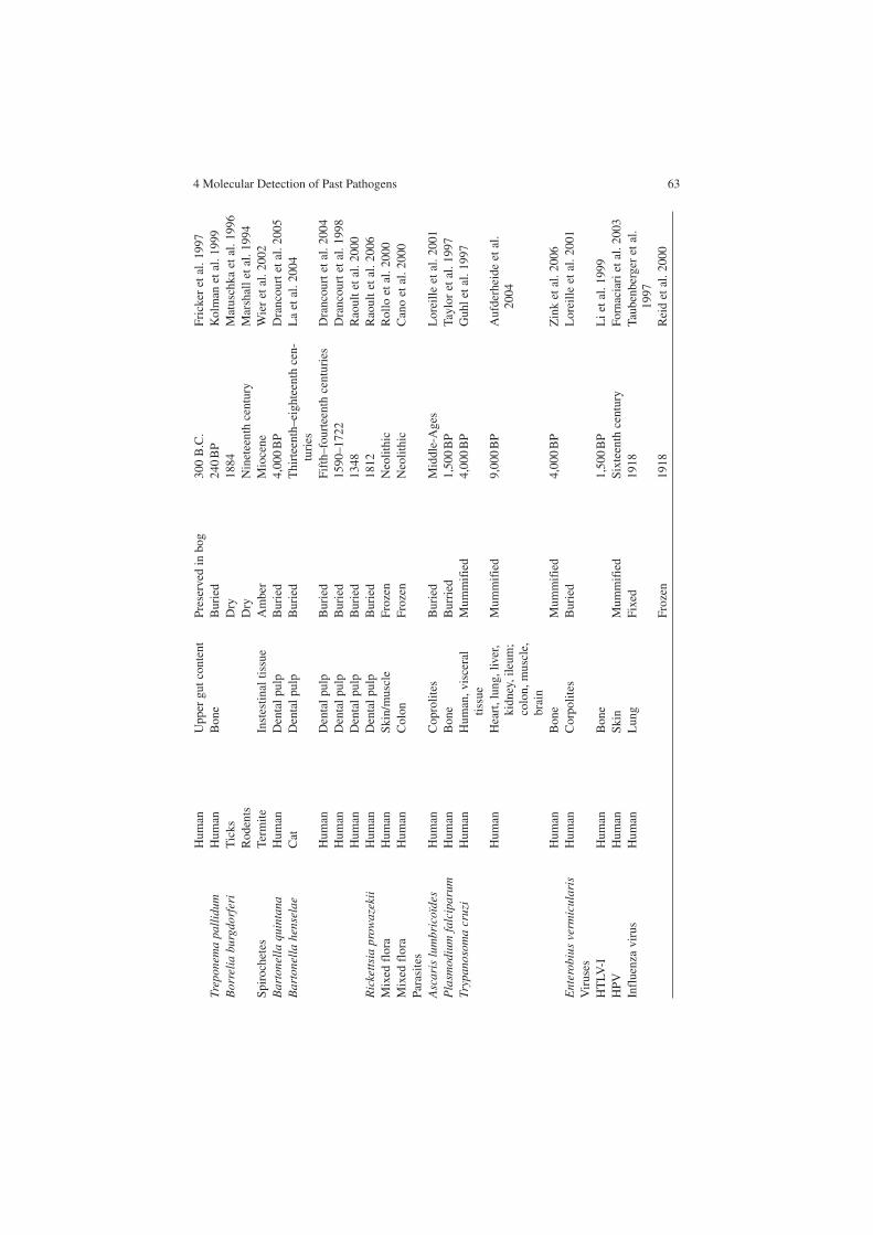
4.6 Molecular Detection of Past Pathogens: Current Data
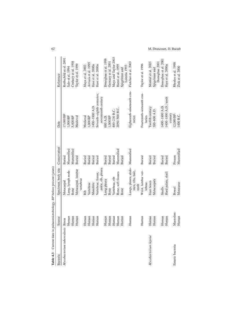
Most published data deal with the detection and molecular characterisation of ancient bacteria, while fewer studies have examined past viruses and parasites. The most significant data are presented in Table 4.3. To complement the molecular detection and identification of past pathogens, some ancient bacteria have been geno-typed. In the case of the M. tuberculosis complex, ancient mycobacteria were genotyped by sequencing the phospholipase-C mtp40 gene, a Mycobacterium tuberculosis-specific region, another Mycobacterium bovis-specific fragment and the oxyR pseudogene (Pääbo 1985). This work demonstrated that medieval myco-bacteria were more closely related to modern Mycobacterium tuberculosis than to Mycobacterium bovis. Similar conclusions were obtained from a spoligotyping analysis of 12 Mycobacterium tuberculosis strains that were characterised among Egyptian mummies dating from 2050 to 500 B.C. (Zink et al. 2003). Spoligotypes obtained from mycobacterial DNA from an extinct bison demonstrated that it was more closely related to the Mycobacterium tuberculosis / Mycobacterium africa-num group than it was to M. bovis (Rothschild et al. 2001). These data indicated that the theory that Mycobacterium tuberculosis had evolved from M. bovis by spe-cific adaptation to the human host was not in fact the case (Stead et al. 1995).
In our laboratory, using multispacer sequence typing (MST), we have successfully genotyped Yersinia pestis in individuals suspected to have died from the Justinian
62 M. Drancourt, D. RaoultTa
ble
4.3
Cur
rent
dat
a in
pal
aeom
icro
biol
ogy.
BP
bef
ore
pres
ent (
year
s)
Bac
teri
aSo
urce
Spec
imen
, bod
y si
teC
onse
rvat
ion
Dat
eR
efer
ence
Myc
obac
teri
um t
uber
culo
sis
Bis
onM
etac
arpa
lB
urie
d17
,000
BP
Rot
hsch
ild e
t al.
2001
Hum
anL
ung,
lym
ph n
ode
Mum
mif
ied
1,00
0 B
PSa
lo e
t al.
1994
Hum
anB
one
Mum
mif
ied
5,40
0 B
PC
rube
zy e
t al.
1998
Hum
anM
etac
arpa
l, lu
mba
r ve
rteb
rae
Bur
ied
Med
ieva
lTa
ylor
et a
l. 19
99
Hum
anR
ibB
urie
dM
edie
val
May
s et
al.
2002
Hum
anV
erte
brae
Bur
ied
1,00
0 B
PA
rria
za e
t al.
1995
Hum
anM
andi
ble
Bur
ied
1400
–180
0 A
.D.
Has
s et
al.
2000
aH
uman
Ver
tebr
ae, f
emur
, an
kle,
rib
, ple
ura
Bur
ied
Seve
nth–
eigh
th c
entu
ries
; se
vent
eent
h ce
ntur
yH
ass
et a
l. 20
00a
Hum
anL
ung
pleu
raB
urie
d60
0 A
.D.
Don
oghu
e et
al.
1998
Hum
anB
one
Bur
ied
1,00
0 B
PG
erna
ey e
t al.
2001
Hum
anV
erte
brae
, rib
Bur
ied
400–
230
B.C
.M
ays
and
Tayl
or 2
003
Hum
anB
one,
sof
t tis
sues
Mum
mif
ied
2050
–500
B.C
.H
anni
et a
l. 19
95H
uman
Bon
eB
urie
dSp
igel
man
and
L
emm
a 19
93H
uman
Lun
gs, p
leur
a, a
bdo-
men
, rib
s, h
air,
teet
h
Mum
mif
ied
Eig
htee
nth–
nine
teen
th c
en-
turi
esFl
etch
er e
t al.
2003
Hum
anW
rist
, lum
bar
ver-
tebr
aeB
urie
dFo
urte
enth
–six
teen
th c
en-
turi
esTa
ylor
et a
l. 19
96
Myc
obac
teri
um l
epra
eH
uman
Foot
bon
esB
urie
dTw
elft
h ce
ntur
yM
ontie
l et a
l. 20
03H
uman
Met
acar
pals
Bur
ied
300–
600
A.D
.Sp
igel
man
and
D
onog
hue
2001
Hum
anSk
ulls
Bur
ied
1400
–180
0 A
.D.
Don
oghu
e et
al.
2001
Hum
anH
ard
pala
te, s
kull
Bur
ied
1400
–180
0 A
.D.;
tent
h ce
ntur
yH
ass
et a
l. 20
00a
Ent
eric
bac
teri
aM
asto
don
Bow
elFr
ozen
12,0
00 B
PR
hode
s et
al.
1998
Hum
anM
etat
arse
Mum
mif
ied
1400
B.C
.Z
ink
et a
l. 20
00
4 Molecular Detection of Past Pathogens 63H
uman
Upp
er g
ut c
onte
ntPr
eser
ved
in b
og30
0 B
.C.
Fric
ker
et a
l. 19
97Tr
epon
ema
pall
idum
Hum
anB
one
Bur
ied
240
BP
Kol
man
et a
l. 19
99B
orre
lia
burg
dorf
eri
Tic
ksD
ry18
84M
atus
chka
et a
l. 19
96R
oden
tsD
ryN
inet
eent
h ce
ntur
yM
arsh
all e
t al.
1994
Spir
oche
tes
Term
iteIn
stes
tinal
tiss
ueA
mbe
rM
ioce
neW
ier
et a
l. 20
02B
arto
nell
a qu
inta
naH
uman
Den
tal p
ulp
Bur
ied
4,00
0 B
PD
ranc
ourt
et a
l. 20
05B
arto
nell
a he
nsel
aeC
atD
enta
l pul
pB
urie
dT
hirt
eent
h–ei
ghte
enth
cen
-tu
ries
La
et a
l. 20
04
Hum
anD
enta
l pul
pB
urie
dFi
fth–
four
teen
th c
entu
ries
Dra
ncou
rt e
t al.
2004
Hum
anD
enta
l pul
pB
urie
d15
90–1
722
Dra
ncou
rt e
t al.
1998
Hum
anD
enta
l pul
pB
urie
d13
48R
aoul
t et a
l. 20
00R
icke
ttsi
a pr
owaz
ekii
Hum
anD
enta
l pul
pB
urie
d18
12R
aoul
t et a
l. 20
06M
ixed
flo
raH
uman
Skin
/mus
cle
Froz
enN
eolit
hic
Rol
lo e
t al.
2000
Mix
ed f
lora
Hum
anC
olon
Froz
enN
eolit
hic
Can
o et
al.
2000
Para
site
sA
scar
is l
umbr
icoï
des
Hum
anC
opro
lites
Bur
ied
Mid
dle-
Age
sL
orei
lle e
t al.
2001
Pla
smod
ium
fal
cipa
rum
Hum
anB
one
Bur
ried
1,50
0 B
PTa
ylor
et a
l. 19
97Tr
ypan
osom
a cr
uzi
Hum
anH
uman
, vis
cera
l tis
sue
Mum
mif
ied
4,00
0 B
PG
uhl e
t al.
1997
Hum
anH
eart
, lun
g, li
ver,
kidn
ey, i
leum
; co
lon,
mus
cle,
br
ain
Mum
mif
ied
9,00
0 B
PA
ufde
rhei
de e
t al.
2004
Hum
anB
one
Mum
mif
ied
4,00
0 B
PZ
ink
et a
l. 20
06E
nter
obiu
s ve
rmic
ular
isH
uman
Cor
polit
esB
urie
dL
orei
lle e
t al.
2001
Vir
uses
HT
LV-I
Hum
anB
one
1,50
0 B
PL
i et a
l. 19
99H
PVH
uman
Skin
Mum
mif
ied
Sixt
eent
h ce
ntur
yFo
rnac
iari
et a
l. 20
03In
flue
nza
viru
sH
uman
Lun
gFi
xed
1918
Taub
enbe
rger
et a
l. 19
97Fr
ozen
1918
Rei
d et
al.
2000
64 M. Drancourt, D. Raoult
plague (Drancourt et al. 2004). After comparison of the two Y. pestis genome sequences available in GenBank, we found that some intergenic spacer sequences were highly variable, and we amplified six of these sequences from the ancient specimens. Sequence analyses showed that the sequences obtained were original sequences owing to the presence of point mutations. These mutations were consist-ently found in several clones, therefore confirming that they were not merely caused by misincorporation of nucleotides by Taq polymerase. Y. pestis has been subdivided into three biovars on the basis of their ability to ferment glycerol and to reduce nitrate. On the basis of their current geographical niche, and on historical records that indicated the geographical origin of the pandemics, it was speculated that each biovar caused a specific pandemic (Devignat 1954). MST data demonstrated that the genotype involved in all three pandemics was associated with the Orientalis biovar, a result recently confirmed by demonstration of a specific deletion in the glpD gene (Drancourt et al. 2007).
4.7 Future Research
The detection of pathogens in their ancient reservoirs, and of vectors, will be a key factor in achieving the goal of a global epidemiology scheme for every trans-missible infectious disease. Such detection will benefit from improved collaboration between palaeozoologists, specialists in ancient ectoparasites and palaeomicro-biologists. Specific issues include the correct collection and identification of buried animals and ectoparasites. With regards to human remains and the remains of other mammals, in our opinion, the broad use of dental pulp could help resolve the aetiology of ancient bloodborne infections, although universal protocols are still required.
The application of the universal 16S rDNA-based detection and identification of bacteria to palaeomicrobiology has been limited by contamination of the ancient material. However, this powerful molecular tool will be invaluable in the study of the nature and epidemiology of unpredicted pathogens. The aetiology of numerous past epidemics remains unknown, despite testing for the presence of one or more bacterial pathogens. Tracing any bacterial pathogen within the remains of this past population could help resolve the question of the aetiology of some mysterious epidemics. Given the small amount of material available in the majority of these cases, testing for all bacterial pathogens simultaneously would be helpful. Studies must be performed to develop a protocol of universal amplification and sequencing that is adapted to ancient bacterial DNA.
Metagenomic analysis of total DNA extracted from ancient specimens is a promising field of research. It relies on the high throughput sequencing made possible by the new generation of pyrosequencers. This new approach has been successfully applied to the study of complex modern flora, and to that of ancient mammoth tissue (Poinar et al. 2006). It may resolve the quest for universal detection, not only of bacteria but also of viruses, in ancient specimens.
4 Molecular Detection of Past Pathogens 65
Genotyping will create the necessary bridge between the detection of microbial DNA in ancient environmental and human specimens and modern microbiology. The availability of a large database of complete microbial genome sequences has already prompted the establishment of suicide PCR and new genotyping methods for past microorganisms, including spoligotyping of M. tuberculosis (Zink et al. 2003) and MST of Y. pestis (Drancourt et al. 2004). Such efforts should be continued.
References
Arriaza BT, Salo W, Aufderheide AC, Holcomb TA (1995) Pre-Columbian tuberculosis in northern Chile: molecular and skeletal evidence. Am J Phys Anthropol 98:37–45
Aufderheide AC, Salo W, Madden M, Streitz J, Buikstra J, Guhl F, Arriaza B, Renier C, Wittmers LE Jr, Fornaciari G, Allison M (2004) A 9,000-year record of Chagas’ disease. Proc Natl Acad Sci USA 101:2034–2039
Bouchet F, Harter S, Le BM (2003) The state of the art of paleoparasitological research in the Old World. Mem Inst Oswaldo Cruz 98[Suppl 1]:95–101
Cano RJ, Tiefenbrunner F, Ubaldi M, Del CC, Luciani S, Cox T, Orkand P, Künzel KH, Rollo F (2000) Sequence analysis of bacterial DNA in the colon and stomach of the Tyrolean Iceman. Am J Phys Anthropol 112:297–309
Crubezy E, Ludes B, Poveda JD, Clayton J, Crouau-Roy B, Montagnon D (1998) Identification of Mycobacterium DNA in an Egyptian Pott’s disease of 5,400 years old. C R Acad Sci III 321:941–951
Devignat R (1954) Biological and biochemical behavior of Pasteurella pestis and Pasteurella pseudotuberculosis. Bull World Health Org 10:463–494
Di BG, Del GS, Cammarota M, Galderisi U, Cascino A, Cipollaro M (2002) Enzymatic repair of selected cross-linked homoduplex molecules enhances nuclear gene rescue from Pompeii and Herculaneum remains. Nucleic Acids Res 30:e16
Donoghue HD, Spigelman M, Zias J, Gernaey-Child AM, Minnikin DE (1998) Mycobacterium tuberculosis complex DNA in calcified pleura from remains 1400 years old. Lett Appl Microbiol 27:265–269
Donoghue HD, Holton J, Spigelman M (2001) PCR primers that can detect low levels of Mycobacterium leprae DNA. J Med Microbiol 50:177–182
Drancourt M, Raoult D (2005) Palaeomicrobiology: current issues and perspectives. Nat Rev Microbiol 3:23–35
Drancourt M, Aboudharam G, Signoli M, Dutour O, Raoult D (1998) Detection of 400-year-old Yersinia pestis DNA in human dental pulp: an approach to the diagnosis of ancient septicemia. Proc Natl Acad Sci USA 95:12637–12640
Drancourt M, Roux V, Dang LV, Tran-Hung L, Castex D, Chenal-Francisque V, Ogata H, Fournier P-E, Crubezy E, Raoult D (2004) Genotyping, Orientalis-like Yersinia pestis, and plague pandemics. Emerg Infect Dis 10:1585–1592
Drancourt M, Tran-Hung L, Courtin J, Lumley H, Raoult D (2005) Bartonella quintana in a 4000-year-old human tooth. J Infect Dis 191:607–611
Drancourt M, Signoli M, Dang LV, Bizot B, Roux V, Tzortzis S, Raoult D (2007) Yersinia pestis orientalis in remains of ancient plague patients. Emerg Infect Dis 13:332–333
Fletcher HA, Donoghue HD, Holton J, Pap I, Spigelman M (2003) Widespread occurrence of Mycobacterium tuberculosis DNA from 18th–19th century Hungarians. Am J Phys Anthropol 120:144–152
Fornaciari G, Zavaglia K, Giusti L, Vultaggio C, Ciranni R (2003) Human papillomavirus in a 16th century mummy. Lancet 362:1160
66 M. Drancourt, D. Raoult
Fricker EJ, Spigelman M, Fricker CR (1997) The detection of Escherichia coli DNA in the ancient remains of Lindow Man using the polymerase chain reaction. Lett Appl Microbiol 24:351–354
Gernaey AM, Minnikin DE, Copley MS, Dixon RA, Middleton JC, Roberts CA (2001) Mycolic acids and ancient DNA confirm an osteological diagnosis of tuberculosis. Tuberculosis 81:259–265
Gilbert MT, Willerslev E, Hansen AJ, Barnes I, Rudbeck L, Lynnerup N, Cooper A (2003) Distribution patterns of postmortem damage in human mitochondrial DNA. Am J Hum Genet 72:32–47
Gilbert MT, Cuccui J, White W, Lynnerup N, Titball RW, Cooper A, Prentice MB (2004) Absence of Yersinia pestis-specific DNA in human teeth from five European excavations of putative plague victims. Microbiology 150:341–354
Golenberg EM, Bickel A, Weihs P (1996) Effect of highly fragmented DNA on PCR. Nucleic Acids Res 24:5026–5033
Guhl F, Jaramillo C, Yockteng R, Vallejo GA, Cardenas-Arroyo F (1997) Trypanosoma cruzi DNA in human mummies. Lancet 349:1370
Hanni C, Brousseau T, Laudet V, Stehelin D (1995) Isopropanol precipitation removes PCR inhibitors from ancient bone extracts. Nucleic Acids Res 23:881–882
Hass CJ, Zink A, Molar E, Szeimies U, Reischl U, Marcsik A, Ardagna Y, Dutour O, Pálfi G, Nerlich AG (2000a) Molecular evidence for different stages of tuberculosis in ancient bone samples from Hungary. Am J Phys Anthropol 2000:293–304
Hass CJ, Zink A, Palfi G, Szeimies U, Nerlich AG (2000b) Detection of leprosy in ancient human skeletal remains by molecular identification of Mycobacterium leprae. Am J Clin Pathol 2000:428–436
Haynes RE, Sanders DY, Cramblett HG (1970) Rocky Mountain spotted fever in children. J Pediatr 76:685–693
Hofreiter M, Jaenicke V, Serre D, Haeseler AA, Pääbo S (2001) DNA sequences from multiple amplifications reveal artifacts induced by cytosine deamination in ancient DNA. Nucleic Acids Res 29:4793–4799
Hoss M, Jaruga P, Zastawny TH, Dizdaroglu M, Pääbo S (1996) DNA damage and DNA sequence retrieval from ancient tissues. Nucleic Acids Res 24:1304–1307
Kolman CJ, Centurion-Lara A, Lukehart SA, Owsley DW, Tuross N (1999) Identification of Treponema pallidum subspecies pallidum in a 200-year-old skeletal specimen. J Infect Dis 180:2060–2063
La VD, Clavel B, Lepetz S, Aboudharam G, Raoult D, Drancourt M (2004) Molecular detection of Bartonella henselae DNA in the dental pulp of 800-year-old French cats. Clin Infect Dis 39:1391–1394
Li HC, Fujiyoshi T, Lou H, Yashiki S, Sonoda S, Cartier L, Nunez L, Munoz I, Horai S, Tajima K (1999) The presence of ancient human T-cell lymphotropic virus type I provirus DNA in an Andean mummy. Nat Med 1428–1432
Lindahl T (1993) Instability and decay of the primary structure of DNA. Nature 362:709–715Loreille O, Roumat E, Verneau O, Bouchet F, Hanni C (2001) Ancient DNA from Ascaris: extrac-
tion amplification and sequences from eggs collected in coprolites. Int J Parasitol 31:1101–1106
Marshall WF, Telford SR, Rys PN, Rutledge BJ, Mathiesen D, Malawista SE, Spielman A, Persing DH (1994) Detection of Borrelia burgdorferi DNA in museum specimens of Peromyscus leucopus. J Infect Dis 170:1027–1032
Matuschka FR, Ohlenbusch A, Eiffert H, Richter D, Spielman A (1996) Characteristics of Lyme disease spirochetes in archived European ticks. J Infect Dis 174:424–426
Mays S, Taylor M (2003) A first prehistoric case of tuberculosis from Britain. Int J Osteoarchaeol 2003:189–196
Mays S, Fysh E, Taylor GM (2002) Investigation of the link between visceral surface rib lesions and tuberculosis in a Medieval skeletal series from England using ancient DNA. Am J Phys Anthropol 119:27–36
4 Molecular Detection of Past Pathogens 67
Montiel R, Garcia C, Canadas MP, Isidro A, Guijo JM, Malgosa A (2003) DNA sequences of Mycobacterium leprae recovered from ancient bones. FEMS Microbiol Lett 226:413–414
Nickle DC, Learn GH, Rain MW, Mullins JI, Mittler JE (2002) Curiously modern DNA for a “250 million-year-old” bacterium. J Mol Evol 54:134–137
Ou CY, Moore JL, Schochetman G (1991) Use of UV irradiation to reduce false positivity in polymerase chain reaction. Biotechniques 10:442, 444, 446
Pääbo S (1985) Molecular cloning of Ancient Egyptian mummy DNA. Nature 314:644–645Papagrigorakis MJ, Yapijakis C, Synodinos PN, Baziotopoulou-Valavani E (2006) DNA examina-
tion of ancient dental pulp incriminates typhoid fever as a probable cause of the Plague of Athens. Int J Infect Dis 10:206–214
Poinar HN, Hoss M, Bada JL, Pääbo S (1996) Amino acid racemization and the preservation of ancient DNA. 272:864–866
Poinar HN, Schwarz C, Qi J, Shapiro B, Macphee RD, Buigues B, Tikhonov A, Huson DH, Tomsho LP, Auch A, Rampp M, Miller W, Schuster SC (2006) Metagenomics to paleogenom-ics: large-scale sequencing of mammoth DNA. Science 311:392–394
Pusch CM, Giddings I, Scholz M (1998) Repair of degraded duplex DNA from prehistoric samples using Escherichia coli DNA polymerase I and T4 DNA ligase. Nucleic Acids Res 26:857–859
Raoult D, Aboudharam G, Crubezy E, Larrouy G, Ludes B, Drancourt M (2000) Molecular iden-tification by “suicide PCR” of Yersinia pestis as the agent of Medieval Black Death. Proc Natl Acad Sci USA 97:12800–12803
Raoult D, Dutour O, Houhamdi L, Jankauskas R, Fournier PE, Ardagna Y, Drancourt M, Signoli M, La VD, Macia Y, Aboudharam G (2006) Evidence for louse-transmitted diseases in soldiers of Napoleon’s Grand Army in Vilnius. J Infect Dis 193:112-120
Reid AH, Fanning TG, Hultin JV, Taubenberger JK (1999) Origin and evolution of the 1918 “Spanish” influenza virus hemagglutinin gene. Proc Natl Acad Sci USA 96:1651–1656
Reid AH, Fanning TG, Janczewski TA, Taubenberger JK (2000) Characterization of the 1918 “Spanish” influenza virus neuraminidase gene. Proc Natl Acad Sci USA 97:6785–6790
Rhodes AN, Urbance JW, Youga H, Corlew-Newman H, Reddy CA, Klug MJ, Tiedje JM, Fisher DC (1998) Identification of bacterial isolates obtained from intestinal contents associated with 12,000-year-old mastodon remains. Appl Environ Microbiol 64:651–658
Rohland N, Hofreiter M (2007) Comparison and optimization of ancient DNA extraction. Biotechniques 42:343–352
Rollo F, Luciani S, Canapa A, Marota I (2000) Analysis of bacterial DNA in skin and muscle of the Tyrolean iceman offers new insight into the mummification process. Am J Phys Anthropol 111:211–219
Rothschild BM, Martin LD, Lev G, Bercovier H, Bar-Gal GK, Greenblatt C, Donoghue H, Spigelman M, Brittain D (2001) Mycobacterium tuberculosis complex DNA from an extinct bison dated 17,000 years before the present. Clin Infect Dis 33:305–311
Salo WL, Aufderheide AC, Buikstra J, Holcomb TA (1994) Identification of Mycobacterium tuberculosis DNA in a pre-Columbian Peruvian mummy. Proc Natl Acad Sci USA 91:2091–2094
Serre D, Hofreiter M, Pääbo S (2004) Mutations induced by ancient DNA extracts? Mol Biol Evol 21:1463–1467
Spencer M, Howe CJ (2004) Authenticity of ancient-DNA results: a statistical approach. Am J Hum Genet 75:240–250
Spigelman M, Donoghue HD (2001) Brief communication: unusual pathological condition in the lower extremities of a skeleton from ancient Israel. Am J Phys Anthropol 114:92–93
Spigelman M, Lemma E (1993) The use of the polymerase chain reaction to detect Mycobacterium tuberculosis in ancient skeletons. Int J Osteoarchaol 3:143
Stead WW, Eisenach KD, Cave MD, Beggs ML, Templeton GL, Thoen CO, Bates JH (1995) When did Mycobacterium tuberculosis infection first occur in the New World? An important question with public health implications. Am J Respir Crit Care Med 151:1267–1268
Taubenberger JK, Reid AH, Krafft AE, Bijwaard KE, Fanning TG (1997) Initial genetic charac-terization of the 1918 “Spanish” influenza virus. Science 275:1793–1796
68 M. Drancourt, D. Raoult
Taubenberger JK, Reid AH, Lourens RM, Wang R, Jin G, Fanning TG (2005) Characterization of the 1918 influenza virus polymerase genes. Nature 437:889–893
Taylor GM, Crossey M, Saldanha JA, Waldron T (1996) Detection of Mycobacterium tuberculosis bacterial DNA in medieval human skeletal remains using polymerase chain reaction. J Archaeol Sci 1996:789–798
Taylor GM, Rutland P, Molleson T (1997) A sensitive polymerase chain reaction method for the detection of Plasmodium species DNA in ancient human remains. Ancient Biomolecules 1:193–203
Taylor GM, Goyal M, Legge AJ, Shaw RJ, Young D (1999) Genotypic analysis of Mycobacterium tuberculosis from medieval human remains. Microbiology 145:899–904
Vreeland RH, Rosenzweig WD, Powers DW (2000) Isolation of a 250 million-year-old halotoler-ant bacterium from a primary salt crystal. Nature 407:897–900
Wier A, Dolan M, Grimaldi D, Guerrero R, Wagensberg J, Margulis L (2002) Spirochete and protist symbionts of a termite (Mastotermes electrodominicus) in Miocene amber. Proc Natl Acad Sci USA 1410–1413
Willerslev E, Cooper A (2005) Ancient DNA. Proc R Soc London B Biol Sci 272:3–16Zink A, Reischl U, Wolf H, Nerlich AG (2000) Molecular evidence of bacteremia by gastrointes-
tinal pathogenic bacteria in an infant mummy from ancient Egypt. Arch Pathol Lab Med 124:1614–1618
Zink AR, Reischl U, Wolf H, Nerlich AG (2002) Molecular analysis of ancient microbial infec-tions. FEMS Microbiol Lett 213:141–147
Zink AR, Sola C, Reischl U, Grabner W, Rastogi N, Wolf H, Nerlich AG (2003) Characterization of Mycobacterium tuberculosis complex DNAs from Egyptian mummies by spoligotyping. J Clin Microbiol 41:359–367
Zink AR, Spigelman M, Schraut B, Greenblatt CL, Nerlich AG, Donoghue HD (2006) Leishmaniasis in ancient Egypt and Upper nubia. Emerg Infect Dis 12:1616–1617