Embed Size (px)
Citation preview

Radiation and Reason 35
Chapter 4 Ionising Radiation
The spectrum of radiation
So what exactly is radiation? The simplest answer is that it is
energy on the move – and there are many kinds. Sunshine, music
and waves on the surface of water are examples. At low levels
many are quite harmless and even beneficial to life. Extreme
levels can cause damage in almost every case – very loud music
can damage hearing, and too much sun causes sunburn.
However, a little sunshine is positively good for the skin by
promoting the production of important vitamins. Similarly music
that is not too loud may be positive and uplifting.
There is an important point here. It is not that gentle music
causes only a little damage, but that it causes no damage to
hearing whatever. When compared with the damage due to
excessively loud sounds, the effect is not proportionate.
Technically such a relationship is termed non-linear and this will
be an important idea in subsequent chapters. In the case of music
and damage to hearing the non-linearity may be obvious, but for
other forms of radiation the distinction between proportionate
and non-proportionate response will need to be looked at using
both experimental data and an understanding of what is
happening.
Most of the radiation from the Sun comes in the form of
electromagnetic waves – this includes light and other parts of a
wide spectrum. Each such wave involves entwined electric and
magnetic fields. It has a frequency and an intensity just as a
sound wave has a pitch and a volume. Our understanding of
electromagnetic waves dates from the work of James Clerk-
Maxwell in the 19th century, who built on the work of Michael
Faraday and others. As for any wave, the speed at which it
moves is equal to the frequency times the wavelength. Since the
speed is essentially constant, the wave may be labelled by its
36 Chapter 4 Ionising Radiation
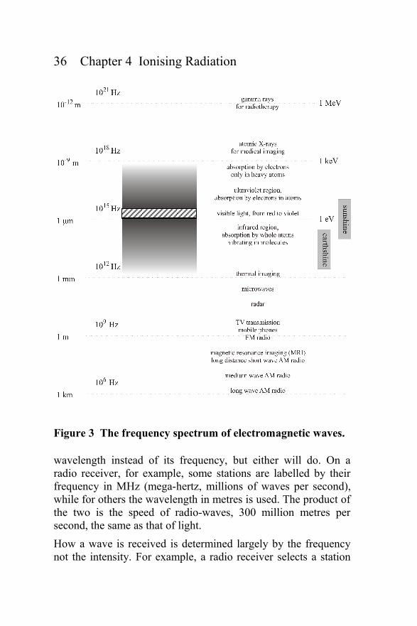
Figure 3 The frequency spectrum of electromagnetic waves.
wavelength instead of its frequency, but either will do. On a
radio receiver, for example, some stations are labelled by their
frequency in MHz (mega-hertz, millions of waves per second),
while for others the wavelength in metres is used. The product of
the two is the speed of radio-waves, 300 million metres per
second, the same as that of light.
How a wave is received is determined largely by the frequency
not the intensity. For example, a radio receiver selects a station
Radiation and Reason 37
by choosing its frequency rather than its loudness. In the same
way that for sound there are frequencies that cannot be heard by
the ear, so for light there are frequencies that are invisible to the
eye. In fact only a tiny range of frequencies of electromagnetic
waves is visible. The whole spectrum is represented in Figure 3
with a logarithmic frequency scale running up the page and
covering more than 15 powers of 10, as shown in the second
column in oscillations per second (Hz). The first column gives
the corresponding wavelength. Visible light with its
characteristic spectrum of rainbow colours is the narrow cross-
hatched band half way up the diagram. The point is that there
really is no fundamental difference between these waves, from
radio through light to X-rays, except the frequency. At the
highest frequencies (and shortest wavelengths) the powers of 10
become harder to cope with and a third scale based on the
electron volt (eV) is often used.11 This is shown on the right of
Figure 3 with the usual prefixes for powers of 10.12
Much benefit has been brought to everyday life through enabling
mankind effectively to see using these other frequencies [4].
Lower in the diagram are radio-waves up to 109 Hz, used for
example in MRI to see inside the human body and in radar to see
ships and planes in fog and darkness. Slightly higher is thermal
imaging, used to see warm bodies accidentally buried or
concealed. Just below the visible frequencies is a region called
the infrared absorption band, shown as shaded in the diagram.
At these frequencies many materials are opaque because the
rotation and vibration of molecules are in tune and resonate with
electromagnetic waves. Above the visible there is another band,
the ultraviolet absorption band. Here it is the more nimble
atomic electrons that are in tune and the cause of the absorption.
So here too materials are opaque, as marked by the shading.
11 The electron volt is 1.610-19 joules. This is a useful scale in the atom. The
electron in the hydrogen atom has an energy of 13.6 eV while typical nuclear
energies are in MeV.
12 μ or micro, one millionth. m or milli, one thousandth.
k or kilo, one thousand. M or mega, one million. G or giga, one billion.
38 Chapter 4 Ionising Radiation
Heavier elements with their more tightly bound electrons have an
ultraviolet absorption band that extends to much higher
frequencies than light elements. This is the frequency range of
the X-rays. Here, metals like copper and calcium absorb
radiation whereas carbon, hydrogen and oxygen are transparent.
Medical images of a patient's teeth or bones (calcium)
illuminated with such radiation show clearly any fracture or
disease because the enveloping tissue (carbon, hydrogen and
oxygen) is transparent.
Above about 100 keV atomic electrons, even those that are most
tightly bound in the heavier elements, cannot move fast enough
to follow the oscillating wave.13 Consequently there is no
resonance and all materials are largely transparent. This region is
called the gamma ray region. Historically the distinction between
X-rays and gamma rays depended on the source – electrons and
nuclei, respectively. This distinction is deceptive because their
effect does not depend on the source, only on their energy (or
frequency). Today this switch of name is usually made at about
100 keV, but the distinction is really only a convention. Gamma
rays are very penetrating, being only weakly absorbed, which is
why they are used in radiotherapy to target energy into a cancer
tumour, deep within a patient's body. This energy may then be
absorbed in the tumour with sufficient intensity that its cells are
killed and it ceases to function. There are practical difficulties in
doing this, as discussed later in Chapter 7.
Damage from radiation
So understanding light, and then learning to see with radiation in
other parts of the spectrum, is really useful. But what of the
risks? The spectrum can be divided roughly into two halves
separated at about 10 eV. Radiation of greater frequency or
13 At such high frequencies the radiation appears less like waves and more
like rays, or particles. In quantum mechanics this distinction has no real
substance, and electromagnetic waves of any frequency f come in bundles of
energy called photons, E = hf, where h is Planck's Constant. Each atom or
nucleus emits one such bundle or particle when it decays.
Radiation and Reason 39
energy is called ionising radiation, that below, non-ionising
radiation. The distinction is that ionising radiation can ionise and
break molecules apart – this is the radiation with which this book
is primarily concerned.
Public concern about weak levels of non-ionising radiation, for
instance from overhead power lines or mobile phones, is
misplaced. The only known way in which such radiation can
cause damage is by heating.14 Put briefly, these radiation sources
are safe if heat is not sensed – even then, benefits may dominate
over any reasonable risk. Warmth from sunshine or a domestic
fire is brought by the same kind of radiation as that in a
microwave oven. While the radiation levels in such an oven can
certainly be dangerous, the heat radiated by a glowing fire on a
cold winter's day is a quite acceptable source of radiation hazard
for most people – in spite of the fact that its heat level can be
sensed, indeed because of it.
But non-ionising radiation still has a crucial environmental
impact. On the right hand side of Figure 3 are two boxes labelled
sunshine and earthshine. Very hot materials like the Sun emit
light in the visible region, but cooler materials also emit, though
predominantly in the infrared frequency range. The sunshine box
indicates the range of frequencies that the Sun emits. Because
this is centred on the visible region for which the atmosphere is
largely transparent, much of this radiation reaches the surface of
the Earth for the benefit of all, including plant life. (Actually the
spectrum of the Sun extends a bit into the infrared and
ultraviolet, too – the infrared part provides warmth, the
ultraviolet causes sunburn, if not filtered by barrier cream and
the small concentration of ozone present in the upper
atmosphere.) The earthshine box indicates the frequency band of
radiation that the surface of the Earth emits with its lower
temperature – but not all of this radiation succeeds in getting out
14 This important statement can be scrutinised but the effect of radio-waves
and microwaves on living tissue is well understood and they are widely used.
For instance, they are used in MRI, safely below the level at which any
significant heating occurs.
40 Chapter 4 Ionising Radiation
of the atmosphere because of infrared absorption by polyatomic
gases,15 in particular carbon dioxide, water vapour and methane.
With an atmosphere containing more of these the Earth is not
able to cool itself nearly as effectively as it is able to absorb the
sunshine. So energy is trapped in the atmosphere and the
temperature increases. Crudely, this is how the Greenhouse
Effect works. If the concentration of these gases rises, the Earth
gets hotter and the climate changes. An extraordinary example is
close at hand – Venus has a surface temperature of 460ºC, thanks
in part to an atmosphere with 97% carbon dioxide.
Like electromagnetic waves, beams of charged particles such as
alpha and beta radiation can also damage molecules, so that they
are classified as ionising radiation – and beams of neutrons and
other ions too, although these are less common in the natural
environment.
Nuclear stability
But what makes a nucleus decay? Or rather, what holds it
together in the first place? The mutual electrical repulsion of the
protons makes large nuclei more unstable than small ones.
Stability only comes from the nuclear force that attracts
neighbouring protons and neutrons together. This nuclear force
overwhelms the electrical repulsion, but only at short distances
within about 10-15 metres. As a result it favours small nuclei for
which the protons and neutrons can huddle close together. The
result is a balance between the preferences for nuclei to be not
too large and not too small, which gives rise to the nuclear
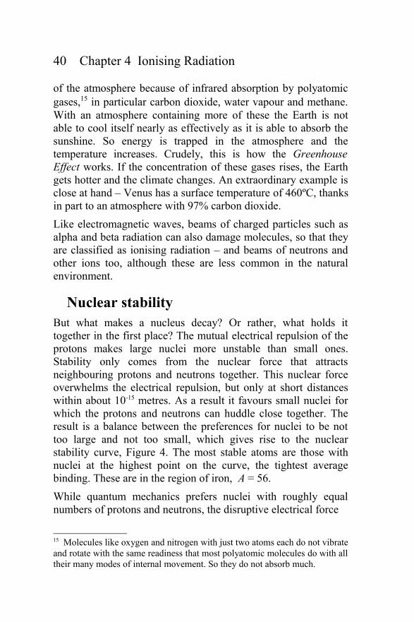
stability curve, Figure 4. The most stable atoms are those with
nuclei at the highest point on the curve, the tightest average
binding. These are in the region of iron, A = 56.
While quantum mechanics prefers nuclei with roughly equal
numbers of protons and neutrons, the disruptive electrical force
15 Molecules like oxygen and nitrogen with just two atoms each do not vibrate
and rotate with the same readiness that most polyatomic molecules do with all
their many modes of internal movement. So they do not absorb much.
Radiation and Reason 41
Figure 4 The average binding energy per proton or neutron
as it depends on the atomic mass number, A.
makes nuclei with too many protons unstable. The result is that
all stable nuclei, except the largest, have roughly equal numbers
of protons and neutrons, so that iron (Z = 26) has 30 neutrons. As
shown in Figure 4, for smaller values of A the binding effect of
the nuclear force is reduced; at larger values of A the disruptive
influence of the electrical effect is increased – either way the
binding is less. Above iron the compromise favours nuclei with
more neutrons than protons because the disruption only acts on
the protons. So for example, the most abundant isotope of lead,
lead-208, has 82 protons but 126 neutrons. There are no naturally
occurring elements above uranium (Z = 92) – those above
actinium (Z = 89) are collectively referred to as the actinides.
The curve shows that in principle nuclei with small A could fuse
together to release energy due to the nuclear force, as shown by
the arrow on the left. This is nuclear fusion and the source of
stellar energy, including that of the Sun. In addition, nuclei with
large A can in principle release energy by splitting apart and
moving towards greater stability as shown by the arrow on the
42 Chapter 4 Ionising Radiation
right. This is nuclear fission.16 Because, like lead, the parent
nucleus has more extra neutrons than its stable fission products,
there are excess free neutrons emitted in the fission process. The
liberation of these extra neutrons is crucial to the nuclear chain
reaction mechanism.
In practice fission is very rare. Alpha decay in which a heavy
nucleus splits into helium and a smaller nucleus is more
common. This is the source of much of the natural radioactive
energy in the Earth's crust – the energy source of natural
geothermal power, in fact. In alpha decay nuclear energy is
released by moving to the left along the curve in steps of four
units in A. As A reduces, the excess proportion of neutrons has
also to be reduced, and this occurs by beta decay in which a
neutron in the nucleus decays emitting an electron and leaving
behind an extra proton within the nucleus.
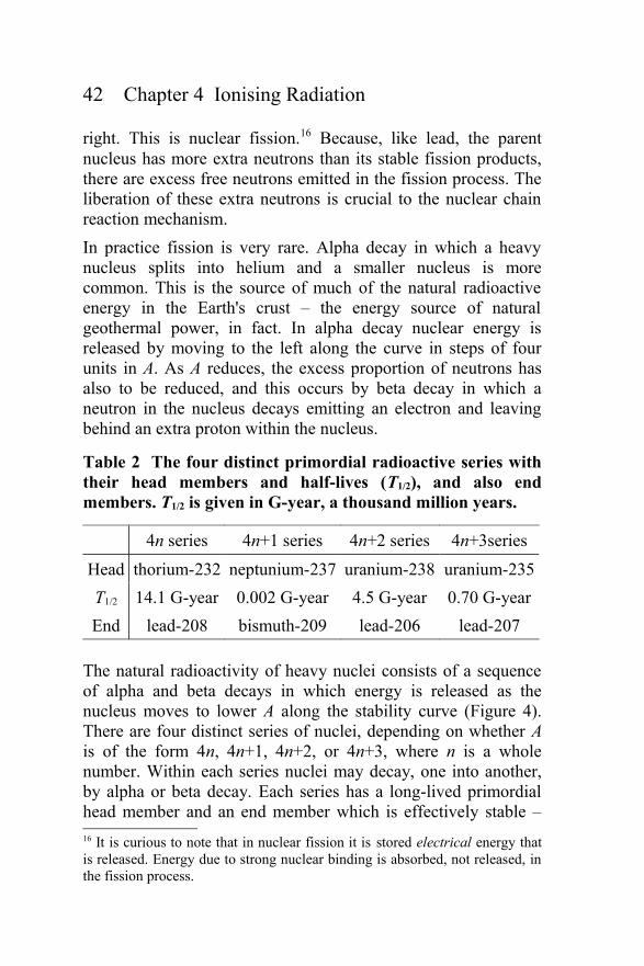
Table 2 The four distinct primordial radioactive series with
their head members and half-lives (T1/2), and also end
members. T1/2 is given in G-year, a thousand million years.
4n series 4n+1 series 4n+2 series 4n+3series
Head thorium-232 neptunium-237 uranium-238 uranium-235
T1/2 14.1 G-year 0.002 G-year 4.5 G-year 0.70 G-year
End lead-208 bismuth-209 lead-206 lead-207
The natural radioactivity of heavy nuclei consists of a sequence
of alpha and beta decays in which energy is released as the
nucleus moves to lower A along the stability curve (Figure 4).
There are four distinct series of nuclei, depending on whether A
is of the form 4n, 4n+1, 4n+2, or 4n+3, where n is a whole
number. Within each series nuclei may decay, one into another,
by alpha or beta decay. Each series has a long-lived primordial
head member and an end member which is effectively stable –
16 It is curious to note that in nuclear fission it is stored electrical energy that
is released. Energy due to strong nuclear binding is absorbed, not released, in
the fission process.
Radiation and Reason 43
these are given in Table 2. The 4n+1 neptunium series has
already died out, but the other three are still active in the natural
environment. The successive members of the 4n+2 series, with
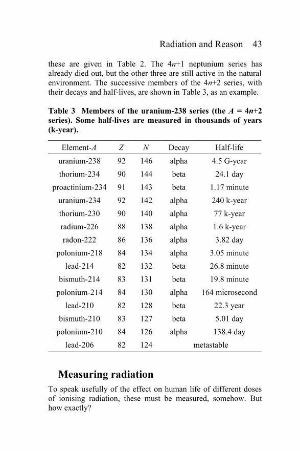
their decays and half-lives, are shown in Table 3, as an example.
Table 3 Members of the uranium-238 series (the A = 4n+2
series). Some half-lives are measured in thousands of years
(k-year).
Element-A Z N Decay Half-life
uranium-238 92 146 alpha 4.5 G-year
thorium-234 90 144 beta 24.1 day
proactinium-234 91 143 beta 1.17 minute
uranium-234 92 142 alpha 240 k-year
thorium-230 90 140 alpha 77 k-year
radium-226 88 138 alpha 1.6 k-year
radon-222 86 136 alpha 3.82 day
polonium-218 84 134 alpha 3.05 minute
lead-214 82 132 beta 26.8 minute
bismuth-214 83 131 beta 19.8 minute
polonium-214 84 130 alpha 164 microsecond
lead-210 82 128 beta 22.3 year
bismuth-210 83 127 beta 5.01 day
polonium-210 84 126 alpha 138.4 day
lead-206 82 124 metastable
Measuring radiation
To speak usefully of the effect on human life of different doses
of ionising radiation, these must be measured, somehow. But
how exactly?
44 Chapter 4 Ionising Radiation
The first step in quantifying a radiation exposure is to measure
how much energy is absorbed per kilogram of living tissue
during the exposure. This energy may cause chemical damage by
breaking molecules apart that leads to biological (cellular)
damage and finally to clinical damage, such as cancer or other
disease. Such clinical damage turns out to be more difficult to
relate to the exposure, especially as it may manifest itself in
different ways, and on long or short timescales, from days to
years.
In earlier decades knowledge of cell biology was too primitive to
provide confident understanding, and adequate evidence of the
effect of radiation on humans was not available to corroborate
any particular view. In their absence, and for lack of anything
better, the knowledge gap was bridged by a rule of thumb – a
model in science-speak. This is the Linear No-Threshold model,
abbreviated LNT. This assumes that clinical damage is in simple
proportion to the initial radiation energy dose. No justification
was given for it, but it was a reasonable working hypothesis at
the time. Despite the poor state of knowledge, a start had to be
made somewhere.
However, given modern biological knowledge and extensive
records of human data, this model is now redundant and many of
its more cautious implications can be ignored. The details are for
discussion in later chapters. First, we return to the questions of
the quantification of radioactivity and absorption of radiation
energy in materials.
The rate at which energy is emitted by a radioactive source
depends on the number of radioactive nuclei N, the energy of the
decay, and the half-life T of the nucleus. The value of N is
reduced by half with every successive time interval T and the
average activity is proportional to N/T. Activity is measured in
decays per second, called becquerel and abbreviated Bq.
Sometimes the activity may be measured in a cubic metre of
material, thus Bq m-3.
So what does this mean in practice? Contamination by
radioactive nuclei with a short half-life results in high activity for

Radiation and Reason 45
a short time; the same contamination with a longer half-life
results in a lower activity, but it continues for longer. Half-life
values vary between a small fraction of a second and many times
the age of the Earth. So sources of radioactive contamination
with short half-lives fade away while others with longer half-
lives continue on. This is in contrast to most chemical pollutants,
such as heavy metals like mercury or arsenic, that remain
hazardous indefinitely. A slightly different situation arises when
a dose of ionising radiation energy comes from an external beam
produced by an accelerator (such as an X-ray machine) or from
an external radioactive source.
Either way the important question is, how far does the radiation
travel in material before being absorbed? Some radiation is so
strongly absorbed in air, or any thin material, that it never
reaches human tissue unless the source is on the skin or inside
the body. Other radiation is weakly absorbed and can pass
through the body. So what is important is not the intensity of the
radiation, but the amount that is absorbed, for instance, per
kilogram of tissue.17 The extent to which it is absorbed depends
on the kind of radiation and its energy (or frequency).
Alpha radiation is stopped even by air, and so the decay energy
is deposited very close to the site of the radioactive
contamination itself, with no dose at all only a little further away.
An example is the energetic, but short range, alpha radiation
emitted by the decay of the radioactive isotope polonium-210. A
large internal dose of this was used allegedly by Russian agents
to kill Alexander Litvinenko in London in 2006. No energy
escaped the immediate location of the poison but there the tissue
received the full radiation energy dose.
Beta decay produces electrons that travel further in material and,
therefore, the deposited energy dose is more diffusely distributed
around the radioactive source. Gamma rays go further still. So
for a radioactive source in rock, for example, any alpha and most
17 Radiation that just passes through and does not deposit any energy is
necessarily harmless – like the neutrino radiation mentioned on page 31.

46 Chapter 4 Ionising Radiation
beta radiation is absorbed internally within the rock, and only the
gamma radiation escapes to give an external energy deposition.
In general a deposited energy dose is quantified as the number of
joules of energy absorbed per kilogram of material, such as
patient tissue. One joule per kilogram is called a gray (Gy).
Typically doses are measured in milligray, with a milligray
(mGy) being one thousandth part of a gray.
The clinical damage caused to living tissue by this deposited
radiation develops as a result of a number of steps.
1. The immediate molecular mayhem left by the radiation.
2. Biological damage in which living cells are put out of
action – this changes with time as the tissue responds to
the radiation dose.
3. The incidence of cancer (and other possible delayed or
heritable effects) related to the exposure, perhaps decades
later.
4. The reduction in life expectancy as a result of such
cancers (this effect on life expectancy is called the
radiation detriment of the exposure).
5. The chance of death shortly after exposure due to acute
radiation sickness brought on by cell death and the
shutdown of the normal biological cycle in one or more
vital organs.
The two lasting consequences for life are described by the
sequences 1-2-3-4 and 1-2-5, and later we will discuss how each
of these outcomes relates to the initial radiation energy dose.
There are other causes of cancer, unrelated to radiation. Some
causes – we shall refer to them generally as stresses – are natural,
others are imposed by choice of lifestyle. Following decades of
study much is known about how these stresses are related to the
occurrence of cancer – to the detriment in fact. An important
question is how the outcome is influenced when there is more
than one stress. These stresses may be quite independent, as in
smoking and radiation, but the result may not be. There remain
some unanswered questions. But the point is that the range of
residual uncertainty is too small to prevent mankind from taking
Radiation and Reason 47
decisions now about how radiation can be used with effective
safety.
For a single acute dose the damage is related to the size of the
dose and the type of radiation. The effects of X-rays, γ-rays and
electrons are found to be roughly the same for the same energy
dose in milligray. However, for other types of ionising radiation
the biological damage is different. Quantitatively, the measured
ratio of damage relative to X-rays is called the relative biological
effectiveness (RBE). So the RBE of a radiation dose indicates
how much more clinical damage it causes than is caused by the
same number of milligray of energetic gamma rays. Essentially
these RBE factors are measured quantities.
RBE factors vary with the clinical end point – that is with the
cancer or disease concerned. Timing effects are important and
we look at these later. The variation with radiation type is
particularly interesting although not too large. For most practical
applications of radiation safety, which we are thinking about in
this discussion, we need to watch the factors of ten, a hundred
and a thousand. RBE factors close to one are less important.
Only in radiotherapy are the effects of radiation very finely
balanced – but in that case gamma rays are usually used and so
RBE is 1.0 anyway. So for this simplified discussion it is
sensible to ignore the RBE factor in the first instance.
Nevertheless the International Commission for Radiological
Protection (ICRP) has felt it necessary to include RBE in some
way. In their radiation safety standards they multiply each energy
dose in gray by a weighting factor, wR, which plays the role of a
broad-brush averaged RBE. [They define wR for protons to be
two; for alpha, fission fragments and other heavy ions to be 20;
for neutrons it depends on the energy; for electrons and photons
it is just one, by definition.] The result they define to be the
equivalent dose, measured in units of sievert (Sv) – or
millisievert (mSv). In ignoring RBE initially we treat doses
measured in milligray and millisievert as equivalent, and come
back later to the distinction when a variation in the type of
48 Chapter 4 Ionising Radiation
radiation has something special to say about how radiation
damage occurs.
These measures of energy deposited (and equivalent dose) may
be for a single acute exposure. It is observed that cell damage is
different if the dose is spread over a period of time, either as a
series of repeated exposures, or as a continuous chronic rate of
exposure. The question is why? What is the radiation detriment
resulting from a chronic rather than an acute radiation exposure?
How does the effect of a single dose of so-and-so many milligray
compare with the effect of a continuous dose rate of a number of
milligray per day – or per year? The matter is not simple,
because dose and dose rate are quite different measures. This is
the subject of Chapter 7.
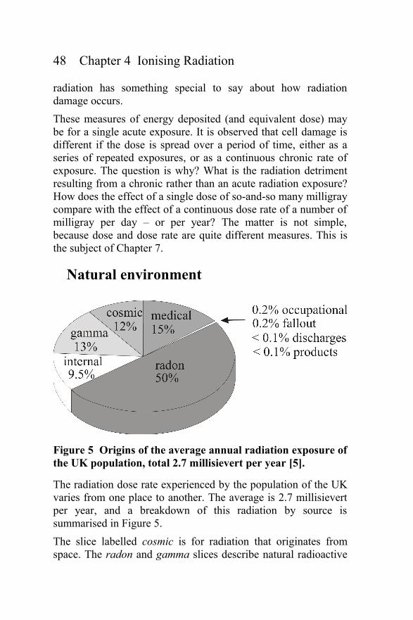
Natural environment
Figure 5 Origins of the average annual radiation exposure of
the UK population, total 2.7 millisievert per year [5].
The radiation dose rate experienced by the population of the UK
varies from one place to another. The average is 2.7 millisievert
per year, and a breakdown of this radiation by source is
summarised in Figure 5.
The slice labelled cosmic is for radiation that originates from
space. The radon and gamma slices describe natural radioactive

Radiation and Reason 49
sources in nearby materials such as water, soil and rock. The
internal radiation slice relates to the decay of radioactive atoms
that occur naturally within the human body. The artificial part of
the exposure is predominantly medical – the average due to all
other man-made sources amounts to less than 0.5%.
The ionising radiation incident on the Earth from space is made
up of electromagnetic radiation, protons and electrons. Some of
the charged particle radiation comes from the Sun where the
erupting magnetic fields of sunspots act as accelerators. At the
top of the atmosphere this radiation causes ionisation, and the
resulting discharges may be seen as the Northern Lights or
aurora. Charged particles with low energy are deflected by the
Earth's magnetic field, except in the magnetic polar regions,
which is why the aurora are seen there. The resulting increased
ionisation of the upper atmosphere affects satellite and radio
communications, and when there is a magnetic storm this
ionisation is high. None of these phenomena has any effect on
health and the ionisation radiation does not reach the ground.
Cosmic radiation also includes protons that are more energetic
and come from outside the solar system, and even outside the
galaxy. These suffer nuclear collisions in the upper atmosphere.
Some collisions create neutrons that then hit nitrogen nuclei high
in the atmosphere to form the famous isotope, carbon-14.
Although only 7.5 kg is created in total in the atmosphere each
year, this is sufficient to maintain the proportion of carbon-14 in
the natural biosphere (1 part in 1012), which provides the basis of
radiocarbon dating. This isotope decays with a half-life of 5,700
years, and its concentration starts to fall as soon as material,
animal or vegetable, dies – that is, stops refreshing its carbon
from the air or digesting other living tissue. By measuring its
concentration, materials can be dated. Famous examples are the
Turin Shroud, the Ice Man from 3,300 BC found in the Otztal
Alps in 1991, and bottles of fake 'old' whisky.
The most energetic protons from space create showers of sub-
atomic particles, most of which decay or are absorbed by the
atmosphere. The only radiation that reaches the ground is a flux
50 Chapter 4 Ionising Radiation
of muons18 and this is responsible for the cosmic slice in Figure
5. At sea level this delivers about 0.6 millisievert per year in
polar latitudes. In equatorial regions the flux is three times
smaller because of the shielding effect of the Earth's magnetic
field, which sweeps incoming protons away into space. The
radiation rises rapidly with height above sea level because of the
reduced absorption by the atmosphere.
In the very distant past the flux of radiation was much greater.
The Universe itself started from a simultaneous explosion,
known as the Big Bang, 13.8 billion years ago. The early stages
were dominated by particle physics of the kind studied on a
small scale at modern research accelerators. After a few minutes
the explosion had cooled sufficiently for the distinct nuclei of
hydrogen and helium to emerge. But, until 300,000 years later it
remained so hot that electrons and nuclei were not bound
together. As it cooled further, the heat radiation became non-
ionising and atoms of hydrogen and helium appeared for the first
time.
Over the next few billion years galaxies and stars formed. These
evolved through nuclear fusion in massive stars, creating the
heavier atoms that we see around us today, a process called
nucleosynthesis. Slowly, as the Universe began to settle down,
systems of planets formed in the neighbourhood of rotating stars,
often composed of lumps of nuclear ash made spherical by
gravity. Interplanetary fragments collided with the larger planets
and their moons, leaving craters on the surfaces of those without
an atmosphere.
This all happened before about 4.5 billion years ago when the
Earth was formed and activity became quieter. Ionising radiation
still reaches the Earth from hot stars in the form of heat radiation
and from exceptional acceleration processes elsewhere in the
Universe.
18 The muon is an unstable subatomic particle with the properties of a heavy
electron, which decays with a half-life of 1.4 microseconds.
Radiation and Reason 51
Meanwhile nuclei of the four radioactive series (described in
Table 2 on page 42) created during the period of nucleosynthesis
continued to decay, although the neptunium series died out long
ago. The other three are still going. The abundance of thorium in
the Earth's crust is 3 to 10 parts per million by weight. For
uranium-238 it is 1.8 to 2.7 parts per million. These values vary
depending on the rock formation. There are significant quantities
of uranium in sea water because its salts are soluble, unlike those
of thorium. Within all natural uranium ores the ratio of uranium-
235 to uranium-238 is currently 0.7%. This varies very little as
the physical and chemical properties of the two isotopes are
almost identical (see page 27) and their relative proportion does
not naturally become diluted or enriched except through decay.
Highly refined materials may be free of radioactivity but they are
exceptional. Wood, concrete, glass and metals are all radioactive
to some degree because they contain traces of natural
radioisotopes.
A few primordial radioactive nuclei are not members of the four
radioactive series. The most abundant is potassium-40 with a
half-life of 1.27 billion years. It decays by beta decay, either to
calcium-40 or to argon-40, both of which are stable. Potassium is
a common element in the Earth's crust (2.1% by weight) and in
sea water (0.044%). The regular stable isotope, potassium-39, is
the most common and the unstable potassium-40 is only a tiny
proportion (0.01117%). Potassium is essential to the electro-
chemistry of living cells and forms about 0.15 kg of human body
weight. Other radioactive isotopes, such as carbon-14, with
shorter lives are found in the environment too, being created
afresh by cosmic radiation. Thus carbon-14 and potassium-40
between them account for 7,500 radioactive decays per second in
an adult human body. The annual dose from such internal
radiation is 0.25 millisievert (see Figure 5).
Two billion years ago the radiation from these nuclei was much
as it is today, except that the proportion of uranium-235 in
natural uranium was higher. In fact, from the measured half-lives
(see Table 2) it is straightforward to calculate that at that time
52 Chapter 4 Ionising Radiation
natural uranium contained 3.5% of the faster decaying
uranium-235. Today, some nuclear fission reactors use uranium
fuel artificially enriched to this proportion, with ordinary water
acting as coolant and moderator, in order to maintain a steady
nuclear chain reaction. Two billion years ago such enriched fuel
and water were available naturally, so that a similar nuclear
reactor could occur by itself under the right circumstances. Clear
evidence that this actually happened has been found in Gabon,
West Africa. This natural nuclear fission reactor, known as the
Oklo Reactor [6, 7], ran on its own for up to a million years. In
our own time the extraordinary evidence came to light with the
discovery that the relative abundance of uranium-235 in this
particularly rich uranium deposit lay outside the narrow range
found elsewhere in the world. It has been shown that the missing
uranium-235 was consumed in the natural reactor cores and that
the remains of the resulting fission products are still to be found
there. This is significant because this reactor was not
decommissioned and buried in a specially selected underground
site at great cost. The residue of the uranium fuel and its fission
products were left where they lay and have not moved in two
billion years. This is an important demonstration of the stability
that nuclear waste deposits can have over extremely long
periods.
Radiation from radioactive sources in materials such as water,
soil or rock reaches the external environment mainly in the form
of gamma radiation and radon – alpha and beta radiation are
mostly absorbed. Radon is a noble gas with little chemical
activity, like helium, neon, argon, krypton and xenon. The
isotope radon-222 has a half-life 3.82 days and is formed in the
uranium-238 series (Table 3). This radioactive gas, once it has
been released into the air, can be inhaled into the lungs where it
may be absorbed and decay by alpha emission leaving polonium-
218, a non-volatile isotope which decays with a sequence of
further alpha emissions. Such alpha radiation has a short range
and deposits all its energy in the lungs. On this basis radon
would be expected to be a significant source of risk to the health
of those who ingest it every day where they live and work.

Radiation and Reason 53
Figure 5 shows radon as a large component of the average
human radiation exposure. In fact, it is larger by factors of five or
more in certain places, depending on the local geology. A
significant question, discussed in Chapter 7, is whether this large
variation is reflected in the local incidence of lung cancer.