Embed Size (px)
Citation preview

CHAPTER 4
Camptothecin from callus cultures of Nofhapodyfes foefida
4.1 Introduction
N. foetida wight) Sleumer (syn: Mapiafoetida Miers) is a small tree
found in the Western Ghats of South India. Roja and Heble 1994 reported
the tissue cultures of N. foeti& and subsequent detection of CPT and 9-
methoxy CPT from these cultures initiated from seed embryos (179). The
present chapter describes the growth and CPT production from callus
cultures of N.foetida as well as them anticancer activity in vitro.
4.2 Materials and methods
4.2.1 Explants:
3-4 weeks old young shoots collected from our green house and
mature berries were collected from Nil@ hius during the month of
February 1997.
4.2.2 Surface sterilization:
Internode and tender leaf were separated from young shoots, soaked
in Tween 20 for Sminutes and washed in running tap water. These explants
were surface sterilized with 0.1% HgC1, as described in the materials and
methods. The surface sterilized (internode 1.5 -2cm, leaf 0.5-lcm and
embryos 0.2-0.4cm) was inoculated into different media with different
concentrations and combinatior~ of hormones (section 2.2.1).
4.2.3 Callus subculture:
Calli were subcultured 4'" week in the same medium, which initiated
on other hormone concentratic~ns of other media
4.2.4 Callus growth measurement:
The gowth of the callus was determined by increase in fresh weight
and dry weight in modified MS niedium supplemented with NAA 2/KN
0.5ml/ lit at loday intervals (section 2.2.5)
4.2.5 Extraction of CPT and HPLC analysis:
1.5 g freeze-dried calli initiated from different combination medium
were ground separately in a mortar and pestle. The powder of each calli
were sonicated in 70% methanol for two minutes, stirred overnight and
filtered. The supernatant of the extract dried and suspended in distilled
water. The resultant suspensiorl was extracted with chloroform. The
chloroform extract dried and was used for the quantification of CPT by
HPLC (Fig 4.1) Section (2.2.9,2.'2.16)
4.2.6 Purification and characterization of CPT:
CPT was purified from the callus developed in mMS QUA2
mg/l+KN 0.5 mg/l) freeze dried callus 9.5g extracted with 70% MeOH
(2.5g), suspended in DD H,O extracted with CHC13 (1.53g). The CHC1,
extract was evaporated to dryness, and resuspended in MeOH and crude
CPT precipitated as pale yellour crystals at 4OC. It was further purified by
TLC (silica gel G) using the solvent system (CHCl,: MeOH 24:l V/V).
The purified CPT with Rf 0.6 was analysed by uv, HPLC, IR, NMR and
ESMS as described in Material:; and methods section (2.2.15, 2.2.16, 2.2.16,
2.2.17, 2.2.18).
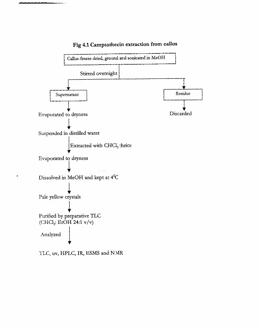
Fig 4.1 Camptothecin extraction from callus
Callus-freeze dlied, ground and sonicated in MeOH I===' Stirred overnight I
Supernatant +--I +
Evaporated to dryness
Suspended in distilled water I
Extracted with CHCl, ]&rice 1 Evaporated to dryness
I Dissolved in MeOH and kept at 4'C
Pale yellow crystals
1 Purified by preparative TLC (CHCl,: EtOH 241 v/v)
Analyzed 1
Residue 0 + Discarded
TLC, uv, HPLC, IR, ESMS and NIMR
4.2.7 Long term Cytotoxicity of the isolated CPT:
50,000 L929 cells were seeded in 96 well plate. Cells were exposed
with different concentrations of CPT dissolved in DMSO, and solvent
reached a concentration not higher than 2% in all experiments. After 72hr
incubation, the viability of the cells was measured by MTT assay as
described in the materials and methods (section 2.2.3).
4.3 Results
4.3.1 Callus induction and callus growth:
MS medium (1962) and modified MS medium (mMS), supplemented
with B5 vitamins gave good response. Of the different explants tried, only
immature embryo responded positively. Immature embryo cultured in 2,4-
D 2mg/l + KN 0.5mg/l induce semi friable callus whereas the internode,
leaf disc explants did not produce callus tissue even after 4-6 weeks of
incubation. Callus induction .was also noted in embryo cultured on
modified MS medium supp1emc:nted with NAA 2mg /I f KN 0.5mg/l
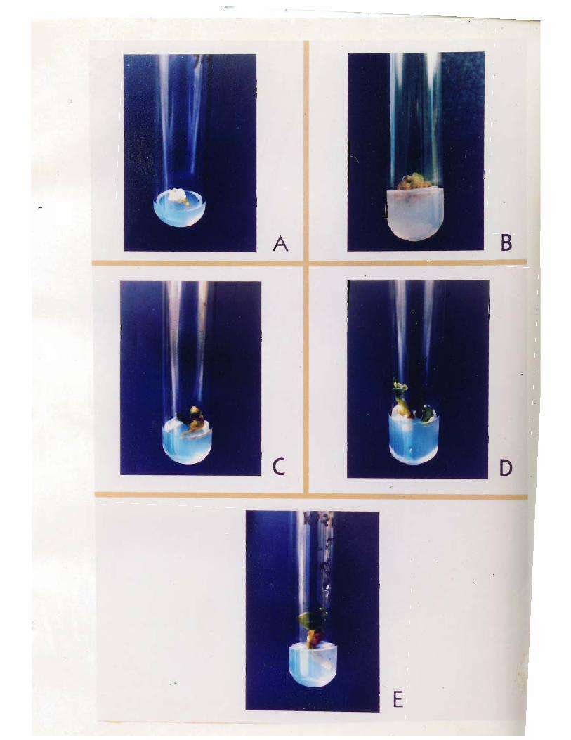
(Table4.4). Plantlets were observed in MS medium containing NAA2mg/l
+ BA 0.5 pig 4.2 C, Dl.
Growth pattern of the callus was studied by subculture of 200mg fw
callus to media (mMS) with different hormonal combinations and results
recorded at 10 days. There was no proliferation of callus recorded in media
supplemented with other than NAA and KN as observed in callus
initiation. Callus proliferation was started only after 10days of culture, and
growth continued to 90 days. .4fter 90 days the growth of callus declined,
as evident by the change in colour to brown and nodular protuberance and
leafy outgrowth were observed (Fig 4.2 B).
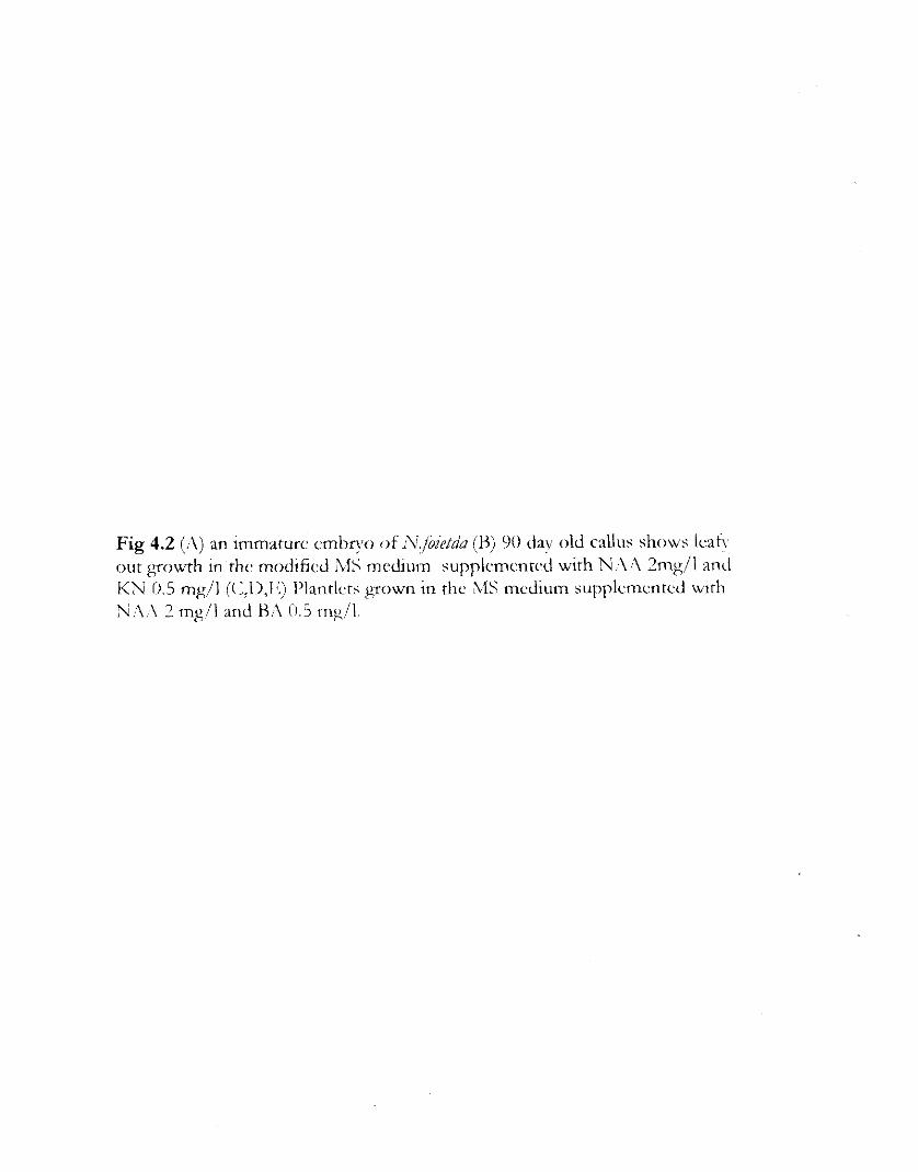
Fig 4.2 (;\) an immature cmbrvo of n'.jbie/d& (H) 90 dav old callus shows icatv out growth in the modifiecl A1S mcdiulm supplcmcntcc! with %I:\ 2mg/l and I<N 0.5 mg/l ((:,ll,l<) I'lantlcts g o w n in the L1S medium supplcmentcd with N:\;\ 2 mg/l and H:\ i1.i mg/l.
l;ig 4.3 uv visihlc al~soq~tion spectra of c~llus dcri\rcd (:isl'.
4.3.2 Thin layer chromatography
The callus extract and standard CPT chromatographed on Silica gel
G showed blue fluorescent spots under uv lamp (254nm) with an Rf 0.60
using the solvent system (CHC1,: MeOH 241 v/v).
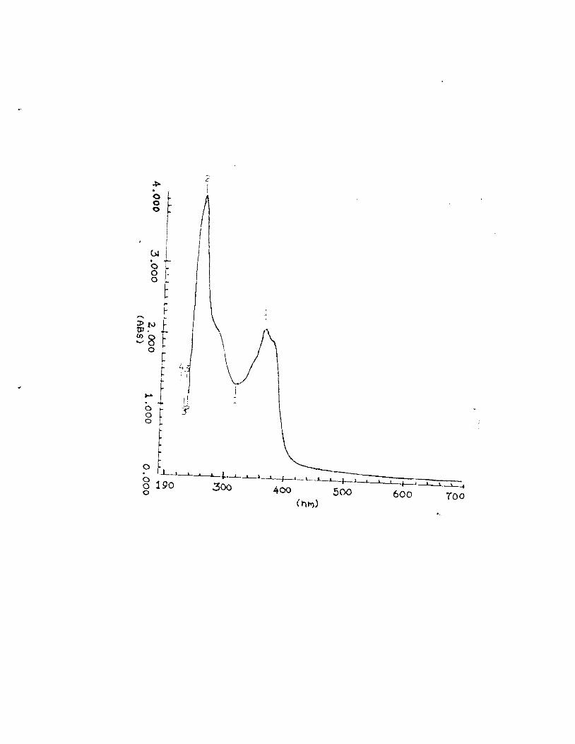
4.3.3 uv absorption
uv absorption spectra of the isolated and standard CPT showed the
Xmax 256,290 and 363nm pig 4.3 A].
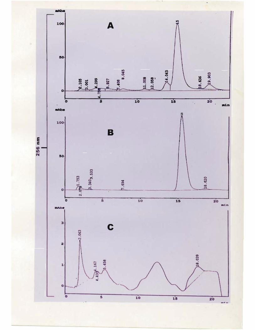
4.3.4 HPLC analysis
The HPLC analysis of callus CPT and standard CPT showed
chromatographically homogenous peak with base h e separation and same
retention time 15 minutespig. 4.4 A, B, C].
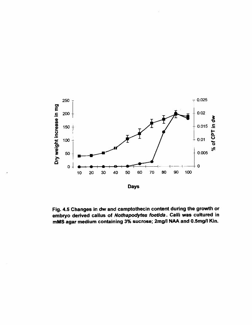
The amount of CPT present in calli is shown in Table 4.2. Of the
different calli analysed, the % (:PT was high in callus grown in the
modified media supplemented with NAA 2/ KN O.Smg/l. The amount of
CPT was 0.02% on 90 day old culture pig 4.51.
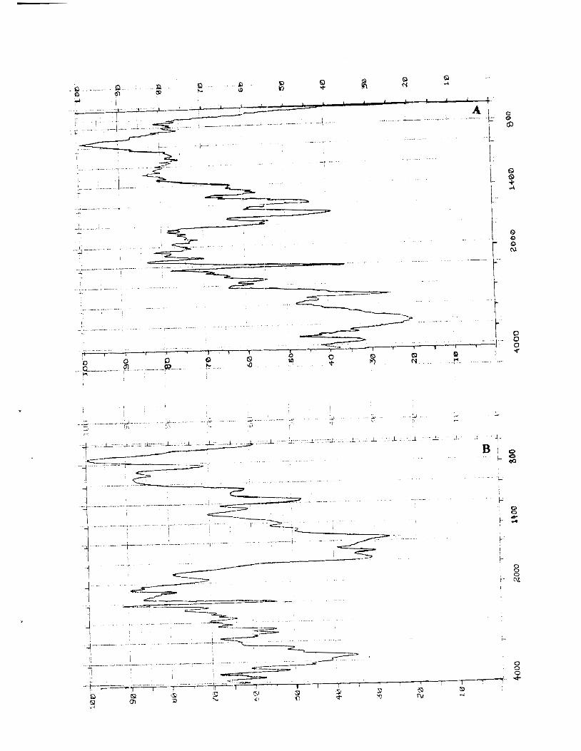
4.3.5 IR spectra of purified CPT
Both standard CPT and callus CPT showed Vmax at 3430(hydroxy),
1938(lactone), 1674(lactam) [Fig 4.6 A, B]
Fig.4.4 HPLC chromatogram of standard CPT, Notha~odvtes foetida seed embryo callus extract with spike (flow rate 1 mumin; details of the mobile phase are found in materials and methods)
a, (I)
g 150 U c .- ,c loo m .- 5 50
Days
Fig. 4.5 Changes in dw and camptothecin content during the growth or embryo derived callus of Nothapodyfes foetida. Calli was cultured in mMS agar medium containing 3% sucrose; 2mgll NAA and 0.5mgll Kin.
I;ig 4.6 IK spectrum ot standard (:IyI' (.\) mid callus dcrivc~l ( : l s l ' (H:I
4.3.6 Mass spectra
Mass spectra of the isolzlted CPT showed m/z 349(MvI+H'),
calculated for molecular formula C:&,,N,O, pig 4.7 A, B] characteristic
of camptothecin.
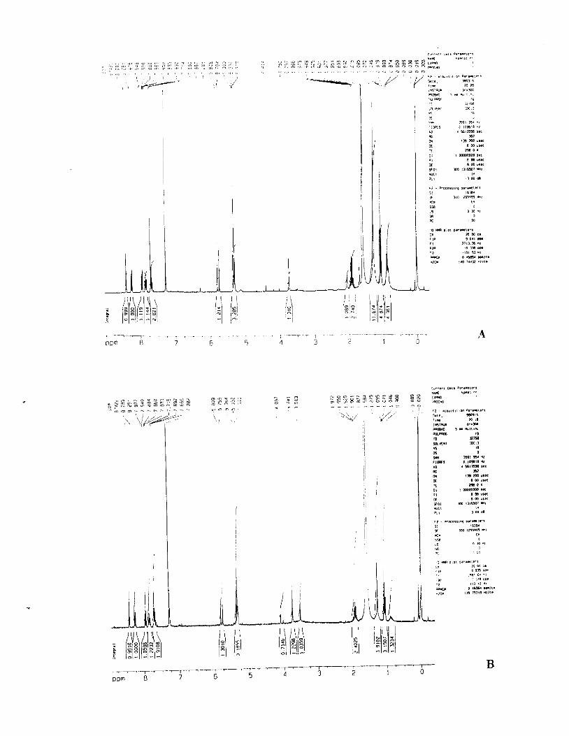
4.3.7 'H-NMR NMR spectra
Showed the number of protons in the isolated CPT. 7.67-8.42(6H,m,
Ar.H), 5.33(3H,m, H-5, H17), 5.80(14,d, J=16.2H-17), 1.07(3H,t, 7.4 H7),
1.93(2H,m). Fig4.8 A,B]
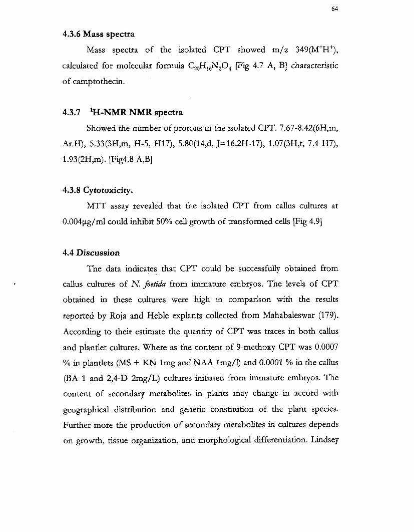
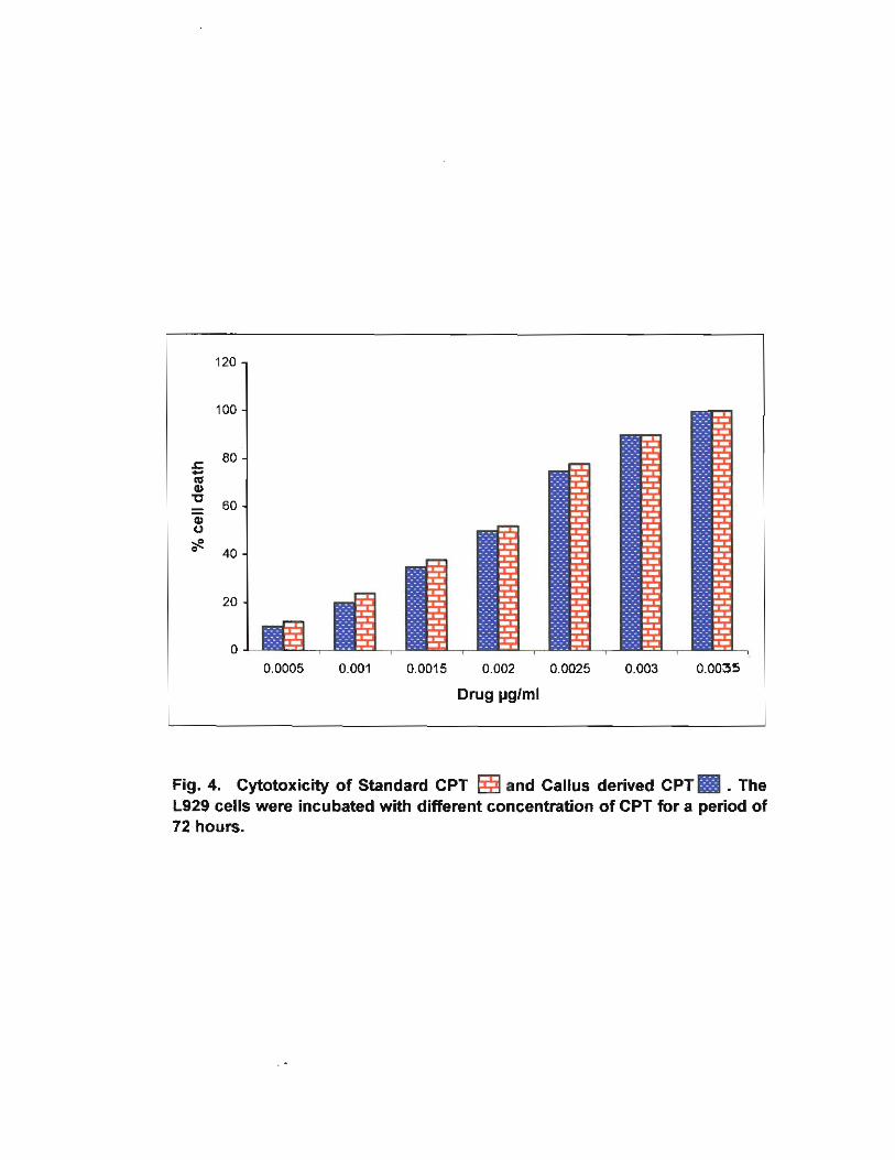
4.3.8 Cytotoxicity.
MTT assay revealed that the isolated CPT from callus cultures at
0.004pg/rnl could inhibit 50% cell gowth of transformed cells [Fig 4.91
4.4 Discussion
The data indicates that CPT could be successfully obtained from
callus cultures of N. foetida from immature embryos. The levels of CPT
obtained in these cultures were hgh in comparison with the results
reported by Roja and Heble explants collected from Mahabaleswar (179).
According to theit estimate the quantity of CPT was traces in both callus
and plantlet cultures. Where as tht: content of 9-methoxy CPT was 0.0007
% in plantlets (MS + KN lmg and NAA Img/l) and 0.0001 % in the callus
(BA 1 and 2,4-D 2mg/L) culture:; initiated from immature embryos. The
content of secondary metabolites in plants may change in accord with
geographical distribution and ge~letic constitution of the plant species.
Further more the production of secondary metabolites in cultures depends
on growth, tissue organization, and morphological differentiation. Lindsey

I;ig 4.7 Llast: spectrum o f srantlartl (:I''I i!\) and callus ctcrrvcci (:IsI' ( H I
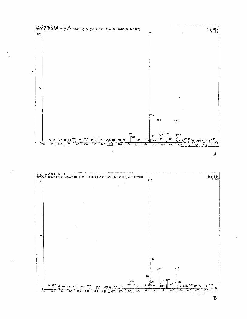
I C H l C N : H 2 0 1:2 ( , \ %.\, , : 'TEST13 1 I6 (7 952)Cn (Cen 8000 HI) Srn (SG. 2x0 751.Cm (107117.175 92,140 162))
; 100 34s Scan ES*
I
~ ~ ~~~ ~~~ ~~~ . .
~ s ~ . ~ , c H ~ ? - N H ~ O l i 2 1 TEST44 115 (7885).Cn (Cen.2. 80 00, Hl). Sm (SG.2x07:i): Cm (1 13:121-(77:102*13816lI! S- ES+ 1
I I
i
I X
I I
i
Y 9
! ! !
0
!
I! !
i' 37, 412
I i I I
"' ":133 136 187 171. 18(1 2M 226 2 1 1 5 2 ~ 1 e 4 279 4 ~ 4 r l e 1 5 5 ~ ~ 6 411: " ,z 1-0 ' \ d o : ,do ' 180 2 d o ' 220 ' zda l-261 ' 260 '~340 320 0 0 360 380 ' ~ 0 0 420 142 :.160 4b0:--
. ~
I
! I I I !
I 1
! !
!350 I

Fig 4.8 i\l hlR spectrum of sranciard (:IY1' (ill and callus Jcrivcd (:1"1' (H)
<",,.", n.,, a,..",...
- - - - - . . . a : -..o g p :z .-., ., ,,, Y ," ~, - - " - m m - - m . ,,, ,, .. ,. ,,, " " ,., ,,, t* - ,\ ,,","* -
3 ..-..,.... 3 2 ; ,,m,ao.uo^RzSi o u - ,, ".. . v - - " "
a",m,., ....*...... -. ,> .II1,,,l,m .11.-111. , ' j" , , ::':. W<t $ ,
'I
; I 1 ;
1 1 I
, 1
' 8
! , ; : I , 1 i
I ;
a 88 1SYU w . m - ,..1111,*
a- Yn.
*%.! s a m m, w "? r,m, OIO.1 .U . .r,m 3.c rn m c" ,m m ",x m . w "'*
a*, , -m ,*c I l ".*
m 8 rn ,,.c YO, m 1111WI -2
-5 %, , m -8
,> ~ e -,,,, ", *"-L... ,b,W
F m ,- -l .,. 5B
a m ., m - > j,*, ,.,.- I...
w <. 1 , .,s 3 3>, >a. ..*, ,. .:
,,, '> *, 0 .- w / c .
- . m m - I;\ W 14 1.1 1: -l-\-i ,,: 5 : I B X 6 N Z - -,,- II I n m _ - - - 1 - : i'i-11-11-1
120
1 00
80 5 1 P) u - 60 - a U
40
20
0 0.0005 0.001 0.0015 0.002 0.0025 0.003 0.0035
Drug pglml
Fig. 4. Cytotoxicity of Standard CPT and Callus derived C P T ~ . The L929 cells were incubated with different concentration of CPT for a period of 72 hours.
e t a/ 1993 reported an increase in alkaloid production correlated with slow
growth rate (197) Tabata et a1 1972 reported that the increased production
secondary metabolites were noticed in the stationary phase of the growth,
which related to tissue organization (198). In our study, it is found that the
growth of callus in mMS niedium declined after 90 days, and
morphological differentiation such as nodular and lea4 out growths
appeared. The % CPT in these cultures were high which may be due to
the above mentioned factors or effect of different environment such as
effect of hormones. In addition it is reported that medium containing NAA
can enhance the production of indole alkaloids (199). According to Van
Hngel 1992 and Weden field 1997 the percentage of CPT was increased in
callus cultures of accumnatu in different concentration of NAA and other
cytokinins (27,28). Our results also show the influence of NAA in the
production of CPT in culture.
In conclusion the present result further open up avenues for the
mass scale production of CPT by cell suspension cultures of N. foetida.
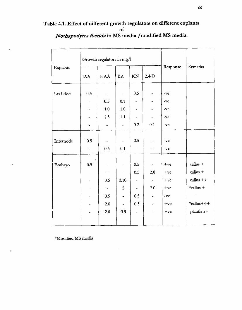
Table 4.1. Effect of different growth regulators on different explants of
Nothapodytes foetida in MS media /modified MS media.
*Modified MS media
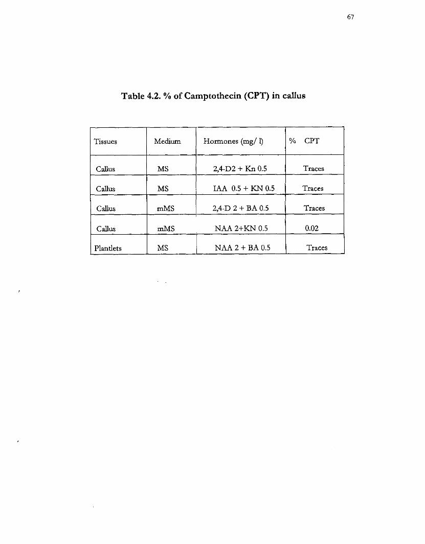
Table 4.2. % of Camptothecin (CPT) in callus
Tissues I Medium I Hormones (mg/ 1) 1 % CPT 1 2,4-D2+ Kn0.5 1 1 If IAA 0.5 + KN 0.5
Callus I mMS 1 2,4-D 2 + BA 0.5 Traces
Callus m M S ' NAA 2+KN 0.5 0.02
Plantlets
I
MS Traces - NAA 2 + BA 0.5