Embed Size (px)
Citation preview
Chapter-3
Studies on certain seaweed - bacterial interaction from Saurashtra coast 39
Chapter-3
Chapter-3
Studies on certain seaweed - bacterial interaction from Saurashtra coast 40
3.1. Introduction
In the marine environment, bacteria are the most common colonizers on the surface of
macroalgae (Armstrong et al., 2001). Seaweed-associated bacteria secrete biologically active,
beneficial compounds that regulate the morphogenesis of marine organisms and help them to
survive under varied environmental conditions. The nature of the bacterial–seaweed interaction
plays an important role and can influence the growth as well as various developmental stages
(e.g. reproduction) of algae. Bacteria are generally assumed to benefit from soluble organic
matter (as a source of energy) produced by host algae (Chandini et al., 2008). Biofilm of certain
bacteria influence the settlement of zoospores of green alga of genus Ulva (Patel et al., 2003).
Spore release in the red algal genus Acrochaetium was enhanced by marine bacteria through N-
acyl homoserine lactone secretion (Weinberger et al., 2007). The plantlet growth-promoting
nature of bacterial isolates associated with Laminaria japonica was reported by Dimitrieva et al.
(2006).
The members of Ulvaceae loose their typical foliose thallus morphology when cultured
axenically in defined synthetic media (Provasoli, 1958; Provasoli & Pintner, 1980). Ulva attain
their natural habit when co-cultured with bacterial isolates capable of inducing differentiation.
This aberrant morphology could successfully been restored to the foliose thallus following the
inoculation of appropriate morphogenesis-inducing bacterial isolates to the culture medium.
Similar findings have also been reported during the subsequent studies in U. pertusa (Nakanishi
et al., 1996), Enteromorpha linza (Fries, 1975), E. compressa (Fries, 1978), U. linza (Marshall
et al., 2006), Monostroma oxyspermum (Tatewaki et al., 1983; Matsuo et al., 2003) and U.
mutabilis (Wichard & Oertel, 2010). However, the necessity of direct physical contact between
the algal and bacterial cells was shown to be prerequisite to complete morphogenesis in U.
pertusa (Nakanishi et al., 1999). Further, Tatewaki et al. (1983) reported that the addition of
culture filtrate of morphogenesis-inducing bacterial isolates was capable of giving the same
effect as that of the addition of bacterial culture itself. Similarly, Marshall et al. (2006) reported
that culture filtrate restored the morphology of the U. linza. It is now well established that
thallusin produced by marine bacteria is effective in regulating the morphogenesis pattern in
seaweeds (Matsou et al., 2005).
Chapter-3
Studies on certain seaweed - bacterial interaction from Saurashtra coast 41
Bacterial genera such as Cytophaga, Flavobacterium, Vibrio, Pseudomonas, Halomonas,
Escherichia and Bacillus have been implicated in promoting the morphogenesis in U. pertusa
(Nakanishi et al., 1999). Further, it has also been shown that morphogenesis in green
macroalgae (members of Ulvaceae and Monostromaceae) is induced by species of Cytophaga,
Flavobacterium, Bacillus and others belonging to the phylum Firmicutes and Bacteroidetes
(Matsuo et al., 2005; Marshall et al., 2006). Along the Indian coast, occurrence of Bacillus,
Vibrio, Micrococcus, Flavobacterium and Cytophaga has been reported from the intertidal
region where seaweeds are one of the dominant benthic communities (Lakshmanaperumelsumy
& Purushothaman, 1982). However, the role of these marine bacterial isolates on seaweed
morphogenesis has not yet been investigated yet.
The green alga Ulva fasciata is one of the most prevalent common green algae and
occurs almost every season on Indian coast (Jha et al., 2009). The high protein content ca. 26 %
on dry weight basis and nutritionally rich omega-3-fatty acids make this seaweed a potential
source of food and feed (Kumari et al., 2010). The previous work on this alga has dealt with
several fundamental studies including phenology, ontogeny (Mantri et al., 2010). Ulva are more
popular among Japanese cuisine and are being cultivated commercially in Japan and Korea
(Hiraoka & Oka, 2008) but the role of associated bacteria on growth, morphology and
reproduction has not been examined in this species.
The present chapter describes the isolation and screening of 53 bacterial isolates
associated with Ulva and Gracilaria species and their morphogenesis-inducing ability in U.
fasciata grown in axenic culture. Five potential bacterial isolates having morphogenesis-
inducing activity were further tested for their ability in induction of growth and reproduction in
the vegetative tissue under axenic culture. Partial 16S rRNA gene sequences of all the 5 strains
with morphogenesis-inducing ability were used to determine their correct taxonomic identity
and phylogenetic relations. Further, the culture filtrates from these isolates were also tested for
their ability to induce U. fasciata morphogenesis and subsequent growth.
3.2. Materials and Methods
Chapter-3
Studies on certain seaweed - bacterial interaction from Saurashtra coast 42
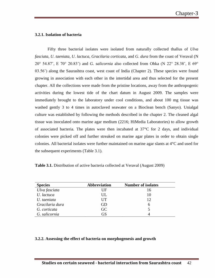
3.2.1. Isolation of bacteria
Fifty three bacterial isolates were isolated from naturally collected thallus of Ulva
fasciata, U. taeniata, U. lactuca, Gracilaria corticata, and G. dura from the coast of Veraval (N
20° 54.87’, E 70° 20.83’) and G. salicornia also collected from Ohka (N 22° 28.38’, E 69°
03.56’) along the Saurashtra coast, west coast of India (Chapter 2). These species were found
growing in association with each other in the intertidal area and thus selected for the present
chapter. All the collections were made from the pristine locations, away from the anthropogenic
activities during the lowest tide of the chart datum in August 2009. The samples were
immediately brought to the laboratory under cool conditions, and about 100 mg tissue was
washed gently 3 to 4 times in autoclaved seawater on a Bioclean bench (Sanyo). Unialgal
culture was established by following the methods described in the chapter 2. The cleaned algal
tissue was inoculated onto marine agar medium (2216; HiMedia Laboratories) to allow growth
of associated bacteria. The plates were then incubated at 37°C for 2 days, and individual
colonies were picked off and further streaked on marine agar plates in order to obtain single
colonies. All bacterial isolates were further maintained on marine agar slants at 4°C and used for
the subsequent experiments (Table 3.1).
Table 3.1. Distribution of active bacteria collected at Veraval (August 2009)
Species Abbreviation Number of isolates
Ulva fasciata UF 16
U. lactuca UL 10
U. taeniata UT 12
Gracilaria dura GD 6
G. corticata GC 5
G. salicornia GS 4
3.2.2. Assessing the effect of bacteria on morphogenesis and growth
Chapter-3
Studies on certain seaweed - bacterial interaction from Saurashtra coast 43
Small aliquots of zoospore suspension with densities of 150 x 103
to 200 x 103 cells cm
-2 were
added to each well containing 5 ml of MP 1 medium in 12 well plates and incubated according
to method described in chapter 2. After 24 h culture, an aliquot of 50 μl bacterial suspension
from each of the 53 bacterial isolates was inoculated into the U. fasciata zoospore culture, and
this was monitored for developmental morphology of spores. The control culture was
maintained without addition of bacterial isolates. Cultures were grown for 15 days and the
medium was replenished at 3 days of intervals. The experiment was repeated three times. Visual
interpretation of images taken at weekly intervals under an Olympus stereo zoom microscope
(model SZ X 16) was adopted as a non-destructive method to study the developmental
morphogenesis of spores and their subsequent growth. Five putative morphogenesis-inducing
bacteria (UF, GC, GS, UL24 and UL, named according to the algal species from which they
were isolated, see Table 3.1) were identified for further study. A consortium of all five isolates
was also subsequently assessed in triplicate for its effect on morphogenesis induction and
growth. Further, culture filtrates (0.22 μm, Millipore) obtained from the five individual isolates
and from the consortium were separately incubated to study their effect on morphogenesis
induction and growth. Growth of fronds was expressed as the relative increase in area.
Microscopic features such as the appearance of cells on the surface, cell area and presence or
absence of marginal spines were also recorded under an Olympus microscope (model BX 60).
ANOVA (1 and 2 way) was used to analyze the morphogenesis-inducing bacteria’s effects on
algal growth, cell size and zoospore induction and significant differences were determined at p ×
0.05. Dunnett’s post hoc analysis was used to analyze bacteria-induced growth in U. fasciata.
To quantify the abundance of associated bacteria during algal growth, about 100 μl of
algal culture medium (10–6
dilution) was spread daily on marine agar medium (standard plate of
90 mm diameter) separately for all the isolates. The colony forming units (CFUs) were counted
manually after 24 h incubation at 37°C.
3.2.3. Induction of spore with bacterial isolates
Chapter-3
Studies on certain seaweed - bacterial interaction from Saurashtra coast 44
Disks of U. fasciata approximately 5 mm2 were excised under axenic conditions from the algal
stock culture. 10 disks with approximate weight of 20 mg were incubated in the multiwell plates
containing 3 ml of sterilized MP1 medium under standard condition described in the chapter 2.
The bacterial cultures (UF, GC, GS, UL24 and UL) and their culture filtrates (0.22 μm,
Millipore) were inoculated in the plates separately while the control was maintained with the
sterile medium at 25°C. The small sized sterile cover slip was put at the bottom of each well and
the zoospores those were released and settled after 24 h were counted under inverted microscope
manually. The data was expressed in number of zoospores per gram of tissue. The experiment
was repeated three times.
3.2.4. Scanning electron and epifluorescence microscopy
The algal fronds grown with the bacterial inoculation were gently washed 3 to 4 times with
autoclaved seawater to remove loosely associated bacteria. Samples were fixed with 2.5%
glutaraldehyde in sterile seawater overnight at 4°C. Then, they were gently washed in sterile
seawater, post fixed in 2% OsO4 in 1:1 ratio (seawater: milli Q) for 2 h. Thereafter, they were
washed in sterile seawater and dehydrated in a graded ethanol series from 20% to 100%. For
scanning electron microscopy (LEO 1430 VP), these specimens were coated with a gold alloy in
a Sputor coater SC 7620. For epi-fluorescent microscopic observation of bacterial cells on the
algal surface, bacterial DNA was stained with 5 μg ml-1
DAPI (4, 6 Diamidino-2-Phenylindole)
and observed using FS 10 (fluorescein isothiocyanate) filter under 40 X objective (Zeiss Imager
M1 Microscope).
3.2.5. Genomic DNA isolation and amplification of 16S rDNA gene sequence
Bacterial colonies were first incubated with lysozyme (10 mg ml-1
) for 2 h at 37°C. Afterwards
genomic DNA was extracted using the cethyl trimethyl ammonium bromide buffer [CTAB 2%,
NaCl 1.4 mM, EDTA 50 mM, Tris 100 mM and PVP 20%] method (Chen & Kuo, 1993).
Purification of genomic DNA was confirmed with 0.8% agarose gel electrophoresis. The
universal forward bacterial primer rf (5’-AGAGTTT GATCCTGGCTCAG-3’; E. coli positions
8–27) and the reverse primer rr (5’-AAGGAGGTGATCCAGCCGCA-3’; E. coli positions
Chapter-3
Studies on certain seaweed - bacterial interaction from Saurashtra coast 45
1541–1522) [Lapara et al., 2000] were used for the PCR amplification of the partial 16S rRNA
gene sequence. The reaction mixture contained 2.5 μl 10X PCR buffer containing MgCl2, 100
ng of each forward and reverse primer, 25 mM of each deoxynucleotide triphosphate (dATP,
dCTP, dGTP and dTTP), 1 unit of Taq DNA polymerase and 10 ng of DNA template. The PCR
protocol included a 5 min initial denaturation at 95°C, followed by 30 cycles at 94°C for 40 s,
55°C for 40 s, 72°C for 2 min, with a final cycle of 10 min at 72°C and incubation at 4°C.
Partial 16S rDNA gene amplification products were loaded on 2% agarose gels and run at 50 V
for 1 h at 25°C and visualized with a UV transilluminator. Bands were excised and purified
using QIAquick PCR purification kit (Qiagen, no. 28104). Forward and reverse DNA
sequencing reactions of PCR amplification were carried out with rf and rr primers using BDT
v3.1 Cycle sequencing kit on ABI 3730 x l Genetic Analyzer. Sequencing of isolates was done
by the Xcleris Laboratories, Ahmedabad (Gujarat, India). A search of the NCBI database
(http://blast.ncbi.nlm.nih.gov/Blast.cgi) for similarity of 16S rDNA gene sequences to isolated
strains was made using software (MEGA-4) (Tamura et al., 2007). Partial sequences of 16S
rDNA gene were generated from forward and reverse sequence data using Aligner software
(DNA Codon code, Florida). Partial 16S rRNA gene sequences were used to carry out BLAST
with NCBI genbank database.
3.3. Results
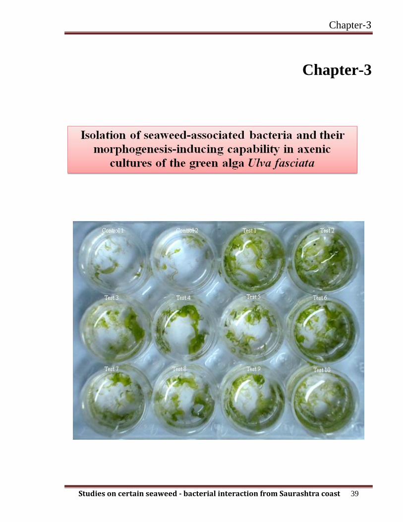
3.3.1. Assessing the effect of bacteria on morphogenesis and growth
A total of 53 bacterial isolates (35 from Ulva spp. and 18 from Gracilaria spp.) were isolated in
this study (Table 3.1). With no bacteria added, the axenic culture derived from U. fasciata
zoospore germination was very aberrant contrasted to that of the natural thallus (Fig. 3.1).
Chapter-3
Studies on certain seaweed - bacterial interaction from Saurashtra coast 46
Fig. 3.1. Morphogenesis of U. fasciata as axenic culture after (A) 4 weeks and (C) 6 weeks in
the absence of morphogenesis inducing bacteria, and after (B) 4 weeks and (D) 6 weeks in the
presence of morphogenesis-inducing bacteria. Scale bars = 2 mm
The compact callus like growth was observed in culture after 2 weeks with no evidence
of branching. The subsequent growth raised the tubular creeping fronds after 4 weeks of culture
(Fig. 3.1A). The highly branched distinct tubular fronds were formed after subsequent culture of
6 weeks (Fig. 3.1C). In all these cases the morphology of germlings was completely dissimilar
to the natural thallus. Conversely, five putative morphogenesis-inducing bacteria (UF, GC, GS,
UL24 and UL) were able to induce considerable morphogenesis in axenic culture of U. fasciata
with similarity to the natural pattern of growth (Fig. 3.1B, D). The cell area of the mature thallus
(after four weeks of culture) was 146 µm2 in the control while variable dimensions of cells in
surface view were recorded in all the five isolates as well as their culture filtrate and inducing
effect of the UF isolate was effective at p ≤ 0.05% (Fig. 3.2 and Table 3.2).
Chapter-3
Studies on certain seaweed - bacterial interaction from Saurashtra coast 47
Fig. 3.2. Effect of 5 different morphogenesis inducing bacterial isolates, their corresponding
culture filtrates, and the control treatment on axenic U. fasciata cells (n = 50 cells). Effect is
significant at p ≤ 0.05. The effects of culture filtrates were not significant (p > 0.05), in
comparison to bacterial isolates. Error bars represent ± SD.
Individual cell sizes of the U. fasciata were larger when morphogenesis took place in the
presence of morphogenesis-inducing bacteria than when axenic cultures were allowed to
develop alone (Fig. 3.3A and B). Out of all five isolates, UF isolate was capable of inducing
spine development on the margins while others failed, in comparison with the culture filtrate
and control (Fig. 3.3C and D). The arrangement of the cells showed a different pattern to the
control in cross section view with five putative morphogenesis-inducing bacteria (Fig. 3.3 E &
F).

Chapter-3
Studies on certain seaweed - bacterial interaction from Saurashtra coast 48
Fig. 3.3. Morphogenesis of U. fasciata as axenic culture in the presence / absence of
morphogenesis-inducing bacteria. (A) Cell size in the absence of bacteria, (B) cell size in the
presence of bacteria, (C) absence of spine in the control, (D) presence of spine in the presence of
bacteria, (E) atypical cell structure in the absence of bacteria, (F) wild-type cell structure in the
presence of bacteria.

Chapter-3
Studies on certain seaweed - bacterial interaction from Saurashtra coast 49
An increase in the thallus area after four weeks was 2.67 mm2 in control while maximum
increase of 20.33 mm2 was observed in consortium with minimum recorded both in GC and GS
(4 mm2). In the case of culture filtrates a similar mode of relative increase in thallus area was
recorded (Fig. 3.4). The induction of morphogenesis pattern was recorded in the following
order, UF=UL=UL24>GC=GS. The consortium of isolates, UF, UL24 and UL significantly
promoted the growth of the plantlets (p ≤ 0.01, Dunnett’s post-hoc analysis after one way
ANOVA), while there was no significant difference recorded for GS and GC morphogenesis-
inducing bacterial isolates at p ≤ 0.05 (Table 3.2, Fig. 3.4). Isolates (UF, GC, GS and UL)
showed growth in the range of 1750 to 2750 CFU ml-1
at the end of 5 days, whereas isolate
UL24 showed growth around 4500 CFU ml-1
.
Fig. 3.4. Effect of five different and consortium of morphogenesis inducing bacteria and their
corresponding culture filtrate, and a control treatment on U. fasciata thallus size. Dunnett’s post
hoc analysis: *represents the 99% confidence interval (CI), # not significant at 99% CI, +
represents the 95% CI. The effects of culture filtrates were not significant at p > 0.05, in
comparison to bacterial isolates. Control: axenic U. fasciata culture; error bars: ± SD.
Chapter-3
Studies on certain seaweed - bacterial interaction from Saurashtra coast 50
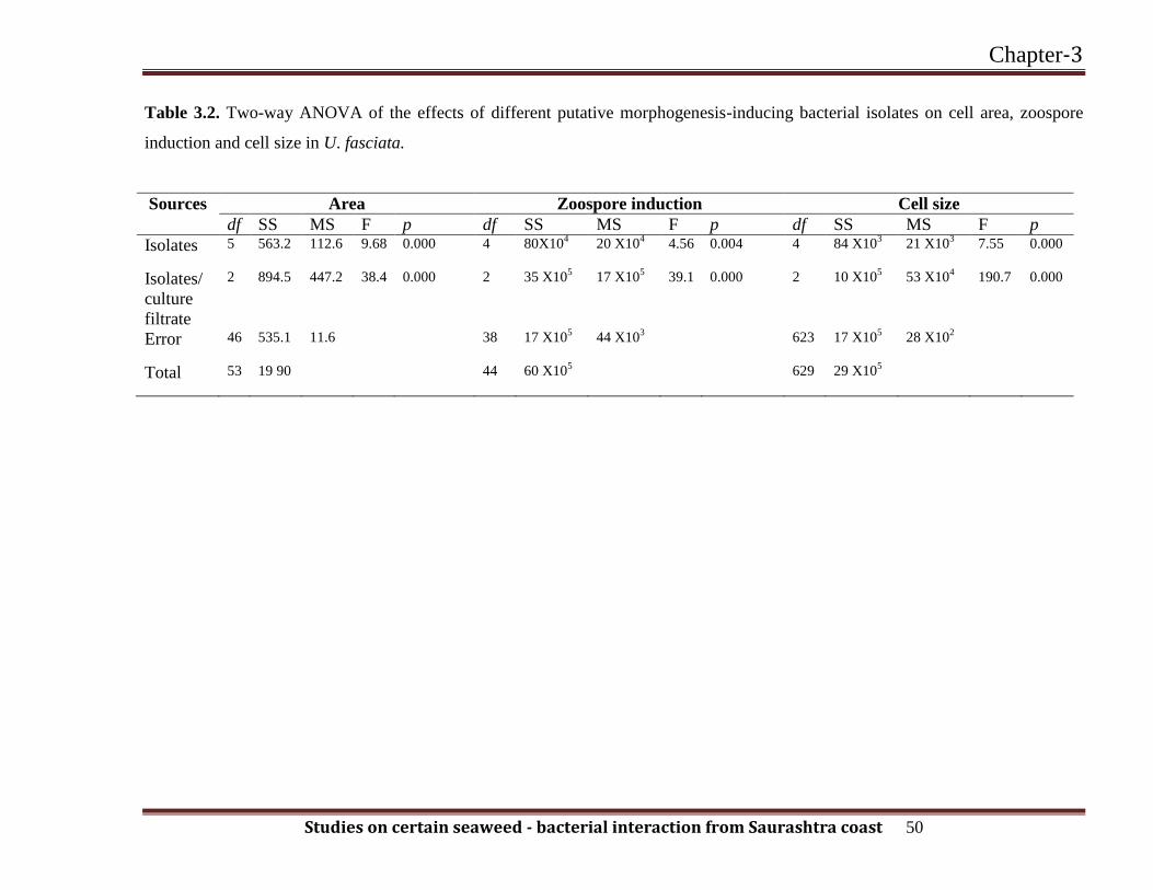
Table 3.2. Two-way ANOVA of the effects of different putative morphogenesis-inducing bacterial isolates on cell area, zoospore
induction and cell size in U. fasciata.
Sources Area Zoospore induction Cell size
df SS MS F p df SS MS F p df SS MS F p
Isolates 5
563.2 112.6 9.68 0.000 4 80X104 20 X10
4 4.56 0.004 4
84 X103 21 X10
3 7.55 0.000
Isolates/
culture
filtrate
2 894.5 447.2 38.4 0.000 2
35 X105 17 X10
5 39.1 0.000 2
10 X105 53 X10
4 190.7 0.000
Error 46
535.1 11.6 38 17 X105 44 X10
3 623 17 X10
5 28 X10
2
Total 53
19 90 44 60 X105 629
29 X105
Chapter-3
Studies on certain seaweed - bacterial interaction from Saurashtra coast 51
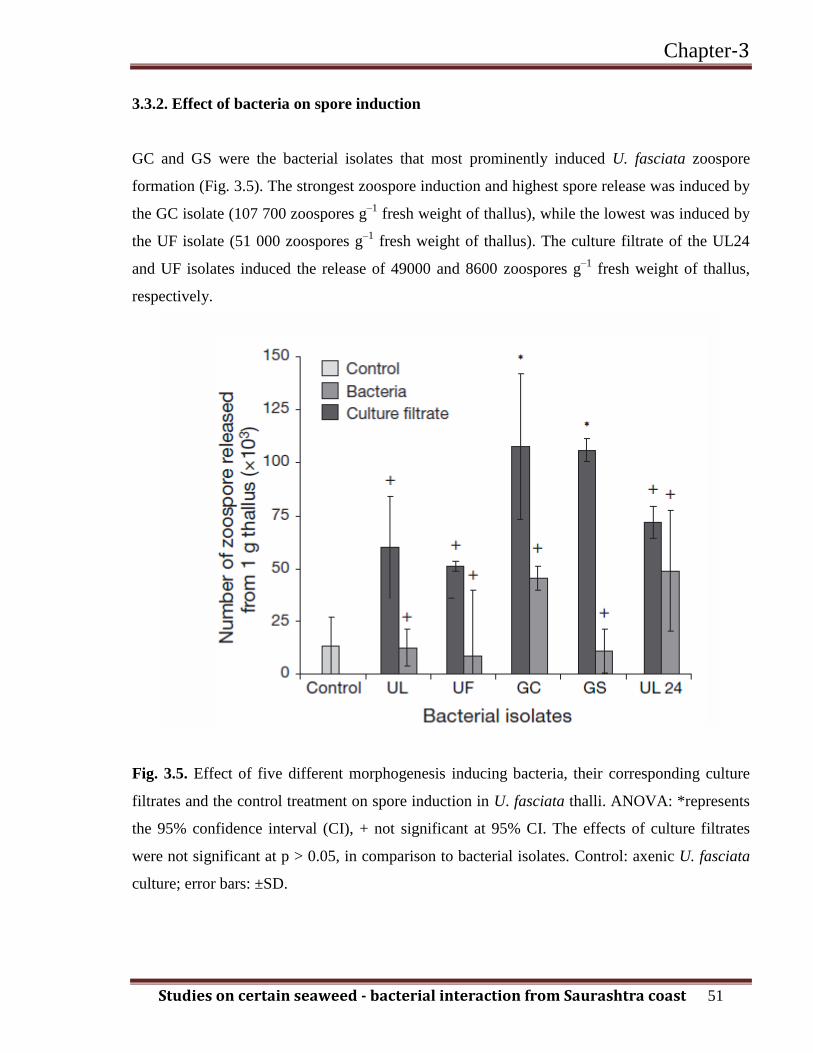
3.3.2. Effect of bacteria on spore induction
GC and GS were the bacterial isolates that most prominently induced U. fasciata zoospore
formation (Fig. 3.5). The strongest zoospore induction and highest spore release was induced by
the GC isolate (107 700 zoospores g–1
fresh weight of thallus), while the lowest was induced by
the UF isolate (51 000 zoospores g–1
fresh weight of thallus). The culture filtrate of the UL24
and UF isolates induced the release of 49000 and 8600 zoospores g–1
fresh weight of thallus,
respectively.
Fig. 3.5. Effect of five different morphogenesis inducing bacteria, their corresponding culture
filtrates and the control treatment on spore induction in U. fasciata thalli. ANOVA: *represents
the 95% confidence interval (CI), + not significant at 95% CI. The effects of culture filtrates
were not significant at p > 0.05, in comparison to bacterial isolates. Control: axenic U. fasciata
culture; error bars: ±SD.
Chapter-3
Studies on certain seaweed - bacterial interaction from Saurashtra coast 52
Therefore, direct addition of certain bacteria to axenic culture was more effective for the
induction of spores compared with the culture filtrate (p ≤ 0.05, 1- and 2 way ANOVA; Table
3.2). The induction and release of zoospores in the presence of the GC and GS isolates was
statistically more significant in comparison with the other isolates (Fig. 3.5).
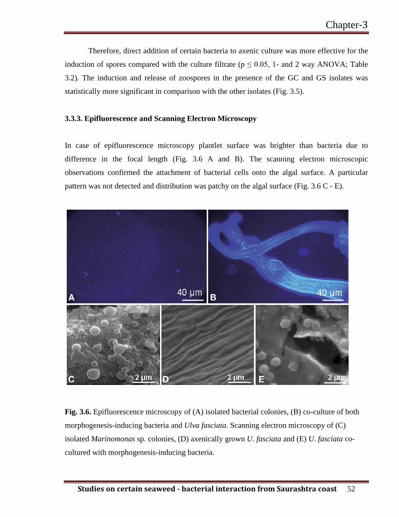
3.3.3. Epifluorescence and Scanning Electron Microscopy
In case of epifluorescence microscopy plantlet surface was brighter than bacteria due to
difference in the focal length (Fig. 3.6 A and B). The scanning electron microscopic
observations confirmed the attachment of bacterial cells onto the algal surface. A particular
pattern was not detected and distribution was patchy on the algal surface (Fig. 3.6 C - E).
Fig. 3.6. Epifluorescence microscopy of (A) isolated bacterial colonies, (B) co-culture of both
morphogenesis-inducing bacteria and Ulva fasciata. Scanning electron microscopy of (C)
isolated Marinomonas sp. colonies, (D) axenically grown U. fasciata and (E) U. fasciata co-
cultured with morphogenesis-inducing bacteria.
Chapter-3
Studies on certain seaweed - bacterial interaction from Saurashtra coast 53
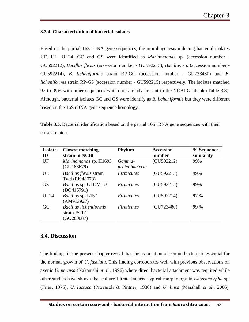
3.3.4. Characterization of bacterial isolates
Based on the partial 16S rDNA gene sequences, the morphogenesis-inducing bacterial isolates
UF, UL, UL24, GC and GS were identified as Marinomonas sp. (accession number -
GU592212), Bacillus flexus (accession number - GU592213), Bacillus sp. (accession number -
GU592214), B. licheniformis strain RP-GC (accession number - GU723480) and B.
licheniformis strain RP-GS (accession number - GU592215) respectively. The isolates matched
97 to 99% with other sequences which are already present in the NCBI Genbank (Table 3.3).
Although, bacterial isolates GC and GS were identify as B. licheniformis but they were different
based on the 16S rDNA gene sequence homology.
Table 3.3. Bacterial identification based on the partial 16S rRNA gene sequences with their
closest match.
Isolates
ID
Closest matching
strain in NCBI
Phylum Accession
number
% Sequence
similarity
UF Marinomonas sp. H1693
(GU183679)
Gamma-
proteobacteria
(GU592212) 99%
UL Bacillus flexus strain
Twd (FJ948078)
Firmicutes (GU592213) 99%
GS Bacillus sp. G1DM-53
(DQ416791)
Firmicutes (GU592215) 99%
UL24 Bacillus sp. L157
(AM913927)
Firmicutes (GU592214) 97 %
GC Bacillus licheniformis
strain JS-17
(GQ280087)
Firmicutes (GU723480) 99 %
3.4. Discussion
The findings in the present chapter reveal that the association of certain bacteria is essential for
the normal growth of U. fasciata. This finding corroborates well with previous observations on
axenic U. pertusa (Nakanishi et al., 1996) where direct bacterial attachment was required while
other studies have shown that culture filtrate induced typical morphology in Enteromorpha sp.
(Fries, 1975), U. lactuca (Provasoli & Pintner, 1980) and U. linza (Marshall et al., 2006).
Chapter-3
Studies on certain seaweed - bacterial interaction from Saurashtra coast 54
However, very few studies were performed with isolates of bacteria that were phylogenetically
well characterized. In the present chapter, isolates were identified by partial 16S rRNA gene
sequences and phylogenetic analysis showed that all five putative bacterial isolates belonged to
morphogenesis inducing bacterial isolates (Matsuo et al., 2003). There were only 5 out of 53
isolates showed statistically significant induction of growth, morphology and zoospore
induction. Here it is reported for the first time that morphogenesis-inducing bacteria are capable
of inducing the differentiation of whole plantlet with respect to induction of spines on the
surface, enlargement of cells and regaining wild type cell structure. On the contrary, culture
filtrate of five isolates and control failed to induce the typical morphogenesis of the U. fasciata.
This suggests that physical contact between bacterial and host cells are necessary for the
development of the particular cell shape, growth and differentiation of U. fasciata. In case of UL
isolate and consortium, culture filtrate slightly helped in the development of morphology of the
U. fasciata (Fig. 3.4) but it might be less significant due to instability of the secreted substances
or signal. In contrast to culture filtrate, tightly associated bacteria might be providing certain
substances which are continuously secreting and enhancing the growth and typical morphology
of U. fasciata.
It has also been reported that morphogenesis in green macroalgae from the families
Ulvaceae and Monostromaceae are controlled by group of bacteria specifically Cytophaga,
Pseudomonas, Staphylococcus, Vibrio and Bacillus, Flavobacterium sp. (Nakanishi et al., 1999;
Duan et al., 1995; Matsau et al., 2003; Marshall et al., 2006). This finding demonstrated that
Marinomonas sp. and Bacillus sp. induced wild type morphology and growth of the U. fasciata.
Thus, differentiation and growth in U. fasciata was found to be depending on Marinomonas sp.
and Bacillus sp which was not previously reported. Gram-positive Bacillus sp. was also found to
affect the morphology or growth of U. fasciata to a greater extent than that reported by
Nakanishi et al. (1996) for U. pertusa. The effect of bacteria on the growth rate of macroalgae
has not been well quantified previously. However subjective indications of inducing growth and
development have been reported for sporelings of U. pertusa, U. conglobata, and U. intestinalis
when incubated with bacterial isolates of the Bacteroidetes phylum (Matsou et al., 2005;
Marshall et al., 2006). The mechanism by which bacteria modulate the morphology of the
plantlet is not well understood so far, although a number of hypothesis have been suggested.
Chapter-3
Studies on certain seaweed - bacterial interaction from Saurashtra coast 55
However, Matsuo et al. (2005) identify and characterize an exogenous growth factor produced
by bacteria belonging to the Bacteriodetes group called thallusin and suggested that is an
essential factor for normal growth of M. oxyspermum. An endosymbiotic bacterium from the
Agrobacterium–Rhizobium group, containing the nifH gene encoding for nitrogenase involved
in nitrogen fixation, was isolated from rhizoids of the green alga, Caulerpa taxifolia (Chisholm
et al., 1996). It was suggested that this isolate might be important for nitrogen supply to the
seaweed. A bacterium of the Roseobacter group was responsible for the gall formation in the red
alga (Prionitis lanceolata) due to overproduction of indole-3-acetic acid (Ashen et al., 2000).
Phosphate solublizing activity of B. licheniformis was found for the growth promoting in
mangrove plant (Rojas, 2001). It has also been suggested that secondary metabolites released by
some epibiotic bacteria may prevent subsequent biofouling by other organisms (Callow et al.,
1998; Armstrong et al., 2001) thereby providing some protection to the host alga. Till date,
these are few data to support these speculations. No correlation was found between isolates that
altered the differentiation of U. fasciata and those that enhanced spore induction. An interesting
result is that those isolates which were inducing differentiation were not inducing spore
production significantly. The consortium of the all five putative morphogenesis-inducing
isolates significantly induced the differentiation (Table 3.2). The positive effect shown by the
consortium suggested that normal morphology is not dependent on the presence of a single
bacterium, but differentiation can be affected by a wide range of different bacteria. The work
has further confirmed that wide range of bacteria that initiate differentiation presumably confers
ecological flexibility to the alga so that it is not dependent on specific bacteria. Morphogenesis-
inducing bacterial isolates indicate that there might be positive interactions between them, but
the mechanism of their interaction is still unclear. However, direct attachment of bacteria to the
plantlet does appear to be essential in comparison with culture filtrate. In addition, this is the
first report providing the evidence of effect of bacterial isolates on the released zoospore.