Embed Size (px)
Citation preview

Nervous System
Chapter 38

Adapted from Holt Biology 2008
Chapter 38 Section 1:
Structures of the Nervous
System
Key Vocabulary
Terms

Adapted from Holt Biology 2008
Neuron
A nerve cell that is
specialized to
receive and conduct
electrical impulses

Central Nervous System
(CNS)
The brain and the
spinal cord; its main
function is to control
the flow of
information in the
body

Peripheral Nervous System
(PNS)
All of the parts of the
nervous system except
for the brain and the
spinal cord (the central
nervous system);
includes the cranial
nerves and nerves of
the neck, chest, lower
back, and pelvis

Brain
The mass of nerve
tissue that is the
main control center
of the nervous
system

Cerebrum
The upper part of the
brain that receives
sensation and
controls movement

Brainstem
The stem like portion of
the brain that connects
the cerebral
hemispheres with the
spinal cord and that
maintains the
necessary functions of
the body, such as
breathing and
circulation
Cerebellum
A posterior portion of
the brain that
coordinates muscle
movement and
controls
subconscious
activities and some
balance functions

Spinal Cord
A column of nerve
tissue running from
the base of the brain
through the vertebral
column
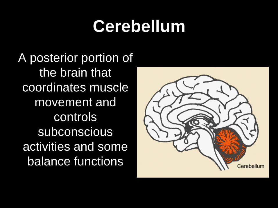
Reflex
An involuntary
and almost
immediate
movement in
response to a
stimulus

Content Objectives
Write these down! I will be able to identify:
• The function of the central nervous
system.
• The two components of the peripheral
nervous system.
• How a spinal reflex is generated.

Adapted from Holt Biology 2008
Chapter 38 - Section 1:
Structures of the Nervous
System
Notes

The Nervous System
Neuron: specialized cell of the nervous
system that carries messages
(signaling cell)

The Nervous System
Key point: The nervous
system controls and
coordinates functions
throughout the body and
responds to internal and
external changes.

Divisions of the nervous system
1. Central Nervous System (CNS)
2. Peripheral Nervous System (PNS)

Central Nervous System (CNS)
a. Brain
b. Spinal cord

Peripheral Nervous System (PNS)
a. Sensory
b. Motor

Divisions of the Nervous System
which
consists of
is divided
into
that
make up
which is
divided into
The Nervous
System
Sensory
nerves Motor
nerves
Autonomic
nervous
system
Somatic
nervous
system
Central
nervous
system
Peripheral
nervous
system
Sympathetic
nervous system
Parasympathetic
nervous system
Brain Spinal
Cord

Structures of the Nervous System

Think, Share, Write #1
What is a neuron?
Think, Share, Write #1
What is a neuron?
A neuron is a specialized cell in
the nervous system, a nerve cell.

The Brain
5
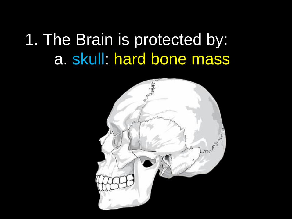
1. The Brain is protected by:
a. skull: hard bone mass

1. The Brain is protected by:
b. meninges:
3 layers of tissue
covering the brain

1. The Brain is protected by:
c. cerebrospinal fluid:
surrounds and absorbs shock

2. Spinal Cord
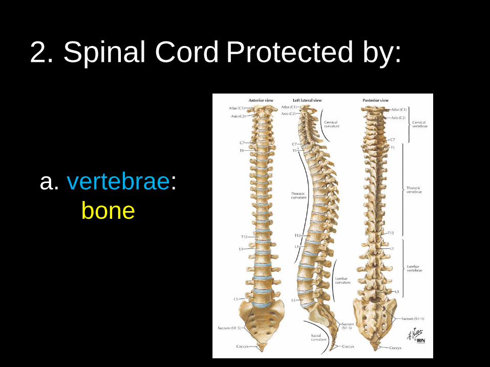
2. Spinal Cord Protected by:
a. vertebrae:
bone

b. meninges:
3 layers of
tissue
2. Spinal Cord Protected by:

c.
cerebrospinal
fluid:
wraps around,
shock
absorber
2. Spinal Cord Protected by:
Adapted from Holt Biology 2008
YOUR TURN
With a partner, read the Chapter 38 Section 1 Active Reading – Structures of the Nervous System
1st - Take turns reading the questions aloud to each other, alternating questions.
2nd - Take turns reading the selection aloud to each other, alternating sentences or paragraphs.

Adapted from Holt Biology 2008 Adapted from Holt Biology 2008
YOUR TURN
As you read discuss the content.
Reread and discuss each question. Write down the best answer to the question using full descriptive sentences.
• Be prepared to share with the class.
Adapted from Holt Biology 2008
2
Content Objectives
Write these down! I will be able to identify:
• The function of the central nervous
system.
• The two components of the peripheral
nervous system.
• How a spinal reflex is generated.
4

Four Main Areas of the Brain:
1. Cerebrum
2. Cerebellum
3. Pons
4. Medulla

1.Cerebrum:
largest, most
prominent part,
voluntary
activities of the
body,
intelligence,
learning,
judgment

Two sections connected by the
corpus callosum
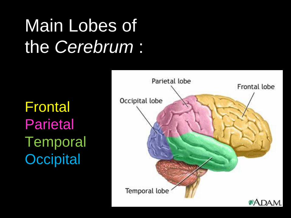
Main Lobes of
the Cerebrum :
Frontal
Parietal
Temporal
Occipital

Surfaces of the Brain:
1. Cortex : outer surface - gray matter;
Process information from senses &
Controls body movement

2. Medulla: center, white matter;
Bundles of myelinated axons
Surfaces of the Brain:

Four Main Areas of the Brain cont. :
2. Cerebellum:
2nd largest area,
coordinates and
balances
actions of
muscles that
move the body
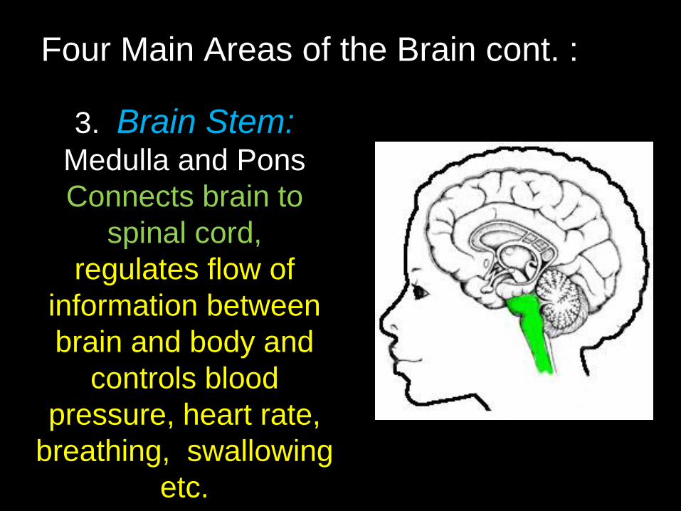
Four Main Areas of the Brain cont. :
3. Brain Stem: Medulla and Pons
Connects brain to
spinal cord,
regulates flow of
information between
brain and body and
controls blood
pressure, heart rate,
breathing, swallowing
etc.

4. Thalamus and Hypothalamus
Between brain and brain stem
Four Main Areas of the Brain cont. :
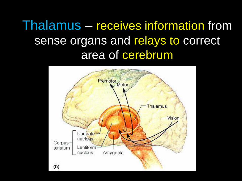
Thalamus – receives information from
sense organs and relays to correct
area of cerebrum
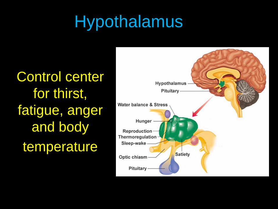
Control center
for thirst,
fatigue, anger
and body
temperature
Hypothalamus

The Brain Section 35-3
Figure 35-9 The Brain

Main
communication link
between brain and
rest of the body.
31 pairs of nerves
out from the spinal
cord and lead to the
rest of the body.
The Spinal Cord
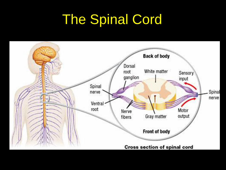
The Spinal Cord

Reflex: automatic response to a
stimulus - It is usually a reaction to
protect body

Reflex arc – The nerve pathway -
1. sensory receptor,
2. spinal cord,
3. muscle or gland (effector)

Reflex

Think, Share, Write #2
What neurons are involved in a
spinal reflex?

Think, Share, Write #2
What neurons are involved in a
spinal reflex?
Sensory neurons, motor neurons,
and interneurons are involved in a
spinal reflex?

Outside the
Central Nervous
System
Transmits from
sense organs to
CNS and back to
muscles or
glands.
Peripheral Nervous System

Two Divisions: Sensory and
Motor
Peripheral Nervous System

The sensory
division transmits
impulses from
sense organs -
such as the ears
and taste buds -
to the CNS.

The motor
division
transmits
impulses from
the CNS
system to the
muscles or
glands.
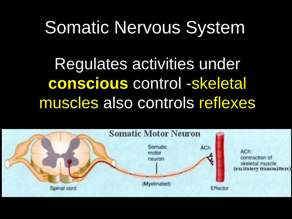
Regulates activities under
conscious control -skeletal
muscles also controls reflexes
Somatic Nervous System

Regulates activities that are
automatic – involuntary
Examples:
- heartbeat
- contraction of smooth muscles
in the digestive system
Autonomic Nervous System

Sympathetic - fight or flight
Parasympathetic – rest and digest
Two Parts: Autonomic Nervous System

Most organs are controlled by both. Why?
Two Parts:
Sympathetic & Parasympathetic

Content Objectives
Write these down! I will be able to identify:
• How the body conducts electricity.
• How a neuron’s structure allows the
neuron to send electrical signals.
• How a nerve impulse is generated
• How neurons communicate with each
other.

Adapted from Holt Biology 2008
Chapter 38 Section 2:
Neurons and the Nerve
Impulse
Key Vocabulary
Terms
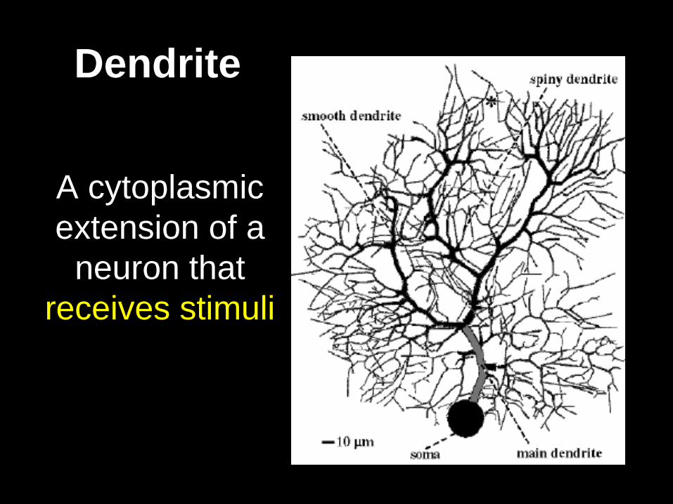
Dendrite
A cytoplasmic
extension of a
neuron that
receives stimuli

Axon
An elongated
extension of a
neuron that
carries
impulses away
from the cell
body

Nerve
A collection of
nerve fibers
through which
impulses travel
between the
central nervous
system and other
parts of the body

Membrane Potential
The difference
in electric
potential
between the
two sides of a
cell membrane

Action Potential
A sudden change
in the polarity of the
membrane of a
neuron, gland cell,
or muscle fiber that
facilitates the
transmission of
electrical impulses

Synapse
The junction at which the end of the axon of
a neuron meets the end of a dendrite or the
cell body of another neuron or meets
another cell

Neurotransmitter
A chemical
substance that
transmits nerve
impulses
across a
synapse

Think, Share, Write #3
What part of the cell sends
electrical signals?

Think, Share, Write #3
What part of the cell sends
electrical signals?
The part of the cell that sends
electrical signals is the axon.

Adapted from Holt Biology 2008
Chapter 38 - Section 2:
Neurons and the Nerve Impulse
Notes

Neurons and the Nerve Impulse
Key Point: The
nervous and
endocrine systems
help the body
maintain
homeostasis by
responding to
change.

Neurons and the Nerve Impulse
Impulses in the
nervous system
are electrical and
chemical. The
electrical signals
are caused by the
movement of ions
across the cell
membrane of
neurons.

Adapted from Holt Biology 2008
YOUR TURN
With a partner, read the Chapter 38 Section 2 Active Reading – Neurons and Nerve Impulses
1st - Take turns reading the questions aloud to each other, alternating questions.
2nd - Take turns reading the selection aloud to each other, alternating sentences or paragraphs.

Adapted from Holt Biology 2008 Adapted from Holt Biology 2008
YOUR TURN
As you read discuss the content.
Reread and discuss each question. Write down the best answer to the question using full descriptive sentences.
• Be prepared to share with the class.
Adapted from Holt Biology 2008
2

Axon terminals
Myelin sheath
Nodes
Cell body
Axon
Nucleus
Dendrites
Section 35-2
A Neuron
The Neuron

The Nerve Impulse
Key Point: An impulse begins
when a neuron is stimulated by
another neuron or by a stimulus in its
environment
Click picture

The Synapse
Space between two neurons or
neuron and muscle or gland.
Vesicle
Axon
Axon
terminal
Synaptic cleft
Neurotransmitter
Receptor
Dendrite of
adjacent neuron
Direction of Impulse
Section 35-2

Impulse - message - crosses the synapse
with the aid of a chemical called a neurotransmitter

Neurotransmitters change the permeability of
the membrane of the next neuron, muscle or
gland to allow moving impulse to go from one
to the other
click

Think, Share, Write #4
How are neurotransmitters
released?

Think, Share, Write #4
How are neurotransmitters
released?
Neurotransmitters are released
from synaptic vesicles at the
presynaptic membrane by
exocytosis.

Content Objectives
Write these down! I will be able to identify:
• How sensory information is detected.
• The five (5) types of sensory
receptors.
• Where the sites of sensory
processing are in the brain.

Adapted from Holt Biology 2008
Chapter 38 Section 3:
Sensory Systems
Key Vocabulary
Terms

Sensory Receptor
A specialized
structure that
contains the ends
of sensory
neurons and that
responds to
specific types of
stimuli

Retina
The light-
sensitive inner
layer of the eye,
which receives
images formed by
the lens and
transmits them
through the optic
nerve to the brain

Cochlea
A coiled tube that
is found in the
inner ear and that
is essential to
hearing

Semicircular Canal
The fluid-filled
canal in the inner
ear that helps
maintain balance
and coordinate
movements

Taste Bud
One of many oval
concentrations of
sensory nerve
endings on the
tongue, palate,
and pharynx

Think, Share, Write #5
What types of cells are sensory
receptors?

Think, Share, Write #5
What types of cells are sensory
receptors?
The types of cells that are sensory
receptors are specialized neurons
located in sensory organs.
.
Adapted from Holt Biology 2008
Chapter 38 - Section 3:
Sensory Systems
Notes
Content Objectives
Write these down! I will be able to identify:
• How sensory information is detected.
• The five (5) types of sensory
receptors.
• Where the sites of sensory
processing are in the brain.

The Senses
Sensory receptors:
specialized neurons
that react directly to
environmental
stimuli

Key point: There are five general
categories of sensory receptors:
pain receptors
thermoreceptors
mechanoreceptors
chemoreceptors
photoreceptors

Pain receptors
Respond to tissue damage

Thermoreceptors
Respond to mild heat and cold
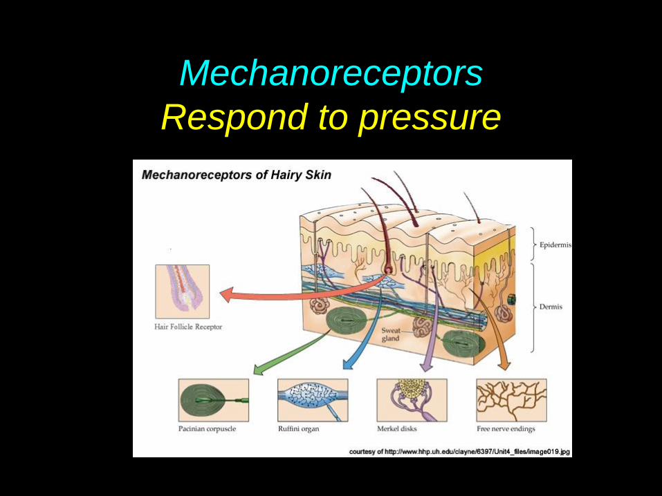
Mechanoreceptors
Respond to pressure

Chemoreceptors
Respond to chemicals

Photoreceptors
Respond to light

Choroid
Retina
Blood vessels
Optic nerve
Fovea
Vitreous humor
Sclera
Ligaments
Iris
Pupil
Cornea
Aqueous humor
Lens
Muscle
Section 35-4
Figure 35-14 The Eye
Four Senses - Vision - The Eye

Cornea - focuses light

Iris - colored part of the eye, contains muscles to control opening
of the pupil

Pupil - allows light into the eye

Lens - adjusts focus, muscles control shape of the lens

Retina - contains photo receptors (rods & cones) that convert light to nerve
impulses

Rods - sensitive to light, no colors Cones -distinguishes color

Optic nerve - carries impulses to the brain

Adapted from Holt Biology 2008
YOUR TURN
With a partner, read the Chapter 38 Section 3 Active Reading – Sensory Systems
1st - Take turns reading the questions aloud to each other, alternating questions.
2nd - Take turns reading the selection aloud to each other, alternating sentences or paragraphs.
Adapted from Holt Biology 2008 Adapted from Holt Biology 2008
YOUR TURN
As you read discuss the content.
Reread and discuss each question. Write down the best answer to the question using full descriptive sentences.
• Be prepared to share with the class.
Adapted from Holt Biology 2008
2
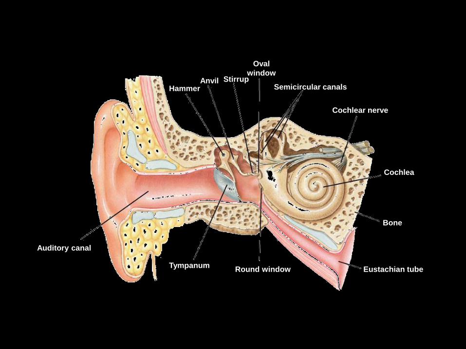
Auditory canal
Tympanum Round window Eustachian tube
Bone
Cochlea
Cochlear nerve
Semicircular canals
Oval
window Stirrup Anvil
Hammer
Section 35-4
Figure 35-15 The Ear
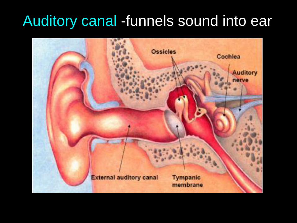
Auditory canal -funnels sound into ear

Tympanic membrane - vibrates to further
transmit sound

Ossicles - small bones, transmit
vibrations into the cochlea

Cochlea - fluid filled chamber that
converts sound waves to nerve impulses

Think, Share, Write #6
How do hair cells detect sound
waves?
Think, Share, Write #6
How do hair cells detect sound
waves?
Hair cells detect sound waves by
resting on a membrane that
vibrates when sound waves enter
the cochlea.
.

Semicircular canals - fluid filled canals
that are involved in balance and
equilibrium

Smell – Nose
Receptors located in nasal
passages
Chemical sense - odor molecules
must be moist

Taste - tongue
Receptors located on
the surface of the
tongue
Four tastes only -
sweet, sour, bitter,
salty

Touch and pressure
Receptors located in skin all over
body surface
Most on fingers, toes and face

Content Objectives
Write these down! I will be able to identify:
• Why psychoactive drugs are
dangerous..
• The neural mechanisms that underly
drug addiction.
• How nervous system function is
damaged.

Adapted from Holt Biology 2008
Chapter 38 - Section 4:
Nervous System Dysfunction
Notes

Used to increase
alertness, relieve fatigue
Used to relieve anxiety,
irritability, tension
Used to relieve pain
Stimulants
Depressants
Opiates
Amphetamines
Barbiturates
Tranquilizers
Morphine
Codeine
Increase heart and respiratory
rates; elevate blood pressure;
dilate pupils; decrease appetite
Slow down the actions of the
central nervous system; small
amounts cause calmness and
relaxation; larger amounts cause
slurred speech and impaired
judgement
Act as a depressant; cause
drowsiness, restlessness, nausea
Section 35-5
Commonly Abused Drugs
Drug Type Medical Use Examples Effects on the body
Commonly Used Drugs

Drug - substance that
causes a change in
the body
Drugs and the Nervous System

Various outcomes from drugs
a. Kill bacteria or treat
disease
b. Affect specific systems
c. Cause changes in brain,
nervous system or
synapse

Stimulants
Depressants
Cocaine
Opiates
Marijuana
Alcohol
Drugs that affect synapse

• Action of nervous
system
• Heart rate, blood
pressure, and
breathing rate
Stimulants – increase:

• Release of
neurotransmitters
at some synapses
in the brain
Stimulants – increase:

• Slow action of the
nervous system
• Slow down
respiration rate &
heart rate – may
cause death
Depressants

• Lower blood
pressure
• Relax muscles
and relieve tension
Depressants

Think, Share, Write #6
How can depressants cause
death?

Think, Share, Write #6
How can depressants cause
death?
Depressants can slow down hart
rate and breathing rate enough to
cause death.
.

Causes the sudden release in the brain
of a neurotransmitter called dopamine
which causes feelings of pleasure that
may lead to depression when it wears
off and can cause heart attack.
Cocaine

Mimic natural chemicals in the brain
known as endorphins, which normally
help to overcome sensations of pain.
Pain killing drugs are addictive
Opiates

Marijuana - active ingredient THC
Euphoria and disorientation; more
destructive to lungs than cigarettes
Loss of memory- inability to concentrate
– less testosterone in males

Alcohol - depressant, even small
amounts will slow down the rate
at which the nervous system
functions
slows reflexes, disrupts
coordination, impairs judgment

Fetal Alcohol Syndrome
Many mental and physical abnormalities.
DO NOT DRINK IF COULD GET
PREGNANT OR IF YOU ARE
PREGNANT.
![The Descriptive Complexity Approach to LOGCFL · the descriptive complexity approach to logcfl 631 1 Independently from the present work, the question from [6] was solved in a draft](https://img.pdfslide.us/doc/110x75/5f11f9d1969121476168320d/the-descriptive-complexity-approach-to-logcfl-the-descriptive-complexity-approach.jpg)