Embed Size (px)
DESCRIPTION
Chapter 37. Electrocardiography. Anatomy of the Heart. Four chambers Atria: two upper chambers Ventricles: two lower chambers Deoxygenated and oxygenated blood Cycle begins with each heartbeat Coronary arteries. Electrical Conduction System of the Heart. - PowerPoint PPT Presentation
Citation preview
© 2014 Cengage Learning. All Rights Reserved. May not be scanned, copied or duplicated, or posted to a publicly accessible website, in whole or in part.
© 2014 Cengage Learning. All Rights Reserved. May not be scanned, copied or duplicated, or posted to a publicly accessible website, in whole or in part.
Chapter 37
Electrocardiography
© 2014 Cengage Learning. All Rights Reserved. May not be scanned, copied or duplicated, or posted to a publicly accessible website, in whole or in part.
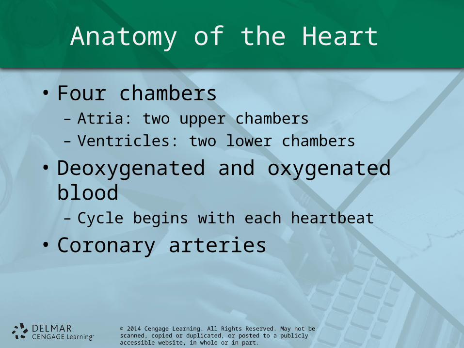
Anatomy of the Heart
• Four chambers– Atria: two upper chambers– Ventricles: two lower chambers
• Deoxygenated and oxygenated blood– Cycle begins with each heartbeat
• Coronary arteries
© 2014 Cengage Learning. All Rights Reserved. May not be scanned, copied or duplicated, or posted to a publicly accessible website, in whole or in part.
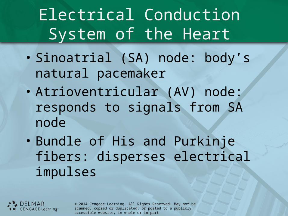
Electrical Conduction System of the Heart
• Sinoatrial (SA) node: body’s natural pacemaker
• Atrioventricular (AV) node: responds to signals from SA node
• Bundle of His and Purkinje fibers: disperses electrical impulses
© 2014 Cengage Learning. All Rights Reserved. May not be scanned, copied or duplicated, or posted to a publicly accessible website, in whole or in part.
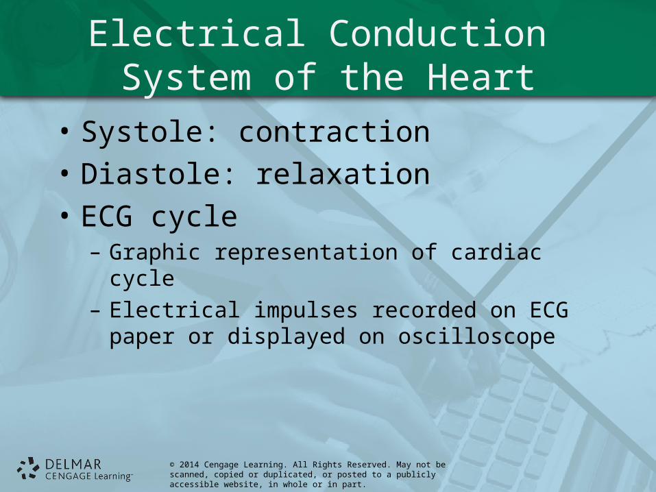
Electrical Conduction System of the Heart
• Systole: contraction
• Diastole: relaxation
• ECG cycle– Graphic representation of cardiac cycle– Electrical impulses recorded on ECG paper or
displayed on oscilloscope
© 2014 Cengage Learning. All Rights Reserved. May not be scanned, copied or duplicated, or posted to a publicly accessible website, in whole or in part.
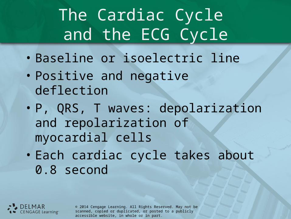
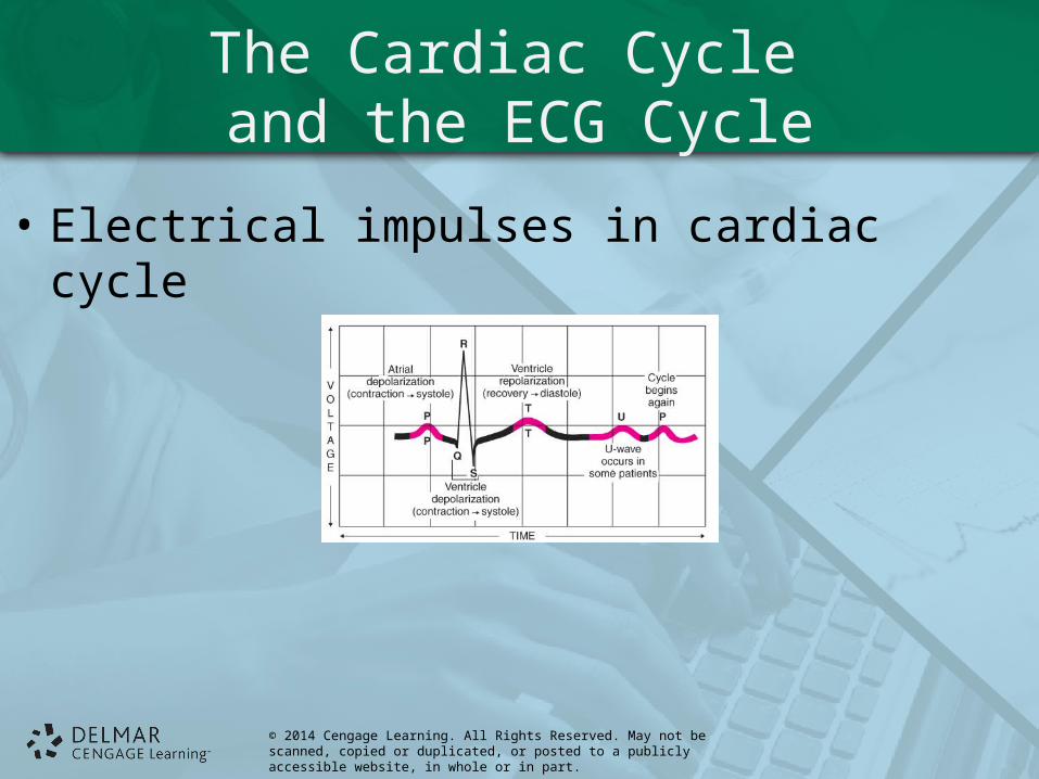
The Cardiac Cycle and the ECG Cycle
• Baseline or isoelectric line
• Positive and negative deflection
• P, QRS, T waves: depolarization and repolarization of myocardial cells
• Each cardiac cycle takes about 0.8 second
© 2014 Cengage Learning. All Rights Reserved. May not be scanned, copied or duplicated, or posted to a publicly accessible website, in whole or in part.
The Cardiac Cycle and the ECG Cycle
• Electrical impulses in cardiac cycle
© 2014 Cengage Learning. All Rights Reserved. May not be scanned, copied or duplicated, or posted to a publicly accessible website, in whole or in part.
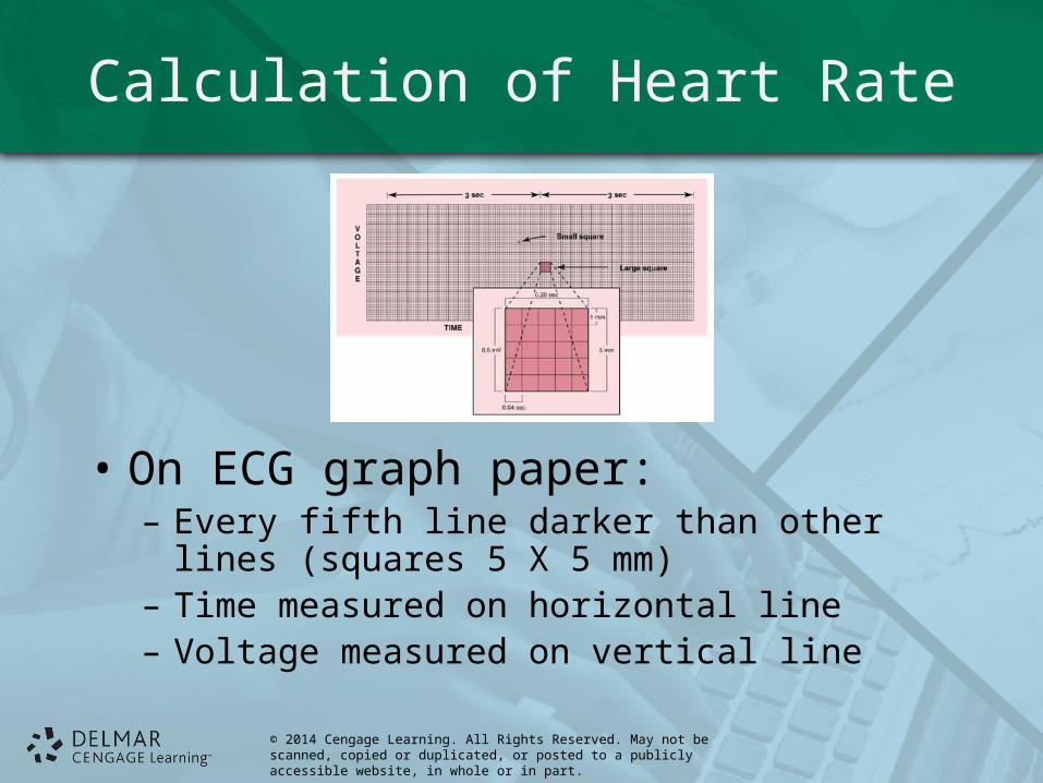
Calculation of Heart Rate
• On ECG graph paper:– Every fifth line darker than other lines (squares 5 X
5 mm)– Time measured on horizontal line– Voltage measured on vertical line
© 2014 Cengage Learning. All Rights Reserved. May not be scanned, copied or duplicated, or posted to a publicly accessible website, in whole or in part.
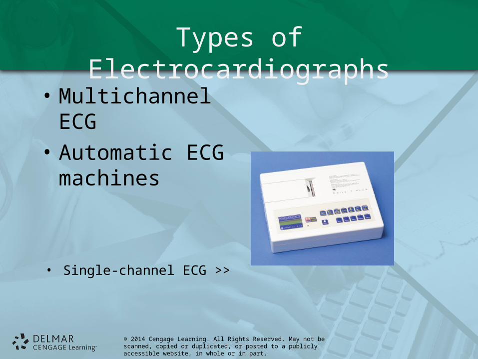
Types of Electrocardiographs
• Multichannel ECG
• Automatic ECG machines
• Single-channel ECG >>
© 2014 Cengage Learning. All Rights Reserved. May not be scanned, copied or duplicated, or posted to a publicly accessible website, in whole or in part.
Types of Electrocardiographs
• ECG telephone transmissions
• Facsimile electrocardiograph
• Interpretive electrocardiograph
© 2014 Cengage Learning. All Rights Reserved. May not be scanned, copied or duplicated, or posted to a publicly accessible website, in whole or in part.
ECG Equipment
• Electrocardiograph paper– Black or dark blue– Wax or plastic coated– Heat and pressure sensitive– Heat of stylus can be adjusted to obtain sharp
tracing
© 2014 Cengage Learning. All Rights Reserved. May not be scanned, copied or duplicated, or posted to a publicly accessible website, in whole or in part.
ECG Equipment
• Electrolyte– Applied with each electrode to pick up electrical
current – In form of gel, lotion, paste, or pre-saturated pads,
contained within adhesive sensors
© 2014 Cengage Learning. All Rights Reserved. May not be scanned, copied or duplicated, or posted to a publicly accessible website, in whole or in part.
ECG Equipment
• Sensors or electrodes– Detect electrical impulses on body surface; relay
them through cables, or lead, wires to ECG machine
– Disposable sensors (electrodes) contain layer of electrolyte gel on adhesive surface; used on limbs and chest
© 2014 Cengage Learning. All Rights Reserved. May not be scanned, copied or duplicated, or posted to a publicly accessible website, in whole or in part.
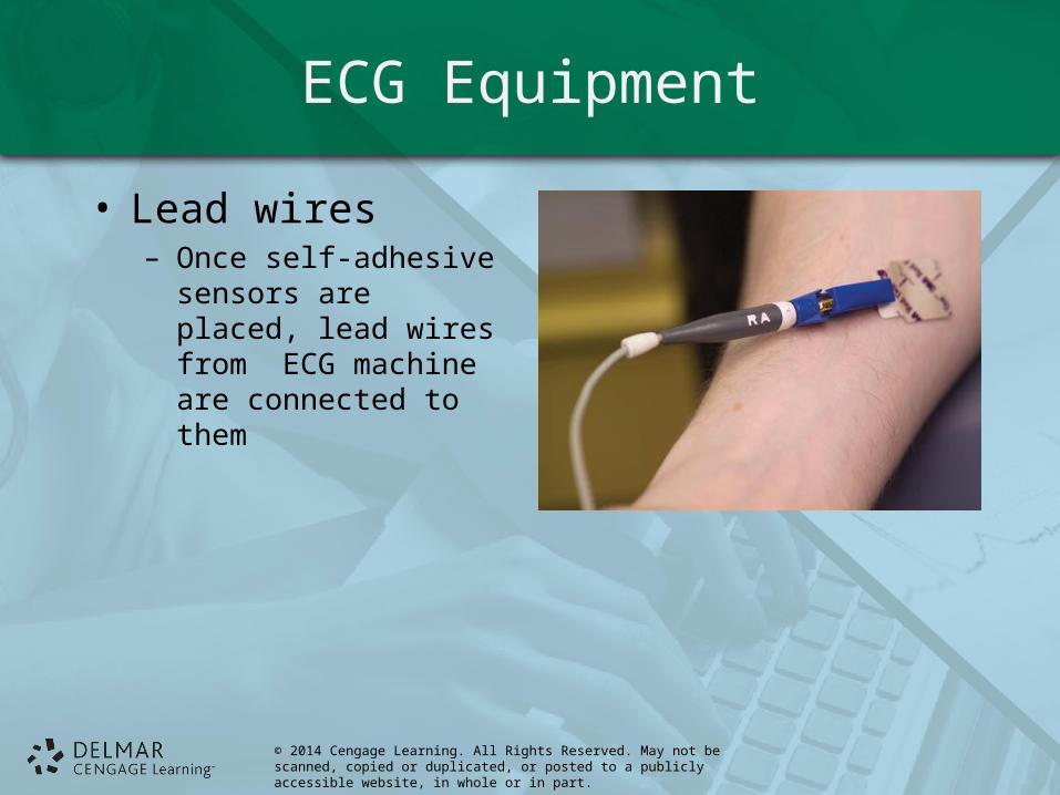
ECG Equipment
• Lead wires– Once self-adhesive
sensors are placed, lead wires from ECG machine are connected to them
© 2014 Cengage Learning. All Rights Reserved. May not be scanned, copied or duplicated, or posted to a publicly accessible website, in whole or in part.
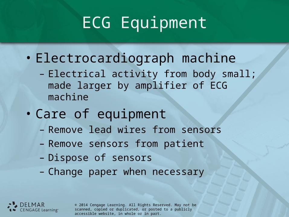
ECG Equipment
• Electrocardiograph machine– Electrical activity from body small; made larger by
amplifier of ECG machine
• Care of equipment– Remove lead wires from sensors– Remove sensors from patient– Dispose of sensors– Change paper when necessary
© 2014 Cengage Learning. All Rights Reserved. May not be scanned, copied or duplicated, or posted to a publicly accessible website, in whole or in part.
Lead Coding
• 12 leads recorded using 10 lead wires
• Necessary for identification and mounting purposes
• Newer ECGs automatically mark (code) each lead
© 2014 Cengage Learning. All Rights Reserved. May not be scanned, copied or duplicated, or posted to a publicly accessible website, in whole or in part.
The Electrocardiograph and Sensor Placement
• 10 sensors that record 12 leads of heart’s electrical activity
• Allows for 3D interpretation of activity
• Electrodes placed on patient’s four limbs and chest
© 2014 Cengage Learning. All Rights Reserved. May not be scanned, copied or duplicated, or posted to a publicly accessible website, in whole or in part.
The Electrocardiographand Sensor Placement
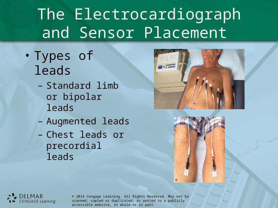
• Types of leads– Standard limb or
bipolar leads– Augmented leads– Chest leads or
precordial leads
© 2014 Cengage Learning. All Rights Reserved. May not be scanned, copied or duplicated, or posted to a publicly accessible website, in whole or in part.
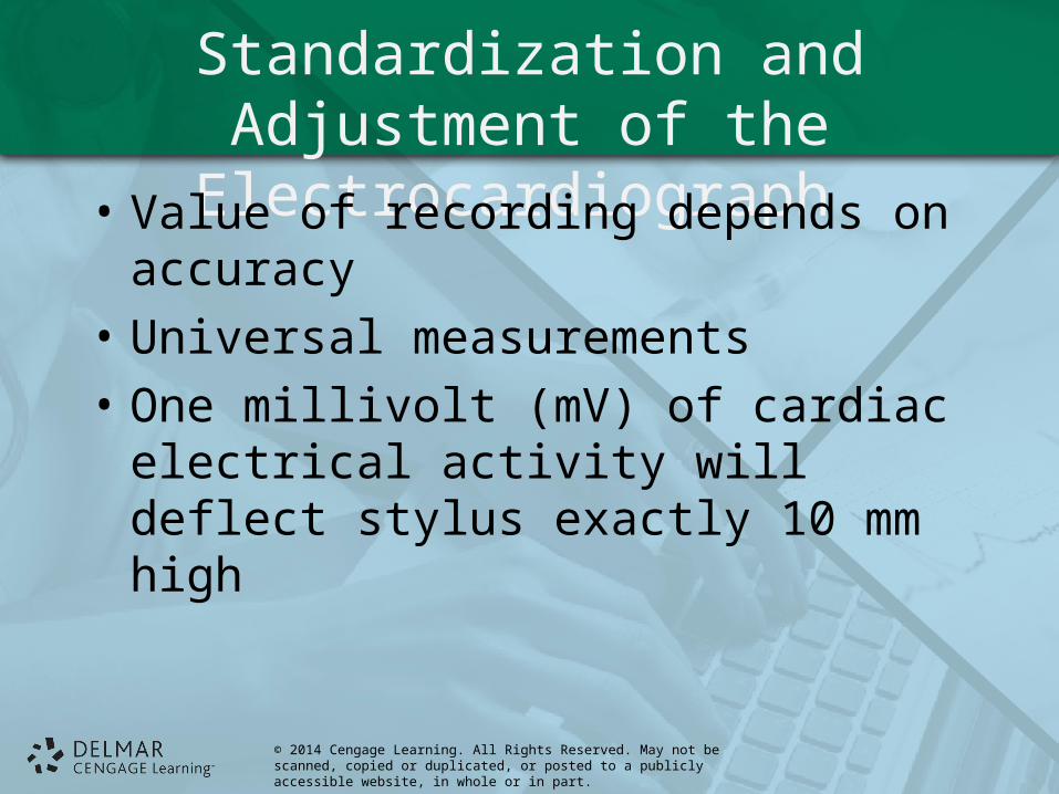
Standardization and Adjustment of the Electrocardiograph
• Value of recording depends on accuracy
• Universal measurements
• One millivolt (mV) of cardiac electrical activity will deflect stylus exactly 10 mm high
© 2014 Cengage Learning. All Rights Reserved. May not be scanned, copied or duplicated, or posted to a publicly accessible website, in whole or in part.
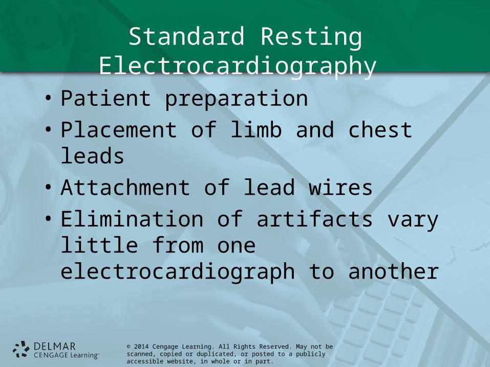
Standard Resting Electrocardiography
• Patient preparation
• Placement of limb and chest leads
• Attachment of lead wires
• Elimination of artifacts vary little from one electrocardiograph to another
© 2014 Cengage Learning. All Rights Reserved. May not be scanned, copied or duplicated, or posted to a publicly accessible website, in whole or in part.
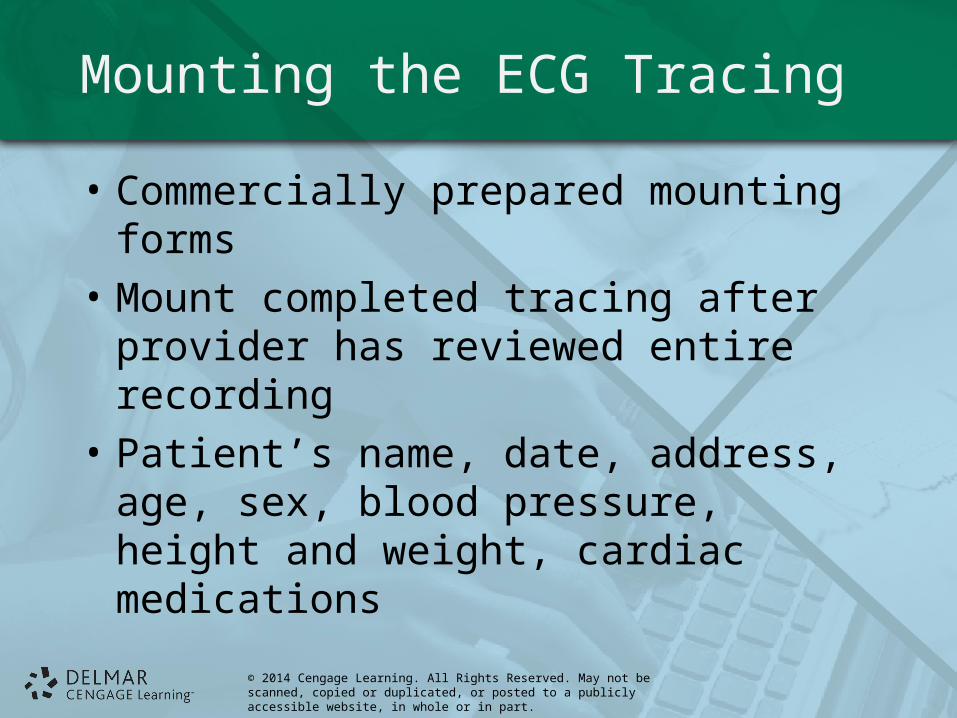
Mounting the ECG Tracing
• Commercially prepared mounting forms
• Mount completed tracing after provider has reviewed entire recording
• Patient’s name, date, address, age, sex, blood pressure, height and weight, cardiac medications
© 2014 Cengage Learning. All Rights Reserved. May not be scanned, copied or duplicated, or posted to a publicly accessible website, in whole or in part.
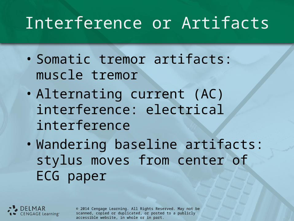
Interference or Artifacts
• Somatic tremor artifacts: muscle tremor
• Alternating current (AC) interference: electrical interference
• Wandering baseline artifacts: stylus moves from center of ECG paper
© 2014 Cengage Learning. All Rights Reserved. May not be scanned, copied or duplicated, or posted to a publicly accessible website, in whole or in part.
Interference or Artifacts
• Interrupted baseline artifacts: break seen between waves
• Patients with unique problems
© 2014 Cengage Learning. All Rights Reserved. May not be scanned, copied or duplicated, or posted to a publicly accessible website, in whole or in part.
Myocardial Infarctions (Heart Attacks)
• Number one cause of death in United States
• Behaviors to adopt for healthy heart

© 2014 Cengage Learning. All Rights Reserved. May not be scanned, copied or duplicated, or posted to a publicly accessible website, in whole or in part.
Cardiac Arrhythmias
• Atrial arrhythmias– Premature atrial contractions (PAC)– Paroxysmal atrial tachycardia (PAT)– Atrial fibrillation
© 2014 Cengage Learning. All Rights Reserved. May not be scanned, copied or duplicated, or posted to a publicly accessible website, in whole or in part.
Cardiac Arrhythmias
• Ventricular arrhythmias– Premature ventricular contractions (PVCs)– Ventricular tachycardia– Ventricular fibrillation
© 2014 Cengage Learning. All Rights Reserved. May not be scanned, copied or duplicated, or posted to a publicly accessible website, in whole or in part.
Defibrillation
• Electrical device that applies countershocks to heart through electrodes or pads placed on chest wall
• Can convert cardiac arrhythmia to normal sinus rhythm
• Automated external defibrillators (AED)
© 2014 Cengage Learning. All Rights Reserved. May not be scanned, copied or duplicated, or posted to a publicly accessible website, in whole or in part.
Other Cardiac Diagnostic Tests
• Holter monitor (portable ambulatory electrocardiograph)– Portable continuous recording of cardiac activity
for 24-hour period – Noninvasive test helps to diagnose cardiac
arrhythmias– Digital, three-channel ECG; Windows-based
software– Electrode placement not the same as for standard
12-lead resting ECG
© 2014 Cengage Learning. All Rights Reserved. May not be scanned, copied or duplicated, or posted to a publicly accessible website, in whole or in part.
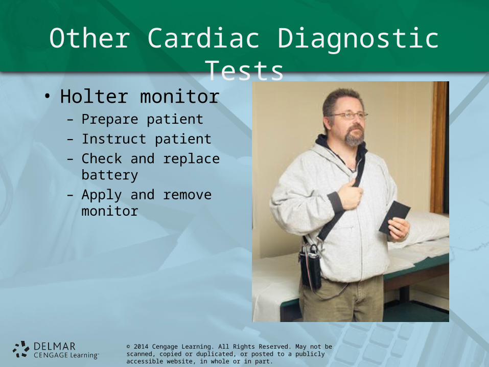
Other Cardiac Diagnostic Tests
• Holter monitor– Prepare patient– Instruct patient– Check and replace
battery– Apply and remove
monitor
© 2014 Cengage Learning. All Rights Reserved. May not be scanned, copied or duplicated, or posted to a publicly accessible website, in whole or in part.
Other Cardiac Diagnostic Tests
• Holter monitor– Patient activity diary
• Record all activities, emotional states, time of their occurrence
• Record chest pain and other symptoms and time of occurrence
– Removal• Patient returns to office• Tape analyzed by scanner or computer
© 2014 Cengage Learning. All Rights Reserved. May not be scanned, copied or duplicated, or posted to a publicly accessible website, in whole or in part.
Other Cardiac Diagnostic Tests
• Loop ECG– Uses only two electrodes– Records few minutes of ECG at a time on
computer chip– Recorded event transmitted by telephone to
provider
© 2014 Cengage Learning. All Rights Reserved. May not be scanned, copied or duplicated, or posted to a publicly accessible website, in whole or in part.
Other Cardiac Diagnostic Tests
• Treadmill stress test or exercise tolerance ECG– Diagnose heart disorders– Probable cause of patient’s chest pain– Assess patient’s cardiac ability following cardiac
surgery– Noninvasive test– Patient exercises on treadmill at prescribed rates
of speed
© 2014 Cengage Learning. All Rights Reserved. May not be scanned, copied or duplicated, or posted to a publicly accessible website, in whole or in part.
Other Cardiac Diagnostic Tests
• Treadmill stress test or exercise tolerance ECG– Complications ( myocardial infarction or serious
arrhythmia) can occur during testing – Further diagnostic tests such as cardiac
catheterization (angiogram) may be necessary

© 2014 Cengage Learning. All Rights Reserved. May not be scanned, copied or duplicated, or posted to a publicly accessible website, in whole or in part.
Other Cardiac Diagnostic Tests
• Thallium stress test– Similar to treadmill stress– Patient given injection of radioactive (thallium)– Test shows how well blood flows to heart muscle
© 2014 Cengage Learning. All Rights Reserved. May not be scanned, copied or duplicated, or posted to a publicly accessible website, in whole or in part.
Other Cardiac Diagnostic Tests
• Echocardiography/ultrasonography– Noninvasive, diagnostic test– Ultrasound to image internal structures of heart
• CT and MRI coronary imaging– Identify location and thickness of cardiac muscle
scars due to damage
© 2014 Cengage Learning. All Rights Reserved. May not be scanned, copied or duplicated, or posted to a publicly accessible website, in whole or in part.
Cardiac Procedures
• Percutaneous transluminal coronary angioplasty (PTCA)– Widens narrowed or blocked coronary artery– Balloon angioplasty – Stents: small mesh tubes compressed around
balloon; permanent intervention
© 2014 Cengage Learning. All Rights Reserved. May not be scanned, copied or duplicated, or posted to a publicly accessible website, in whole or in part.
Cardiac Procedures
• Atherectomy and laser angioplast
• Coronary artery bypass
• Heart valve: repaired or replaced
• Procedures for arrhythmias