Embed Size (px)
Citation preview
399
Acute Management of Pharyngoesophageal Trauma
Chapter 30
ACUTE MANAGEMENT OF PHARYNGOESOPHAGEAL TRAUMA
NICI EDDY BOTHWELL, MD*
INTRODUCTION
ANATOMY
INITIAL EVALUATION
DIAGNOSTIC STUDIES
ACUTE MANAGEMENT
POSTOPERATIVE CARE
OUTCOMES
SUMMARY
CASE PRESENTATIONSCase Study 30-1Case Study 30-2
*Lieutenant Colonel, Medical Corps, US Army; Department of Otolaryngology–Head and Neck Surgery, Madigan Army Medical Center, 9040 Jackson Avenue, Tacoma, Washington 98431; Assistant Professor of Surgery, Uniformed Services University of the Health Sciences
400
Otolaryngology/Head and Neck Combat Casualty Care
INTRODUCTION
In a large literature review by McConnell and Trun-key,1 combining published data from 1963 to 1990 on penetrating neck trauma, the pharynx and esophagus were injured in 9.6% of 1,275 total neck injuries. A more recent literature review of 1,560 patients with blunt and penetrating neck trauma reported a 2% rate for pharyngoesophageal injuries.2 The incidence of pha-ryngoesophageal injuries is higher in military penetrat-ing neck trauma compared with civilian neck trauma. A trauma registry review of 75 British soldiers with penetrating neck trauma from 2004 to 2008 reported a 15% incidence of pharyngoesophageal injuries.3 There is also a much higher rate of pharyngoesophageal injuries from fragments (eg, improvised explosive devices [IEDs]) rather than gunshot wounds.3 A review by Brennan et al4 of 112 patients with penetrating neck trauma requiring neck exploration during Operation Iraqi Freedom had a similar rate of 14% for pharyn-goesophageal injuries. As the relative percentage of gunshot trauma compared with stab and blunt neck trauma increases in civilian trauma (approaching >50%), the incidence of pharyngoesophageal injuries approaches that seen in the military experience.5 Be-cause of the advances in body armor, military casual-ties are surviving injuries, but are prone to a relative increase in injuries to the face, neck, and extremities. Also, the pattern of injury from the IEDs results in multiple, penetrating, high-velocity fragments to these exposed regions and equates to an increased chance of one of them causing a pharyngoesophageal injury.
Trauma to the pharynx and cervical esophagus is rare, difficult to diagnose, and may only become ap-parent days after the injury when a patient manifests with signs of an infection. Accurately identifying a pharyngoesophageal injury even in the era of high-resolution computed tomography (CT) scans and magnetic resonance imaging scans can be challenging. A majority of the literature discusses how to reliably identify a pharyngoesophageal injury without having to perform a neck exploration on every patient with neck trauma. In contrast, there is very little information on how to surgically manage the injury itself. When significant injuries are not identified or the repair is unsuccessful, the ensuing morbidity and mortality are certain unless there is immediate secondary inter-vention. Because of the relative rarity, a majority of the data on pharyngoesophageal trauma comes from the civilian trauma literature rather than the military literature. The primary distinction between civilian and military trauma is the low-velocity versus high-velocity trauma. Although the injury described in the literature may be similar between low-velocity and high-velocity trauma, the degree of tissue devastation in a high-velocity injury compared with a low-velocity injury is significantly higher. Therefore, it is important for the military provider to consider this fundamen-tal difference in injury patterns when approaching a patient with neck trauma. Ultimately, this results in a lower threshold for imaging, endoscopy, and surgical intervention.
ANATOMY
The pharynx consists of three separate segments: (1) the nasopharynx, (2) the oropharynx, and (3) the hypopharynx. The nasopharynx extends from the skull base to the inferior aspect of the soft palate. It includes the posterior nasopharyngeal wall, Eusta-chian tube orifices, and the fossa of Rosenmüller.6
The oropharynx lies between the nasopharynx and the hypopharynx from the level of the soft palate to the tip of the epiglottis. It includes the posterior oropharyngeal wall, the base of the tongue, the hard and soft palate, tonsils, and retromolar trigone.6
The hypopharynx is the region of the pharynx that extends from the epiglottis superiorly to the cervical esophagus inferiorly.6 The hypopharynx is bordered anteriorly by the larynx and posteriorly by the ret-ropharyngeal space. It is divided into three regions: (1) the pyriform sinuses, (2) the postcricoid region, and (3) the posterior pharyngeal wall. The superior extent of the hypopharynx is approximately at the
level of the hyoid bone or the pharyngoepiglottic folds. Inferiorly, the hypopharynx tapers to the upper esophageal sphincter at the cricopharyngeus muscle. The cricopharyngeus muscle represents the transition between the hypopharynx and cervical esophagus, which starts at the level of the sixth cervical vertebra and lies posterior and slightly left of the trachea. The cervical esophagus is that portion of the esophagus that extends to the thoracic inlet. Just posterior to the cervical esophagus is the anterior paraspinous muscles and cervical spine. At the proximal margin of the esophagus is the upper esophageal sphincter, which is formed by the muscular fibers of the inferior pharyngeal constrictor condensing to form the crico-pharyngeus muscle. The trachea and thyroid gland are anterior to the cervical esophagus with the lobes of the thyroid also extending laterally. The retroesopha-geal space is contiguous with the retropharyngeal space above and the posterior mediastinum below.
401
Acute Management of Pharyngoesophageal Trauma
The location of pharyngoesophageal injury is important because of the distinct difference in in-jury pattern and risk of complications between the hypopharynx and the esophagus. As the pharyngo-esophageal funnel descends, it becomes narrowed, the muscular reinforcement decreases, and it becomes more intimately bound to the laryngotracheal appara-tus that increases its vulnerability to shearing forces.7
The hypopharynx can be subdivided into two levels based on the level of the arytenoids. Above the level of the arytenoids, the hypopharynx is reinforced by the middle and inferior constrictor muscles; it has low intraluminal pressure and redundant mucosa. The region of the hypopharynx above the arytenoids is inherently more flexible, resistant to extensive in-
jury, and has a decreased incidence of complications compared with the region below the level of the ary-tenoids.7 Below the level of the arytenoids, the funnel narrows and is enveloped by the inferior pharyngeal constrictor with inherent areas of weakness (Killian’s Triangle) where the fibers organize to form the crico-pharyngeal muscle. There is an increased intraluminal pressure resulting in pooling of secretions and gastric contents above this region, which can be a provoca-tive or perpetuating factor leading to complications from pharyngoesophageal injury.7 Awareness of the proximity and association that these potential spaces have with the pharynx and esophagus is critical in the correct medical and therapeutic management of patients with pharyngoesophageal trauma.
INITIAL EVALUATION
The initial evaluation and resuscitation of a patient with any trauma follow Advanced Trauma Life Support (ATLS) protocol. Patients who are unstable or who have hard signs of a serious neck injury will require a neck exploration with or with-out panendoscopy or surgical airway regardless of preoperative imaging or endoscopy. Symptoms requiring immediate neck exploration are listed in Exhibit 30-1. If the patient is stable, then this is an opportunity to complete a secondary survey and imaging studies focusing on acute signs of pharyngoesophageal injury. Exhibit 30-2 lists the most common signs and symptoms of acute pha-ryngoesophageal injury. Patients having any of these findings are presumed to have a pharyngo-esophageal injury and should complete additional
diagnostic studies to determine whether surgical intervention is indicated. The additional diagnostic studies to consider are CT neck with or without CT angiogram, panendoscopy (esophagoscopy), and contrast esophagram (esophagography). Typically, if a patient is being taken to the operating room emergently for a neck exploration, then it is likely that a panendoscopy will also be performed. If the patient is stable enough to have a preoperative CT angiogram, this will benefit the surgeon with regard to tracing the trajectory of the penetrating trauma and planning the surgical approach.
EXHIBIT 30-1
INDICATIONS FOR IMMEDIATE NECK EXPLORATION
Indications
• Majorairwayinjury • Unstableairway • Massivesubcutaneousemphysema • Airextravasation • Signsofmajorvascularinjury • Severe,activebleeding • Hemodynamicallyunstable • Refractoryshock • Evolvingshock • Rapidlyexpandinghematoma
EXHIBIT 30-2
SIGNS AND SYMPTOMS OF PHARYNGOESOPHAGEAL INJURY
Signs and Symptoms
• Dysphagia • Hematemesis • Hoarseness • Hemoptysis • Chestpain • Neckpain • Airbubblingthroughwound • Subcutaneousemphysema • Retropharyngealedema • Retropharyngealair • Hematoma • Deviatedtrachea • Pneumomediastinum • Airwaycompromise
402
Otolaryngology/Head and Neck Combat Casualty Care
There is some controversy whether pharyngo-esophageal trauma presents with predictable signs and symptoms to prompt surgical intervention and whether that is defined as neck exploration or repair of a pharyngoesophageal injury. A few authors have provided opposing evidence regarding the presenta-tion of pharyngoesophageal injuries. Yet, some authors believe that most patients with esophageal trauma are asymptomatic or that early signs and symptoms of esophageal injury are few.1,8 This would imply a very high false-negative rate. A more conservative false-negative rate of 20%, using only physical examination findings, was reported by Weigelt et al.9 Taken from the opposing viewpoint, Vassiliu et al2 claimed that the diagnosis of aerodigestive trauma can be based solely on clinical examination and that the absence of clinical findings reliably excludes aerodigestive tract trauma. In support of this claim, Demetriades et al10
had previously established a high negative predictive value using a systematic algorithm on 152 patients with neck trauma. When using this algorithm, the absence of clinical signs reliably excluded significant aerodigestive tract injuries requiring surgical interven-tion.10 Of the 64 patients who were symptomatic, only 10 patients required surgical intervention.10 Although there may be dissension about the reliability of physical examination findings and predictable symptoms, the key to management of pharyngoesophageal trauma is using a systematic approach to evaluation, diagnosis, and treatment.
What cannot be overemphasized is the subtlety of the signs and symptoms of an occult injury and the importance of intervention at the earliest suspicion of a pharyngoesophageal injury. Those patients with negative signs and symptoms and a negative workup should be observed closely for at least 24 hours with serial examinations by the same provider. For the US combat casualty patient, this means that serial examinations may occur during the medical evacu-ation from theater to the nearest Role 4 facility. In contrast, the local national or coalition force casualty patient is most likely admitted to the hospital for days or weeks; therefore, a minimum 24-hour observation period does not change his length of stay. Many occult pharyngoesophageal injuries present 2 to 3 days after the original injury with minimal symptoms, espe-cially if the patient is intubated. Signs and symptoms of ambulatory patients with iatrogenic esophageal perforations can be loosely applied to how a patient with an occult pharyngoesophageal injury may pres-ent. In a review of 36 ambulatory patients with mostly iatrogenic esophageal perforations, the most common presenting symptoms were pain (66.7%), dyspnea (38.9%), fever (33.3%), and dysphagia (5.5%).11 Sub-cutaneous emphysema was the most common sign (66%).11 In the setting of an intubated patient with polytrauma, the only detectable signs are fever and subcutaneous emphysema. This emphasizes the importance of a dedicated observation period with serial examinations by the same provider.
DIAGNOSTIC STUDIES
Maintaining a diagnostic algorithm is the key to ac-curate and timely diagnosis and treatment. Accurate in the sense that negative neck explorations are kept to a minimum and timely in the sense that the diagnostic modality does not inappropriately delay interven-tion. As more studies are applied, the sensitivity and specificity increase and the rate of negative neck ex-plorations decreases, but this comes at the expense of a delay in diagnosis. The most common radiological studies for neck trauma are high-resolution CT and contrast esophagography.
Plain chest X-rays and lateral neck films are com-monly used in the trauma setting as part of the ATLS protocol, but are not defined in the diagnostic algo-rithm for pharyngoesophageal trauma. Signs on plain X-ray films that are consistent with a pharyngoesopha-geal injury include prevertebral air, mediastinal air, mediastinal widening, hemothorax, pneumothorax, and pleural effusion.6,11,12 Of course, plain X-rays alone are not especially helpful in evaluating patients with neck trauma since most X-ray films will not show
signs of an injury. Of 19 patients presenting with neck crepitus with neck trauma (penetrating and blunt), 75% had a normal chest X-ray film, 10% had pneumo-mediastinum, and 5% had a pneumothorax.13 There is little utility in using a plain X-ray film for the diagnosis and workup of pharyngoesophageal trauma; but, in a mass casualty setting, it may be the only imaging that is readily available, and positive findings may prompt a more expeditious workup.
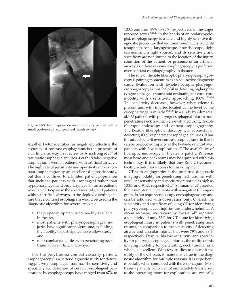
Contrast esophagography is used primarily in the civilian setting on stable, ambulatory patients and is especially helpful in identifying the persistence or resolution of a leak. (Figure 30-1). When used alone, the sensitivity of contrast esophagography ranges from 48% to 100%, and the specificity is near or at 100%.9,14–19 When combined with esophagoscopy, sen-sitivity becomes 100%.9 Location of the injury may affect the sensitivity of contrast esophagography. In a study by Ahmed et al,20 contrast esophagography detected 100% of esophageal injuries, but missed 100% of oropharyngeal and hypopharyngeal injuries.
403
Acute Management of Pharyngoesophageal Trauma
Figure 30-1. Esophagram on an ambulatory patient with a small posterior pharyngeal leak (white arrow).
Another factor identified as negatively affecting the accuracy of contrast esophagrams is the presence of an artificial airway. In a review by Armstrong et al16 of traumatic esophageal injuries, 4 of the 5 false-negative esophagrams were in patients with artificial airways. The high rate of sensitivity and specificity makes con-trast esophagography an excellent diagnostic study, but this is confined to a limited patient population that includes patients with esophageal rather than hypopharyngeal and oropharyngeal injuries, patients who can participate in the swallow study, and patients without artificial airways. In the deployed setting, it is rare that a contrast esophagram would be used in the diagnostic algorithm for several reasons:
• theproperequipmentisnotreadilyavailablein theater;
• mostpatientswithpharyngoesophageal in-juries have significant polytrauma, excluding their ability to participate in a swallow study; and
• mostcombatcasualtieswithpenetratingnecktrauma have artificial airways.
For the polytrauma combat casualty patient, esophagoscopy is a better diagnostic study for detect-ing pharyngoesophageal trauma. The sensitivity and specificity for detection of cervical esophageal pen-etrations by esophagoscopy have ranged from 67% to
100% and from 89% to 95%, respectively, in the larger reported series.9,16,19 In the hands of an otolaryngolo-gist, esophagoscopy is a safe and highly sensitive di-agnostic procedure that requires minimal instruments (esophagoscope, laryngoscope, bronchoscope, light carriers, and a light source), and its sensitivity and specificity are not limited to the location of the injury, condition of the patient, or presence of an artificial airway. For these reasons, esophagoscopy is preferred over contrast esophagography in theater.
The role of flexible fiberoptic pharyngoesophagos-copy is gaining momentum as an adjunctive diagnostic study. Evaluation with flexible fiberoptic pharyngo-esophagoscopy is most helpful in detecting higher pha-ryngoesophageal trauma and evaluating for vocal cord mobility with a sensitivity approaching 100%.15,16,20 The sensitivity decreases, however, when edema is present and with injuries located at the level of the cricopharyngeus muscle.15,16,20 In a study by Ahmed et al,20 33 patients with pharyngoesophageal injuries from penetrating neck trauma were evaluated using flexible fiberoptic endoscopy and contrast esophagography. The flexible fiberoptic endoscopy was successful in detecting 100% of pharyngoesophageal injuries. It has the added benefit over contrast esophagrams in that it can be performed rapidly at the bedside on intubated patients with few complications.20 The availability of fiberoptic endoscopy in theater is patchy. Whereas most head and neck teams may be equipped with this technology, it is unlikely that any Role 2 treatment facility would have access to this equipment.
CT with angiography is the preferred diagnostic imaging modality for penetrating neck trauma, with excellent sensitivity and specificity reported as high as 100% and 94%, respectively.21 Soliman et al5 asserted that asymptomatic patients with a negative CT angio-gram do not require endoscopy or esophagoscopy, and can be followed with observation only. Overall, the sensitivity and specificity of using CT for identifying pharyngoesophageal injuries are underwhelming. A recent retrospective review by Kazi et al22 reported a sensitivity of only 53% for CT alone for identifying esophageal injury in patients with penetrating neck trauma, in comparison to the sensitivity of detecting airway and vascular injuries that were 79% and 88%, respectively. Despite this low sensitivity and specific-ity for pharyngoesophageal injuries, the utility of this imaging modality for penetrating neck trauma, as a whole, is excellent. With few studies to discredit the utility of the CT scan, it maintains value in the diag-nostic algorithm for multiple reasons. It is expedient, especially when compared with the esophagram. Most trauma patients, who are not immediately transferred to the operating room for exploration, are typically
404
Otolaryngology/Head and Neck Combat Casualty Care
sent through the CT scan for a head-to-toe “trauma-gram,” and therefore a CT angiogram is completed concurrently and without a delay in intervention. The results of the CT scan are not operator-dependent like an ultrasound, but can be interpreted by the surgeon. The CT is also helpful for surgical planning by trac-ing the trajectory of the penetrating injury, localizing fragments, and identifying potential injuries to other vital structures.4,6,23 Unfortunately, not every medical treatment facility in theater has a CT scanner. As a general rule, forward surgical teams are not equipped with CT scanners. The lowest level of care that owns a CT scanner is a selected or augmented Role 2 and all Role 3 medical treatment facilities. Of course, the highly mobile and modular units, such as a forward surgical team, do not have CT scanners. Therefore,
their diagnostic algorithm for neck trauma relies more heavily on history, physical examination, plain X-ray films, and neck explorations.
If one is to rely on evidence-based medicine, none of the diagnostic modalities when used in isolation are sensitive or specific enough to effectively rule out a pharyngoesophageal injury. For this reason, a combination of physical examination, diagnostic imaging, and panendoscopy are used in a systematic approach. It is important to recognize the advantages and disadvantages of each modality to avoid missing this injury. This algorithm may change depending on the level of the treatment facility and the equipment that is available. If the injury cannot be ruled out using these modalities, then either neck exploration or serial examinations is required.
ACUTE MANAGEMENT
The first branching point in the acute management of patients with neck trauma is separating the asymp-tomatic from the symptomatic patients. Patients who are unstable with hard signs of neck trauma are taken to the operating room immediately for panendoscopy and neck exploration. Symptomatic patients who are stable will complete diagnostic studies (preferably CT angiogram) rapidly, prior to going to the operat-ing room for panendoscopy and neck exploration. Patients who are asymptomatic should complete a CT angiogram at the very least, and possibly rigid/fiberoptic endoscopy, with or without an esophagram. Thus, some people would argue that esophagography and esophagoscopy are low-yield diagnostic studies in the asymptomatic patient.5 If there are positive or equivocal findings on diagnostic studies in an asymp-tomatic patient suggestive of a pharyngoesophageal injury, the patient is taken to the operating room for panendoscopy and possible neck exploration. Those asymptomatic patients with negative diagnostic stud-ies can be observed for 24 to 48 hours until clinically cleared.
Although the assessment of pharyngoesophageal injury focuses on diagnostic studies and surgical in-tervention, one of the most important interventions is starting antibiotics. To avoid the morbidity of mediasti-nitis, abscesses, and deep neck space infections, broad-spectrum antibiotics should be initiated in the emer-gency room or theater trauma bay.16 In an extensive review on traumatic external penetrating esophageal injuries, 50% of patients who received antibiotics 13 or more hours after their injury developed purulence at the site of injury versus none of the patients who received antibiotics within 12 hours of injury.16 There were three deep neck space infections in this series of
23 patients, all of whom had received antibiotics 10 or more hours after the injury.16 The timing of antibiotics is perhaps just as important as the timing of surgical intervention, but is oftentimes delayed, thus leading to preventable increases in complications and morbidity.
A systematic panendoscopy should include a thor-ough examination of the nasopharynx, oropharynx, hypopharynx, larynx, trachea, and esophagus. If a CT scan is completed prior to panendoscopy, it is helpful to trace the trajectory of the missiles and focus endos-copy to those particular sides and levels in the neck. It is assumed that patients with neck trauma have a concomitant head or spinal injury until cleared either radiologically or clinically. Ideal head positioning for panendoscopy can be challenging in patients with vertebral injuries and/or spinal precautions, and may force the surgeon to abandon this diagnostic evaluation if adequate visualization cannot be achieved safely.
Neck exploration in a severely injured patient may be one of the first and only interventions, but it is usu-ally used in conjunction with panendoscopy. There are absolute indications for immediate neck exploration:
• hemodynamicinstability, • hematemesis, • expandinghematoma, • airwaycompromise, • airleakagefromthewound,or • neurologicaldeficit(seeExhibit30-1).5
In a retrospective review of 163 patients with penetrating neck trauma, a majority of patients with pharyngoesophageal injuries met indications for im-mediate exploration.5 Of all patients with positive neck explorations during Operation Iraqi Freedom, 14.2%
405
Acute Management of Pharyngoesophageal Trauma
were found to have digestive tract injuries.4 Thus, if a neck exploration is being performed, especially on a combat casualty patient, it behooves the surgeon to interrogate the pharynx and cervical esophagus.
Depending on the physical examination findings and preoperative imaging available, a unilateral or bilateral neck exploration may be necessary to identify and properly repair a pharyngoesophageal injury. For this reason, both sides of the neck should be prepped and draped. There are several incisions that can be used to gain access to the neck. A vertical incision along the anterior border of the sternocleidomastoid muscle is preferred for a unilateral neck exploration and allows rapid access to the carotid sheath. For ac-cess to bilateral necks using one incision, one can use either a U-shaped apron incision across the midline or a wide collar incision. Once access to the neck has been obtained, the vital structures have been inter-rogated and a pharyngoesophageal injury identified, the devitalized tissue is resected and the wound is thoroughly irrigated. It is important to debride all devitalized tissue, but also limit the amount of dissec-tion and mobilization of the pharynx and esophagus, especially along the lateral aspect because this can in-advertently lead to devascularization.6,16 Although the intent is to achieve a tensionless, water-tight closure, the maneuvers required to achieve this result may, in and of themselves, lead to a leak or fistula.16 To aid in identification of the esophagus, a soft nasogastric tube or bougie can be placed.6 If a perforation is difficult to identify during neck exploration, air or methylene blue can be pushed through a nasogastric tube.1 The majority of acute pharyngoesophageal injuries can be repaired simply with resection of compromised tissue and primary closure, resection with reanastomosis, or resection and diversion. Suturing technique var-ies widely, but the most preferred method of closure includes approximation and inversion of the mucosal edges using an absorbable suture and a second layer of closure through the muscle using a permanent suture.6 For reinforcement of a primary repair, local or regional muscle flaps from the strap muscles or sternocleidomastoid muscle may be mobilized and rotated into the wound. Using a vascularized muscle flap can reduce the incidence of fistula formation, especially in patients with combined laryngotracheal and pharyngoesophageal trauma.2,16 Prior to skin closure, the integrity of the mucosal closure can be tested using a transoral irrigation with a bulb syringe.6 Finally, place one or several drains (preferably passive) avoiding direct approximation over a suture or anas-tomosis.16,23 Diversion through a pharyngostomy or esophagostomy is reserved for repairs delayed beyond 12 to 24 hours7,16 or those injuries with extensive loss
of viable tissue whereby an adequate primary closure cannot be achieved.1,6 The goal, in this scenario, is to divert saliva away from the great vessels to avoid the risk of a blowout.6 The three key components in acute surgical management are:
1. a thorough neck exploration; 2. resection of devitalized tissue; and 3. repair via primary closure, reanastomosis, or
diversion.
There are several situations, usually dictated by location and size, when pharyngoesophageal trauma does not require surgical intervention. In a retrospec-tive review by Vassiliu et al2 on aerodigestive tract inju-ries from neck trauma, 12% were managed nonopera-tively for small pharyngeal or laryngotracheal injuries without an increase in morbidity or mortality. Vassiliu et al2 also calculated that only 15% of symptomatic patients had injuries that were significant enough to require treatment. In a study of 109 patients with penetrating neck trauma from stab and low-velocity gunshot wounds, four patients with confirmed pha-ryngoesophageal leaks by contrast esophagography were successfully managed conservatively with intra-venous fluids, NPO (nil per os [or nothing by mouth]), intravenous antibiotics, and serial esophagrams.24 The primary mechanism of injury being stab wounds and low-velocity gunshot wounds may have predisposed the success of using a conservative approach in this study. In a similar study with primarily stab and low-velocity penetrating neck trauma, 17 patients with confirmed leaks on contrast esophagography were managed nonoperatively. Only one patient developed an infection requiring incision and drainage for a complication rate of 6%.25 The location of the leak or injury may also dictate whether surgical intervention is indicated. All oropharyngeal and nasopharyngeal lesions, regardless of size, and hypopharyngeal lesions located above the level of the arytenoids and less than 2 cm can be managed nonoperatively.6,7 In the study by Ahmed et al20 on 33 patients with penetrating pha-ryngoesophageal trauma, all esophageal and hypo-pharyngeal injuries were repaired primarily whereas none of the oropharyngeal injuries were repaired without any increase in complications. Of course, if one should choose conservative management of a small, well-contained pharyngoesophageal injury, close monitoring is required with immediate intervention if a patient develops signs of a complication.4,26 Those patients with minor pharyngoesophageal injuries who are treated conservatively should be monitored for 2 to 3 days, kept NPO, placed on antibiotics, and given nutrition via a Dobhoff tube or intravenously.
406
Otolaryngology/Head and Neck Combat Casualty Care
POSTOPERATIVE CARE
diet. Serial esophagography is also used to track the resolution of small leaks being managed nonsurgi-cally. Preferably, broad-spectrum antibiotics covering normal oropharyngeal flora should be started within 12 hours of the injury, if not sooner, to reduce the mor-bidity associated with occult injuries progressing to deep neck space infections.7,16 During this 5- to 7-day NPO period and for 24 to 48 hours after resuming an oral diet, the patient should be followed for signs and symptoms of a leak. These findings include neck pain, odynophagia, dysphagia, neck crepitus, fever, neck swelling, and increased drain output. After a patient successfully clears this period of observation and oral challenge, the drain(s) can be pulled.
The postoperative care of a patient with pharyngo-esophageal trauma includes the following:
• NPO, • broad-spectrumantibiotics, • parenteralnutritionortheDobhofffeeding
tube, and • H2 blockers for a period of 5 to 7 days post-
operatively.
The patient is monitored for signs of an infection and increased drain output.6 At the discretion of the surgeon, contrast esophagography can be used to evaluate for an occult leak prior to resuming a normal
OUTCOMES
The most common complications from pharyn-goesophageal trauma are leaks, abscess, deep neck infection, sepsis, fistula, mediastinitis, and death. In a metaanalysis on neck trauma by Asensio et al,8 most of the pharyngoesophageal complications were a result of infection manifesting as abscess, mediastinitis, and empyema. Of 43 esophageal injuries from penetrating trauma, there was a mean complication rate of 1.66 per patient, 49% of which were specifically esophageal-related.8 The calculated independent risk factors for developing esophageal-related complications were the following: (a) delay in definitive management, (b) increased esophageal injury grade > 2, and (c) compli-cated repair.8 In a larger series of 1,560 patients with neck trauma, a complication rate of 3.1% was found in patients with aerodigestive tract trauma.2 In contrast to the metaanalysis by Asensio et al,8 the complications in this series were due to leaks from the repairs rather than the original injury.2 The increased rate of compli-cations in the series by Vassiliu et al2 may be from the combination of laryngotracheal and pharyngoesopha-geal trauma. It has been noted in other reports that the complication rate increases in patients with both laryngotracheal and pharyngoesophageal trauma as a result of an increased rate of fistula formation.27 Early intervention is critical to avoiding complications with pharyngoesophageal trauma. Multiple studies have demonstrated that early identification and early inter-vention, including antibiotics and surgical repair, result in decreased infections, leaks, and fistula formation.16,28 In a retrospective review of 23 penetrating esophageal injuries, the rate of prolonged leaks (>7 days) went from 20% in those patients treated within 12 hours up to 100% in those patient treated after 24 hours.16 In a review of 70 patients with pharyngoesophageal injuries from penetrating neck trauma, 29% of patients developed
deep neck space infections that required surgical drain-age ranging from 32 hours to 11 days after injury.7 This study highlights the significant delay in presentation inherent to this complication. The authors also com-pared the complication rates of pharyngoesophageal trauma in the upper versus lower hypopharynx, and demonstrated a higher rate of abscess and fistula forma-tion in lower hypopharyngeal injuries compared with upper hypopharyngeal injuries.7 It was hypothesized that this was from the higher pressure and pooling of secretions in the lower hypopharynx compared with the upper hypopharynx. The common denominator is that most complications ultimately manifest as an infec-tion, and the surgeon should be vigilant with monitor-ing for signs of infection in the postoperative period.
The mortality rate for pharyngoesophageal inju-ries is also noteworthy. In a review of 43 penetrating esophageal injuries, the overall mortality rate was 19%, with most deaths occurring in the emergency room or the operating room due to airway or vascular injuries.7 A more recent report in 2012 quoted a mortality rate of 15% for laryngotracheal and pharyngoesophageal injuries.5 The morbidity and mortality directly as-sociated with a pharyngoesophageal injury are often because of a delay in diagnosis or intervention. In a review on complications of esophageal perforations in ambulatory patients, the mortality of perforations di-agnosed within 24 hours versus after 24 hours revealed a mortality of 3.7% versus 44%, respectively.11 Primary closure within 24 hours resulted in the most favorable outcome. Of those patients treated surgically, the mor-tality rate was 11.5%. The mortality rate nearly doubled to 20% in patients treated conservatively without surgical intervention.11 This study also highlights that the location of the pharyngoesophageal injury dictates the degree of mortality. Not surprisingly, there is a
407
Acute Management of Pharyngoesophageal Trauma
higher rate of mortality for patients with thoracic (18%) and abdominal esophageal injuries (23%) versus cervi-cal esophageal injuries (0%).11 There are three phases in managing patients with pharyngoesophageal injuries to reduce mortality and morbidity:
1. initial management in the emergency room, 2. in the operating room when the injury is be-
ing assessed and repaired, and 3. during the postoperative or observation pe-
riod when monitoring for signs of a leak.
Figure 30-2. Large esophageal injury retracted with Bab-cock’s and tracheal laceration (with endotracheal tube in distal lumen) from an improvised explosive device blast.
SUMMARY
Pharyngoesophageal injuries can be elusive and due diligence is required by the surgeon to apply a systematic algorithm to the evaluation, explora-tion, and repair of these injuries. Although there is little uncertainty about management of a patient with hard signs of pharyngoesophageal injury and the necessity of surgical intervention, the decision to go to the operating room when there is a patient with soft signs or indeterminate findings can be more confusing. Data cannot single out one diag-nostic modality as the panacea because none have a sensitivity or specificity high enough to be used
in isolation. But in theater, when time or resources are limited, panendoscopy serves as the definitive diagnostic study for pharyngoesophageal trauma. The majority of surgeons in theater use CT scans as the imaging modality of choice, but there are other modalities to consider in the appropriate set-ting. Treatment of these injuries is predicated upon preventing leaks and infections, and avoiding the untoward complications that ensue. This is accom-plished through timely diagnosis and treatment of the injury whether it is the initial presentation or the delayed manifestation of an occult injury.
CASE PRESENTATIONS
Case Study 30-1
Presentation
A 30-year-old male Iraqi soldier presented with penetrating neck trauma from an IED. The patient had a rapid primary ATLS survey and was then taken to the operating room for a surgical airway and neck exploration.
Preoperative Workup/Radiology
None.
Operative Planning/Timing of Surgery
This patient had hard signs of significant zone II penetrating neck trauma with active bleeding from the neck and airway compromise. The patient was taken immediately to the operating room to secure the airway, control bleeding, and perform a neck ex-ploration. Standard ATLS protocol was followed with a secondary survey performed after the airway was secured and bleeding was controlled. C-spine precau-tions were maintained.
Operation
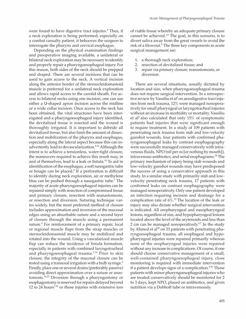
The neck was immediately prepped for emergent surgical airway and neck exploration. A large 7-cm tra-cheal laceration and esophageal perforation were iden-tified (Figure 30-2). The distal portion of the trachea
was secured and intubated with an endotracheal tube. A formal neck exploration was then performed. Hemorrhage was controlled by clamping and tying off bleeding vessels. Bilateral neck exploration into the carotid sheaths confirmed no bleeding or hematoma. A formal tracheostomy was created distal to the tracheal defect and brought out through a separate midline skin incision. The devitalized tissue was resected. The tra-cheal and esophageal injuries were repaired primarily in dual layers. The endotracheal tube was pulled and a Shiley tracheostomy tube (Covidien, Mansfield, MA)
408
Otolaryngology/Head and Neck Combat Casualty Care
placed into the formal tracheostomy stoma. Thorough irrigation was performed. Multiple Penrose drains were placed prior to closure.
Complications
None.
Lessons Learned
This case highlights the process for immediate surgical airway access. This patient would have likely had an unsuccessful transoral intubation because of significant laryngotracheal injury, resulting in delayed airway access. The patient had no postoperative com-plications partly because of immediate exploration and repair.
Case Study 30-2
Presentation
A 22-year-old US Army dog handler was shot in the right shoulder with a suspected AK-47. The trajectory of the bullet continued through the right neck (zone II) across the midline and exited on the left (zone III). The patient was stable on arrival at the trauma bay. There was blood coming from the oropharynx and subcutaneous emphysema on examination.
Preoperative Workup/Radiology
The CT angiogram showed no great vessel injury, moderate subcutaneous emphysema, and a trajectory across the midline through the pharynx.
Operative Planning/Timing of Surgery
Because the patient was stable, the CT angiogram was performed prior to surgical intervention. Physical examination and CT scan showed signs of a pharyn-goesophageal injury, so he was taken to the operating room for panendoscopy and neck exploration.
Operation
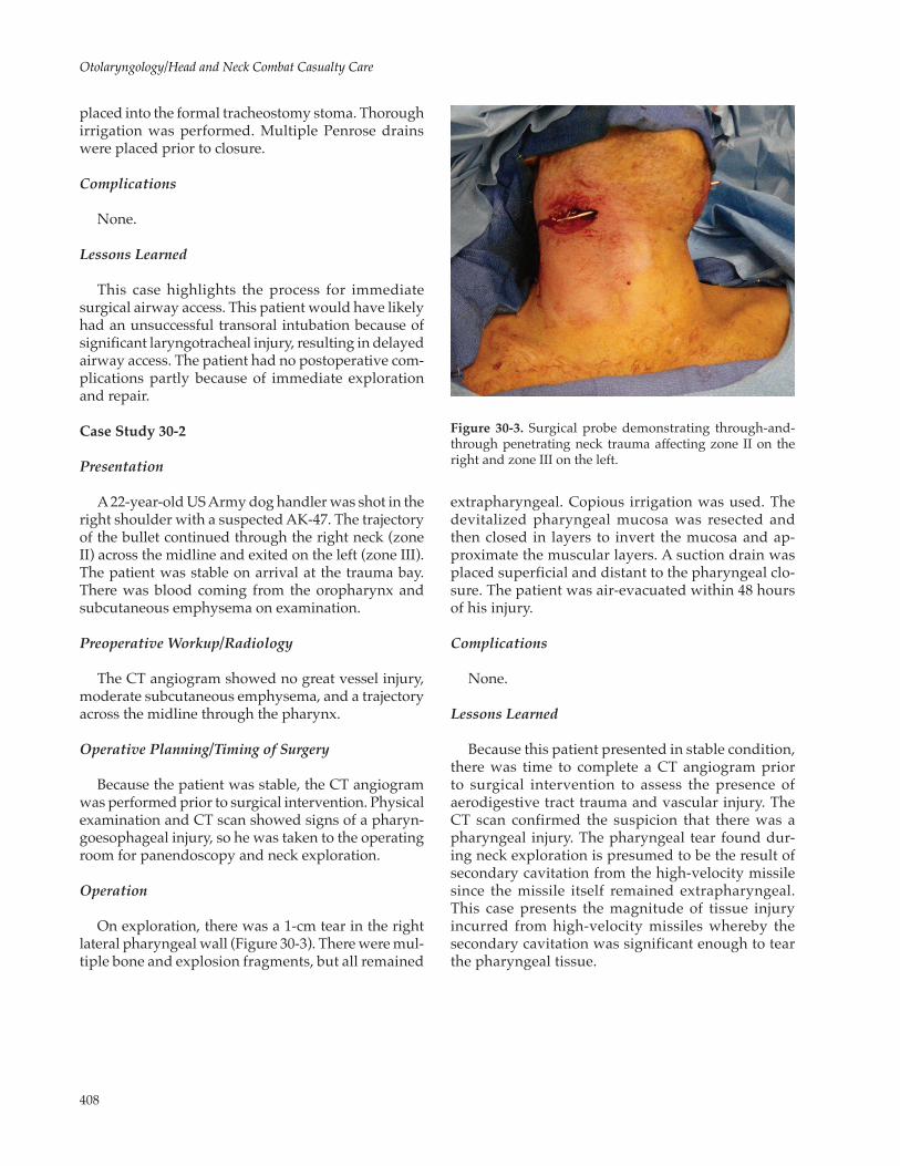
On exploration, there was a 1-cm tear in the right lateral pharyngeal wall (Figure 30-3). There were mul-tiple bone and explosion fragments, but all remained
extrapharyngeal. Copious irrigation was used. The devitalized pharyngeal mucosa was resected and then closed in layers to invert the mucosa and ap-proximate the muscular layers. A suction drain was placed superficial and distant to the pharyngeal clo-sure. The patient was air-evacuated within 48 hours of his injury.
Complications
None.
Lessons Learned
Because this patient presented in stable condition, there was time to complete a CT angiogram prior to surgical intervention to assess the presence of aerodigestive tract trauma and vascular injury. The CT scan confirmed the suspicion that there was a pharyngeal injury. The pharyngeal tear found dur-ing neck exploration is presumed to be the result of secondary cavitation from the high-velocity missile since the missile itself remained extrapharyngeal. This case presents the magnitude of tissue injury incurred from high-velocity missiles whereby the secondary cavitation was significant enough to tear the pharyngeal tissue.
Figure 30-3. Surgical probe demonstrating through-and-through penetrating neck trauma affecting zone II on the right and zone III on the left.
409
Acute Management of Pharyngoesophageal Trauma
REFERENCES
1. McConnell DB, Trunkey DD. Management of penetrating trauma to the neck. Adv Surg. 1994;27:97–127.
2. Vassiliu P, Baker J, Henderson S, Alo K, Velmahos G, Demetriades D. Aerodigestive injuries of the neck. Am Surg. 2001;67:75–79.
3. Breeze J, Masterson L, Banfield, G. Outcomes from penetrating ballistic cervical injury. J R Army Med Corps. 2012;158:96–100.
4. Brennan J, Lopez M, Gibbons MD, et al. Penetrating neck trauma in Operation Iraqi Freedom. Otol Head Neck Surg. 2011;144:180–185.
5. Soliman AMS, Ahmad SM, Roy D. The role of aerodigestive tract endoscopy in penetrating neck trauma. Laryngoscope. 2012;Oct 15.doi:10.1002/lary.23611. [Epub ahead of print.]
6. Kesser BW, Chance E, Kleiner D, et al. Contemporary management of penetrating neck trauma. Am Surg. 2009;75:1–10.
7. Stanley RB, Armstrong WB, Fetterman BL, Shindo ML. Management of external penetrating injuries into the hypo-pharyngeal-cervical esophageal funnel. J Trauma. 1997;42:675–679.
8. Asensio JA, Chahwan S, Forno W, et al. Penetrating esophageal injuries: multicenter study of the American Associa-tion for the Surgery of Trauma. J Trauma. 2001;50:289–296.
9. Weigelt JA, Thal ER, Snyder WH III, et al. Diagnosis of penetrating cervical esophageal injuries. Am J Surg. 1987;154:619–622.
10. Demetriades D, Theodorou D, Cornwell E, et al. Evaluation of penetrating injuries of the neck: prospective study of 223 patients. World J Surg. 1997;21:41–48.
11. Eroglu A, Kurkcuoglu IC, Karaglanoglu N, Tekinbas C, Yimaz O, Basoglu M. Esophageal perforation: the importance of early diagnosis and primary repair. Dis Esophagus. 2004;17:91–94.
12. Burgess CA, Dale OT, Almeyda R, Corbridge RJ. An evidence based review of the assessment and management of penetrating neck trauma. Clin Otolaryngol. 2012;37:44–52.
13. Goudy SL, Miller FB, Bumpous JM. Neck crepitance: evaluation and management of suspected upper aerodigestive tract injury. Laryngoscope. 2002;112:791–795.
14. Back MR, Baumgartner FJ, Klein SR. Detection and evaluation of aerodigestive tract injuries caused by cervical and transmediastinal gunshot wounds. J Trauma. 1997;42:680–686.
15. Flowers JL, Graham SM, Ugarte MA, et al. Flexible endoscopy for the diagnosis of esophageal trauma. J Trauma. 1996;40:261–265.
16. Armstrong WB, Detar TR, Stanley RB. Diagnosis and management of external penetrating cervical esophageal injuries. Ann Otol Rhinol Laryngol. 1994;103:863–871.
17. Yap RG, Yap AG, Obeid FN, et al. Traumatic esophageal injuries: 12 year experience at Henry Ford Hospital. J Trauma. 1984;24:623–625.
18. Ordog GJ, Albin D, Wasserberger J, Schlater TL, Balasubramaniam S. 110 bullet wounds to the neck. J Trauma. 1985;25:238–246.
19. Noyes LD, McSwain NE, Markowitz IP. Panendoscopy with arteriograophy versus mandatory exploration of penetrat-ing wounds of the neck. Ann Surg. 1986;204:21–31.
410
Otolaryngology/Head and Neck Combat Casualty Care
20. Ahmed N, Massier C, Tassie J, et al. Diagnosis of penetrating injuries of the pharynx and esophagus in the severely injured patient. J Trauma. 2009;67:152–154.
21. Inaba K, Munera F, McKenney M, et al. Prospective evaluation of screening multislice helical computed tomographic angiography in the initial evaluation of penetrating neck injuries. J Trauma. 2006;61:144–149.
22. Kazi M, Junaid M, Khan MJ, Ali NS, Masoom A. Utility of clinical examination and CT scan in assessment of penetrat-ing neck trauma. J Coll Physicians Surg Pak. 2013;23:308–309.
23. Shiroff AM, Gale SC, Martin ND, et al. Penetrating neck trauma: a review of management strategies and discussion of the “no zone” approach. Am Surg. 2013;79:23–29.
24. Ngakane H, Muckart DJJ, Luvuno FM. Penetrating visceral injuries of the neck: results of a conservative management policy. Br J Surg. 1990;77:908–910.
25. Madiba TE, Muckart DJJ. Penetrating injuries to the cervical oesophagus: is routine exploration mandatory? Ann R Coll Surg Engl. 2003;85:162–166.
26. Biffl WL, Moore EE, Dagmar H, et al. Selective management of penetrating neck trauma based on cervical level of injury. Am J Surg. 1997;174:678–682.
27. Feliciano DV, Bitondo CC, Mattox KL, et al. Combined tracheoesophageal injuries. Am J Surg. 1985;150:710–715.
28. Richardson JD, Martin LF, Borzotta AP, Polk HC. Unifying concepts in treatment of esophageal leaks. Am J Surg. 1985;149:157–162.


![RESEARCH Open Access Pharyngoesophageal reconstruction … · 2017. 8. 27. · reconstruction techniques, the complication rates are still relatively high [5]. Many studies declared](https://img.pdfslide.us/doc/110x75/60d4ac94215b5468f50ec20b/research-open-access-pharyngoesophageal-reconstruction-2017-8-27-reconstruction.jpg)