Embed Size (px)
Citation preview
Dr. Bruce Forciea Page 41
Chapter 3
Cells
Dr. Bruce Forciea Page 42
Cells Cells are a major part of our bodies. In this section we will review the major parts of a cell and investigate cellular transport mechanisms. The human body has something on the order of 10 trillion cells all working in harmony to keep us alive. Cells are fundamental building blocks for many of the tissues and organs of our bodies. In this section we will primarily be concerned with studying cells that contain a nucleus known as eukaryotic cells. The lowest level of organization was the atom followed by molecules and tissues. Then there were organelles and cells. So cells contain smaller structures called organelles. These are much like the organs in our bodies. The organelles have various functions that are important in maintaining the cell (fig. 3.1).

Dr. Bruce Forciea Page 43
Let’s look at a few of the major parts of the cell and its organelles.
Figure 3.1. The cell contains a variety of organelles
http://commons.wikimedia.org/wiki/Image:Animal_cell_structure_en.svg

Dr. Bruce Forciea Page 44
Cell Membrane We will start by looking at the cell membrane. The structure of the cell membrane has a lot to do with its function. The cell membrane is composed of molecules called phospholipids (fig. 3.2). The phosphate head of the phospholipid likes water so it is called hydrophilic while the lipid tail is called hydrophopic or “water hating.” Because of the water loving and hating characteristics of the heads and tails, phospholipids arrange themselves in what is known as a bilayer (fig. 3.3).
Figure 3.2. Phospholipids contain a phosphate head and a lipid tail.
http://commons.wikimedia.org/wiki/Image:Phospholipid_structure.png
Dr. Bruce Forciea Page 45
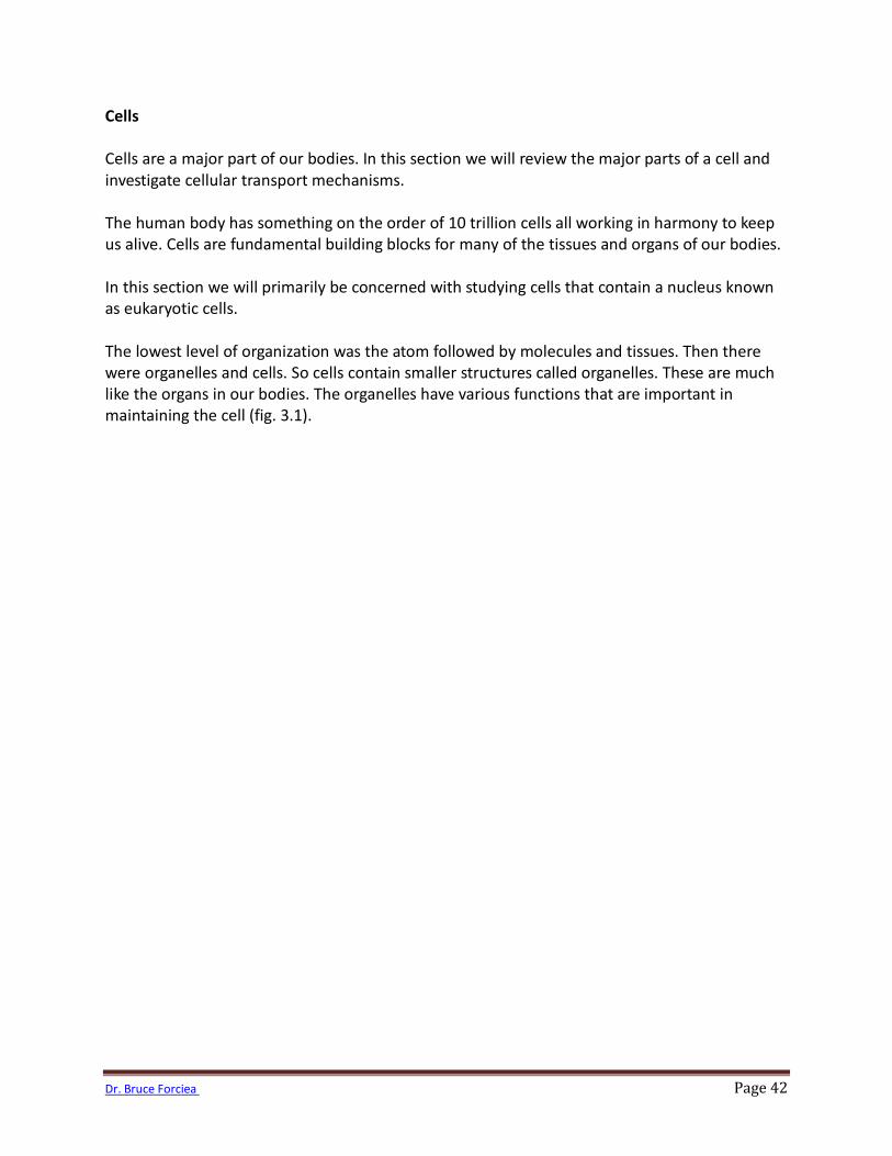
The cell membrane also lets certain substances in or out. We say that it is selectively permeable. For example, lipid soluble substances can pass right through the cell membrane. Examples of lipid soluble substances include oxygen, carbon dioxide and steroids. Water soluble substances cannot pass through the cell membrane and require carrier proteins in order to get in or out of the cell. The cell membrane also contains a number of proteins. Some of these proteins are imbedded on the surface of the cell and some go all the way through the cell membrane. Some proteins act as channels to allow substances to pass through the membrane. Others act as receptors that receive information carried by proteins. Still others act as connection points for other cells to attach. These are known as intercellular junctions. Let’s look at some of the other parts of a cell.
Figure 3.3. Phopholipids arrange themselves into a bilayer.
http://commons.wikimedia.org/wiki/Image:Fluid_Mosaic.svg
Jerome Walker

Dr. Bruce Forciea Page 46
Cytoplasm The cytoplasm or cytosol is the fluid inside the cell. It contains a network of channels and support structures called the cytoskeleton. This is much like the skeleton in your body. Endoplasmic Reticulum Another important organelle is the endoplasmic reticulum (fig. 3.4). The endoplasmic reticulum comes in two varieties; rough and smooth. Rough endoplasmic reticulum is studded with ribosomes. The ribosomes function in making proteins (protein synthesis). Smooth endoplasmic reticulum does not contain ribosomes. It functions in making lipids (lipid synthesis). Ribosomes contain RNA, protein and the enzymes needed for protein synthesis.
Figure 3.4. The endoplasmic reticulum (3) contains ribosomes (5) that function in making proteins. The Golgi apparatus (11)then packages the proteins in vesicles (12). http://commons.wikimedia.org/wiki/Image:Nucleus_ER_golgi.jpg
Dr. Bruce Forciea Page 47
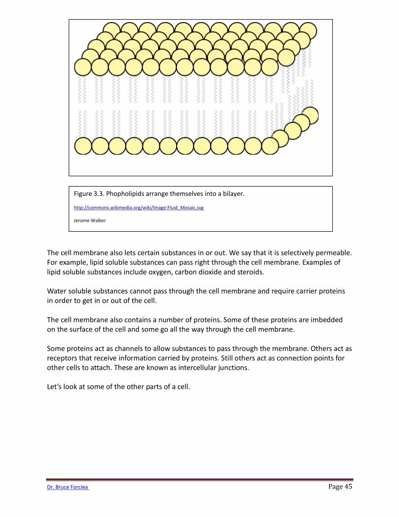
After the endoplasmic reticulum synthesizes the proteins they need to be packed up and shipped out to other parts of the cell or to other cells. That’s where the Golgi apparatus takes over. The Golgi apparatus packs up the proteins. Besides the vesicles from the Golgi apparatus, there are other vesicles containing enzymes for breaking up debris in the cell. These are called lysosomes. Mitochondrion The next organelle we will investigate is very important because it produces energy that is needed throughout the body. It is known as the “powerhouse” of the cell and is called the mitochondrion (fig. 3.5). The mitochondrion takes in fuel such as glucose and extracts the energy from it to make ATP. The inner portion of the mitochondrion is folded into shelves called cristae. These are studded with enzymes needed for the many chemical reactions used to make ATP.
Figure 3.5. The mitochondrion.
http://commons.wikimedia.org/wiki/Image:Diagram_of_an_animal_mitochondrion.svg
Dr. Bruce Forciea Page 48
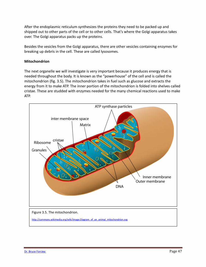
Centrosome The centrosome is important in producing a structure called the mitotic spindle that helps to separate the chromosomes during mitosis. The centrosome consists of 2 hollow cylinders called centrioles. The centrioles are constructed from tubular proteins (fig. 3.6).
Figure 3.6. This is a picture of a centriole. Notice the circular structure in the lower right-hand corner. The centriole consists of tubular proteins.
http://commons.wikimedia.org/wiki/Image:Spindle_centriole_-_embryonic_brain_mouse_-_TEM.jpg

Dr. Bruce Forciea Page 49
Cilia and Flagella The cell contains other protein structures called cilia and flagella. Cilia and flagella are important in cellular movements (fig. 3.7). Cilia are protein structures that move substances across cells. A flagellum is a long protein structure that moves the cell. Cells may have many cilia but will only have one flagellum.
Figure 3.7. Notice the hair-like structures on B, E, H and I. These are cilia. Cilia can move substances along the surface of cells.
http://commons.wikimedia.org/wiki/Image:Tkanka_nablonkowa.png
Dr. Bruce Forciea Page 50
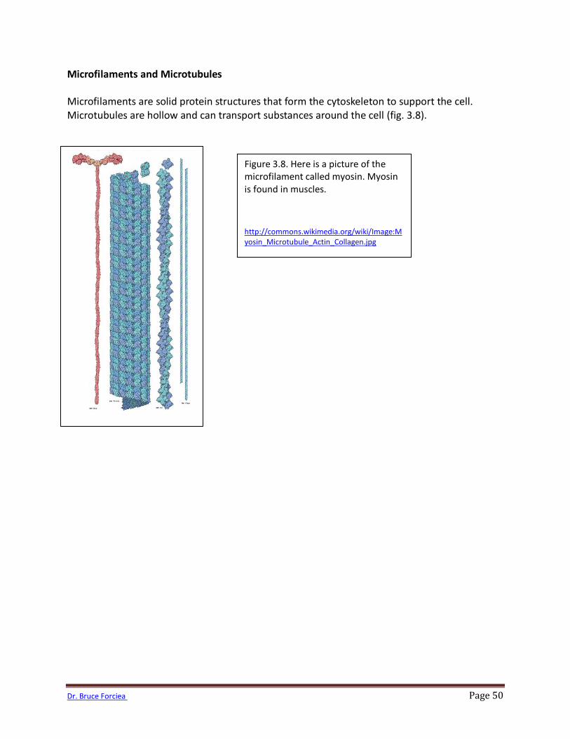
Microfilaments and Microtubules Microfilaments are solid protein structures that form the cytoskeleton to support the cell. Microtubules are hollow and can transport substances around the cell (fig. 3.8).
Figure 3.8. Here is a picture of the microfilament called myosin. Myosin is found in muscles.
http://commons.wikimedia.org/wiki/Image:Myosin_Microtubule_Actin_Collagen.jpg
Dr. Bruce Forciea Page 51
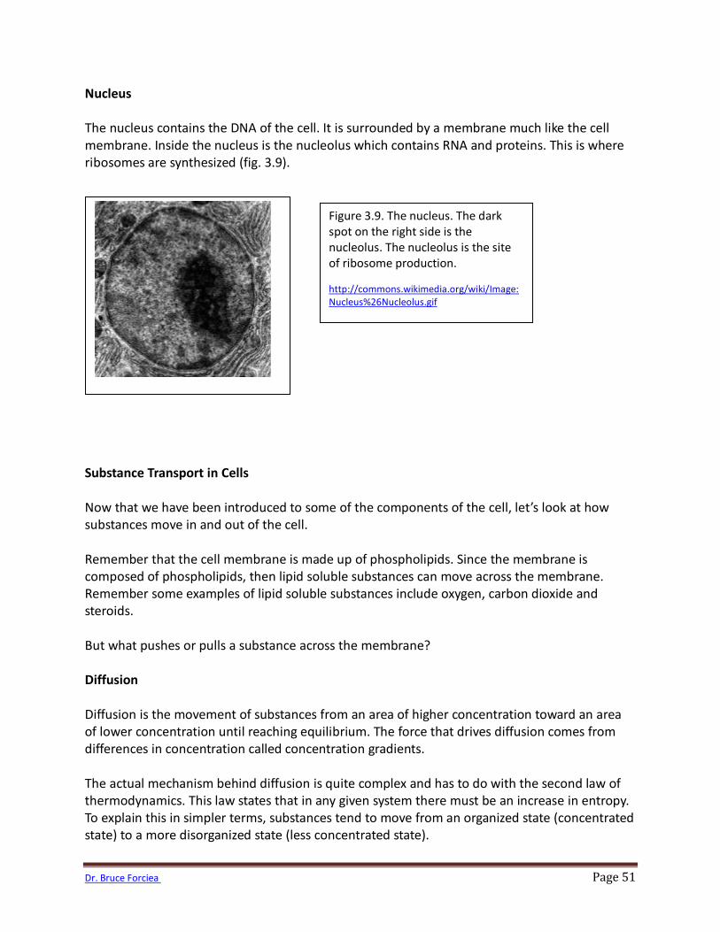
Nucleus The nucleus contains the DNA of the cell. It is surrounded by a membrane much like the cell membrane. Inside the nucleus is the nucleolus which contains RNA and proteins. This is where ribosomes are synthesized (fig. 3.9). Substance Transport in Cells Now that we have been introduced to some of the components of the cell, let’s look at how substances move in and out of the cell. Remember that the cell membrane is made up of phospholipids. Since the membrane is composed of phospholipids, then lipid soluble substances can move across the membrane. Remember some examples of lipid soluble substances include oxygen, carbon dioxide and steroids. But what pushes or pulls a substance across the membrane? Diffusion Diffusion is the movement of substances from an area of higher concentration toward an area of lower concentration until reaching equilibrium. The force that drives diffusion comes from differences in concentration called concentration gradients. The actual mechanism behind diffusion is quite complex and has to do with the second law of thermodynamics. This law states that in any given system there must be an increase in entropy. To explain this in simpler terms, substances tend to move from an organized state (concentrated state) to a more disorganized state (less concentrated state).
Figure 3.9. The nucleus. The dark spot on the right side is the nucleolus. The nucleolus is the site of ribosome production.
http://commons.wikimedia.org/wiki/Image:Nucleus%26Nucleolus.gif
Dr. Bruce Forciea Page 52
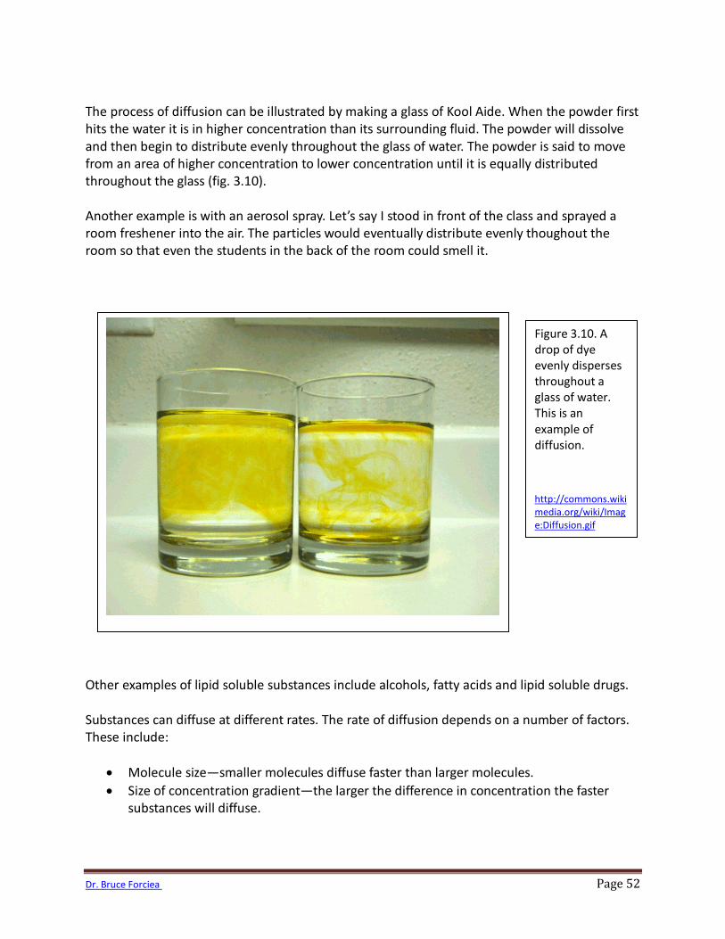
The process of diffusion can be illustrated by making a glass of Kool Aide. When the powder first hits the water it is in higher concentration than its surrounding fluid. The powder will dissolve and then begin to distribute evenly throughout the glass of water. The powder is said to move from an area of higher concentration to lower concentration until it is equally distributed throughout the glass (fig. 3.10). Another example is with an aerosol spray. Let’s say I stood in front of the class and sprayed a room freshener into the air. The particles would eventually distribute evenly thoughout the room so that even the students in the back of the room could smell it. Other examples of lipid soluble substances include alcohols, fatty acids and lipid soluble drugs. Substances can diffuse at different rates. The rate of diffusion depends on a number of factors. These include:
• Molecule size—smaller molecules diffuse faster than larger molecules. • Size of concentration gradient—the larger the difference in concentration the faster
substances will diffuse.
Figure 3.10. A drop of dye evenly disperses throughout a glass of water. This is an example of diffusion.
http://commons.wikimedia.org/wiki/Image:Diffusion.gif
Dr. Bruce Forciea Page 53
• Temperature—because diffusion relies on the movement of molecules, higher temperatures will cause more movement and speed up diffusion. For example, substances will diffuse faster at body temperature than room temperature.
• Distance—the shorter the distance, the faster substances will diffuse. • Electrical forces—Cells generally carry a negative charge on the inside. Negative charges
will attract positive electrolytes and repel negative ones. This can speed up or slow down the rate of diffusion.
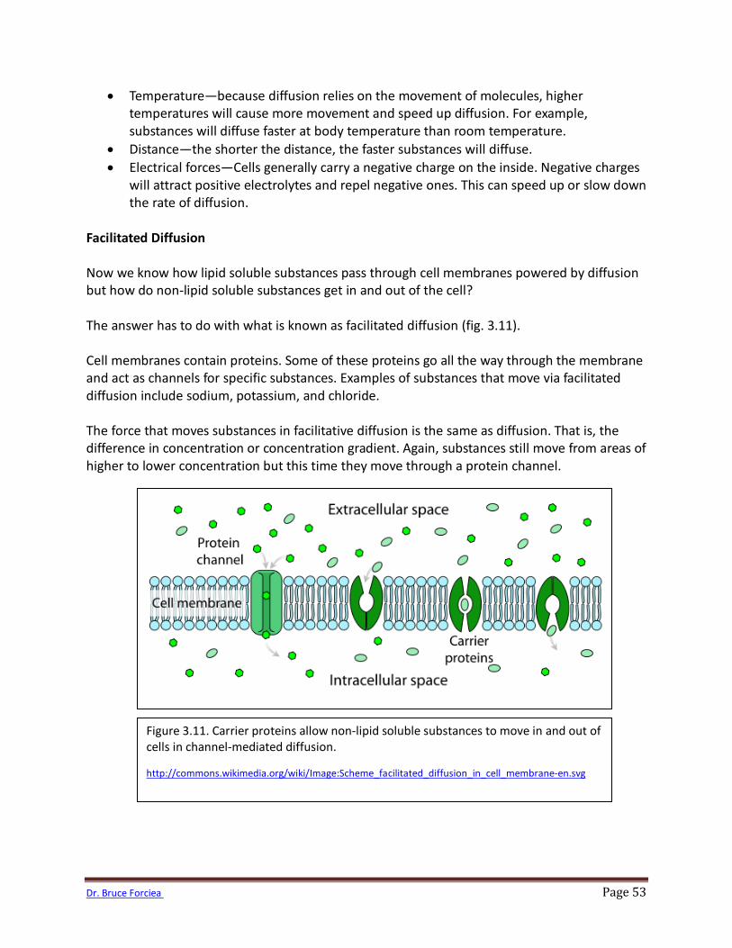
Facilitated Diffusion Now we know how lipid soluble substances pass through cell membranes powered by diffusion but how do non-lipid soluble substances get in and out of the cell? The answer has to do with what is known as facilitated diffusion (fig. 3.11). Cell membranes contain proteins. Some of these proteins go all the way through the membrane and act as channels for specific substances. Examples of substances that move via facilitated diffusion include sodium, potassium, and chloride. The force that moves substances in facilitative diffusion is the same as diffusion. That is, the difference in concentration or concentration gradient. Again, substances still move from areas of higher to lower concentration but this time they move through a protein channel.
Figure 3.11. Carrier proteins allow non-lipid soluble substances to move in and out of cells in channel-mediated diffusion.
http://commons.wikimedia.org/wiki/Image:Scheme_facilitated_diffusion_in_cell_membrane-en.svg
Dr. Bruce Forciea Page 54
Substances moving in and out of cells by facilitated diffusion must bind to receptors on the protein channel. Once they bind, the protein changes its shape allowing the substance in or out of the cell. Since proteins only have so many receptors, once the receptors become saturated there cannot be movement of any additional substances. Therefore in some cases a larger concentration gradient will not move substances at a faster rate (unlike diffusion). The rate of diffusion then partially depends on the saturation of the receptors on the protein channel. One example of a substance transported into the cell via facilitated diffusion is glucose. Glucose is used by cells to make ATP, an important energy molecule in the body. Muscle cells require glucose to make ATP for muscle contraction. In order for glucose to move into a muscle cell it not only needs to connect to a receptor on the protein channel but another hormone called insulin also needs to connect to a special insulin receptor on the protein. In some cases the insulin receptors become resistant to insulin causing blood glucose levels to rise. This occurs in what is known as insulin resistant diabetes. We will learn more about diabetes in a later chapter. Osmosis Water moves within the human body across a variety of membranes. The membranes are called semipermeable because they only allow water to move across them, not solute. The movement of water across a semipermeable membrane has a special name; osmosis. Water moves like every other substance in our universe, from an area of higher concentration to lower concentration. However, we typically do not talk about concentration in terms of water. We usually talk about concentration in terms of solute. So you could think of osmosis in 2 ways:
1. Water moves across a semipermeable membrane from a higher area of concentration of water to a lower concentration of water.
2. Water moves across a semipermeable membrane from an area of lower concentration of solute to an area of higher concentration of solute.
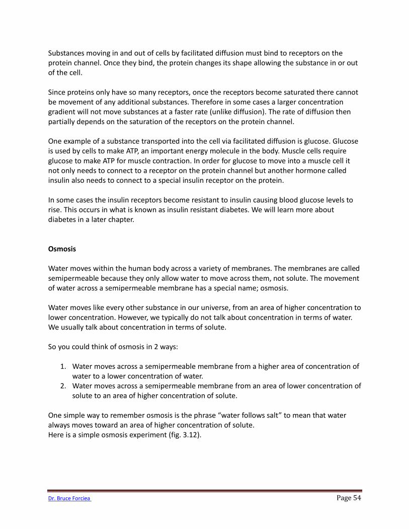
One simple way to remember osmosis is the phrase “water follows salt” to mean that water always moves toward an area of higher concentration of solute. Here is a simple osmosis experiment (fig. 3.12).
Dr. Bruce Forciea Page 55
Isotonic/Hypotonic/Hypertonic Solutions The force exhibited by osmosis is called osmotic pressure. This pressure is related to the solute concentration of the solution. In chemistry we describe concentration in terms of osmolarity. However, in physiology when we are concerned with concentration with regard to cells we use the term tonicity. Tonicity is related to the human cell whereby osmolarity is the number of osmoles per liter. Osmolarity depends on the number of particles of solute. For example, one mole of glucose in water would equate to 1 osmole since glucose remains as 1 molecule in water. However, one mole of sodium chloride would equate to 2 osmoles because sodium chloride dissociates in water to form 2 particles. If a solution has the same osmolarity as body fluids we say the solution is isotonic. The human body’s osmolarity is close to .30 osmoles or 300 milliosmoles.
Figure 3.12. Water moves toward an area of higher solute concentration in osmosis.
Dr. Bruce Forciea Page 56
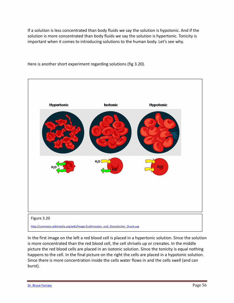
If a solution is less concentrated than body fluids we say the solution is hypotonic. And if the solution is more concentrated than body fluids we say the solution is hypertonic. Tonicity is important when it comes to introducing solutions to the human body. Let’s see why. Here is another short experiment regarding solutions (fig 3.20). In the first image on the left a red blood cell is placed in a hypertonic solution. Since the solution is more concentrated than the red blood cell, the cell shrivels up or crenates. In the middle picture the red blood cells are placed in an isotonic solution. Since the tonicity is equal nothing happens to the cell. In the final picture on the right the cells are placed in a hypotonic solution. Since there is more concentration inside the cells water flows in and the cells swell (and can burst).
Figure 3.20
http://commons.wikimedia.org/wiki/Image:Erythrozyten_und_Osmotischer_Druck.svg
Dr. Bruce Forciea Page 57
Filtration Sometimes cells arrange themselves in thin layers and substances can move between the cells. These layers or membranes work the same way as filters. Filters sort substances based on size. Smaller substances move through the spaces and larger substances do not. Think of a coffee filter. The filter has very small holes that only allow the water to move through. The grounds are too large to fit through the holes. The force that drives filtration is fluid pressure. This pressure is also known as hydrostatic pressure. In order to move substances through a filter they must move from an area of higher pressure to lower pressure. There are many examples of filters in the body. These include the capillaries and kidneys. Active Transport So far we have seen how substances move down their respective concentration gradients in diffusion and facilitative diffusion. But what if a substance needs to be moved against its concentration gradient? In active transport substances are moved against their concentration gradients by carrier proteins. However, there is an energy cost to be paid for this action. So the carrier proteins use ATP as an energy source. An example of an active transport protein is the sodium potassium pump (fig. 3.21). Normally there is more sodium outside of the cell than in so sodium would move from outside to in. Also, there is usually more potassium inside the cell than out, so potassium would follow its concentration gradient and move out of the cell. However, we want to move these molecules against their concentration gradients. So this can be done but energy must be used to do so. Energy is used by the pump in the form of ATP.
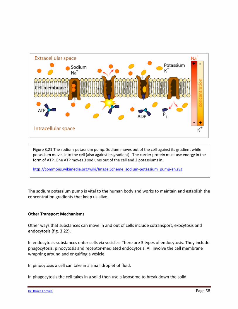
Dr. Bruce Forciea Page 58
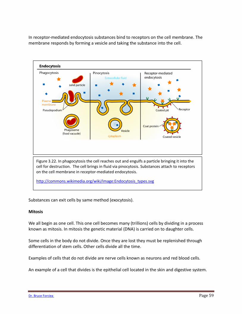
The sodium potassium pump is vital to the human body and works to maintain and establish the concentration gradients that keep us alive. Other Transport Mechanisms Other ways that substances can move in and out of cells include cotransport, exocytosis and endocytosis (fig. 3.22). In endocytosis substances enter cells via vesicles. There are 3 types of endocytosis. They include phagocytosis, pinocytosis and receptor-mediated endocytosis. All involve the cell membrane wrapping around and engulfing a vesicle. In pinocytosis a cell can take in a small droplet of fluid. In phagocytosis the cell takes in a solid then use a lysosome to break down the solid.
Figure 3.21.The sodium-potassium pump. Sodium moves out of the cell against its gradient while potassium moves into the cell (also against its gradient). The carrier protein must use energy in the form of ATP. One ATP moves 3 sodiums out of the cell and 2 potassiums in.
http://commons.wikimedia.org/wiki/Image:Scheme_sodium-potassium_pump-en.svg
Dr. Bruce Forciea Page 59
In receptor-mediated endocytosis substances bind to receptors on the cell membrane. The membrane responds by forming a vesicle and taking the substance into the cell. Substances can exit cells by same method (exocytosis). Mitosis We all begin as one cell. This one cell becomes many (trillions) cells by dividing in a process known as mitosis. In mitosis the genetic material (DNA) is carried on to daughter cells. Some cells in the body do not divide. Once they are lost they must be replenished through differentiation of stem cells. Other cells divide all the time. Examples of cells that do not divide are nerve cells known as neurons and red blood cells. An example of a cell that divides is the epithelial cell located in the skin and digestive system.
Figure 3.22. In phagocytosis the cell reaches out and engulfs a particle bringing it into the cell for destruction. The cell brings in fluid via pinocytosis. Substances attach to receptors on the cell membrane in receptor-mediated endocytosis.
http://commons.wikimedia.org/wiki/Image:Endocytosis_types.svg
![STEM CELLS EMBRYONIC STEM CELLS/INDUCED PLURIPOTENT STEM CELLS Stem Cells.pdf · germ cell production [2]. Human embryonic stem cells (hESCs) offer the means to further understand](https://img.pdfslide.us/doc/110x75/6014b11f8ab8967916363675/stem-cells-embryonic-stem-cellsinduced-pluripotent-stem-cells-stem-cellspdf.jpg)