Embed Size (px)
Citation preview

65
Chapter 3
Cardiovascular system
PLR3 12/7/04 8:16 PM Page 65
Chapter 3 Cardiovascular system
66
Figure 3.1 Ultrasound of the heart showing all four chambers.
Cardiovascular investigations
Plain films
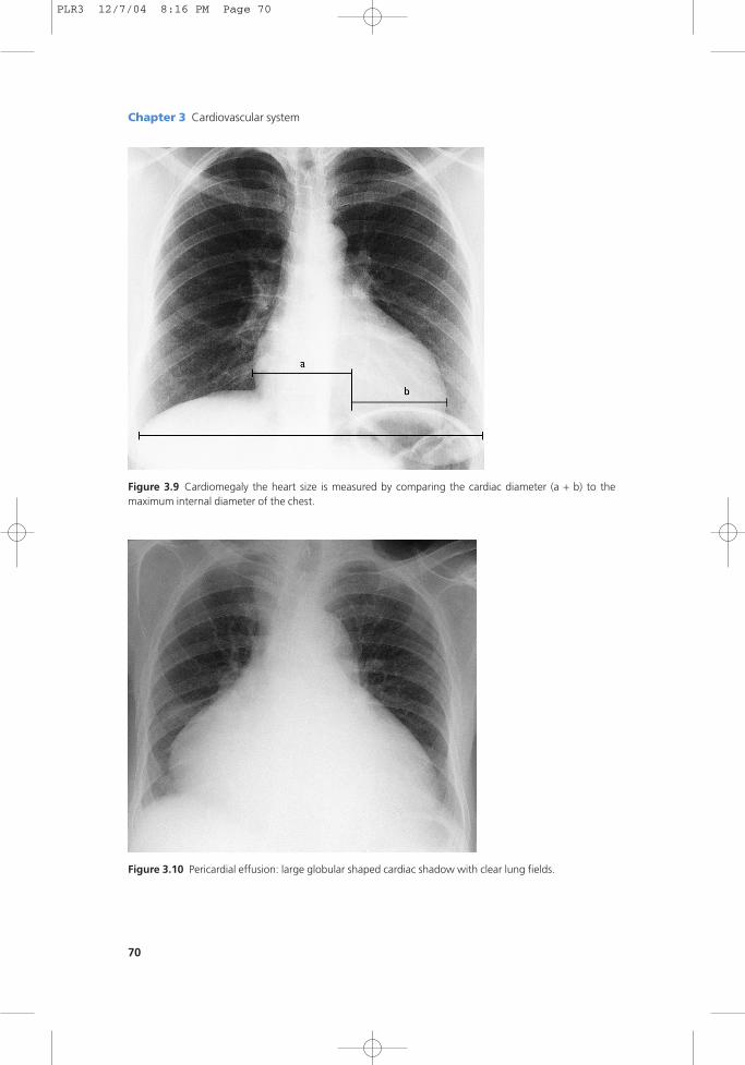
Evaluate heart size and chamber enlargement. On a standard PA chest projection, the ratio
of the cardiac diameter to that of the maximum internal diameter of the chest should be no
greater than 50% on a full inspiratory film. Expiratory films may falsely give the impression
of cardiomegaly and pulmonary congestion. Supine films may also give a similar
appearance.
Ultrasound
Echocardiography and Doppler examination reveal anatomical abnormalities as well as flow
disturbances and assist in the study of incompetent and stenotic valves and ventricular func-
tion; aortic arch aneurysms, dissecting aneurysms, cardiomyopathy and pericardial effu-
sions can also be diagnosed using echocardiography.
PLR3 12/7/04 8:16 PM Page 66
Isotope scanning
Technetium-99m pyrophosphate accumulates in damaged myocardium whereas thallium-
201 produces a deficient uptake in territories supplied by occluded or narrowed arteries.
Thallium is most commonly used as a screening technique in patients with suspected cor-
onary artery disease.
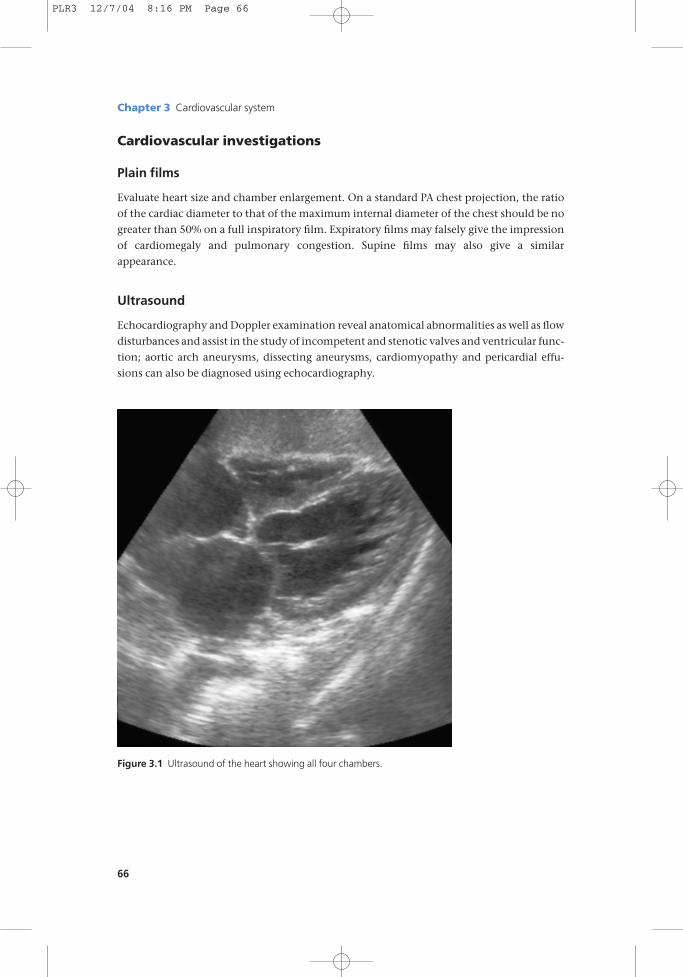
Computed tomography (CT)
Relevant applications include the further evaluation and diagnosis of dissecting thoracic
aneurysms, pericardial effusions and myocardial tumours. A recent non-invasive applica-
tion of CT calculates the total calcium deposition in the coronary arteries giving a predictive
value of coronary artery disease.
Cardiovascular system Chapter 3
67
Figure 3.2 CT abdomen. Leaking abdominal aortic aneurysm with periaortic haematoma (arrow).
Figure 3.3 Dissection of the descendingaorta. Note the intimal flap in the contrast-filled aorta (arrow) and blood inthe mediastinum.
PLR3 12/7/04 8:16 PM Page 67
Chapter 3 Cardiovascular system
68
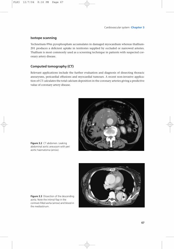
Figure 3.4 Right-upper limb venography showing thrombusin the axillary and subclavian veins (arrows) with a large collat-eral circulation.
Magnetic resonance imaging (MRI)
MRI can be gated to the cardiac cycle to reduce motion artefact. It can examine the heart in
any plane and is of value in many clinical situations including pericardial effusions, hyper-
trophic cardiomyopathy, and congenital and valvular heart disease. Magnetic resonance an-
giography (MRA) has the capability of providing a non-invasive method of imaging many
vascular abnormalities such as aneurysm, dissection, stenoses, occlusions and congenital
anomalies.
Venography
The venous system can be studied by proximal contrast injection. The most common indi-
cations are injection into:● a foot vein to look at the lower limb venous system;● antecubital vein to assess the axillary and subclavian vein and the superior vena cava;● femoral vein to study the inferior vena cava.
PLR3 12/7/04 8:16 PM Page 68
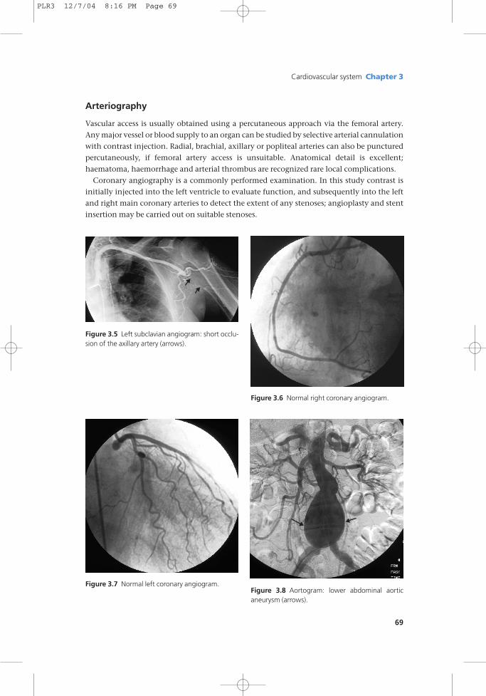
Figure 3.6 Normal right coronary angiogram.
Figure 3.7 Normal left coronary angiogram.Figure 3.8 Aortogram: lower abdominal aorticaneurysm (arrows).
Cardiovascular system Chapter 3
69
Arteriography
Vascular access is usually obtained using a percutaneous approach via the femoral artery.
Any major vessel or blood supply to an organ can be studied by selective arterial cannulation
with contrast injection. Radial, brachial, axillary or popliteal arteries can also be punctured
percutaneously, if femoral artery access is unsuitable. Anatomical detail is excellent;
haematoma, haemorrhage and arterial thrombus are recognized rare local complications.
Coronary angiography is a commonly performed examination. In this study contrast is
initially injected into the left ventricle to evaluate function, and subsequently into the left
and right main coronary arteries to detect the extent of any stenoses; angioplasty and stent
insertion may be carried out on suitable stenoses.
Figure 3.5 Left subclavian angiogram: short occlu-sion of the axillary artery (arrows).
PLR3 12/7/04 8:16 PM Page 69
Chapter 3 Cardiovascular system
70
Figure 3.9 Cardiomegaly the heart size is measured by comparing the cardiac diameter (a + b) to the maximum internal diameter of the chest.
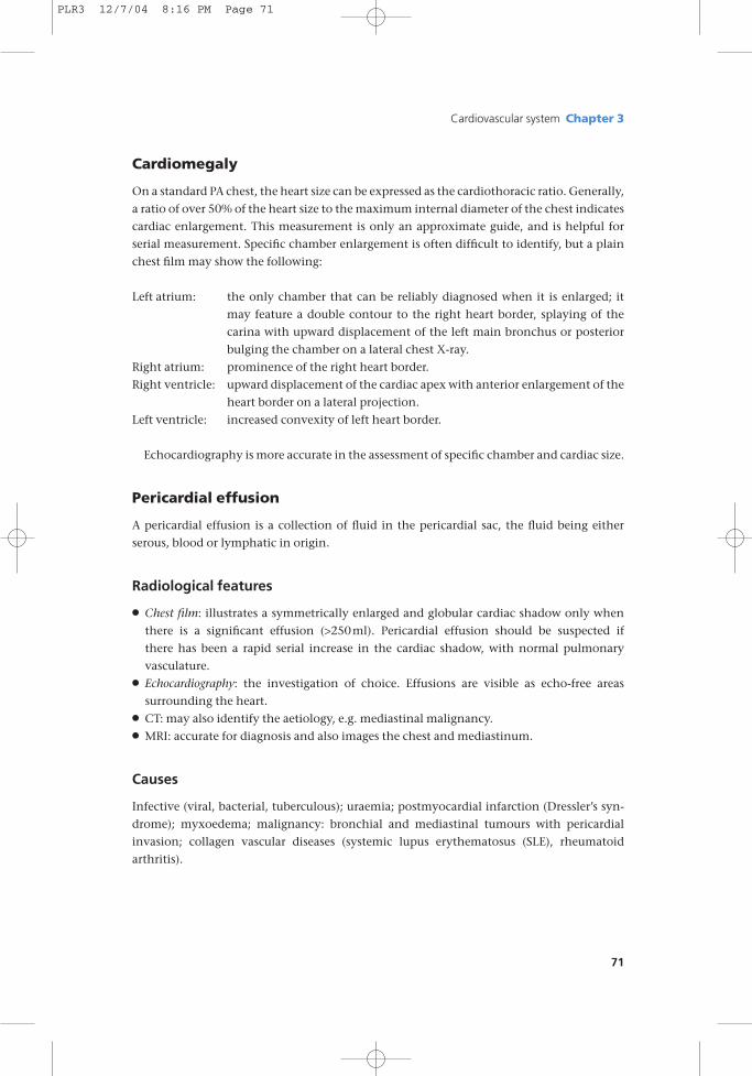
Figure 3.10 Pericardial effusion: large globular shaped cardiac shadow with clear lung fields.
PLR3 12/7/04 8:16 PM Page 70
Cardiomegaly
On a standard PA chest, the heart size can be expressed as the cardiothoracic ratio. Generally,
a ratio of over 50% of the heart size to the maximum internal diameter of the chest indicates
cardiac enlargement. This measurement is only an approximate guide, and is helpful for
serial measurement. Specific chamber enlargement is often difficult to identify, but a plain
chest film may show the following:
Left atrium: the only chamber that can be reliably diagnosed when it is enlarged; it
may feature a double contour to the right heart border, splaying of the
carina with upward displacement of the left main bronchus or posterior
bulging the chamber on a lateral chest X-ray.
Right atrium: prominence of the right heart border.
Right ventricle: upward displacement of the cardiac apex with anterior enlargement of the
heart border on a lateral projection.
Left ventricle: increased convexity of left heart border.
Echocardiography is more accurate in the assessment of specific chamber and cardiac size.
Pericardial effusion
A pericardial effusion is a collection of fluid in the pericardial sac, the fluid being either
serous, blood or lymphatic in origin.
Radiological features
● Chest film: illustrates a symmetrically enlarged and globular cardiac shadow only when
there is a significant effusion (>250ml). Pericardial effusion should be suspected if
there has been a rapid serial increase in the cardiac shadow, with normal pulmonary
vasculature.● Echocardiography: the investigation of choice. Effusions are visible as echo-free areas
surrounding the heart.● CT: may also identify the aetiology, e.g. mediastinal malignancy.● MRI: accurate for diagnosis and also images the chest and mediastinum.
Causes
Infective (viral, bacterial, tuberculous); uraemia; postmyocardial infarction (Dressler’s syn-
drome); myxoedema; malignancy: bronchial and mediastinal tumours with pericardial
invasion; collagen vascular diseases (systemic lupus erythematosus (SLE), rheumatoid
arthritis).
Cardiovascular system Chapter 3
71
PLR3 12/7/04 8:16 PM Page 71
Chapter 3 Cardiovascular system
72
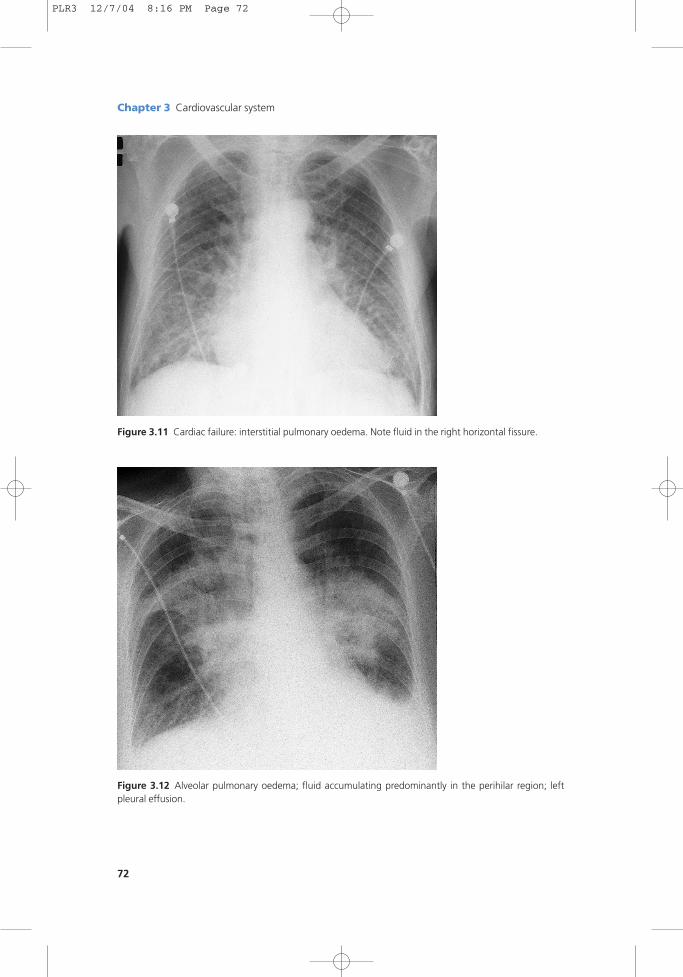
Figure 3.11 Cardiac failure: interstitial pulmonary oedema. Note fluid in the right horizontal fissure.
Figure 3.12 Alveolar pulmonary oedema; fluid accumulating predominantly in the perihilar region; leftpleural effusion.
PLR3 12/7/04 8:16 PM Page 72
Cardiac failure
Cardiac failure is said to be present when tissue demands cannot be adequately supplied by
the heart. It is usually due to low output from ischaemic heart disease but, paradoxically,
may rarely result from high output as a consequence of excessive tissue needs in conditions
such as thyrotoxicosis or Paget’s disease.
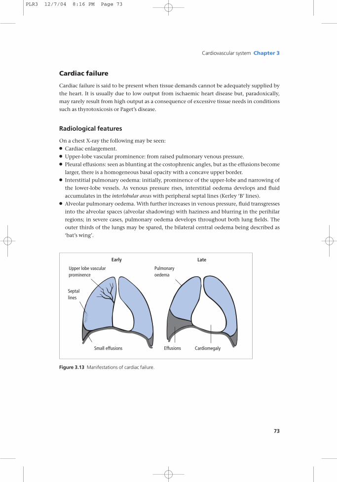
Radiological features
On a chest X-ray the following may be seen:● Cardiac enlargement.● Upper-lobe vascular prominence: from raised pulmonary venous pressure.● Pleural effusions: seen as blunting at the costophrenic angles, but as the effusions become
larger, there is a homogeneous basal opacity with a concave upper border.● Interstitial pulmonary oedema: initially, prominence of the upper-lobe and narrowing of
the lower-lobe vessels. As venous pressure rises, interstitial oedema develops and fluid
accumulates in the interlobular areas with peripheral septal lines (Kerley ‘B’ lines).● Alveolar pulmonary oedema. With further increases in venous pressure, fluid transgresses
into the alveolar spaces (alveolar shadowing) with haziness and blurring in the perihilar
regions; in severe cases, pulmonary oedema develops throughout both lung fields. The
outer thirds of the lungs may be spared, the bilateral central oedema being described as
‘bat’s wing’.
Cardiovascular system Chapter 3
73
Upper lobe vascular prominence
Septal lines
Small effusions
Pulmonary oedema
CardiomegalyEffusions
Early Late
Figure 3.13 Manifestations of cardiac failure.
PLR3 12/7/04 8:16 PM Page 73
Chapter 3 Cardiovascular system
74
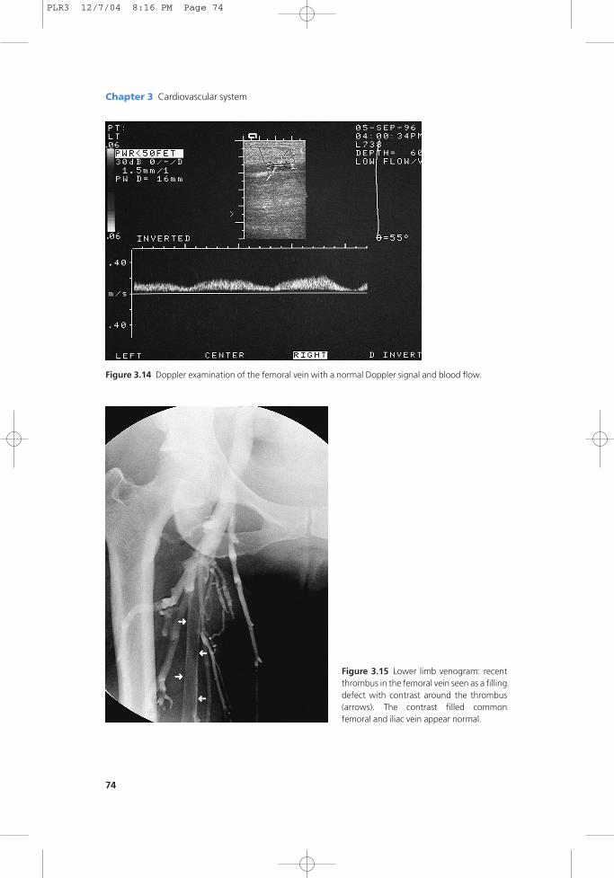
Figure 3.14 Doppler examination of the femoral vein with a normal Doppler signal and blood flow.
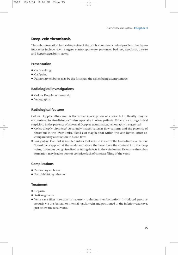
Figure 3.15 Lower limb venogram: recentthrombus in the femoral vein seen as a fillingdefect with contrast around the thrombus(arrows). The contrast filled commonfemoral and iliac vein appear normal.
PLR3 12/7/04 8:16 PM Page 74
Deep-vein thrombosis
Thrombus formation in the deep veins of the calf is a common clinical problem. Predispos-
ing causes include recent surgery, contraceptive use, prolonged bed rest, neoplastic disease
and hypercoaguability states.
Presentation
● Calf swelling.● Calf pain.● Pulmonary embolus may be the first sign, the calves being asymptomatic.
Radiological investigations
● Colour Doppler ultrasound.● Venography.
Radiological features
Colour Doppler ultrasound is the initial investigation of choice but difficulty may be
encountered in visualizing calf veins especially in obese patients. If there is a strong clinical
suspicion, in the presence of a normal Doppler examination, venography is suggested.● Colour Doppler ultrasound. Accurately images vascular flow patterns and the presence of
thrombus in the lower limbs. Blood clot may be seen within the vein lumen, often ac-
companied by a reduction in blood flow.● Venography. Contrast is injected into a foot vein to visualize the lower-limb circulation.
Tourniquets applied at the ankle and above the knee force the contrast into the deep
veins, thrombus being visualized as filling defects in the vein lumen. Extensive thrombus
formation may lead to poor or complete lack of contrast filling of the veins.
Complications
● Pulmonary embolus.● Postphlebitic syndrome.
Treatment
● Heparin.● Anticoagulants.● Vena cava filter insertion in recurrent pulmonary embolization. Introduced percuta-
neously via the femoral or internal jugular vein and positioned in the inferior vena cava,
just below the renal veins.
Cardiovascular system Chapter 3
75
PLR3 12/7/04 8:16 PM Page 75
Chapter 3 Cardiovascular system
76
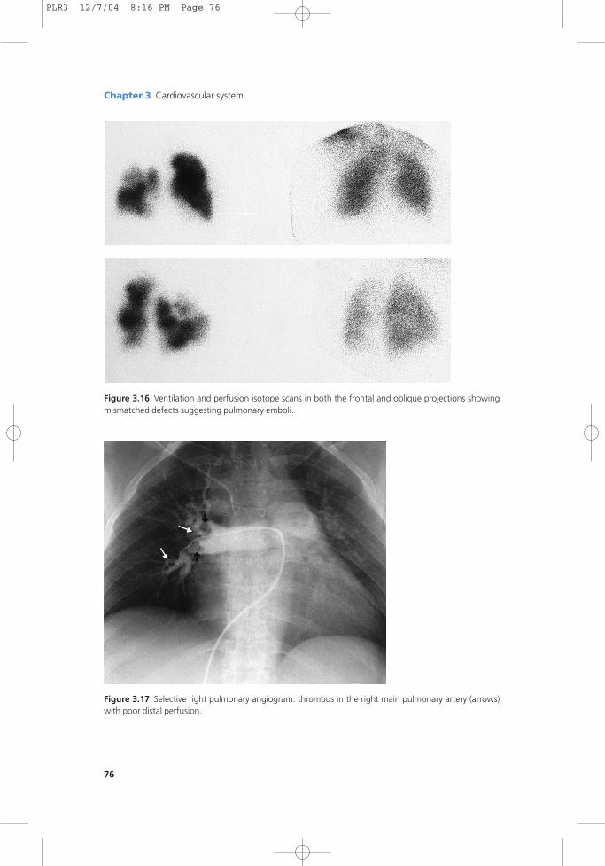
Figure 3.16 Ventilation and perfusion isotope scans in both the frontal and oblique projections showingmismatched defects suggesting pulmonary emboli.
Figure 3.17 Selective right pulmonary angiogram: thrombus in the right main pulmonary artery (arrows)with poor distal perfusion.
PLR3 12/7/04 8:16 PM Page 76
Pulmonary embolus
Pulmonary embolismPulmonary embolism occurs when a blood clot detaches from the peripheral venous system
and lodges in the pulmonary artery or its branches.
Pulmonary infarctionPulmonary infarction is the lesion that develops secondary to pulmonary embolus. Predis-
posing causes include prolonged bed rest, recent surgery, pregnancy, hypercoagulable states
and lower limb deep-vein thrombosis.
Radiological features
The blood clot usually originates from the pelvic or lower-limb veins and migrates into the
pulmonary circulation. The chest X-ray is normal in the majority of cases, but if pulmonary
infarction develops as a consequence of embolus, any of the following may be seen:● raised diaphragm;● small pleural effusions;● basal collapse or plate-like atelectasis;● consolidation, often segmental, peripherally situated and wedge-shaped.● Isotope scan. Pulmonary embolus results in a segmental defect in perfusion with preserved
ventilation (ventilation/perfusion mismatch).● CT pulmonary arteriography (CTPA). A rapid series of scans are taken through the lungs after
intravenous injection of a large bolus of contrast; emboli are seen as filling defects in the
contrast column. This is proving to be a highly accurate technique and is now often em-
ployed as the first line of investigation for a suspected pulmonary embolus.● Pulmonary angiography. This is an invasive investigation; direct contrast injection into the
pulmonary arteries reveals blood clot as intraluminal filling defects with obstruction and
attenuation of the pulmonary arterial branches. Infusion of thrombolytics through the
catheter may lyse the clot.
Complications
Pulmonary hypertension: resolves in the acute stage when thrombi disintegrate. However, it
may persist with recurrent embolization.
Types of embolism
● Fat embolism. Usually seen after severe skeletal trauma with fat globules entering the cir-
culation and obstructing pulmonary vessels.● Septic embolism. Arising from tricuspid endocarditis or infected material from central
venous pressure (CVP) lines, pacing wires, etc.● Amniotic fluid embolism. Commonest cause of postpartum maternal death. Amniotic
debris may gain access to the maternal circulation with subsequent embolization.
Cardiovascular system Chapter 3
77
PLR3 12/7/04 8:16 PM Page 77
Chapter 3 Cardiovascular system
78
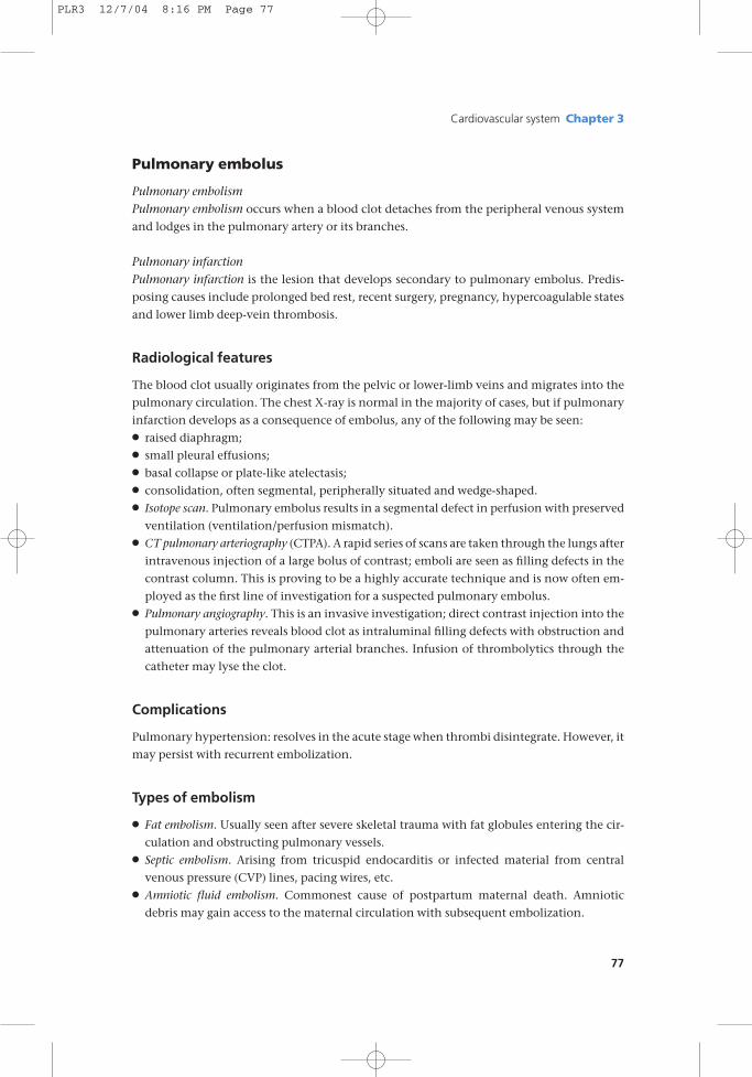
Figure 3.18 Plain abdominal film showing curvilinear calcification (arrows) in a large abdominal aorticaneurysm.
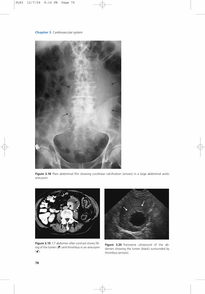
Figure 3.19 CT abdomen after contrast shows fill-ing of the lumen ( ) and thrombus in an aneurysm( ).
➜ ➜
Figure 3.20 Transverse ultrasound of the ab-domen showing the lumen (black) surrounded bythrombus (arrows).
PLR3 12/7/04 8:16 PM Page 78
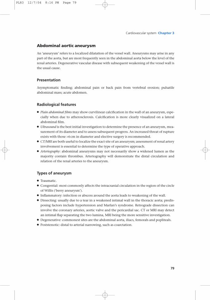
Abdominal aortic aneurysm
An ‘aneurysm’ refers to a localized dilatation of the vessel wall. Aneurysms may arise in any
part of the aorta, but are most frequently seen in the abdominal aorta below the level of the
renal arteries. Degenerative vascular disease with subsequent weakening of the vessel wall is
the usual cause.
Presentation
Asymptomatic finding; abdominal pain or back pain from vertebral erosion; pulsatile
abdominal mass; acute abdomen.
Radiological features
● Plain abdominal films may show curvilinear calcification in the wall of an aneurysm, espe-
cially when due to atherosclerosis. Calcification is more clearly visualized on a lateral
abdominal film.● Ultrasound is the best initial investigation to determine the presence of an aneurysm, mea-
surement of its diameter and to assess subsequent progress. An increased threat of rupture
exists with those >6cm in diameter and elective surgery is recommended.● CT/MRI are both useful to localize the exact site of an aneurysm; assessment of renal artery
involvement is essential to determine the type of operative approach.● Arteriography: abdominal aneurysms may not necessarily show a widened lumen as the
majority contain thrombus. Arteriography will demonstrate the distal circulation and
relation of the renal arteries to the aneurysm.
Types of aneurysm
● Traumatic.● Congential: most commonly affects the intracranial circulation in the region of the circle
of Willis (‘berry aneurysm’).● Inflammatory: infection or abscess around the aorta leads to weakening of the wall.● Dissecting: usually due to a tear in a weakened intimal wall in the thoracic aorta; predis-
posing factors include hypertension and Marfan’s syndrome. Retrograde dissection can
involve the coronary arteries, aortic valve and the pericardial sac. CT or MRI may detect
an intimal flap separating the two lumina, MRI being the more sensitive investigation.● Degenerative: commonest sites are the abdominal aorta, iliacs, femorals and popliteals.● Poststenotic: distal to arterial narrowing, such as coarctation.
Cardiovascular system Chapter 3
79
PLR3 12/7/04 8:16 PM Page 79
Chapter 3 Cardiovascular system
80
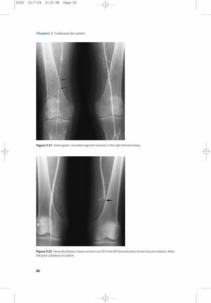
Figure 3.21 Arteriogram: occluded segment (arrows) in the right femoral artery.
Figure 3.22 Femoral embolus: sharp contrast cut off in the left femoral artery (arrow) due to embolus. Notethe poor collateral circulation.
PLR3 12/7/04 8:16 PM Page 80
Peripheral vascular disease
Arterial insufficiency commonly develops in the lower limbs from atheromatous involve-
ment of the aorta and lower-limb arteries. Pain in the calves or buttocks on exercise (inter-
mittent claudication), cold limbs and ulceration are the commonest clinical features.
Predisposing causes include diabetes and smoking.
Radiological investigations
Doppler ultrasound; arteriography; MRI.
Radiological features
Ultrasound will diagnose major occlusions but arteriography is required for the accurate
visualization of diseased vessels, stenoses and occlusions; resolution of MR angiography has
recently shown such significant improvements that it may eventually replace conventional
arteriography.
Treatment
● Balloon angioplasty.● Metallic stent insertion under radiological control.● Surgical bypass grafts: aorto-iliac, femoropopliteal and femorofemoral.
Arterial embolus
An arterial embolus occurs when blood clot, originating elsewhere in the cardiovascular sys-
tem, travels more peripherally and occludes an artery. The lower limbs are affected in the
majority of cases. Symptoms are of rapid onset and consist of a cold, pale, numb leg with
absent pulses distal to the occlusion. Predisposing factors include recent myocardial infarc-
tion with mural thrombus and atrial fibrillation. If there is co-existing vascular disease, a
diagnosis of acute thrombosis should be considered.
Radiological features
Peripheral arteriography demonstrates the contrast column in the artery with a sharp, well-
defined cut-off point, usually a convex upper border projecting into the lumen of the vessel.
Further evidence of an acute episode is provided by a deficient collateral circulation.
Treatment
● Surgical embolectomy.● Thrombolysis: perfusion of streptokinase or tissue plasminogen activator (TPA) directly
into the arterial thrombus in order to lyse the clot.
Cardiovascular system Chapter 3
81
PLR3 12/7/04 8:16 PM Page 81
Chapter 3 Cardiovascular system
82
Figure 3.23 Pulmonary hypertension: bilateral hilar vascular enlargement and prominence of the pulmonary outflow tract.
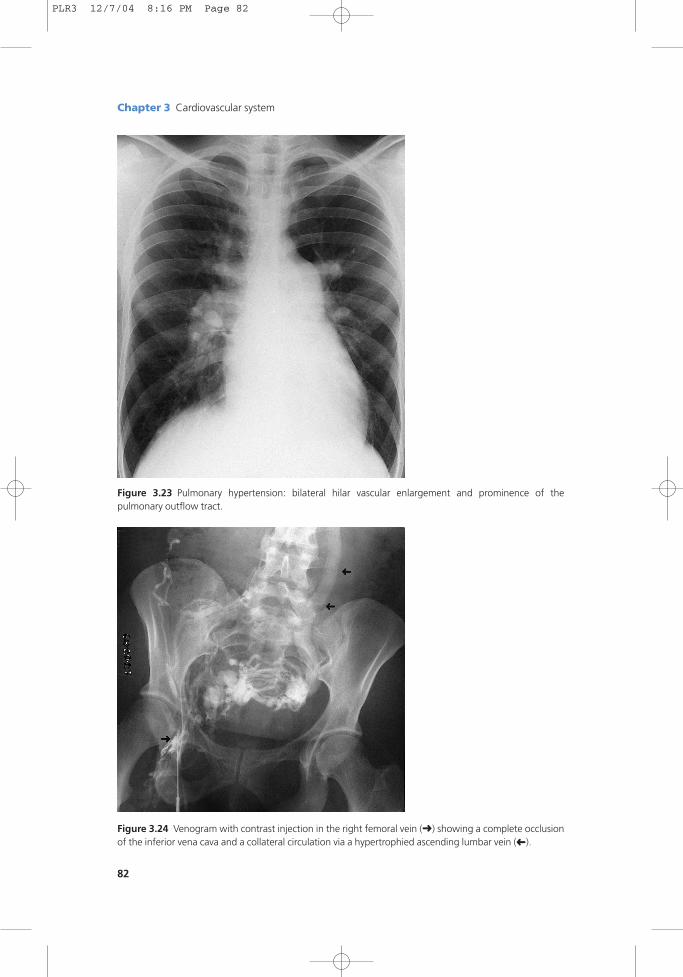
Figure 3.24 Venogram with contrast injection in the right femoral vein (➜) showing a complete occlusionof the inferior vena cava and a collateral circulation via a hypertrophied ascending lumbar vein ( ).
➜
PLR3 12/7/04 8:16 PM Page 82
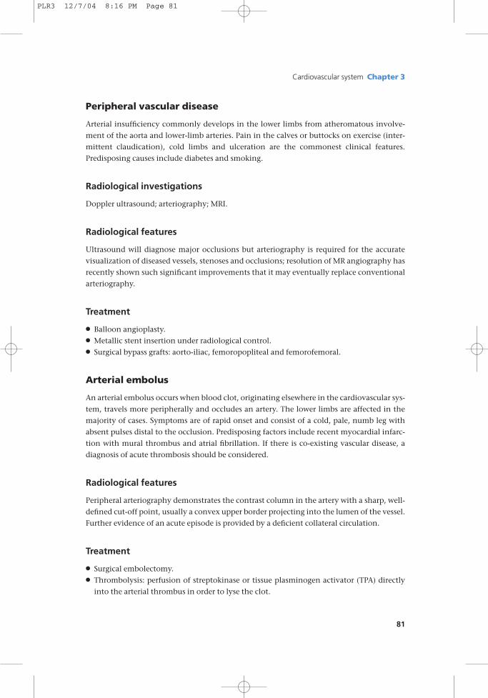
Pulmonary arterial hypertension
Pulmonary arterial hypertension refers to increased pulmonary artery pressure from its
normal value of 25/10mmHg to greater than 30/15mmHg.
Radiological features
Hypertension has to be quite marked before changes on a chest X-ray.● Cardiac enlargement with right ventricular hypertrophy.● Dilatation of the pulmonary hilar vessels with distal attenuation.● Distension of the main pulmonary artery with a bulge below the aortic knuckle.
Causes
● Increased pulmonary blood flow in congenital heart disease.● Obstruction of the pulmonary circulation, e.g. pulmonary emboli, parenchymal lung
disease.● Secondary to pulmonary venous hypertension from left-heart failure or mitral stenosis.
Superior and inferior vena cava obstruction
The superior (SVC) and inferior vena cava (IVC) may obstruct from many causes resulting in
distal venous distension.
Presentation
● SVC obstruction: facial and neck oedema, visible collateral veins.● IVC obstruction: lower limb oedema, scrotal oedema.
Radiological investigations
● Doppler ultrasound: verifies decreased or lack of a blood flow pattern.● CT/MRI: confirms occlusion and often identifies the cause.● Venography: demonstrates anatomical detail, especially useful if stenting is to be
considered.
Causes of SVC obstruction
● Neoplastic: bronchial carcinoma, lymphoma, radiotherapy.● Benign: mediastinal disease due to tuberculosis, sarcoid.
Causes of IVC obstruction
● Tumour invasion from abdominal neoplasms, most commonly renal.● Retroperitoneal fibrosis; radiotherapy.
Cardiovascular system Chapter 3
83
PLR3 12/7/04 8:16 PM Page 83
Chapter 3 Cardiovascular system
84
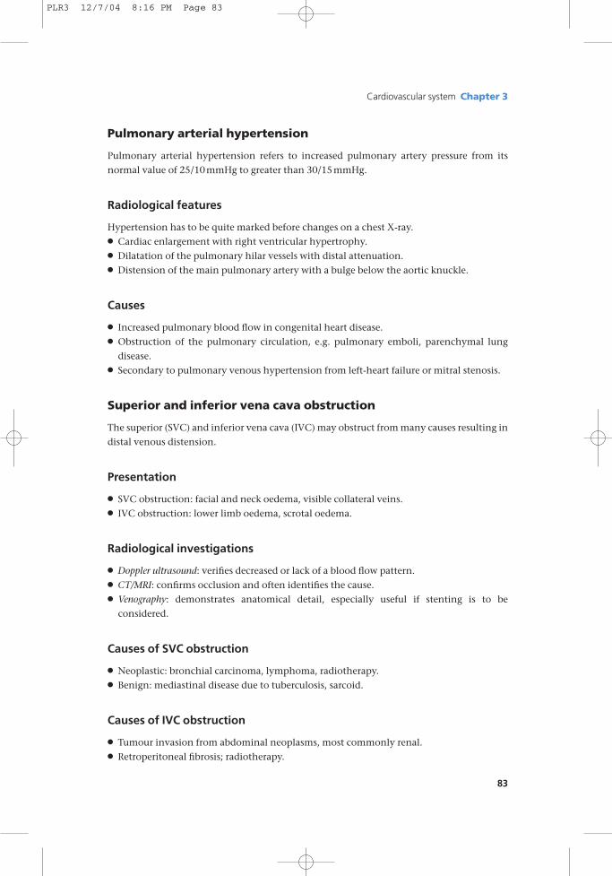
Figure 3.25 Pericardial calcifica-tion (arrow).
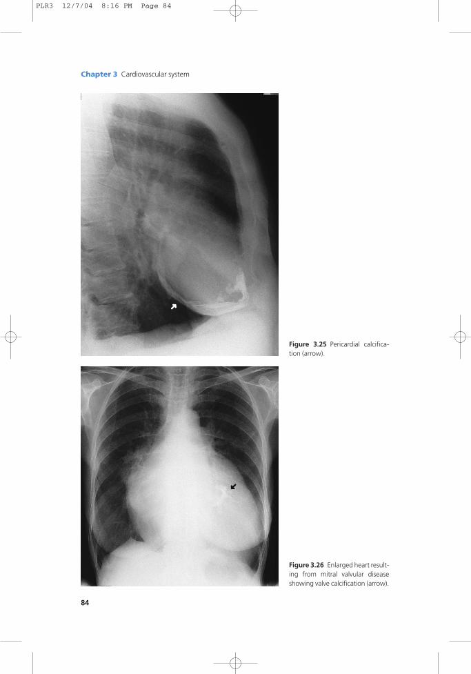
Figure 3.26 Enlarged heart result-ing from mitral valvular diseaseshowing valve calcification (arrow).
PLR3 12/7/04 8:16 PM Page 84
Cardiovascular system Chapter 3
85
Cardiac calcification
Pericardial calcification
Usually follows pericarditis: tuberculosis, rheumatoid arthritis, pyogenic, viral or rheumatic
fever; the aetiology may be unknown.
Myocardial calcification
Occurs typically at the apex of the left ventricle; the common causes are myocardial infarc-
tion and ventricular aneurysm.
Valve calcification
Calcification in the valves is common, but has to be quite extensive before being evident on
plain films. Calcification usually means an element of stenosis, with the aortic and mitral
valves most commonly affected. Causes include atheroma, rheumatic valvular disease and
congenital bicuspid valve.
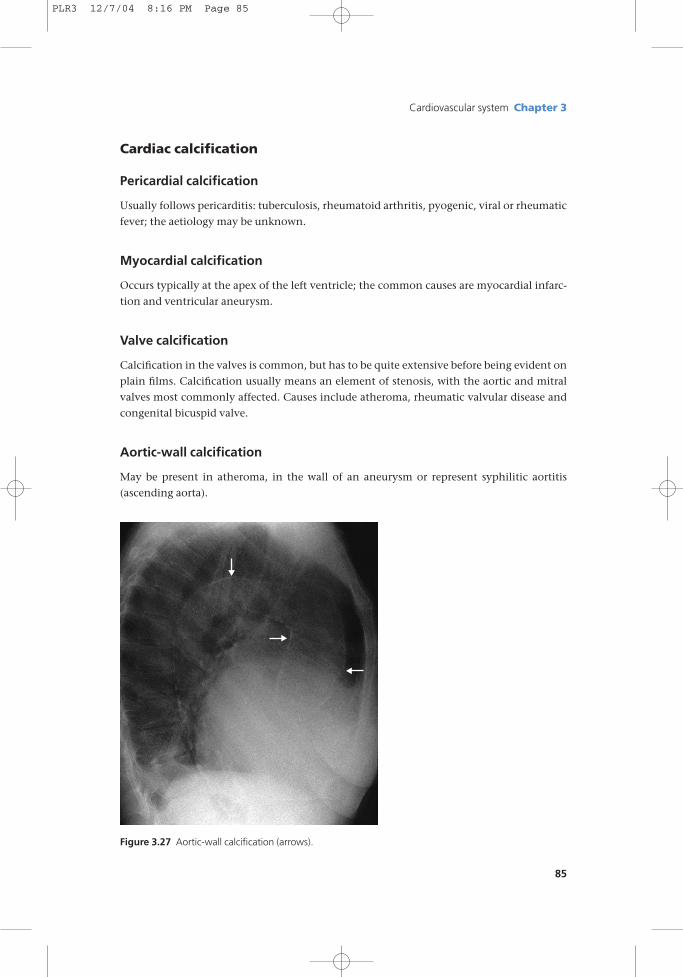
Aortic-wall calcification
May be present in atheroma, in the wall of an aneurysm or represent syphilitic aortitis
(ascending aorta).
Figure 3.27 Aortic-wall calcification (arrows).
PLR3 12/7/04 8:16 PM Page 85
Chapter 3 Cardiovascular system
86
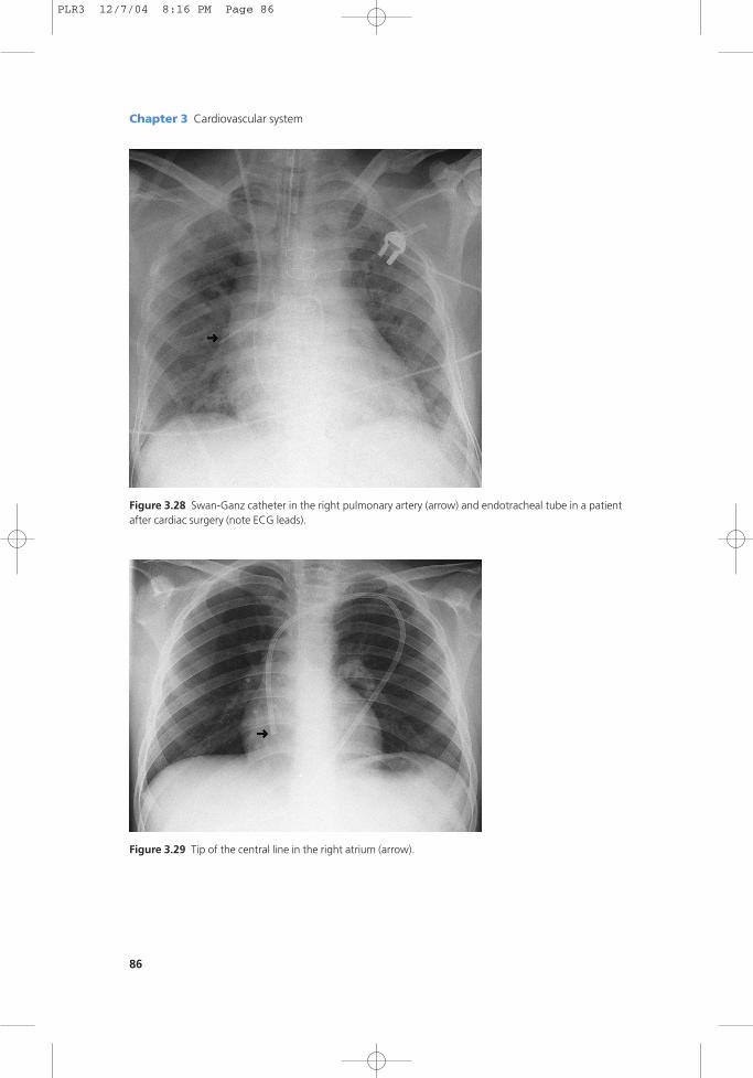
Figure 3.28 Swan-Ganz catheter in the right pulmonary artery (arrow) and endotracheal tube in a patientafter cardiac surgery (note ECG leads).
Figure 3.29 Tip of the central line in the right atrium (arrow).
PLR3 12/7/04 8:16 PM Page 86
Tube and catheter placement
Endotracheal tube
The tip should be positioned 5–7cm above the carina. When sited too far, the endotracheal
tube may advance into a main bronchus, causing collapse of the opposite lung.
Central line
Inserted via the jugular or subclavian veins into a large intrathoracic vein. For accurate mea-
surement of right atrial pressure, the tip of the catheter must lie in a large intrathoracic vein
such as the superior vena cava.
Swan-Ganz catheter
The catheter is inserted via the jugular, subclavian or femoral vein and manipulated through
the right heart into either the right or left pulmonary artery. The end-diastolic left-
ventricular pressure is estimated from a reading taken at the distal tip of the catheter.
Nasogastric tube
The radio-opaque tip should be visualized in the region of the stomach on plain films. An
X-ray is usually necessary to ensure that the tube is not malpositioned, especially into the
trachea or bronchus.
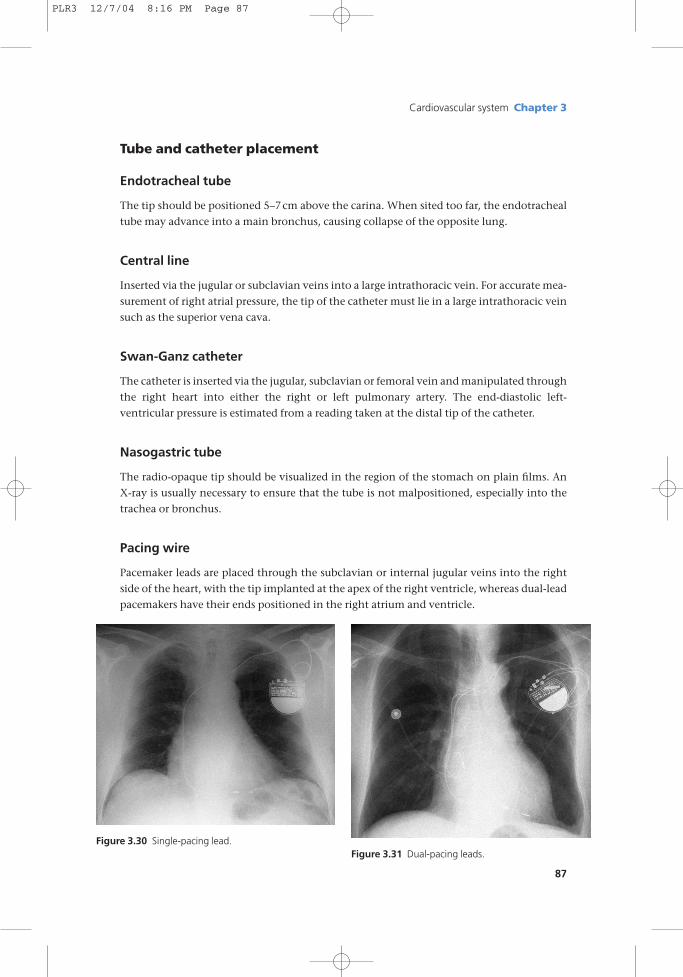
Pacing wire
Pacemaker leads are placed through the subclavian or internal jugular veins into the right
side of the heart, with the tip implanted at the apex of the right ventricle, whereas dual-lead
pacemakers have their ends positioned in the right atrium and ventricle.
Cardiovascular system Chapter 3
87
Figure 3.30 Single-pacing lead.Figure 3.31 Dual-pacing leads.
PLR3 12/7/04 8:16 PM Page 87

PLR3 12/7/04 8:16 PM Page 88