Embed Size (px)
Citation preview

CHAPTER 2
Primary evaluation of medicinal plant extracts and isolated compounds againstchloroquine sensitive strain of Plasmodium falciparum Page 24
2.1 Introduction
Malaria is common in poor and less developed countries of the world.
According to an estimate, about 350-500 million people are infected from malarial
parasite Plasmodium falciparum and approximately one to three million deaths
recorded annually, which represents at least one death every 30 sec (Breman, J., 2001,
Trafford, H., 2005). The vast majority of cases occur in children under the age of 5
years (Greenwood, B .M. et. al., 2005, Winstanley, P. A., 2000). Countries like Africa
faces its hard impact (Bull. WHO, 1993; Ekthawatchai, S. et al., 1999), while other hit
hard hit tropical areas are South East Asia, India and China. Experts estimate that
around 40 % of the cases of malarial fever in India are of Plasmodium falciparum
(Kumar, S., 1994).
The first gold standard drug for treatment of malaria was quinine, a
compound isolated from the bark of a Rubiaceae tree called Cinchona and the drug
is still in use. Later on, its derivative was synthesized having more improvised
action and lesser side effects like nausea, vomiting and gastric refluxes. The drug is
in therapeutic use from around 40 years and is still the cheapest and most effective
therapy for malaria. However, due to massive use, resistance with gold standard
chloroquine has been widely spread and mortality is on increase in Africa.
Treatment of chloroquine-resistant malaria is with alternative drugs or drug
combinations, which are rather expensive and sometimes toxic. Furthermore, these
combinations are not always based on pharmacokinetic principles due to inadequate
knowledge of metabolism and mechanism of action of most antimalarial drugs.
Resistance and viability are still problems with another choices of treatment like
sulfadoxine pyrimethamine and artemisinins (Boland, P., 2001, Fidock, D. A., 2004,
Ridley, R. G., 2002).
Chinese herb “Quinghao” solved the problem of resistance. The latin name of
this herb is Artemisia annua and artemisinin, a sesquiterpene lactone isolated from
the same became block buster anti malarial drug. Unfortunately, the therapy had
drawbacks like drug resistance and economical viability.
CHAPTER 2
Primary evaluation of medicinal plant extracts and isolated compounds againstchloroquine sensitive strain of Plasmodium falciparum Page 25
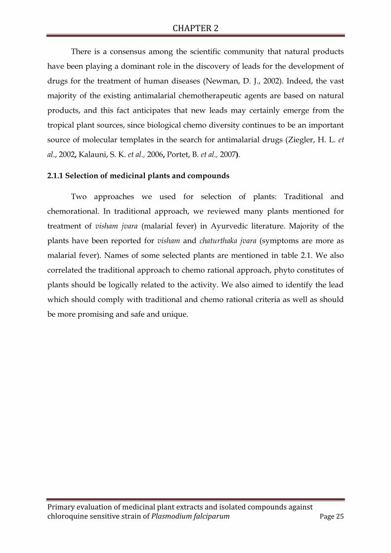
There is a consensus among the scientific community that natural products
have been playing a dominant role in the discovery of leads for the development of
drugs for the treatment of human diseases (Newman, D. J., 2002). Indeed, the vast
majority of the existing antimalarial chemotherapeutic agents are based on natural
products, and this fact anticipates that new leads may certainly emerge from the
tropical plant sources, since biological chemo diversity continues to be an important
source of molecular templates in the search for antimalarial drugs (Ziegler, H. L. et
al., 2002, Kalauni, S. K. et al., 2006, Portet, B. et al., 2007).
2.1.1 Selection of medicinal plants and compounds
Two approaches we used for selection of plants: Traditional and
chemorational. In traditional approach, we reviewed many plants mentioned for
treatment of visham jvara (malarial fever) in Ayurvedic literature. Majority of the
plants have been reported for visham and chaturthaka jvara (symptoms are more as
malarial fever). Names of some selected plants are mentioned in table 2.1. We also
correlated the traditional approach to chemo rational approach, phyto constitutes of
plants should be logically related to the activity. We also aimed to identify the lead
which should comply with traditional and chemo rational criteria as well as should
be more promising and safe and unique.

CHAPTER 2
Primary evaluation of medicinal plant extracts and isolated compounds againstchloroquine sensitive strain of Plasmodium falciparum Page 26
Table 2.1: Antimalarial plants reported in traditional literature
Name of theplant
Commonname
Detailsmentioningassociation withmalaria
ClassicalReferences
Scientific Reportstatus(References)
Adhatodazeylenica
Vasaka,Ardusi
Used in vishamjvara formulationslike Vatsakadikwatha,Vasadighritam
Charaka Samhita,Cikitsa, 3/223-224
Reported(Henrik ToftSimonsen et al.,2001)
Embelia ribes Vidanga Used as krimignain Vidanga churna
Bhavapraksha,Krimirogadhikara,7-21
Not reported
Enicostemmalittorale
Mamejavaka,Katu chirata,Chhota chirata
Mentionedspecifically forvisham jvara
Dravyagunavigyana, Part II ,704
Not reported
Picrorhizakurrao
Katuka Used in vishamjvar nashak panchakwatha for santataand satata jvara
Charaka Samhita,Cikitsa, 3/200-203
Reported(Singh, V andBanyal, HS,2011)
Holorhennaantidysentrica
Indrayava Used in vishamjvar nashak panchakwatha for santataand anyedyushkajvara
Charaka Samhita,Cikitsa, 3/200-203
Reported(Verma, G. et al.,2011)
Azadirachtaindica
Neem Used in vishamjvar nashak panchakwatha for satatajvara
Charaka Samhita,Cikitsa, 3/200-203
Reported(Iwu, MM et al.,1986)
Cyprusrotundens
Motha Used in vishamjvar nashak panchakwatha for satata,anyesyushka andchaturthaka jvara
Charaka Samhita,Cikitsa, 3/200-203
Reported(Thebtaranonth,C. et al., 1995)
Cissumpalosparrera
Patha Used in vishamjvar nashak panchakwatha for santatajvara
Charaka Samhita,Cikitsa, 3/200-203
Reported(Singh, V andBanyal, HS,2011)
Santalumalbum
Chandana Used in vishamjvar nashak panchakwatha forchaturthaka jvara
Charaka Samhita,Cikitsa, 3/200-203
Reported(Henrik ToftSimonsen et al.,2001)
From review of traditional literature, we identified three plants reported for
treatment of fever and related symptoms of malaria: Embelia ribes, Adhatoda vasica
and Enicostemma littorale. Adhatoda vasica was reported for treatment of visham jvara

CHAPTER 2
Primary evaluation of medicinal plant extracts and isolated compounds againstchloroquine sensitive strain of Plasmodium falciparum Page 27
in Charaka Samhita but Enicostemma littorale and Embelia ribes are not mentioned in
Brihat Trayi – Three main books of Ayurveda viz. Charaka Samhita, Sushruta Samhita
and Astanga hridayam. Laghu Trayi mentions their uses in jvara and visham jvara. We
also found no records mentioning antimalarial activities of these plants in scientific
literature except Adhatoda vasica which was previously reported by few authors
(Henrik Toft Simonsen et al., 2001).
We also applied a chemo rational approach on the selected plants and
reviewed correlations between main compounds of selected plants and their
probable role for antimalarial activities. Parasite requires electron transfer for
energy. Existence of coenzyme Q8 redox entity, in Plasmodium is responsible for
electron transfer of the parasite. Quinines possess antimalarial action by inhibiting
this enzyme (Yieh-Ping Wan et al., 1974). Quinones are also highly active against P.
falciparum in vitro (Carvalho, L. H. et al., 1992). From these, we hypothesized
probable antimalarial action of benzoqinone; embelin, a major phytochemical of
Embelia ribes.
Vasicine, a quinazoline alkaloid of Adhatoda vasica resembles with
phenomenon, that weak bases play a major role in antimalarial action by raising
intravesicular pH (Yayon, A. et al., 1984; Paul, H. Schlesinger et al., 1988) and thereby
affecting transport of macromolecules as well as membranes and phospholipase
activity (Stahl, P. And Schwart, A. L., 1986).
Flavonoids are the largest group of phytochemicals. Earlier studies have
reported that flavonoids possess antimalarial activity and demonstrate marked and
selective potentiating effect on the antiplasmodial activity of artemisinin (Liu, K. C.
C., 1992). We selected a flavonoid hesperidin as it is nowhere reported for
antimalarial activity and can be easily obtained by sources like waste orange peel.
Irridoids are mono or sequi terpenoidal glycosidal compounds from the
plants and also called bitter glycosides. One of the best examples of similar class of
compound is artemisinin from Artemisia annua. Many irridoids has been identified
for antimalarial activity of many plants. Swertiamarin from Enicostemma littorale is a

CHAPTER 2
Primary evaluation of medicinal plant extracts and isolated compounds againstchloroquine sensitive strain of Plasmodium falciparum Page 28
compound of similar category; hence we selected the same for study (Cláudio R. et
al., 2011).
These selected plants and their relevant compounds were screened to define
their antimalarial activity on chloroquine sensitive strain of Plasmodium falciparum.
We also used alkaloids isolated from cinchona bark and quinine dihydrochloride as
respective standards. Viable methods of isolation were established by us to achieve
proposed compounds (particularly embelin, hesperidin and cinchona alkaloids;
where working standards to be used).
2.2 Materials and methods
Part A: Preparation of extracts and isolation of compounds
2.2.1 Chemicals
All the chemicals used were of analytical grade.
2.2.2 Extracts / compounds selected for study
Extracts: Methanolic extract of Enicostemma littorale, Embelia ribes and Adhatoda vasica
Compounds: Embelin (benzoquinone), vasicine (isoquinoline alkaloid), swertiamarin
(secoirridoid glucoside), hesperidin (a flavonol glucoside) and mixture of alkaloids
isolated from cinchona bark.
Embelin was isolated from Embelia ribes, Hesperidin was isolated from orange
peel and cinchona alkaloids were isolated from Cinchona officinalis bark, while,
another compounds were procured as gift from VED Pharmaceuticals Pvt Ltd.,
Baroda.
2.2.3 Plant material and preparation of extracts
The whole herb of Enicostemma littorale was collected from Bhavnagar, the
coastal area of Gujarat in the month of August; Adhatoda vasica leaves were collected
from our campus in month of August. While, Embelia ribes fruits were purchased
from a local supplier in Baroda. The specimen were sent for identification to Botany

CHAPTER 2
Primary evaluation of medicinal plant extracts and isolated compounds againstchloroquine sensitive strain of Plasmodium falciparum Page 29
department of M S University of Baroda and identified as an authentic material. The
plant material collected were dried in oven at 50 °C, crushed in a mixer to yield
powder of 40 – 60 mesh which was further used for isolation. The dried leaf powder
(1 kg) was extracted with methanol (1 litre x 3). The resulting methanol soluble
fractions were pooled together and concentrated under reduced pressure. The
extracts were stored in air tight vial.
2.2.4 Isolation of compounds
Embelin, hesperidin and cinchona alkaloids were used as working standards,
after confirming purity and proper characterization. We developed rapid method of
isolation of these compounds in a way to make them commercially viable. Brief
procedures of isolation of respective compounds are mentioned below.
2.2.4.1 Embelin from Embelia ribes
Hundred grams of coarsely powdered berries of Embelia ribes were extracted
with chloroform (500 ml x 3). The chloroform soluble fractions were pooled together
and evaporated to reduce volume up to 500 ml. This was covered and kept in freeze
(4 – 8 ºC) for overnight. The golden colored needles so obtained were filtered by
watmann no. 1 filter paper. The needles were purified by washing them with
petroleum ether (60 – 80 ºC) and dried at room temperature. The resulting
compound was immediately transferred to air tight vial and stored in refrigerator.
2.2.4.2 Hesperidin from orange peel
Twenty five gm of Orange peel powder was defatted with petroleum ether
(60-80 ºC) (500 ml x 3). The resulting marc was extracted with methanol (500 ml x 3)
repetitively and concentrated to a syrupy mass. Glacial acetic acid (25 ml) was added
to the concentrated extract, and diluted with 250 ml of water. The white fluffy
precipitates so obtained were mixture of hesperidin and rutin. This mixture was
washed repetitively with methanol and pure product so obtained was dried under
vacuum.

CHAPTER 2
Primary evaluation of medicinal plant extracts and isolated compounds againstchloroquine sensitive strain of Plasmodium falciparum Page 30
2.2.4.3 Cinchona alkaloids
Bark of cinchona was received from a supplier from Amazon. Powder of
cinchona bark 20 gm was defatted with petroleum ether (60-80ºC) (3 x 100 ml) and
the marc was extracted with methanol repetitively until gives negative dragendorff’s
test. The methanol soluble portion was concentrated and dissolved in 10%
orthophosphoric acid. This orthophosphoric acid solution was again basified with
ammonia up to pH 9 and extracted with dichloromethane and concentrated to
dryness. The dried portion was rinsed with cyclohexane and the solvent was
evaporated. The compound was obtained as pale yellowish white and off white
amorphous crystal form.
2.2.5 Characterization of extracts/compounds
2.2.5.1 Characterization of Enicostemma littorale/swertiamarin; Adhatoda
vasica/vasicine and Embelia ribes/embelin
The extracts and their relevant compounds were evaluated by using High
Performance Thin Layer Chromatography (HPTLC). We used optimized
chromatographic conditions as reported by authors previously. For Enicostemma
littorale/swertiamarin, method mentioned by Vishwakarma, S.L. (Vishwakarma, S.L.
et al., 2004); Adhatoda vasica/vasicine by S. Soni (Soni, S. et al., 2009) and Embelia
ribes/embelin by Kukkar, R. (Kukkar, R. et al, 2010) were used. The detailed HPTLC
conditions are as mentioned below.
TLC plates
Pre coated silica gel 60 F254 (E. Merck) of uniform thickness (0.2 mm) were used.
Preparation of test solution
Twenty five mg of each extract of drug was dissolved in methanol. The
methanol soluble portions were pooled together and concentrated to make up the
volume of 50 ml in a volumetric flask. Same way, 2 mg of each standard was
dissolved in 1 ml of methanol.

CHAPTER 2
Primary evaluation of medicinal plant extracts and isolated compounds againstchloroquine sensitive strain of Plasmodium falciparum Page 31
Procedure
Ten µl of each solution was applied on TLC plate with help of Linomat V
spotter. The plated were allowed to run in respective optimized solvent system up to
8 cm height and scanned at 254 nm by using TLC scanner 3, attached with winCATS
software to obtain densitogram. Optimized solvent system conditions are mentioned
as below.
Name of extract and relevantcompound
Solvent system
Enicostemma littorale and swertiamamrin Ethyl acetate : methanol (8:2)Embelia ribes and embelin n propanol: n butanol: ammonia (7:1:2)
Adhatoda vasica and vasicine Ethyl acetate: methanol: ammonia(8:2:0.2)
2.2.5.2 Characterization of hesperidin
Hesperidin was confirmed by various methods as mentioned below.
Evaluation by Ultraviolet spectrometry:
The solution 1 was drawn to a quartz cuvette of 1 ml and scanned for the
wavelength ranging between 200 – 800 nm on Helios UV spectrophotometer. The
wavelength at maxima peak was recorded.
Evaluation by melting point
One mg of compound was taken in a capillary tube and placed in silicon oil.
The temperature was continuously recorded and the point at which the compound
melts was recorded.
Evaluation of the compound by mass fragmentation pattern
For mass spectral identification by ESI-MS, 1 mg of compound was dissolved in
methanol and was introduced for direct fragmentation in ionization chamber in
positive and negative ionization mode. The base peak was recorded and compared
with the molecular weight.

CHAPTER 2
Primary evaluation of medicinal plant extracts and isolated compounds againstchloroquine sensitive strain of Plasmodium falciparum Page 32
2.2.5.3 Characterization of cinchona alkaloids
The liquid chromatography of mixture of alkaloids was carried out by
following a validated HPLC, a most convenient technique for mixture of isomers.
Method reported by Ravishankara (Ravishankara, M. N. et al., 2001) was followed
for the same. In brief, the mixture containing alkaloids was dissolved in 10 percent
sulphuric acid to obtain concentration of 1 mg/ml and chromatographed by using
stationary phase of C18 column (Restek, 250 mm length, 4.6 mm internal diameter
and 5 µm particle size) and mobile phase of 85 mM potassium dihydrogen ortho
phosphate solution containing 40 mM of triethyl amine adjusted to pH 2.8 with
orthophosphoric acid solution. The resulting solution was mixed with acetonitrile in
ratio of 90:10 v/v, flow rate adjusted was of 1.2 ml/min in HPLC system (Perkin
Elmer Series 200) with UV/VIS detector adjusted at wavelength of 226 nm. Quinine
sulphate was used as a standard by dissolving quinine sulphate tablets in water to
make 1 mg/ml of solution.
Part B: Antimalarial activity
2.3 Parasite culturing and antiplasmodial assay of extracts/compounds
2.3.1 Culturing Plasmodium falciparum
Cultivation of malaria parasites is an important tool for the understanding of
parasite biology, biochemistry, molecular biology, immunology, and pharmacology.
We followed technique of the erythrocytic stages of P. falciparum as that originally
described by Trager and Jensen as well as Schuster (Trager, W. and Jensen, J. B.,
1976; Schuster, F. L., 2002). Detailed methodologies for parasite culturing are
mentioned as below.
2.3.1.1 Malarial parasites
Two strains of Plasmodium falciaprum namely MRC 20 and RKL 19 were used
in the study. The MRC 20 strain was isolated from the patient infected with
Plasmodium falciparum before any medication, known as sensitive to chloroquine
while, RKL 19 was isolated and maintained from the patient with relapse of malaria

CHAPTER 2
Primary evaluation of medicinal plant extracts and isolated compounds againstchloroquine sensitive strain of Plasmodium falciparum Page 33
after treatment of chloroquine, was known as resistant to chloroquine. These both
strains were procured from National Institute of Malaria Research (NIMR), New
Delhi.
2.3.1.2 Culture medium
The RPMI medium was routinely used for in vitro culture of Plasmodium
falciparum throughout this study. It was prepared by dissolving 10.4 gm of RPMI
1640 powder containing L- glutamine but without sodium bicarboanate (Hi Media)
and 5.94 gms of HEPES buffer (SIGMA) in 960 ml of double distilled water. Two
militres of gentamycin sulphate (40 mg/ml) was added to the medium solution
before sterilization by filtration of millipore filter of 0.22 µm porosity. One hundred
millilitres of the sterile medium was transferred in to sterile glass bottle as a stock
medium, which was stored for a period up to one month at 4 °C.
Each stock medium (100 ml) was added with 4.2 ml of 5 % (w/v) NaHCO3
(Hi Media) to give a pH of 7.4, 1 ml of 10 mM D – glucose (SIGMA), and 1 ml of 2
mM L – glutamine (SIGMA). This medium was referred to as complete medium
without serum (C-RPMI without serum). The complete RPMI medium was
supplimented with 10 % (v/v) human inactivated serum (group A, B, and O) before
using for cultivation and this medium was referred to as complete medium with
serum (C-RPMI with serum). This medium was stored at 4 °C and used within one
month.
2.3.1.3 Normal human serum
Normal human serum of all blood groups was collected from healthy donors
who had no history of malaria infection. Moreover, donors did not receive any drug
within two weeks prior to blood collection. Sera group A, B and O were used for
routine culture of Plasmodium falciparum, whereas group O serum was used for
assessment of antimalarial activity.
Blood was collected intravenously through a bag (after removing anti
coagulant solutions) and allowed to clot at room temperature for 30 min. The bag
CHAPTER 2
Primary evaluation of medicinal plant extracts and isolated compounds againstchloroquine sensitive strain of Plasmodium falciparum Page 34
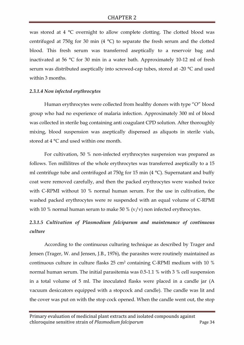
was stored at 4 °C overnight to allow complete clotting. The clotted blood was
centrifuged at 750g for 30 min (4 °C) to separate the fresh serum and the clotted
blood. This fresh serum was transferred aseptically to a reservoir bag and
inactivated at 56 °C for 30 min in a water bath. Approximately 10-12 ml of fresh
serum was distributed aseptically into screwed-cap tubes, stored at -20 °C and used
within 3 months.
2.3.1.4 Non infected erythrocytes
Human erythrocytes were collected from healthy donors with type “O” blood
group who had no experience of malaria infection. Approximately 300 ml of blood
was collected in sterile bag containing anti coagulant CPD solution. After thoroughly
mixing, blood suspension was aseptically dispensed as aliquots in sterile vials,
stored at 4 °C and used within one month.
For cultivation, 50 % non-infected erythrocytes suspension was prepared as
follows. Ten millilitres of the whole erythrocytes was transferred aseptically to a 15
ml centrifuge tube and centrifuged at 750g for 15 min (4 °C). Supernatant and buffy
coat were removed carefully, and then the packed erythrocytes were washed twice
with C-RPMI without 10 % normal human serum. For the use in cultivation, the
washed packed erythrocytes were re suspended with an equal volume of C-RPMI
with 10 % normal human serum to make 50 % (v/v) non infected erythrocytes.
2.3.1.5 Cultivation of Plasmodium falciparum and maintenance of continuous
culture
According to the continuous culturing technique as described by Trager and
Jensen (Trager, W. and Jensen, J.B., 1976), the parasites were routinely maintained as
continuous culture in culture flasks 25 cm2 containing C-RPMI medium with 10 %
normal human serum. The initial parasitemia was 0.5-1.1 % with 3 % cell suspension
in a total volume of 5 ml. The inoculated flasks were placed in a candle jar (A
vacuum desiccators equipped with a stopcock and candle). The candle was lit and
the cover was put on with the stop cock opened. When the candle went out, the stop

CHAPTER 2
Primary evaluation of medicinal plant extracts and isolated compounds againstchloroquine sensitive strain of Plasmodium falciparum Page 35
cock was immediately closed. By this method, an atmosphere with low oxygen and
high carbon dioxide content, i.e., 17 % O2, 3 % CO2 and 80 % N2 was produced. The
candle jar was incubated at 37 °C in an incubator.
The medium was changed daily; old medium was being removed with a
pipette. The thin blood smear was made and stained with Giemsa stain. Percentage
parasitemia was calculated by counting the infected erythrocytes in a total of 10,000
erythrocytes. Sub culturing was performed when parasitemia is higher than 5-6%.
Briefly, the infected erythrocytes were collected in to a centrifuge tube and
centrifuged at 500g for 7 min (4 °C). The packed erythrocytes were washed twice
with C-RPMI without 10 % human serum. After the final wash, the packed
erythrocytes were suspended to make 50 % (v/v) suspension in to flasks, percent cell
suspension were reduced from 50 % to 3 % with C-RPMI medium with 10% normal
human serum. Approximately 5 ml of this suspension was then placed in to culture
flasks as mentioned above.
2.3.1.6 Synchronization of culture
Generally, continuous culture of P. falciparum results in a loss of synchronicity
characteristic, which can complicate the evaluation of experimental results. Thus, it
is important to start all experiments with a synchronous ring stage. Briefly,
synchronous cultures of MRC 20 and RKL 19 strains were collected in to centrifuge
tube and centrifuged at 500g for 10 min (4 °C). After the removal of supernatant, 5 %
(w/v) sterile D – sorbitol was added to the packed erythrocytes at the ratio of 4:1
and gently mixed. The mixture was incubated at 37 °C for 20 min in the waterbath.
During the period of incubation, only erythrocytes harbouring late trophozoites and
schizonts were selectively lysed, leaving only the erythrocytes infected with ring
stage among non-infected erythrocytes. Sorbitol was removed by centrifugation and
the packed erythrocytes were then washed twice with C – RPMI without 10 %
normal human serum. After final wash, the packed erythrocytes was adjusted to 3 %
cell suspension and cultured in a candle jar as described above.

CHAPTER 2
Primary evaluation of medicinal plant extracts and isolated compounds againstchloroquine sensitive strain of Plasmodium falciparum Page 36
2.3.1.7 Freezing and storage of parasites
Plasmodium falciparum parasites in continuous culture were harvested
whenever at least 5 % ring stage was obtained. The culture suspension was collected
into a centrifuge tube and centrifuged at 500g for 7 min (4 °C). After the removal of
supernatant, the packed erythrocytes were resuspended in an equal volume of
freezing solution (sorbitol, 0.9 % NaCl and 99 % glycerine). An aliquot of 1-1.5 ml of
the suspension was carefully added into a cryopreserved tube and frozen at -80 °C
refrigerator. These frozen parasites were stored as stock culture.
2.3.1.8 Thawing of parasites
Frozen cryopreservative tubes were removed from -80 °C refrigerator and
thawed at 37 °C in a waterbath. The suspension was transferred to a sterile
centrifuge tube, to which an equal volume of sterile 2.5 % sodium chloride solution
was added, and then centrifuged at 500g for 7 min (4°C). After the removal of
supernatant, the packed erythrocytes were washed three times with C-RPMI without
10 % normal human serum. After the final wash, the packed erythrocytes was
resuspended in C-RPMI with 10 % normal human serum to make 3 % cell
suspension, and transferred in to sterile culture flask and cultured in a candle jar as
described above.
2.3.1.9 Preparation of parasite inoculums
Up to 5 % ring stage P. falciparum obtained by several rounds of sorbitol lysis
were pooled ascpetically to a 15 ml centrifuge tube and centrifuged at 500g for 7 min
(4°C). After the removal of supernatant, the packed erythrocytes were resuspended
in an equal volume of C-RPMI medium without 10 % normal human serum to make
a 50 % (v/v) cell suspension. The 50 % non-infected erythrocytes were added to
adjust percentage of parasitemia approximately 1 to 1.5 %. The erythrocyte
suspension was diluted to 3 % cell suspension with C-RPMI medium with 20 %
normal human serum. This mixture of suspension is referred to as parasite
inoculums.

CHAPTER 2
Primary evaluation of medicinal plant extracts and isolated compounds againstchloroquine sensitive strain of Plasmodium falciparum Page 37
2.3.1.10 Giemsa staining of parasites
The stain preparation was optimized by us. In brief, 25 mg of giemsa powder
was triturated in a mortar pastel with 5 ml of glycerol, 15 ml of methanol was added
to it and filtered. The resulting solution was immediately transferred to a amber
colored reagent bottle and stored in dark. For parasite staining, 5 ml of the solution
was diluted with 25 ml of cold water. The smeared culture slides were stained by
fixing with methanol first and then allowed to be dipped in prepared giemsa
staining solution for 15 minutes. Then the slides were washed with water to remove
excess stain, allowed to dry in air and observed under microscope by putting a drop
of immersion oil.
2.3.1.11 Schizont Maturation Inhibition Assay
Various techniques for antiplasmodial activity of plant extracts/compounds
were followed as previously reported by the authors ((Desjardins, R. E. et al, 1979;
Ringwald, P. et al, 1996; Wernsdorfer, W. H., 1994).
We evaluated the extracts and compounds for antimalarial activity by a stage
specific assay method called schizont maturation inhibition assay (SMI). In brief,
culture Plasmodium falciparum was maintained as mentioned above and observed
after 3, 6 and 24 h for regular development of parasite stages. The cultures were
synchronized using sorbitol (4.2%) and parasitaemia was adjusted to 3-4 % by
diluting with fresh human erythrocytes. The cells were diluted with complete media
to make 8% haematocrit. Again the slides of culture were prepared and observed for
the calculation of parasitemia, particularly for young trophozoites or ring stages.
One milligram of each extract/compound was dissolved in 100 µl of DMSO
and then 900 µl of incomplete RPMI 1640 medium (without serum) was added. A
series of eleven concentrations were prepared from the stock solutions by 2 fold
dilutions in 96 well micro titre plates. After interval of 24 hr and 48 hr, thin films of
the contents of each well were prepared and examined under the microscope.
Parasite count for each blood film was made using a compound microscope under

CHAPTER 2
Primary evaluation of medicinal plant extracts and isolated compounds againstchloroquine sensitive strain of Plasmodium falciparum Page 38
oil immersion with X 100 objective after staining the film with giemsa solution. Each
film was observed at 20 different visual fields and at three different parts of slide.
The number of schizonts per 200 parasites were noted and compared between
control and test wells for the determination of the % inhibition. All doses were
studied in cultures and the mean was observed for purposes of inferences. The
inhibition of parasite growth in the drug-treated groups was calculated as follows:
Parasitaemia in the control (non-treated) group minus parasitaemia in the drug
treated group, divided by parasitaemia in the control (non-treated) group, expressed
as percentages. All values are expressed as percentage growth inhibition. Dose
response curves of the fractions were obtained by plotting percentage inhibition
against log concentration. The values of the compounds provided a mid-point value
where parasite growth would be 50%. Linear regression analysis was applied to the
linear portion of the sigmoidal curve was plotted and IC50 values were derived for
each extract/compound.

CHAPTER 2
Primary evaluation of medicinal plant extracts and isolated compounds againstchloroquine sensitive strain of Plasmodium falciparum Page 39
2.4 Results
2.4.1 Characterization of extracts/compounds by HPTLC
Comparative TLC pattern (figure 2.1 a) and densitograms (figure 2.1 b) of
Adhatoda vasica leaf extract and vasicine represented a band at Rf 0.40 corresponding
to vasicine in both reference and test solution when the plate was visualized under
UV 254 nm. Presence of a single peak of vasicine (track 2 of figure 2.1 b) in
densitogram showed its purity and corresponding peak in test sample represented
the same as a major compound of the herb.
Figure 2.2 (a & b) mentions TLC pattern and densitograms of Enicostemma
littorale extract and swertiamarin. Swertiamarin was observed at 0.55 Rf in both test
and standard tracks when the plate was observed under UV254 nm. From TLC and
densitograms, it was observed that swertiamarin was in pure form and was a major
compound of the herb.
A band of Rf 0.12 corresponded to embelin in both test and standard solutions
were compared by TLC and obtained densitograms as mentioned in figure 2.3 a, b &
c. Figure 2.3 a represents TLC pattern when the plate was observed under UV 254
nm, while figure 2.3 b represents pink spots of embelin observed at natural light
after derivatization of the same plate with 5 % methnolic solution of potassium
hydroxide. The isolated compound was also found to be pure form recorded
densitogram (figure 2.3 c, track 2).
2.4.2 Characterization of hesperidin
The hesperidin was isolated as a white amorphous powder with the yield of 8
percent. The melting point ranged between 258 ° to 262 °C. It was insoluble in water
and sparingly soluble in methanol, clearly soluble in pyridine and dimethyl
sulfoxide while insoluble in acetone, chloroform and petroleum ether. The UV max
of compound was ranged between 286-289 nm. Further it was confirmed by Liquid
chromatographic (figure 2.4 a) and mass pattern (figure 2.4 b) showing molecular
weight of 610 m/z.

CHAPTER 2
Primary evaluation of medicinal plant extracts and isolated compounds againstchloroquine sensitive strain of Plasmodium falciparum Page 40
2.4.3 Characterization of cinchona alkaloids
The mixture was found to contain four distinct peaks corresponding to
cinchonine, cinchonidine, quinidine and quinine at retention time (Rt) of 2.09, 4.39,
6.43 and 7.32 min. respectively. Quinine standard was also resolved at Rt of 7.32 min.
From the relative reference (Ravishankara, M. N., 2001), we standardized this
mixture and found that quinine was a major compound 59.75%, while, quinidine,
cinchonidine and cinchonine were present in 14.28, 15.53 and 10.43 % respectively.
Figure 2.5 a represents HPLC pattern of mixture, while figure 2.5 b represents that of
standard quinine.

CHAPTER 2
Primary evaluation of medicinal plant extracts and isolated compounds againstchloroquine sensitive strain of Plasmodium falciparum Page 41
1
2
Figure: 2.1a TLC profile observed under UV 254 nm; 2.1 b densitograms oftracks 1: Adhatoda vasica extract, 2: Vasicine
1 2
Fig. 2.1a Fig. 2.1b
Vasicine
Vasicine
AU
Rf
Rf1 2
1
2
AU
Figure: 2.2a TLC profile observed under UV 254 nm; 2.2 b densitograms of tracks1: Enicostemma littorale extract, 2: swertiamarin
Fig. 2.2a Fig. 2.2b
Swertiamarin
Swertiamarin
1 2

CHAPTER 2
Primary evaluation of medicinal plant extracts and isolated compounds againstchloroquine sensitive strain of Plasmodium falciparum Page 42
Fig. 2.3a Fig. 2.3b
1 2
Embelin
1 2
Figure: 2.3a TLC profile observed under UV 254 nm; 2.3b plate derivatizedwith KOH and observed under natural light; 2.3 c densitograms of tracks 1:
Embelia ribes extract, 2: embelin
Embelin
1
2

CHAPTER 2
Primary evaluation of medicinal plant extracts and isolated compounds againstchloroquine sensitive strain of Plasmodium falciparum Page 43
Fig. 2.4a
Fig. 2.4b
Figure 2.4a: Liquid chromatogram; 2.4 b: Mass spectra of of hesperidin

CHAPTER 2
Primary evaluation of medicinal plant extracts and isolated compounds againstchloroquine sensitive strain of Plasmodium falciparum Page 44
Fig. 2.5a
Fig. 2.5b
Fig. 2.5 a Liquid chromatogram of cinchona alkaloids; 2.5 b Liquid
chromatogram of quinine
Cinchonine (CN); Cinchonidine (CD); Quinidine (QD) and Quinine (QN)

CHAPTER 2
Primary evaluation of medicinal plant extracts and isolated compounds againstchloroquine sensitive strain of Plasmodium falciparum Page 45
2.4.4 Parasite culture
All the stages of parasite development were found distinct from trophozoits
to schizonts. Parasite stages were observed regularly after 48 hr intervals. Some
distinct features of Plasmodium falciparum were seen like double chromatin (nucleus),
multiple invasions (more than one parasite entry in RBC), maurer’s dots (brown
colored crystals of metabolized haemoglobin in to parasite food vacuole). The size
and shape of RBCs were not altered. Figure 2.6 a, b, c and d represents stages like
merozoit invasion, trophozoit, mature schizont and trophozoits with double
chromatin.
Merozoit invasion to RBCFig. 2.5 a
Trophozoit in RBCFig. 2.5 b
Mature schizontFig. 2.5 c
Trophozoit with doublechromatin
Fig. 2.5 d
Figure 2.6: Different stages of parasite development observed duringculturing of Plasmodium falciparum

CHAPTER 2
Primary evaluation of medicinal plant extracts and isolated compounds againstchloroquine sensitive strain of Plasmodium falciparum Page 46
2.4.5 Antiplasmodial assay of extracts/compounds
Detailed antiplasmodial activity of medicinal plant extracts and relevant
compounds are mentioned in table 2.2.
Table 2.2: Antiplasmodial activities of selected extracts and compounds
Name of extract and
relevant compound
Inhibitory concentrations (IC) in µg/ml from
Schizont maturation inhibition (SMI) assay
IC50 IC90
Adhatoda vasica
Vasicine
15.48 ± 0.34
40.95 ± 2.11
38.21 ± 0.23
73.01
Embelia ribes
Embelin
42.19 ± 4.57
9.67 ± 0.0
ND
31.19
Enicostemma littorale
Swertiamarin
529 ± 24.93
0.82 ± 0.0
7390 ± 44.96
1.86 ± 0.0
Cinchona alkaloids
Quinine dihydrochloride
7.54 ± 0.11
0.03 ± 0.002
14.11 ± 0.13
0.066 ± 0.006
Hesperidin 55.4 ± 0.24 140.86
n=3; Mean ± S.E.M; ND: Not detected
From results for antiplasmodial activity of extracts, we found that extract of
Adhatoda vasica leaf inhibited 50 % of the schizont formation at concentration of 15.78
µg/ml, while, 90 % of the activity was achieved at concentration of 38.21 µg/ml
(figure 2.7a). Above this range, the extract was found to lyse the culture cells. Embelia
ribes extract also showed a significant activity having IC50 and IC75 values of 42.19
and 67 µg/ml respectively. We could not detect the IC90 value because there was
lysis of culture cells at that range (figure 2.7b). Enicostemma littorale extract showed
75 % and 90 % inhibition of schizont formation from trophozoits at dose range of

CHAPTER 2
Primary evaluation of medicinal plant extracts and isolated compounds againstchloroquine sensitive strain of Plasmodium falciparum Page 47
1980 and 7390 µg/ml respectively (figure 2.7c). Dose dependent evaluations of all
the extracts for inhibiting schizont formation from trophozoit stages are mentioned
in figure 2.6. Cinchona alkaloid mixture was used as reference standard (figure 2.7d)
exhibited antimalarial activity by exhibiting IC50 and IC90 of 7.54 and 14.11 µg/ml
respectively.
In case of compounds, vasicine inhibited 50 % and 90 % inhibition of
schizonts at dose range of 40.95 and 73.01 µg/ml respectively. Embelin was also
effective at respective concentrations of 9.67 and 31.19 µg/ml. Swertiamarin
exhibited highest antimalarial activity with respective concentrations of 0.82 and
1.86 µg/ml. While, hisperidin exhibited activity of 50 and 90 % at 55.4 and 140.86
µg/ml doses respectively. Quinine was used as a reference standard, also exhibited
inhibition of schizont formation in dose dependent manner.

CHAPTER 2
Primary evaluation of medicinal plant extracts and isolated compounds againstchloroquine sensitive strain of Plasmodium falciparum Page 48
Conc. g/ml
% s
chiz
onts
0.00
1.70
3.70
7.50
15.00
31.00
38.21
0
20
40
60
80
100Adhatoda vasica
Conc.g/ml%
sch
izon
ts0.0
010
.0042
.1955
.0067
.000
20
40
60
80
100Embelia ribes
Conc mg/ml
% s
chiz
onts
0.00
0.12
0.24
0.49
0.99
1.94
3.96
7.39
15.86
0
20
40
60
80
100Enicostemma littorale
Conc. g/ml
% s
chiz
onts
0.00
0.50
1.05
2.10
8.43
14.11
0
20
40
60
80
100Cinchona alkaloids
Figure 2.7: Antimalarial activity of extracts against chloroquine sensitivestrain of Plasmodium falciparum MRC 20
Figure 2.7a: Dose dependent inhibition ofschizont formation by of Adhatoda vasica
extract
Figure 2.7b: Dose dependent inhibition ofschizont formation by of Embelia ribes
extract
Figure 2.7c: Dose dependent inhibition ofschizont formation by of Enicostemma
littorale extract
Figure 2.7d: Dose dependent inhibition ofschizont formation by of Cinchona
alkaloids

CHAPTER 2
Primary evaluation of medicinal plant extracts and isolated compounds againstchloroquine sensitive strain of Plasmodium falciparum Page 49Figure 2.7: Comparative antimalarial activity of extracts against chloroquine
sensitive strain of Plasmodium falciparum MRC 20
conc. g/ml
% s
chiz
onts
0.00
3.90
7.80
40.95
61.50
73.00
0
20
40
60
80
100Vasicine
conc.g/ml
% s
chiz
onts
0.00
2.50
5.00
9.67
31.19
0
20
40
60
80
100Embelin
Conc.g/ml
% s
chiz
onts
0.00
0.60
0.12
0.50
1.06
3.12
6.25
0
20
40
60
80
100Swertiamarin
conc.g/ml
% s
chiz
onts
0.00
3.12
6.25
12.50
50.00
89.39
111.5
50
20
40
60
80
100Hesperidin
conc.g/ml
% s
chiz
onts
0.000
0.005
0.010
0.030
0.045
0.066
0.090
0
20
40
60
80
100
Quinine
Figure 2.7a: Dose dependent inhibition ofschizont formation by vasicine
Figure 2.7b: Dose dependent inhibition ofschizont formation by embelin
Figure 2.7c: Dose dependent inhibition ofschizont formation by swertiamarin
Figure 2.7d: Dose dependent inhibition ofschizont formation by hesperidin
Figure 2.7e: Dose dependent inhibition of schizont formationby quinine

CHAPTER 2
Primary evaluation of medicinal plant extracts and isolated compounds againstchloroquine sensitive strain of Plasmodium falciparum Page 50
2.5 Discussion
Regarding the criteria for considering the in vitro antiplasmodial activity of
extracts as “good”, “moderate”, “low” or “inactive”, earlier studies by Basco and co-
workers (Basco, L. et al., 1994; Dolabela, M. F. et al., 2008) had been adopted with the
following criteria: IC50 < 10 μg/mL, good activity; IC50 of 10-50 μg/mL, moderate
activity; IC50 of 50-100 μg/mL, low activity; and IC50 > 100 μg/mL, inactive. On the
other hand, criteria for compounds were considered as having IC50 < 1 μM,
excellent/potent activity; IC50 of 1-20 μM, good activity; IC50 of 20-100 μM, moderate
activity; IC50 of 100-200 μM, low activity and IC50 > 200 μM, inactive (Batista, R. et.
al., 2009).
Among all the tested extracts, we found extracts Adhatoda vasica and Embelia
ribes were moderately active as per Basco criteria because their IC50 values were
15.48 and 42.19 µg/ml respectively. While, Enicostemma littorale extract was found as
inactive. Order of antimalarial activity of extracts was Adhatoda vasica >Embelia ribes
>Enicostemma littorale. We also found that Adhatoda vasica leaf extract was more
effective than vasicine; a major phytoconstituent of the plant. Sometimes pure drugs
that are industrially produced or isolated from plants may be chosen for their high
activity against a human disease, but they have disadvantages. They rarely have the
same degree of activity as the unrefined extract at comparable concentrations or dose
of the active component (Wagner, H. and Ulrich-Merzenich, G. 2009). Extracts of
Adhatoda vasica and Embelia ribes were also found to lyse plasmodium infected RBCs
when tested further at subsequent higher concentrations than that of their respective
IC75 and IC90. So, it was the reason not to take those plants at further antiplasmodial
study. Standardized mixture of cinchona alkaloids was used as a positive control in
this study and represented higher activity.
While, in case of compounds, swertiamarin was found as most effective
amongst all, as the IC50 of the compound was 2.19 µM which was in the defined
range of good acting compounds as mentioned above. Embelin and vasicine were
found to have IC50 values of 32.19 and 46.54 µM respectively, referred as moderately
active. The order of activity of compounds was as swertiamarin > embelin > vasicine
CHAPTER 2
Primary evaluation of medicinal plant extracts and isolated compounds againstchloroquine sensitive strain of Plasmodium falciparum Page 51
> hesperidin. In case of infectious diseases, it is always advisable to prefer lowest
concentration of therapeutic drug to avoid developing resistance. Swertiamarin was
found to fulfil same phenomenon. Detailed microscopic observations of parasite
morphology in swertiamarin treated groups also represented immature trophozoits.
Hence, swertiamarin not only inhibited schizont formation but also affected
maturity of young trophozoits and this was not observed with another tested
compounds. Detailed literature search of embelin shows that the compound
represents antifertility activity in both male and female rats (Prakash, A. O., 1981).
Toxicity of the compound has also been reported as long term oral administration
cause damage to liver and kindney (Prakash, A. O., 1994).
Traditional literature mentions plant Enicostemma littorale as “chota
chirayata”; normally a substitute of chirayata (Swertia chirata) (Bamber, C.J., 1916)
belonging to Gentianaceae family and is a well documented drug for treatment of
malaria and also as a main ingredient of widely used polyherbal antimalarial
formulations viz. Sudarshan ghanvati, AYUSH 64 etc. (Valecha, N. et al., 2001).
Swertia chirata contains swertiamarin as a major chemical constituent. The amount of
swertiamarin reported in the plant is 0.94 % w/w (Yujoro, N. et al., 2006), while in
Enicostemma littorale it is reported to be present in 7.7 % w/w (Vishwakarma, S. L. et
al., 2004). Our present study postulated potential activity of the major compound
swertiamarin, a secoirridoid glycoside of Enicostemma littorale. Generally irridoids
are responsible for bitter taste of herbs, so also called as bitter glycosides. So, our
present study also proves traditional usage of bitter plants for treatment of fever and
having febrifuge properties. However, extract of Enicostemma littorale could not
produce significant activity as compared to another samples tested in our study but
its main compound produced potential activity reflects certain probabilities of
antagonism by some another compounds as plant extracts are mixture of several
compounds.

CHAPTER 2
Primary evaluation of medicinal plant extracts and isolated compounds againstchloroquine sensitive strain of Plasmodium falciparum
Lipinski rule of 5
Lipinski rule of 5 helps in distinguishing between drug like and non drug likemolecules. It predicts high probability of success or failure due to drug likeness formolecules complying with 2 or more of the following rules:
Molecular mass less than 500 Dalton High lipophilicity (expressed as LogP less than 5) Less than 5 hydrogen bond donors Less than 10 hydrogen bond acceptors Molar refractivity should be between 40-130
These filters help in early preclinical development and could help avoid costly
late-stage preclinical and clinical failures.
In this context, isolated compounds used in study viz. swertiamarin,
hesperidin, quercetin, curcumin, vasicine and embelin were also checked for
their compliance to match above mentioned five criteria of Lipinski.
Calculations were carried out with help of software designed and service
provided by Supercomputing facility for Bioinformatics and Computational
Biology – IIT Delhi. Results mentioning criteria of compounds for Lipinski
rule of 5 are mentioned in below table:
Compoundname
Molecularweight
Dalton
H bonddonor
H bondacceptor
LogP
MolarRefractivity
Swertiamarin 340 0 10 0.000 0.000
Hesperidin 572 0 14 0.000 0.000
Quercetin 292 0 7 1.866 63.329
Curcumin 384 0 3 0.000 0.000
Vasicine 176 0 1 0.000 0.000
Embelin 268 0 4 0.080 69.150
From results, it was found that all compounds were following criteria mentioned by
Lipinski except hesperidin as it has more H bond acceptors and high molecular
weight. Rest of the compounds were predicted for high probability of success.

CHAPTER 2
Primary evaluation of medicinal plant extracts and isolated compounds againstchloroquine sensitive strain of Plasmodium falciparum Page 52
2.6 Conclusion
The plant Enicostemma littorale had not been reported in any scientific
literature for treatment of malaria. Traditional people used the same as a substitute
of chirayata (Swertia chirata), swertiamarin is a major constituent reported in the
same. We also referred that Enicostemma littorale is richest source of swertiamarin
amongst all the Gentianaceae plants. Our results represented swertiamarin as best
active antimalarial principle against chloroquine sensitive strain of Plasmodium
falciparum (MRC 20) and these results are also supported by traditional backbone as
usage of the plant in treatment of malarial fever; we decided to focus for detailed
investigation of Enicostemma littorale (Chapter 3).

CHAPTER 2
Primary evaluation of medicinal plant extracts and isolated compounds againstchloroquine sensitive strain of Plasmodium falciparum Page 53
References
Bamber, C. J. (1916) "Plants of Punjab", 157.
Basco, L., Mitaku, S., Skaltsounis, A. L., Ravelomanantsoa, N., Tillequin, R., Koch,
M., Le Bras, J. (1994) In vitro activities of furoquinoline and acridone alkaloids
against Plasmodium falciparum. Antimicrob. Agents Chemother. 38, 1169-1171.
Batista, R., Silva, A. D. J., and De Oliveira, A. B. (2009) Plant-derived antimalarial
agents: new leads and efficient phytomedicines. Part II. Non-alkaloidal natural
products., Molecules Basel Switzerland 14, 3037-3072.
Boland, P. (2001) Drug resistance in malaria, WHO/CDS/CSR/DRS, World Health
Organization.
Breman, J. (2001), The ears of the hippopotamus: Manifestations, determinants and
estimates of the malaria burden., Am. J. Trop. Med. Hyg., 64, 1-11.
Carvalho, L. H., Ferrari, W. M. S., Krettli, A. U. (1992) A method for screening drugs
against the liver stages of malaria using Plasmodium gallinaceum and Aedes
mosquitoes., Braz. J. Med. Biol. Res. 25, 247–255.
Cláudio R. Nogueira and Lucia M. X. Lopes (2011) Antiplasmodial Natural
Products., Molecules, 16, 2146-2190.
Desjardins, R. E., Canfield, C. J., Haynes, J. D., Chulay, J. D. (1979) Quantitative
assessment of antimalarial activity in vitro by a semiautomated microdilution
technique., Antimicrob. Agents Chemother. 16, 710–718.
Dolabela, M. F., Oliveira, S. G., Nascimento, J. M., Peres, J. M., Wagner, H., Póvoa,
M. M., Oliveira, A. B. (2008) In vitro antiplasmodial activity of extract and
constituents from Esenbeckia febrifuga, a plant traditionally used to treat malaria in
the Brazilian Amazon., Phytomedicine, 15, 367-372.
Ekthawatchai, S. et al. (1999) Synthetic and naturally occurring antimalarials. J.
Heterocyclic Chem., 36, 1599–1605.

CHAPTER 2
Primary evaluation of medicinal plant extracts and isolated compounds againstchloroquine sensitive strain of Plasmodium falciparum Page 54
Fidock, D. A., Rosenthal, P. J., Croft, S. L., Brun, R. and Nwaka, S. (2004)
Antimalarial Drug Discovery: Efficacy Models For Compound Screening, nature
reviews, 3, 511.
Global malaria control bulletin., Bull. WHO (1993) 71, 281– 284.
Greenwood, B. M., Bojang, K., Whitty, C. J. and Targett, G.A. (2005) Malaria., Lancet,
365, 1487-1498.
Iwu, M. M, Obidoa, O., Anazodo, M. (1986) Biochemical mechanism of the
antimalarial activity of Azadirachta indica leaf extract., Pharmacology Research
Communications, 18(1), 81-91.
Kalauni, S. K., Awale, S., Tezuka, Y., Banskota, A. H., Linn, T. Z., Asih, P. B.
Syafruddin, D., Kadota, S. (2006) Antimalarial activity of cassane- and norcassane-
type diterpenes from Caesalpinia crista and their structure-activity relationship.,
Biol. Pharm. Bull., 29, 1050-1052.
Kukkar, R., Saluja, A. K., Shah U. D, Kukkar M. R. (2010) Estimation of Embelin And
Strychnine in Krimimudgara Rasa by HPTLC Method., International Journal of
Pharmaceutical Quality Assurance, 2(1), 1-4.
Kumar, S. (1994) Malaria runs amok in India. New Sci., 9.
Liu, K. C. C., Yang, S. L., Roberts, M. F., Elford, B. C., Phillipson, J. D. (1992)
Antimalarial activity of Artemisia annua flavonoids from whole plants and cell
cultures. Planta Cell Rep. 11, 637–640.
Muriithi, M. W., Abraham, W. R., Addae-Kyereme, J., Scowen, I., Croft, S. L., Gitu, P.
M., Kendrick, H., Njagi, E. N. M., Wright, C. W. (2002) Isolation and in vitro
antiplasmodial activities of alkaloids from Teclea trichocarpa: in vivo antimalarial
activity and X-ray crystal structure of normeliopicine., J. Nat. Prod., 65, 956-959.
Newman, D. J., Cragg, G. M., Snader, K. M. (2003) Natural products as sources of
new drugs over the period 1981-2002., J. Nat. Prod, 66, 1022-1037.

CHAPTER 2
Primary evaluation of medicinal plant extracts and isolated compounds againstchloroquine sensitive strain of Plasmodium falciparum Page 55
Paul, H. Schlesinger., Donald J. Krogstad., Barbara L.Herwaldt. (1988) Antimalarial
agents: Mechanisms of action., Antimicrobial Agents and Chemotherapy, 793-798.
Pillai, C. R. and Usha Devi, A protocol for in vitro cultivation of malaria parasites,
Malaria Parasite Bank, Malaria Research Centre (ICMR), New Delhi.
Portet, B., Fabre, N., Roumy, V., Gornitzka, H., Bourdy, G., Chevalley, S., Sauvain,
M., Valentin, A., Moulis, C. (2007) Activity-guided isolation of antiplasmodial
dihydrochalcones and flavanones from Piper hostmannianum var. berbicense.,
Phytochemistry, 68, 1312-1320.
Prakash, A. O. (1981). Antifertility investigation of embelin on oral contraceptive of
plant origin, Part 1, Biological properties., Planta Med. 41, 259-266.
Prakash, A. O. (1994) Short term toxicity of embelin in female rats., Phytotherapy
research, 8, 257-264.
Ravishankara, M. N., Shrivastava, N., Padh, H., Rajani, M. (2001) HPTLC method for
the estimation of alkaloids from Cinchona officinalis stem bark and its marketed
formulations., Planta Med, 67, 294-296.
Ridley, R. G. (2002) Medical need, scientific opportunity and the drive for
antimalarial drugs., nature, 415, 686.
Ringwald, P., Bickii, J., Basco, L. K. (1996) In vitro activity of antimalarials against
clinical isolates of Plasmodium falciparum in Yaoundé, Cameroon., Am. J. Trop.
Med. Hyg. 55, 254–258.
Schuster, F.L. (2002) "Cultivation of plasmodium spp"., Clin Microbiol Rev. 15 (3),
355–64.
Singh, V. and Banyal, H. S. (2011) Antimalarial effect of Tinospora cordifolia and
Cissampelos pareira in Plasmodium berghei., Current Science, 101 (10), 1356-1358.
Singh, V. and Banyal, H. S. (2011) Asian Journal of Experimental Biology, 2(3), 529-532.

CHAPTER 2
Primary evaluation of medicinal plant extracts and isolated compounds againstchloroquine sensitive strain of Plasmodium falciparum Page 56
Soni, S., Anandjiwala, S., Patel, G. and Rajani, M. (2009) Validation of different
methods of preparation of Adhatoda vasica leaf juice by quantification of total
alkaloids and vasicine., Ind J Pharm Sci, 70 (1), 34-35.
Stahl, P., Schwart, A.L. (1986) Receptor – mediated endocytosis., J. Clin. Invest.
77:657-662.
The Merck Index, 7th edition, Merck & Co, Rahway, New Jersey, USA, 1960.
Thebtaranonth, C., Thebtaranonth, Y., Wanauppathamkul, S. and Yuthavong, Y.
(1995) Antimalarial sesquiterpenes from tubers of Cyperus rotundus: structure of
10, 12-peroxycalamenene, a sesquiterpene endoperoxide., Phytochemistry, 40, 125–
128.
Trafford, H. (2005) Anti malarial therapies., Drug Discov. Today, 10, 1588-1590.
Trager W. and Jensen J. B. (1976) Human malaria parasites in continuous culture.,
Science 193, 673–675.
Valecha, N., Usha, C. D., Hema, J., Shahi, V. K., Sharma, V. P. and Shiv, L. (2001)
Comparative efficacy of ayush-64 vs chloroquine in vivax malaria., Curr. Sci, 78,
1120-1122.
Verma, G., Dua, V. K., Agarwal, D. D. and Atul, P. K. (2011) Antimalarial activity of
Holarrhena antidysentrica and Viola canescens plants traditionally used against
malaria in Garhwal region of north – west Himalaya., Malaria Journal, 2 (10), 20.
Vishwakarma, S. L., Rajani, M., Bagul, M. S. and Goyal, R. K. (2004) A Rapid Method
for the Isolation of Swertiamarin from Enicostemma littorale., Pharm. Biol, 42 (6),
400–403.
Vishwakarma, S. L., Rajani, M., Bagul, M. S. and Goyal, R. K. (2004) A sensitive
HPTLC method for estimation of swertiamarin in Enicostemma littorale Blume,
Swertia chirata (Wall) Clarke, and in formulations containing E. littorale., JPC –
Journal of planar chromatography Modern TLC, 17 (2), 128 – 131.

CHAPTER 2
Primary evaluation of medicinal plant extracts and isolated compounds againstchloroquine sensitive strain of Plasmodium falciparum Page 57
Wagner, H. and Ulrich-Merzenich, G. (2009) Synergy research: approaching a new
generation of phytopharmaceuticals., Phytomedicine, 16:97-110.
Wernsdorfer, W. H. (1994) Epidemiology of drug resistance in malaria., Acta Trop.,
56, 143–156.
Winstanley, P. A. (2000) Chemotherapy for falciparum malaria: The armoury, the
problems and the prospects., Parasitol. Today, 16, 146-152.
Yayon, A., Cabantchik, Z. I., Ginsburg, H. (1984) Identification of the acidic
compartment of Plasmodium flasiparum- infected human erythrocytes as the target
of the antimalarial drug chloroquine., The EMBO Journal, 3(11), 2695-2700.
Yieh-Ping Wan., Thomas H., Porter. , Karl Folkers (1974) Antimalarial quinines for
prophylaxis based on a rationale of inhibition of electron transfer in Plasmodium.,
Proc. Nat. Acad. Sci., USA, 71(3), 952-956.
Yujiro, N., Yamazaki, T., Nakajima, Y., Yamamoto, T., Ando, H., Hirai, Y., Torrizuka,
K. and Ida, Y. (2006) Gastro protective effects of bitter principles isolated from
Gentian root and Swertia herb on experimentally-induced gastric lesions in rats.,
J. Nat. Med, 60, 82-88.
Ziegler, H. L., Staerk, D., Christensen, J., Hviid, L., Hagerstrand, H., Jaroszewski, J.
W. (2002) In vitro Plasmodium falciparum drug sensitivity assay: inhibition of
parasite growth by incorporation of stomatocytogenic amphiphiles into the
erythrocyte membrane., Antimicrob. Agents Chemother., 46, 1441-1446.