Embed Size (px)
Citation preview
Chapter 28The Female Reproductive System
• Reproductive anatomy
• Puberty and menopause
• Oogenesis and the sexual cycle
• Female Sexual Response
• Pregnancy and childbirth
• Lactation
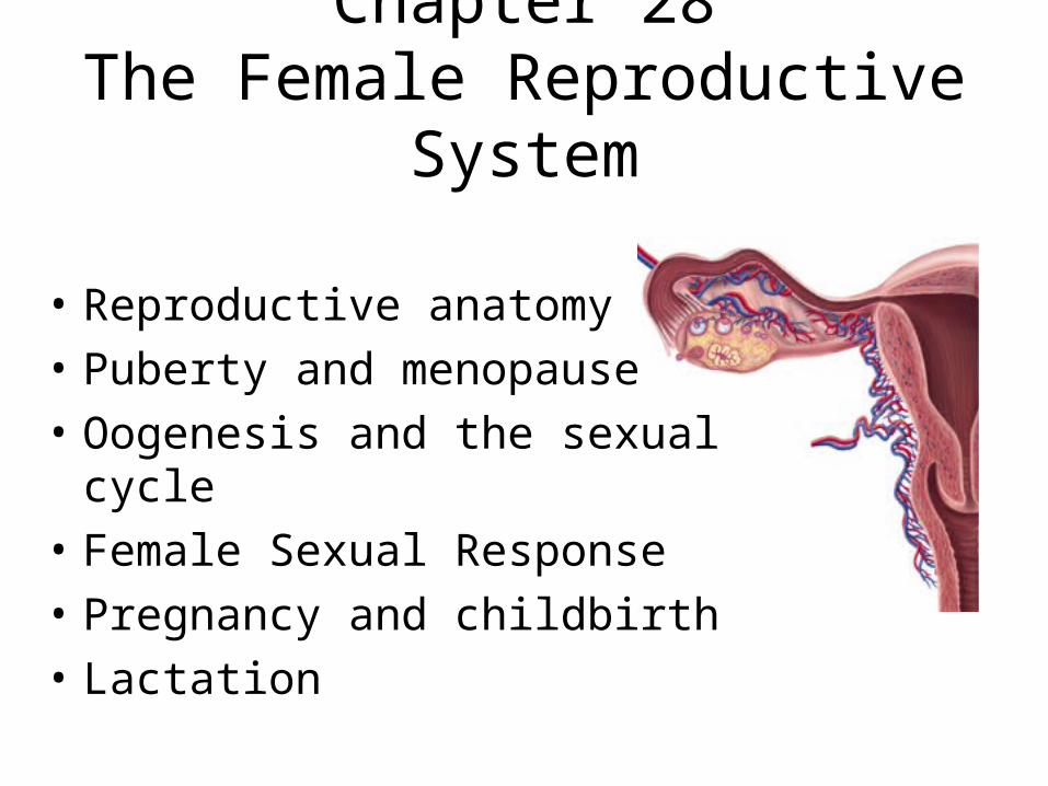
• Produce & deliver gametes
• Provide nutrition & room for fetal development
• Give birth• Nourish the
infant
Female Reproductive System
Sex Differentiation
• Male & female are indistinguishable for the first 8 to 10 weeks of development
• Female develops due to absence of hormones– absence of testosterone & müllerian-inhibiting factor
causes degeneration of (male) mesonephric duct– phallus becomes clitoris, urogenital folds develop into
labia minora & labioscrotal folds into labia majora– paramesonephric duct develops into uterine tubes,
uterus and vagina
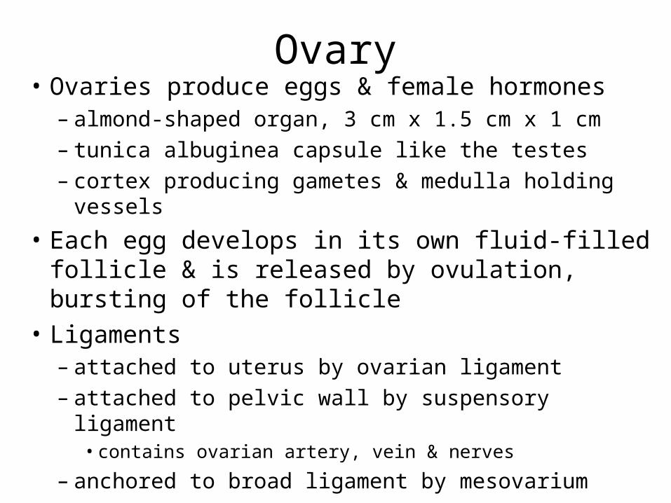
Ovary• Ovaries produce eggs & female hormones
– almond-shaped organ, 3 cm x 1.5 cm x 1 cm– tunica albuginea capsule like the testes– cortex producing gametes & medulla holding vessels
• Each egg develops in its own fluid-filled follicle & is released by ovulation, bursting of the follicle
• Ligaments– attached to uterus by ovarian ligament– attached to pelvic wall by suspensory ligament
• contains ovarian artery, vein & nerves
– anchored to broad ligament by mesovarium
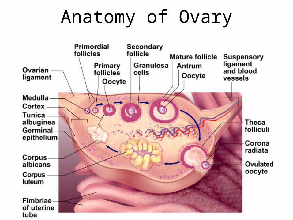
Anatomy of Ovary
Secondary Sex Organs (Genitalia)
• Internal genitalia– duct system consisting of uterine tubes, uterus &
vagina
• External genitalia– clitoris, labia minora, and labia majora– occupy the perineum– accessory glands beneath the skin provide lubrication
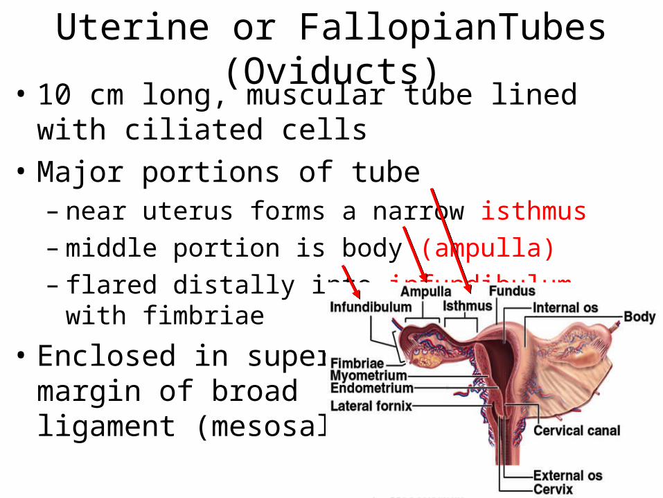
• 10 cm long, muscular tube lined with ciliated cells
• Major portions of tube– near uterus forms a narrow isthmus– middle portion is body (ampulla)– flared distally into infundibulum
with fimbriae
• Enclosed in superior margin of broad ligament (mesosalpinx)
Uterine or FallopianTubes (Oviducts)
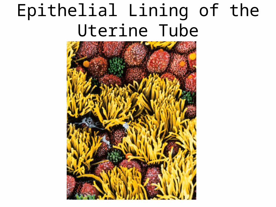
Epithelial Lining of the Uterine Tube
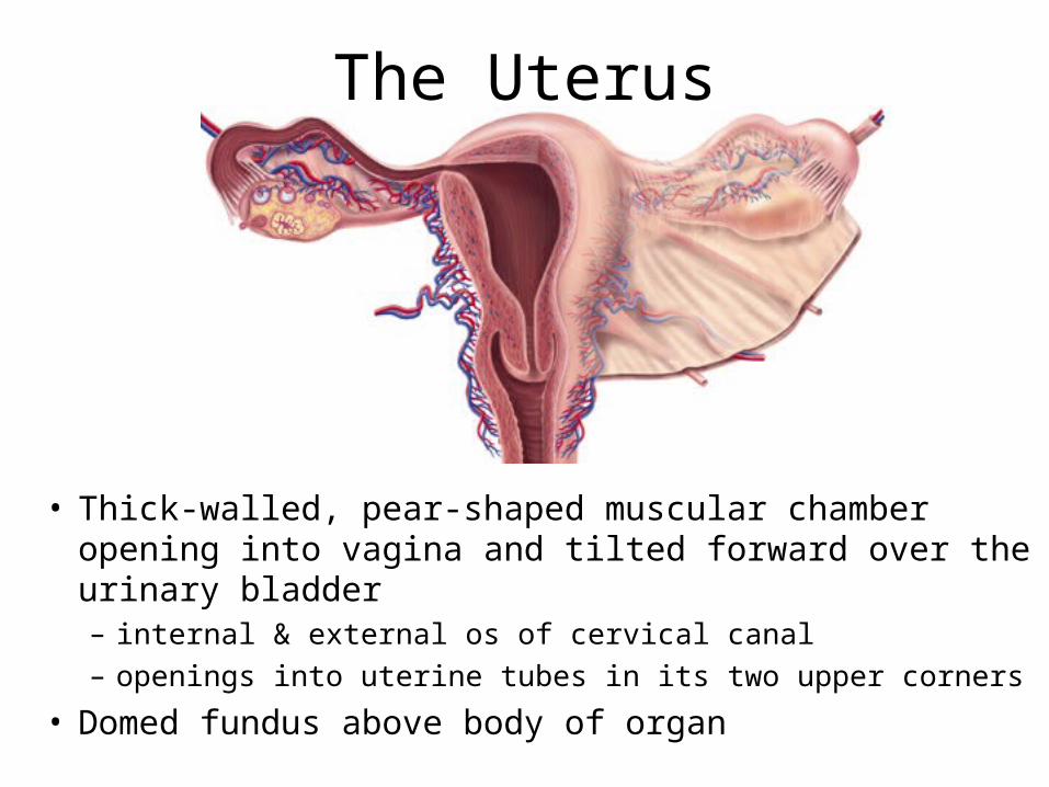
The Uterus
• Thick-walled, pear-shaped muscular chamber opening into vagina and tilted forward over the urinary bladder– internal & external os of cervical canal
– openings into uterine tubes in its two upper corners
• Domed fundus above body of organ
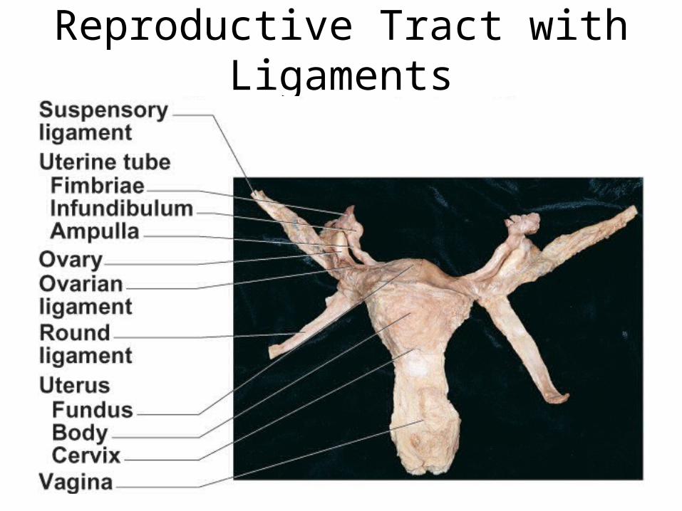
Reproductive Tract with Ligaments
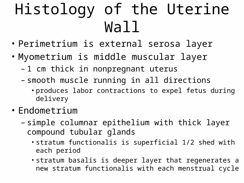
Histology of the Uterine Wall
• Perimetrium is external serosa layer
• Myometrium is middle muscular layer – 1 cm thick in nonpregnant uterus– smooth muscle running in all directions
• produces labor contractions to expel fetus during delivery
• Endometrium– simple columnar epithelium with thick layer compound
tubular glands• stratum functionalis is superficial 1/2 shed with each period• stratum basalis is deeper layer that regenerates a new stratum
functionalis with each menstrual cycle
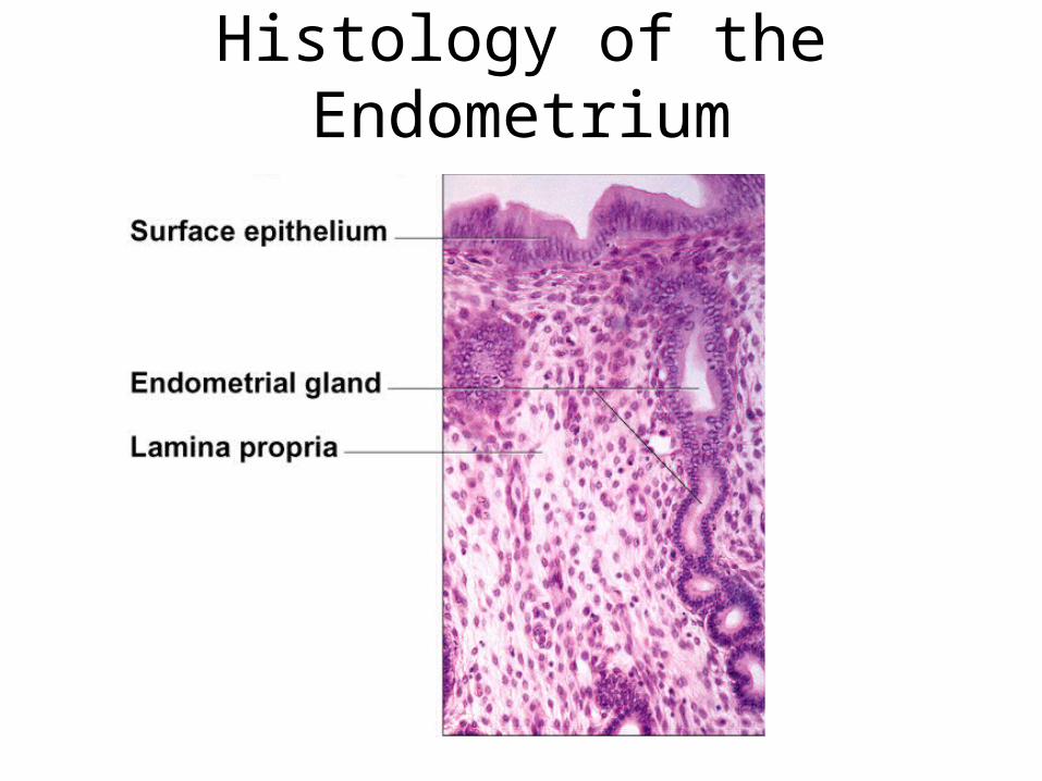
Histology of the Endometrium
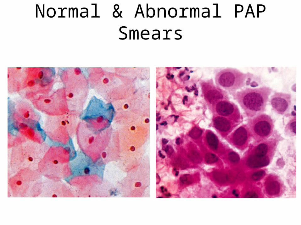
Normal & Abnormal PAP Smears
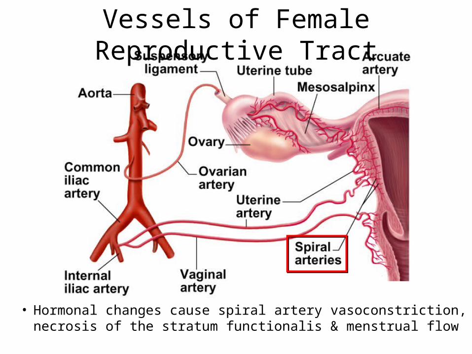
Vessels of Female Reproductive Tract
• Hormonal changes cause spiral artery vasoconstriction, necrosis of the stratum functionalis & menstrual flow
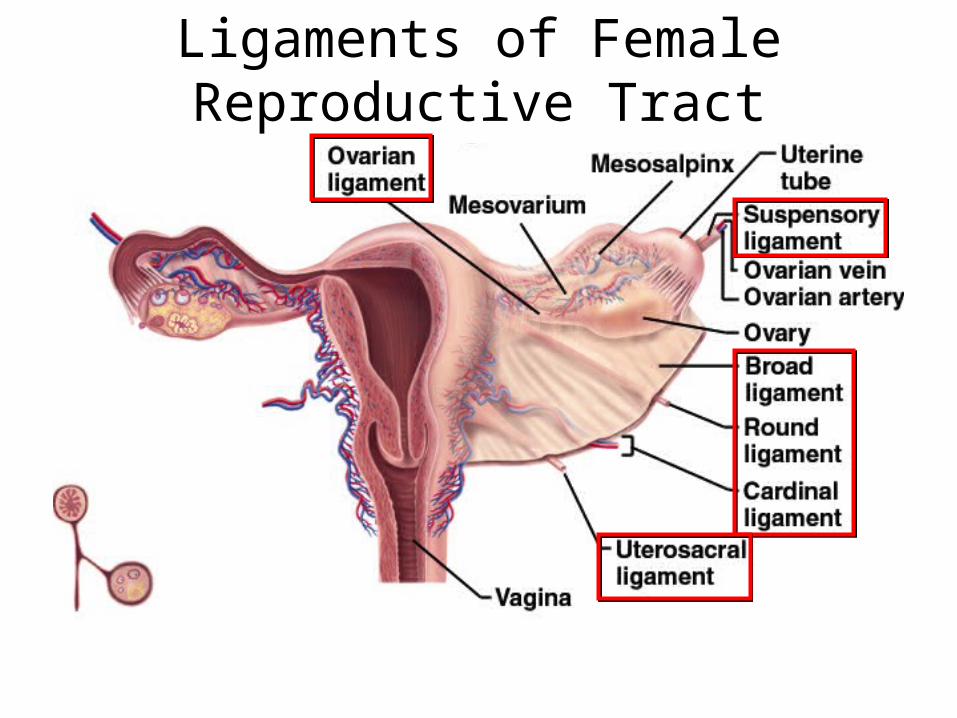
Ligaments of Female Reproductive Tract
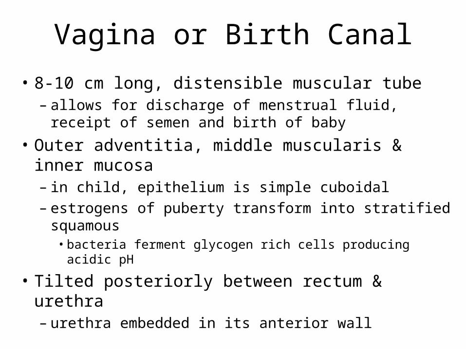
Vagina or Birth Canal
• 8-10 cm long, distensible muscular tube– allows for discharge of menstrual fluid, receipt of
semen and birth of baby
• Outer adventitia, middle muscularis & inner mucosa– in child, epithelium is simple cuboidal– estrogens of puberty transform into stratified squamous
• bacteria ferment glycogen rich cells producing acidic pH
• Tilted posteriorly between rectum & urethra– urethra embedded in its anterior wall
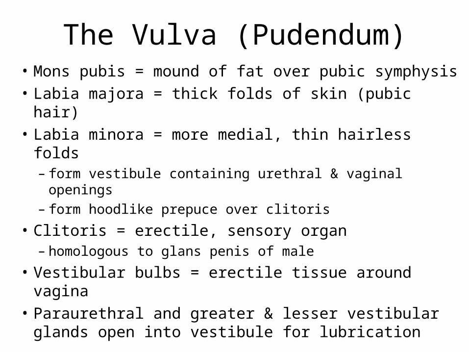
The Vulva (Pudendum)• Mons pubis = mound of fat over pubic symphysis
• Labia majora = thick folds of skin (pubic hair)
• Labia minora = more medial, thin hairless folds– form vestibule containing urethral & vaginal openings– form hoodlike prepuce over clitoris
• Clitoris = erectile, sensory organ– homologous to glans penis of male
• Vestibular bulbs = erectile tissue around vagina
• Paraurethral and greater & lesser vestibular glands open into vestibule for lubrication
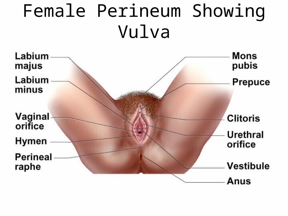
Female Perineum Showing Vulva
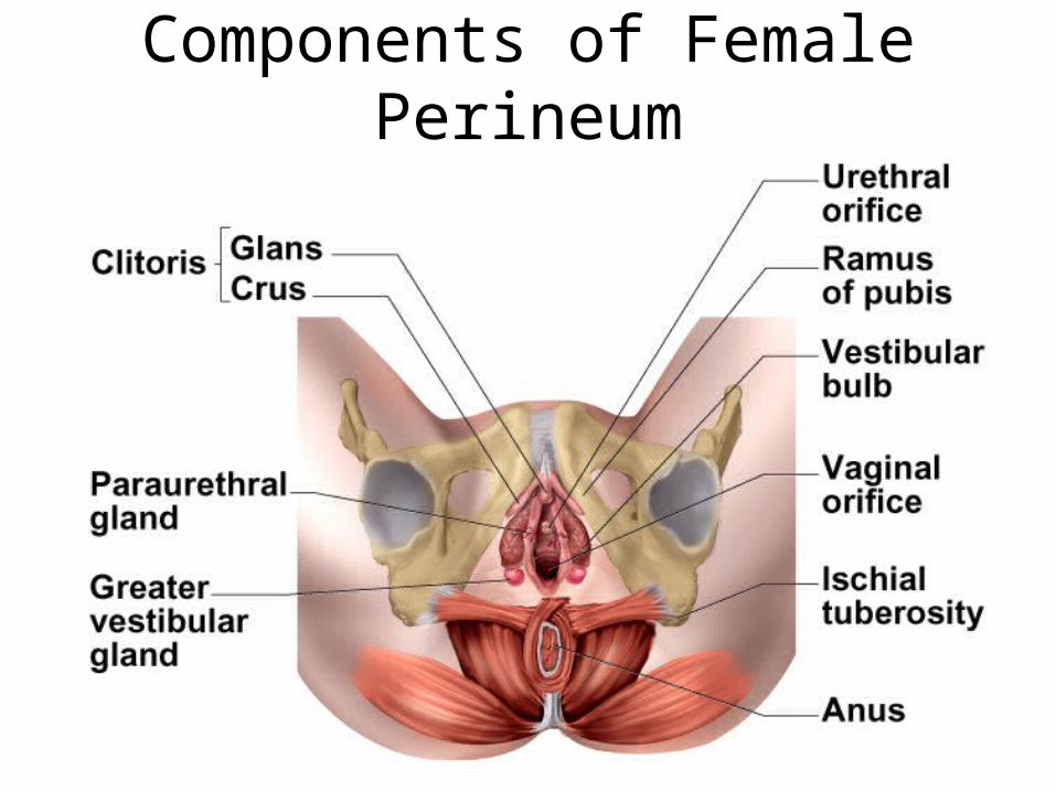
Components of Female Perineum
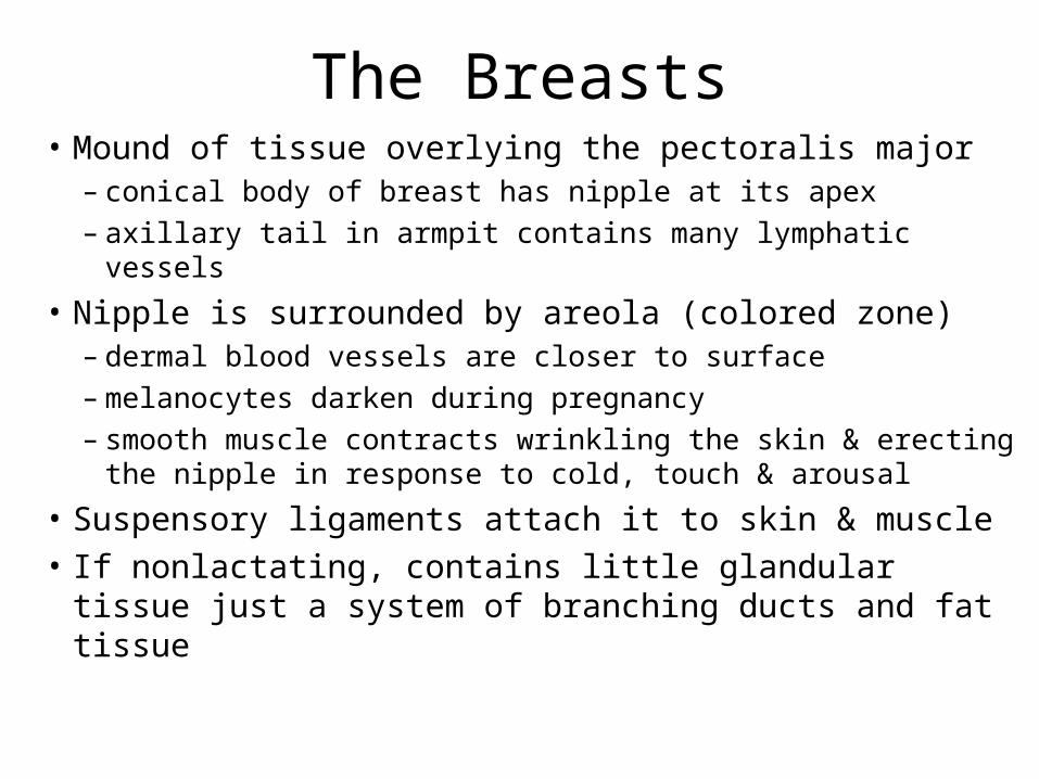
The Breasts• Mound of tissue overlying the pectoralis major
– conical body of breast has nipple at its apex– axillary tail in armpit contains many lymphatic vessels
• Nipple is surrounded by areola (colored zone)– dermal blood vessels are closer to surface– melanocytes darken during pregnancy– smooth muscle contracts wrinkling the skin & erecting
the nipple in response to cold, touch & arousal
• Suspensory ligaments attach it to skin & muscle
• If nonlactating, contains little glandular tissue just a system of branching ducts and fat tissue
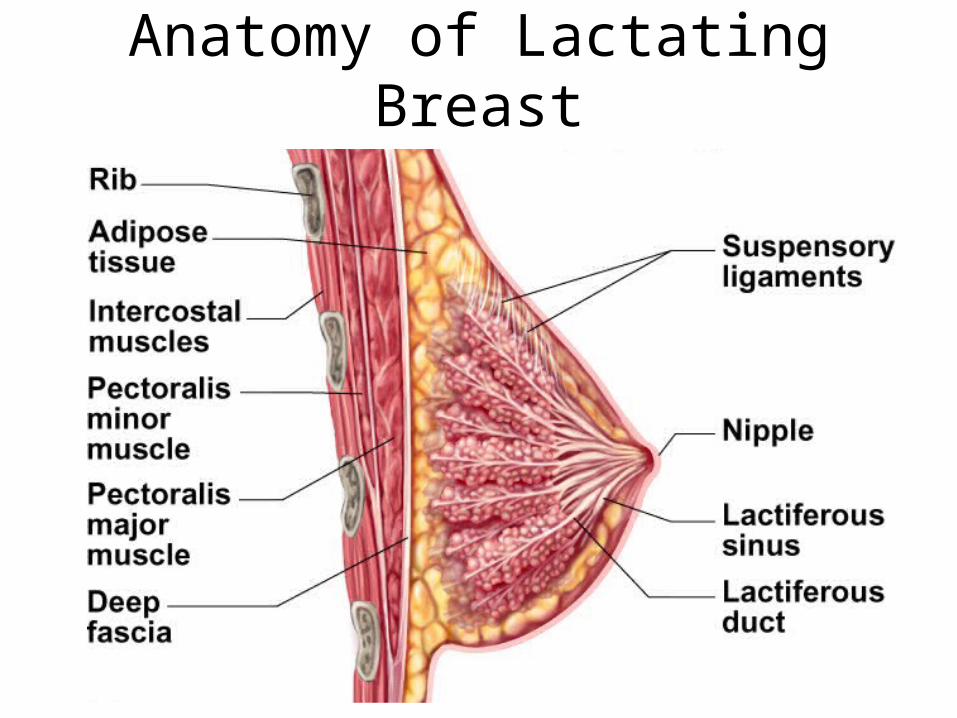
Anatomy of Lactating Breast

Anatomy of Lactating Breast
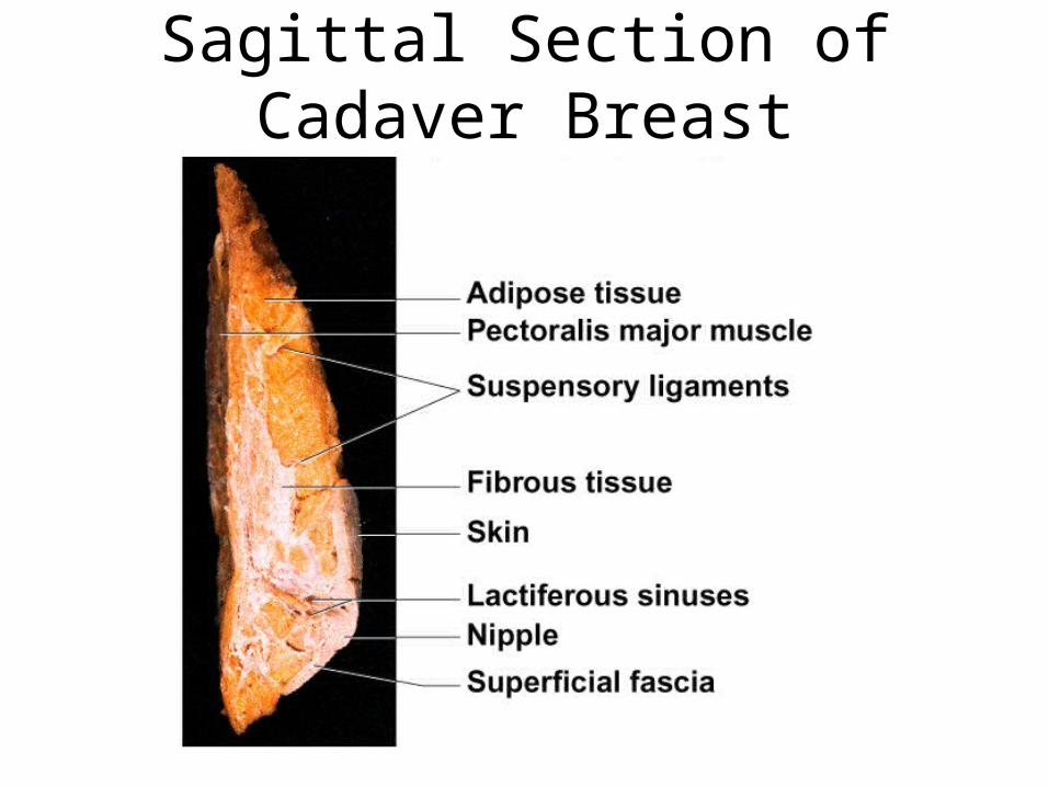
Sagittal Section of Cadaver Breast
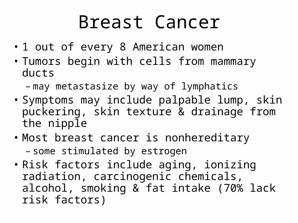
Breast Cancer• 1 out of every 8 American women• Tumors begin with cells from mammary ducts
– may metastasize by way of lymphatics
• Symptoms may include palpable lump, skin puckering, skin texture & drainage from the nipple
• Most breast cancer is nonhereditary– some stimulated by estrogen
• Risk factors include aging, ionizing radiation, carcinogenic chemicals, alcohol, smoking & fat intake (70% lack risk factors)
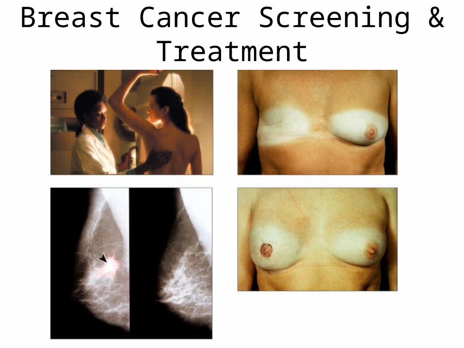
Breast Cancer Screening & Treatment

Puberty• Begins at age 9 or 10 for most girls in the U.S.• Triggered by rising levels of GnRH which stimulate
anterior lobe of pituitary to produce FSH & LH (follicle-stimulating & luteinizing hormone)– FSH stimulates follicles to secrete estrogen & progesterone
• 2nd sex organs maturation, in height & width of pelvis• prepares uterus for pregnancy
• Thelarche = development of breasts• Pubarche = growth of pubic & axillary hair, apocrine &
sebaceous glands • Menarche = first menstrual period (age 12)
– requires at least 17% body fat in teenager, 22% in adult
• Female hormones secreted cyclically & in sequence
Climacteric and Menopause• Midlife change in hormone secretion accompanied
by menopause (cessation of menstruation)– average age of 52
• Age related depletion of follicles means less secretion of estrogen & progesterone– atrophy of uterus, vagina & breasts– skin becomes thinner, bone mass declines, and risks of
cardiovascular disease increase– hot flashes (sudden dilation of cutaneous arteries)
occur several times a day
• HRT = low dose estrogen & progesterone therapy

Oogensis and the Sexual Cycle
• Reproductive cycle - events occurring between fertilization and birth
• Sexual cycle - events recurring every month when pregnancy does not occur– ovarian cycle = events in the ovaries– menstrual cycle = parallel changes in the uterus
Oogenesis• Monthly event producing 1 haploid egg by
meiosis• Embryonic development of ovary
– female germ cells arise from yolk sac of embryo– differentiate into oogonia & multiply in number– transform into primary oocytes(eggs) -- early meiosis I– most degenerate (atresia) by time reach childhood– by puberty 400,000 oocytes remain
• FSH stimulates completion of meiosis I, produces secondary oocyte & 1st polar body– proceeds to meiosis II & ceases until fertilization– after fertilization , releases 2nd polar body
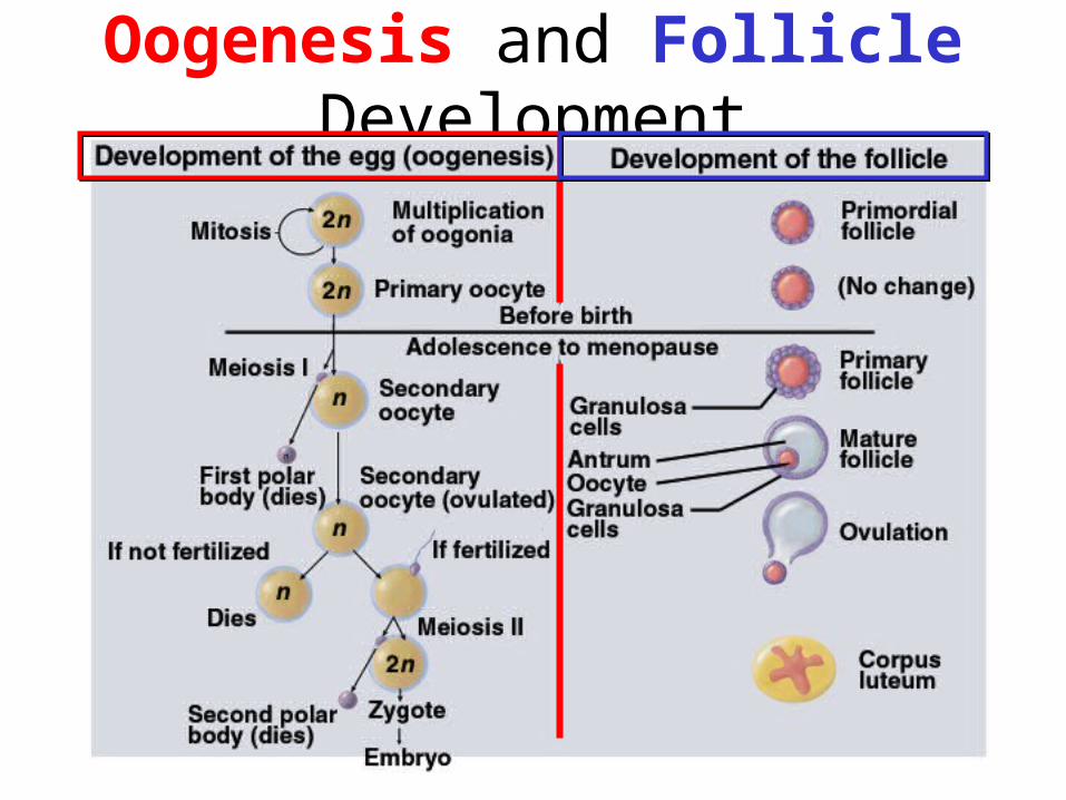
Oogenesis and Follicle Development
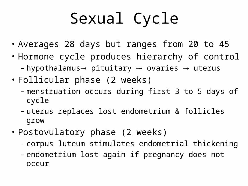
Sexual Cycle
• Averages 28 days but ranges from 20 to 45
• Hormone cycle produces hierarchy of control– hypothalamus pituitary ovaries uterus
• Follicular phase (2 weeks) – menstruation occurs during first 3 to 5 days of cycle– uterus replaces lost endometrium & follicles grow
• Postovulatory phase (2 weeks)– corpus luteum stimulates endometrial thickening– endometrium lost again if pregnancy does not occur
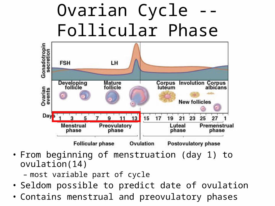
Ovarian Cycle -- Follicular Phase
• From beginning of menstruation (day 1) to ovulation(14) – most variable part of cycle
• Seldom possible to predict date of ovulation• Contains menstrual and preovulatory phases
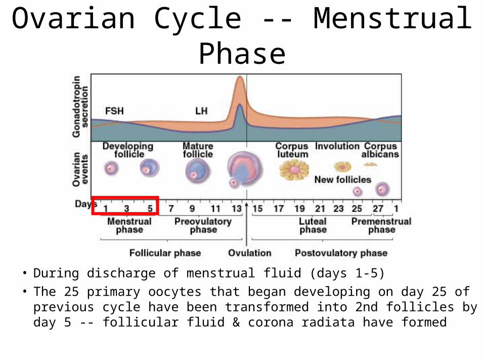
Ovarian Cycle -- Menstrual Phase
• During discharge of menstrual fluid (days 1-5)• The 25 primary oocytes that began developing on day 25 of
previous cycle have been transformed into 2nd follicles by day 5 -- follicular fluid & corona radiata have formed
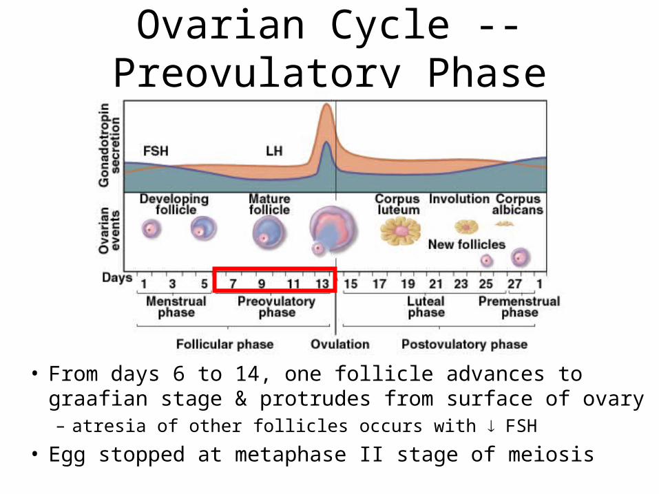
Ovarian Cycle -- Preovulatory Phase
• From days 6 to 14, one follicle advances to graafian stage & protrudes from surface of ovary– atresia of other follicles occurs with FSH
• Egg stopped at metaphase II stage of meiosis
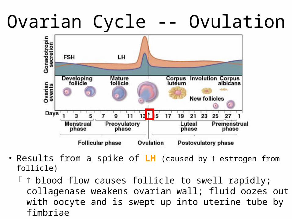
Ovarian Cycle -- Ovulation
• Results from a spike of LH (caused by estrogen from follicle)
blood flow causes follicle to swell rapidly; collagenase weakens ovarian wall; fluid oozes out with oocyte and is swept up into uterine tube by fimbriae
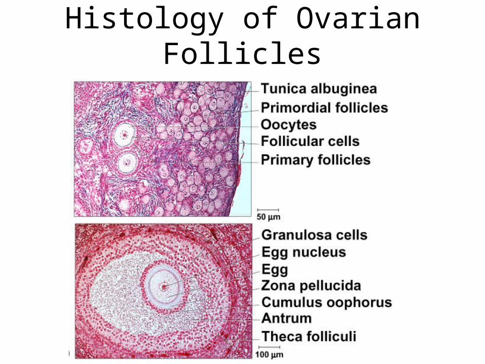
Histology of Ovarian Follicles
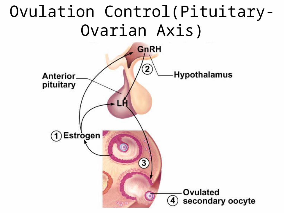
Ovulation Control(Pituitary-Ovarian Axis)
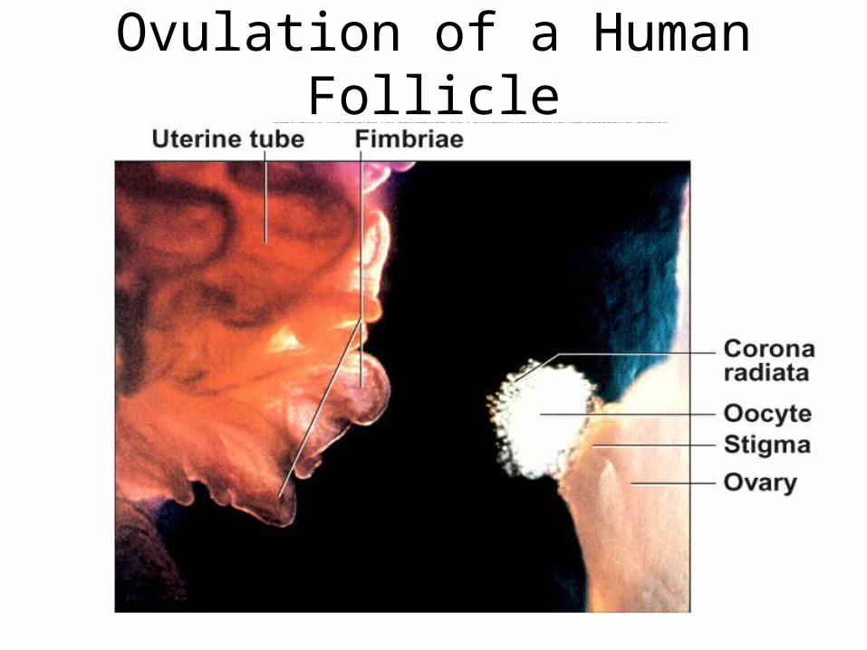
Ovulation of a Human Follicle
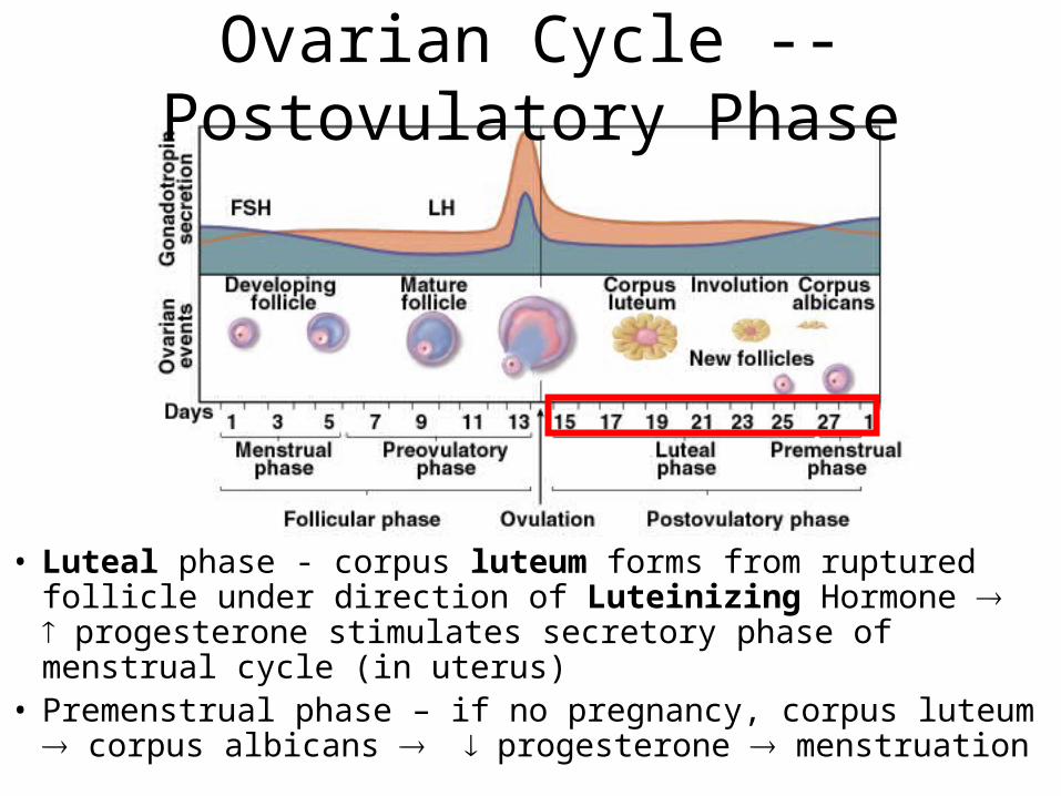
Ovarian Cycle -- Postovulatory Phase
• Luteal phase - corpus luteum forms from ruptured follicle under direction of Luteinizing Hormone progesterone stimulates secretory phase of menstrual cycle (in uterus)
• Premenstrual phase – if no pregnancy, corpus luteum corpus albicans progesterone menstruation
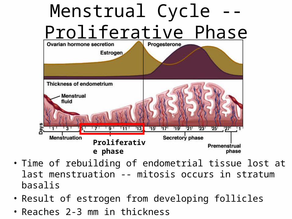
Menstrual Cycle -- Proliferative Phase
• Time of rebuilding of endometrial tissue lost at last menstruation -- mitosis occurs in stratum basalis
• Result of estrogen from developing follicles• Reaches 2-3 mm in thickness
Proliferative phase
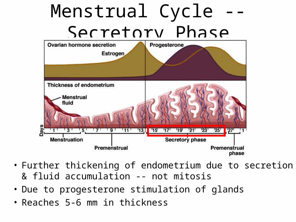
• Further thickening of endometrium due to secretion & fluid accumulation -- not mitosis
• Due to progesterone stimulation of glands• Reaches 5-6 mm in thickness
Menstrual Cycle -- Secretory Phase
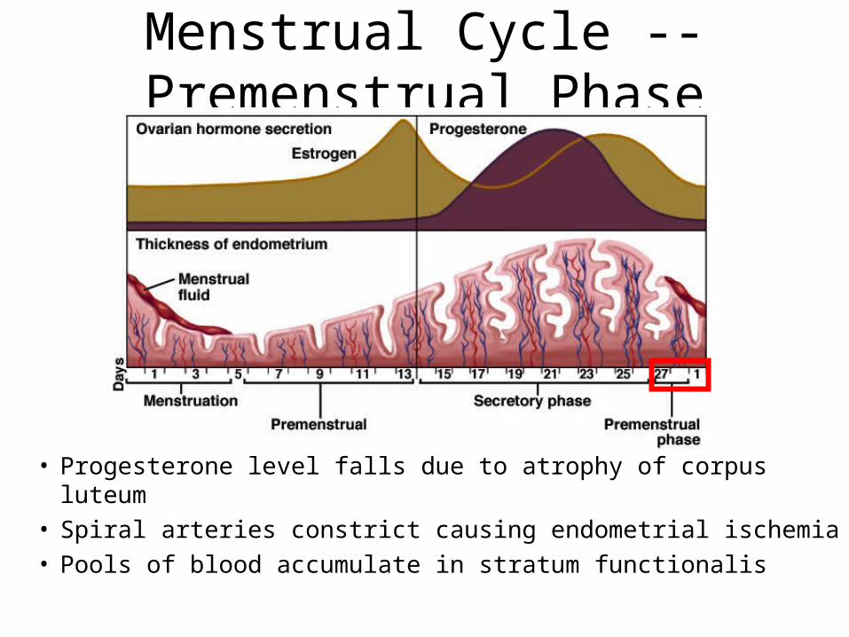
Menstrual Cycle -- Premenstrual Phase
• Progesterone level falls due to atrophy of corpus luteum• Spiral arteries constrict causing endometrial ischemia• Pools of blood accumulate in stratum functionalis
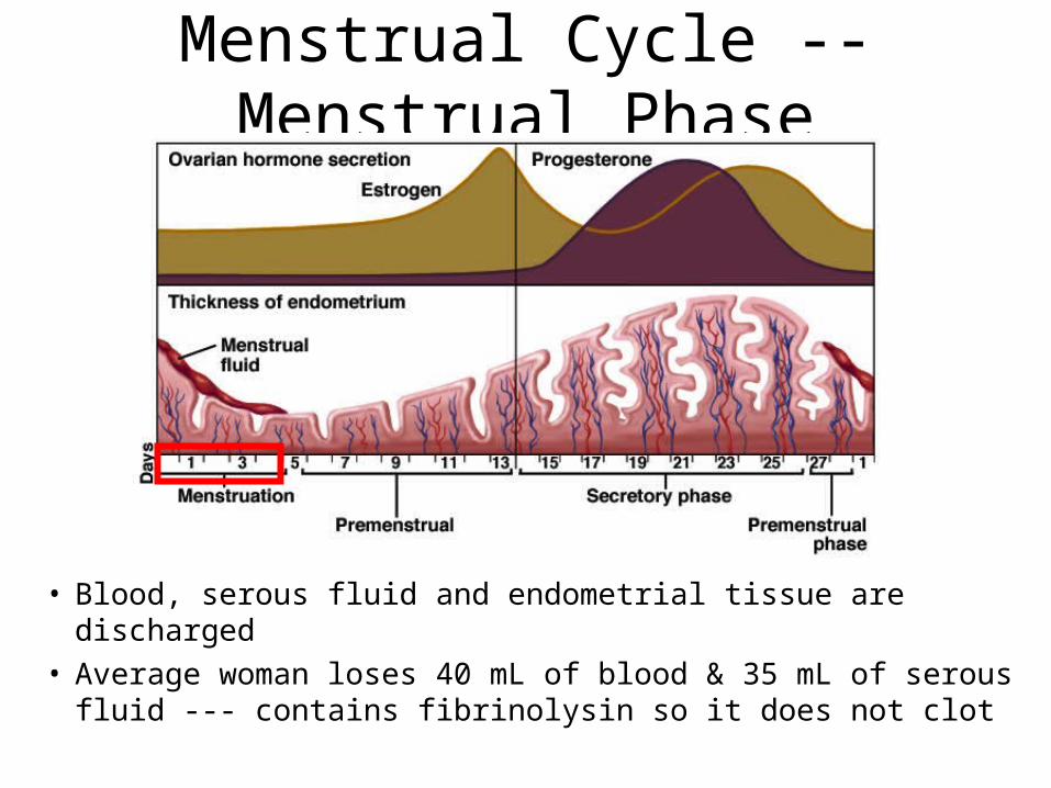
Menstrual Cycle -- Menstrual Phase
• Blood, serous fluid and endometrial tissue are discharged• Average woman loses 40 mL of blood & 35 mL of serous
fluid --- contains fibrinolysin so it does not clot
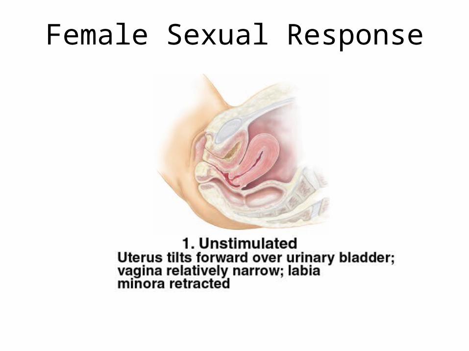
Female Sexual Response
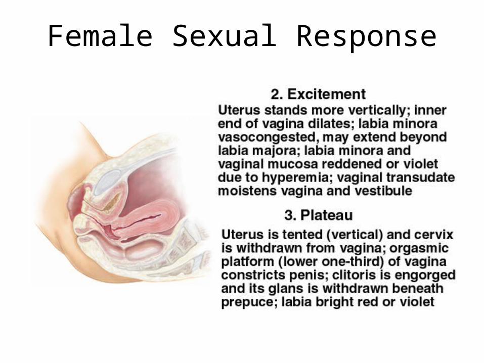
Female Sexual Response
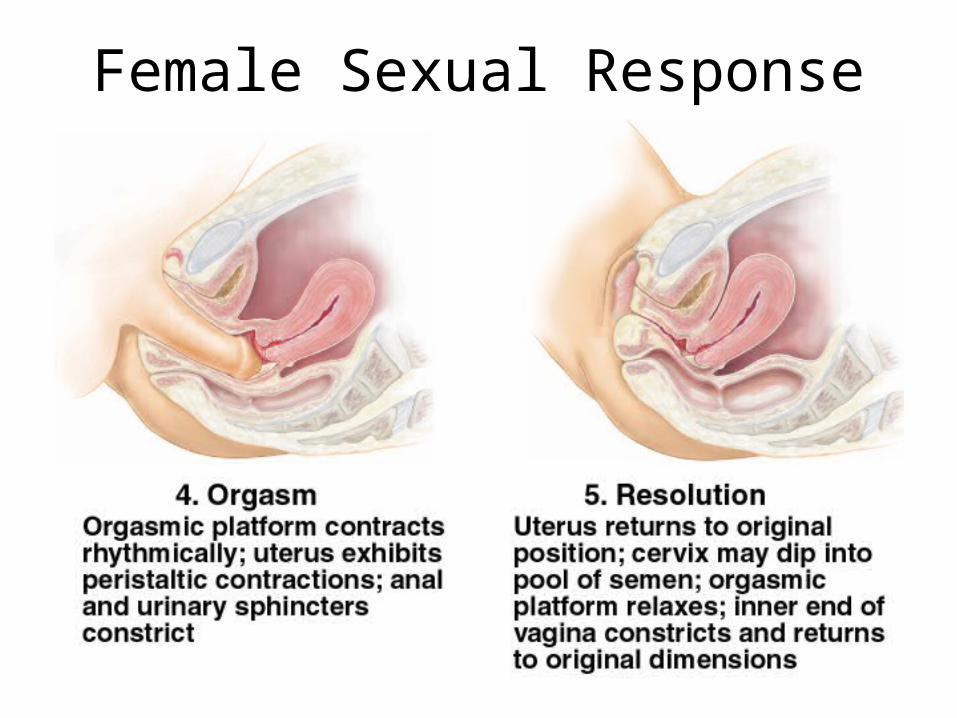
Female Sexual Response
Pregnancy and Childbirth
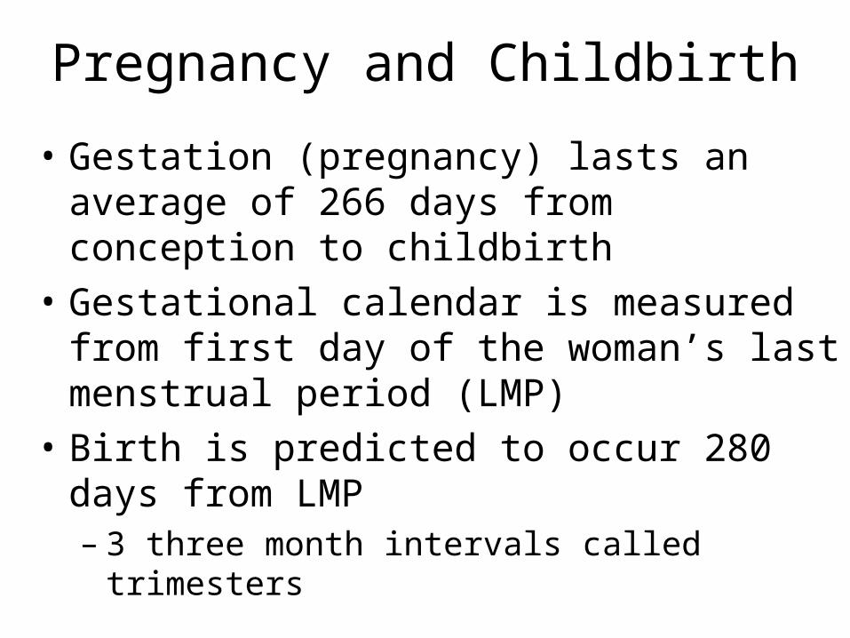
• Gestation (pregnancy) lasts an average of 266 days from conception to childbirth
• Gestational calendar is measured from first day of the woman’s last menstrual period (LMP)
• Birth is predicted to occur 280 days from LMP– 3 three month intervals called trimesters
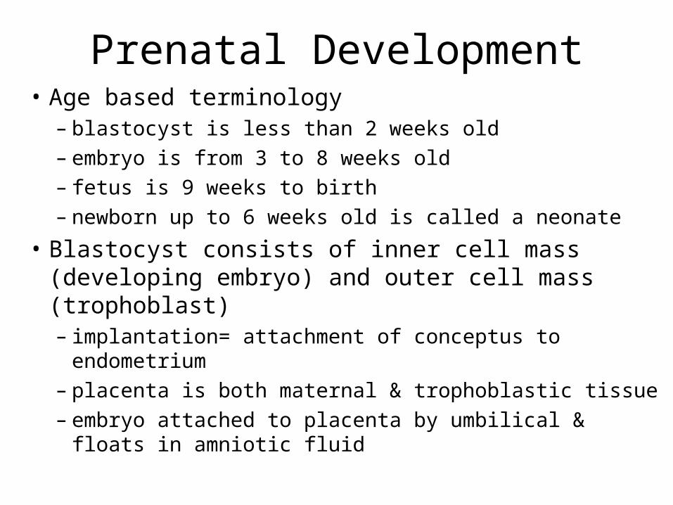
Prenatal Development• Age based terminology
– blastocyst is less than 2 weeks old– embryo is from 3 to 8 weeks old– fetus is 9 weeks to birth– newborn up to 6 weeks old is called a neonate
• Blastocyst consists of inner cell mass (developing embryo) and outer cell mass (trophoblast) – implantation= attachment of conceptus to endometrium– placenta is both maternal & trophoblastic tissue– embryo attached to placenta by umbilical & floats in
amniotic fluid
Hormones of Pregnancy (1)
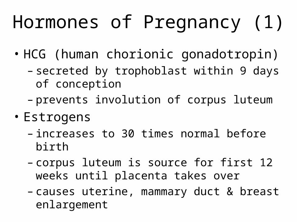
• HCG (human chorionic gonadotropin)– secreted by trophoblast within 9 days of conception– prevents involution of corpus luteum
• Estrogens – increases to 30 times normal before birth– corpus luteum is source for first 12 weeks until
placenta takes over– causes uterine, mammary duct & breast enlargement
Hormones of Pregnancy (2)
• Progesterone secreted by placenta & corpus luteum• suppresses secretion of FSH & LH preventing follicular development• prevents menstruation & thickens endometrium• stimulates development of acini in breast tissue
• HCS (human chorionic somatomammotropin)• called human placental lactogen • secreted from placenta in direct proportion to its size mother’s glucose usage and release of fatty acids
• Other endocrine organs• thyroid gland increases 50% in size BMR of mother• parathyroid glands enlarge & stimulate osteoclasts to release additional
calcium from mother’s bones• Aldosterone secretion fluid retention & leads to in mother’s blood
volume
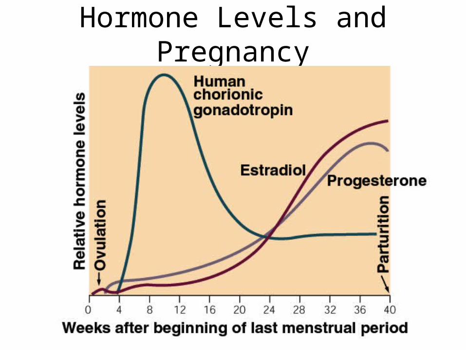
Hormone Levels and Pregnancy
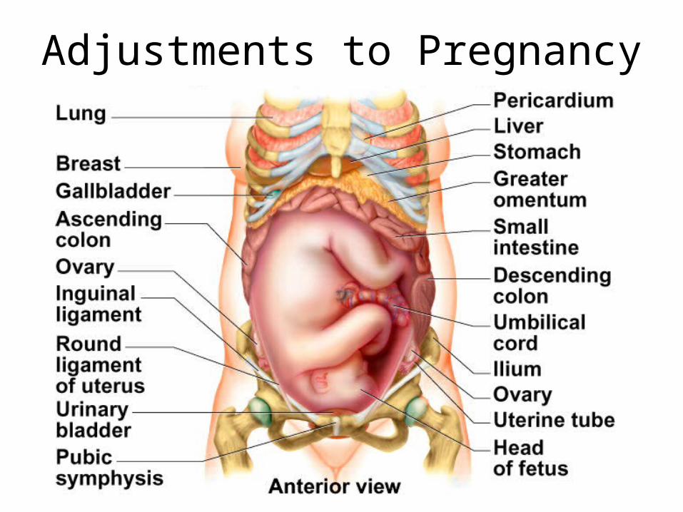
Adjustments to Pregnancy
Adjustments to Pregnancy -- Digestive System, Nutrition & Metabolism
• Nausea during first few months• Constipation & heartburn due to intestinal motility &
pressure on stomach• Basal metabolic rate may stimulate appetite
– healthy weight gain is 24 lb.
• Placenta stores nutrients prior to 3rd trimester– high demand for protein, iron, calcium & phosphates
– supplemental vitamin K reduces risk of neonatal hemorrhages in the brain during delivery
– early supplemental folic acid prevents neurological disorders• spina bifida and anencephaly
Adjustments to Pregnancy -- Circulatory and Respiratory Systems
• Mother’s blood volume rises 30% due to fluid retention & hemopoiesis– by full term, placenta requires 625 mL of blood/minute
• Cardiac output rises about 30% by 27 weeks
• Pressure on large pelvic blood vessels can produce hemorrhoids and varicose veins
• Minute respiratory ventilation about 50%– demands of fetus and higher maternal metabolic rate
– ventilation adjusted to keep PCO2 lower than normal
• Respiratory rate due to inability to breathe as deeply
Adjustments to Pregnancy -- Urinary & Integumentary Systems
• Aldosterone & steroids of pregnancy promote salt and water retention
• GFR by 50% & output is slightly elevated– enabling women to dispose of additional metabolic wastes
• Bladder compression frequency of urination• Skin must grow -- dermal stretching stretch marks• Linea alba may become dark line (linea nigra)• Blotchy darkening of the skin over the nose & cheeks
may temporarily occur(chloasma or “mask of pregnancy”)
Childbirth & Uterine Contractility
• Parturition is process of giving birth by means of contraction of mother’s uterine & abdominal muscles
• Braxton Hicks are weak contractions occurring throughout gestation = false labor
• Progesterone inhibits contractions while estrogen stimulates contractions
• Nearing full term -- posterior pituitary release more oxytocin & uterus produces more receptors– directly stimulates myometrial contractions– stimulates fetal membranes to produce prostaglandins which are
synergists of oxytocin
• Fetus secretes cortisol enhancing estrogen secretion & oxytocin stimulating prostaglandin secretion
Labor Contractions• Contractions begin 30 minutes apart and eventually occur
every 1-3 minutes– periodically relax to blood flow to placenta (fetus)
– contractions strongest in the fundus & body pushing the fetus into the cervix
• Self-amplifying cycle of stretch & contraction (reflex)– positive feedback cycle increasing contractions
• cervical stretching oxytocin secretion uterine contraction cervical stretching
• 2nd reflex arc is from uterus spinal cord abdominal skeletal muscles producing contractions that help expel fetus
• Pain of labor is ischemia of myometrium, stretching of cervix, vagina & perineum (episiotomy prevents tearing)– large head & narrow pelvic outlet because of bipedal locomotion
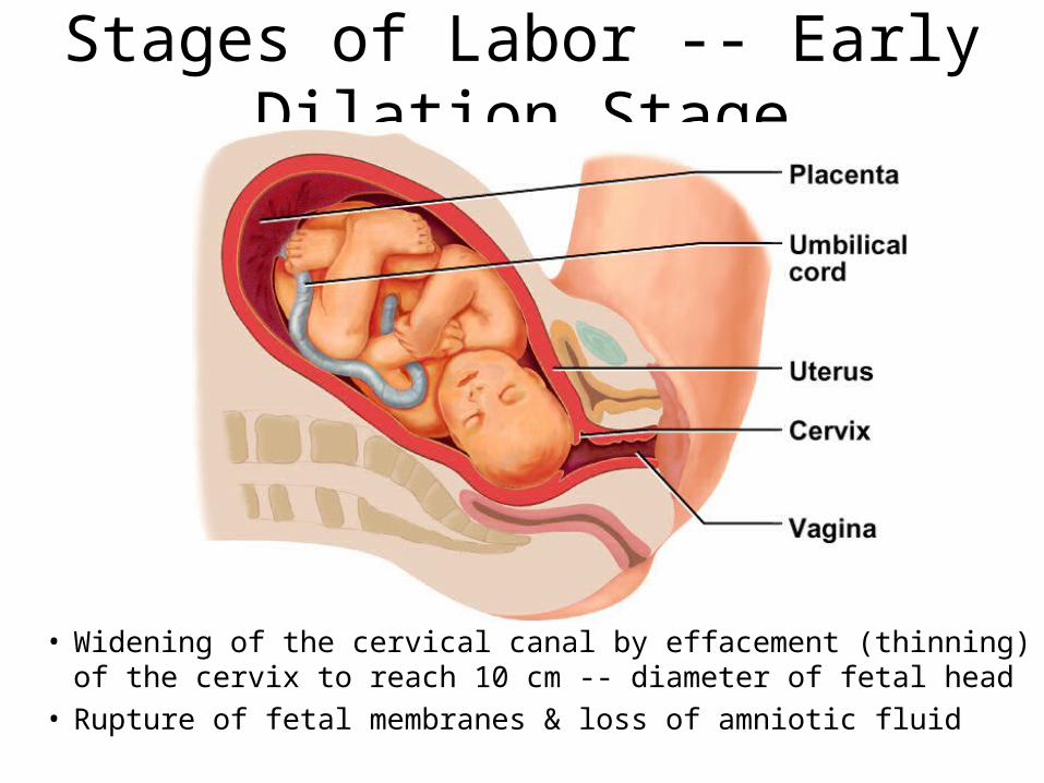
Stages of Labor -- Early Dilation Stage
• Widening of the cervical canal by effacement (thinning) of the cervix to reach 10 cm -- diameter of fetal head
• Rupture of fetal membranes & loss of amniotic fluid
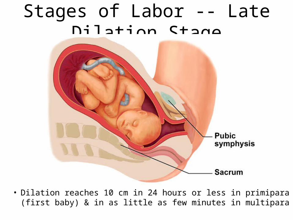
Stages of Labor -- Late Dilation Stage
• Dilation reaches 10 cm in 24 hours or less in primipara (first baby) & in as little as few minutes in multipara
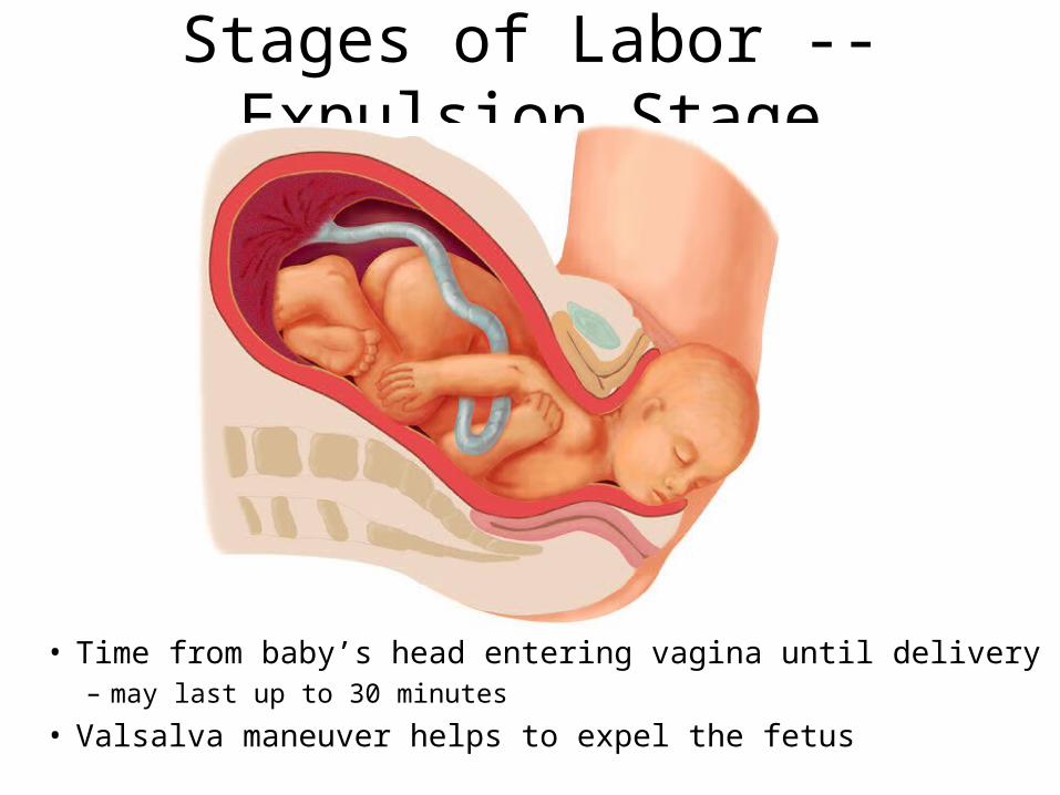
Stages of Labor -- Expulsion Stage
• Time from baby’s head entering vagina until delivery– may last up to 30 minutes
• Valsalva maneuver helps to expel the fetus
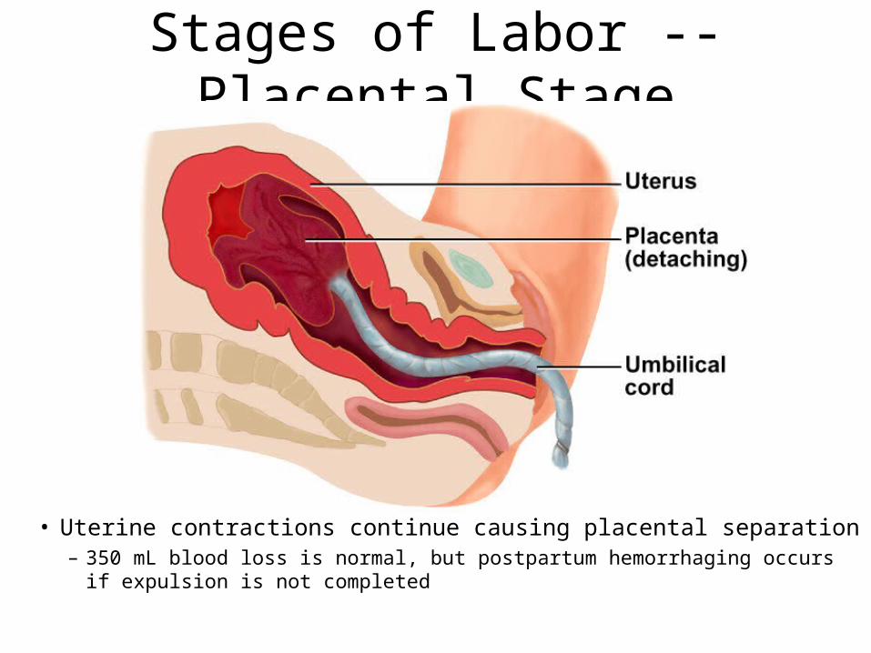
Stages of Labor -- Placental Stage
• Uterine contractions continue causing placental separation– 350 mL blood loss is normal, but postpartum hemorrhaging occurs if
expulsion is not completed
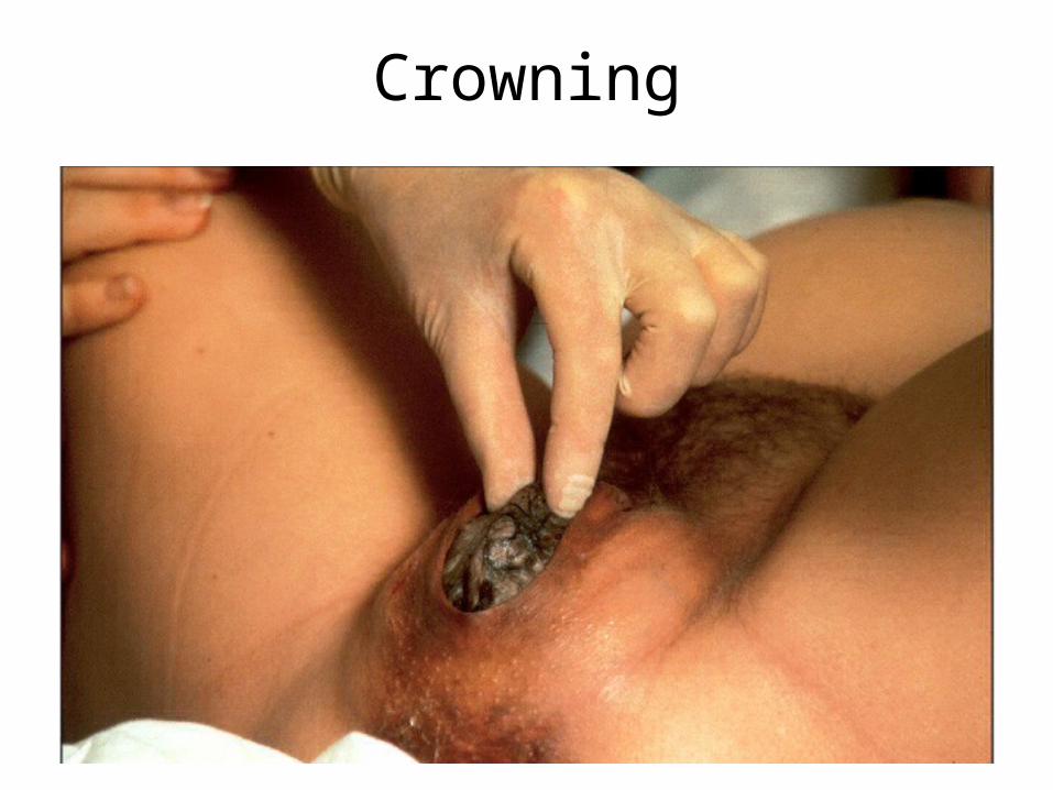
Crowning
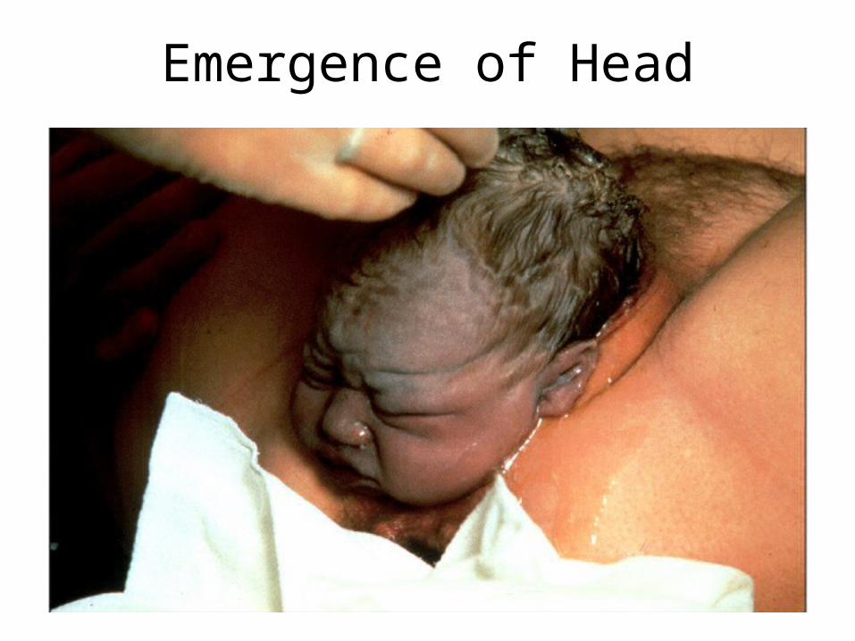
Emergence of Head
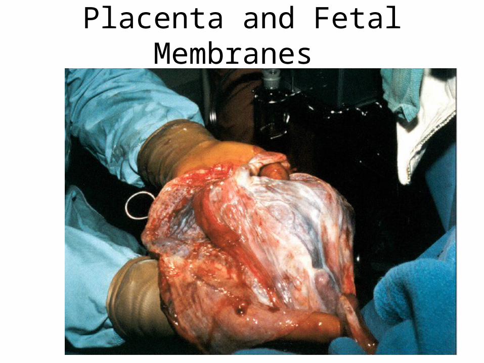
Placenta and Fetal Membranes
Puerperium
• First 6 weeks after delivery = puerperium
• Mother’s anatomy & physiology return to normal– shrinkage of the uterus is called involution
• at pregavid weight in 4 weeks
• accomplished by autolysis by lysosomal enzymes– vaginal discharge called lochia
– breastfeeding promotes involution• suppresses estrogen secretion
• stimulates oxytocin which causes myometrial contraction
Development of Mammary Glands
• Lactation = synthesis and ejection of milk from mammary glands
• High estrogen levels in pregnancy cause ducts to grow and branch
• Progesterone stimulates budding & development of the acini at the ends of the ducts
Colostrum & Milk Synthesis• Colostrum forms in late pregnancy
– similar to breast milk but contains 1/3 less fat, thinner
– nutrition for first 1 to 3 days after birth
– contains IgA protection from gastroenteritis
• Synthesis is promoted by prolactin (from pituitary)– synthesis of hormone begins 5 weeks into pregnancy, by full
term it is 20x normal level
– steroid hormones from placenta oppose it until birth
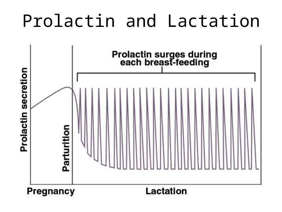
• At birth, prolactin secretion drops to nonpregnant levels, but 20 times that after nursing event ceases– without continuous nursing, production stops in 1 week
• 5-10% of women become pregnant again while nursing– inhibition of GnRH & reduced ovarian cycling
Prolactin and Lactation
Milk Ejection
• Milk ejection or let-down is controlled by a neuroendocrine reflex– infant’s suckling stimulates sensory receptors in nipple,
signaling the hypothalamus & posterior pituitary to release oxytocin
– oxytocin stimulates myoepithelial cells
• Myoepithelial cells surround each gland acinus– epithelial cells packed with actin – contract like smooth muscle to squeeze milk into duct
• milk flow within 30-60 seconds after suckling begins
Breast Milk• Cow’s milk is not a good substitute for human
milk– 1/3 less lactose but 3 times as much protein– harder to digest & more nitrogenous waste (diaper rash)
• Colostrum & milk have a laxative effect that clears intestine of meconium (green, bile-filled fecal material in newborn)
• Breast milk supplies antibodies & colonizes intestine with beneficial bacteria
• Nursing woman can produce 1.5L per day calorie intake by 300, Ca+2 & vitamin D
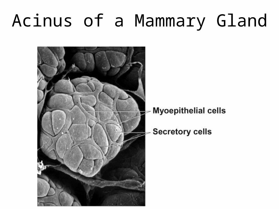
Acinus of a Mammary Gland
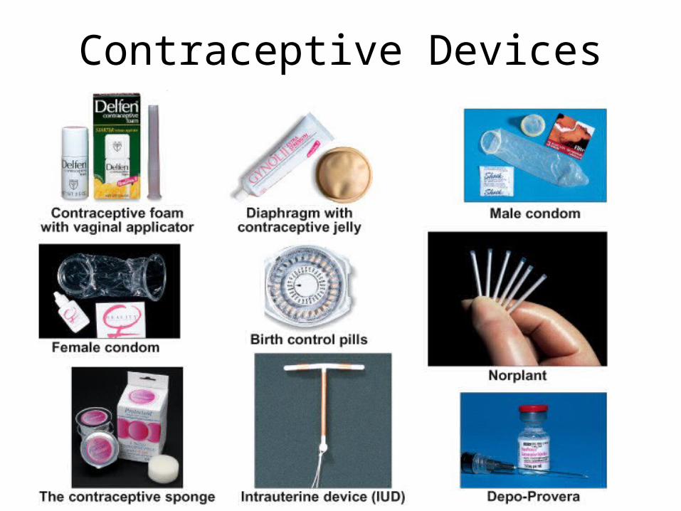
Contraceptive Devices