Embed Size (px)
Citation preview
Chapter 25 The Skin
The Skin: More than a Mechanical Barrier• Squamous epithelial cells• Melanocytes• Dendritic cells• Lymphocytes• Neural end organs and axonal processes• Adnexal components• Sweat glands• Hair follicles
• Definitions of Macroscopic Terms• Definitions of microscopic Terms
Definition of Macroscopic Terms
Excoriation Traumatic lesion breaking the epidermis and causing a raw
linear area( deep scratch): often self-induced
Lichenification Thickened and rough skin characterized by
prominent skin markings: usually the result of repeated rubbing
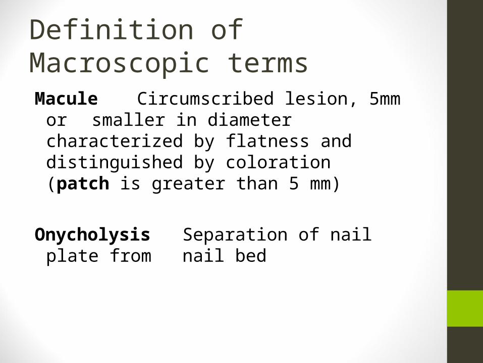
Definition of Macroscopic terms
Macule Circumscribed lesion, 5mm or smaller in diameter
characterized by flatness and distinguished by coloration (patch is greater than 5 mm)
Onycholysis Separation of nail plate from nail bed
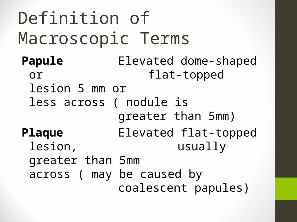
Definition of Macroscopic Terms
Papule Elevated dome-shaped or flat-topped lesion 5 mm or
less across ( nodule is greater than
5mm)Plaque Elevated flat-topped lesion,
usually greater than 5mm across ( may be caused
by coalescent papules)
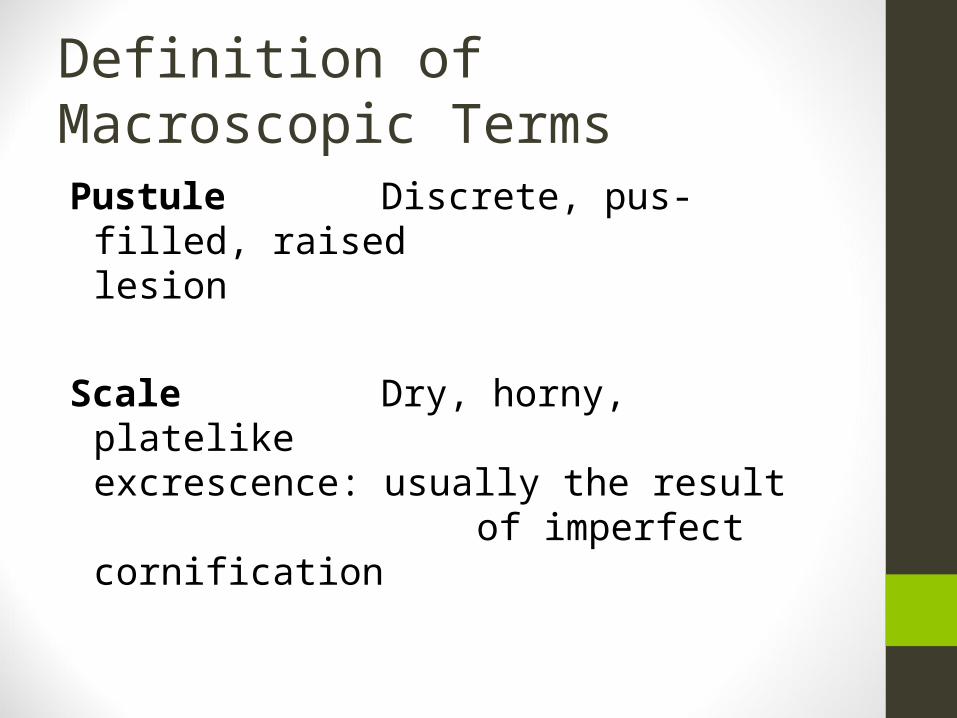
Definition of Macroscopic Terms
Pustule Discrete, pus-filled, raised lesion
Scale Dry, horny, platelike excrescence: usually
the result of imperfect cornification
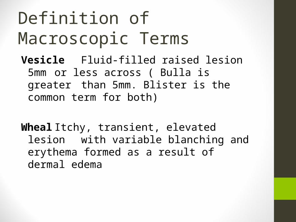
Definition of Macroscopic Terms
Vesicle Fluid-filled raised lesion 5mm or less across ( Bulla is greater
than 5mm. Blister is the common term
for both)
Wheal Itchy, transient, elevated lesion with variable blanching
and erythema formed as a result of dermal edema
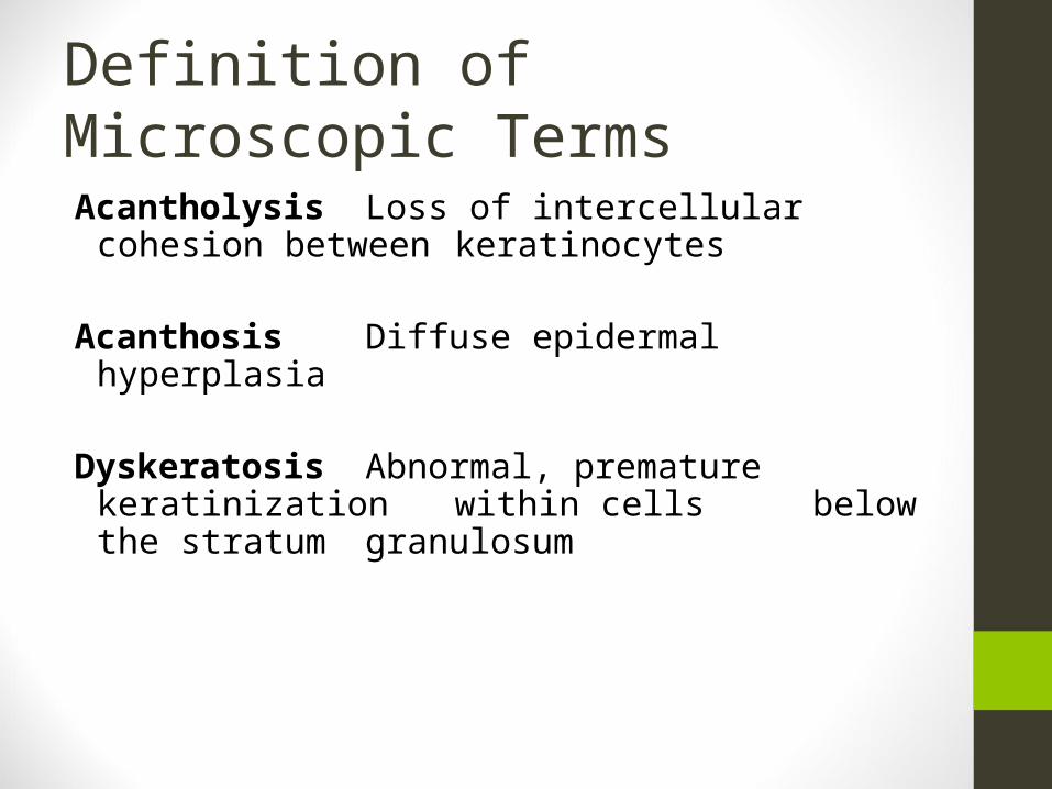
Definition of Microscopic Terms
Acantholysis Loss of intercellular cohesion between keratinocytes
Acanthosis Diffuse epidermal hyperplasia
Dyskeratosis Abnormal, premature keratinization within cells
below the stratum granulosum
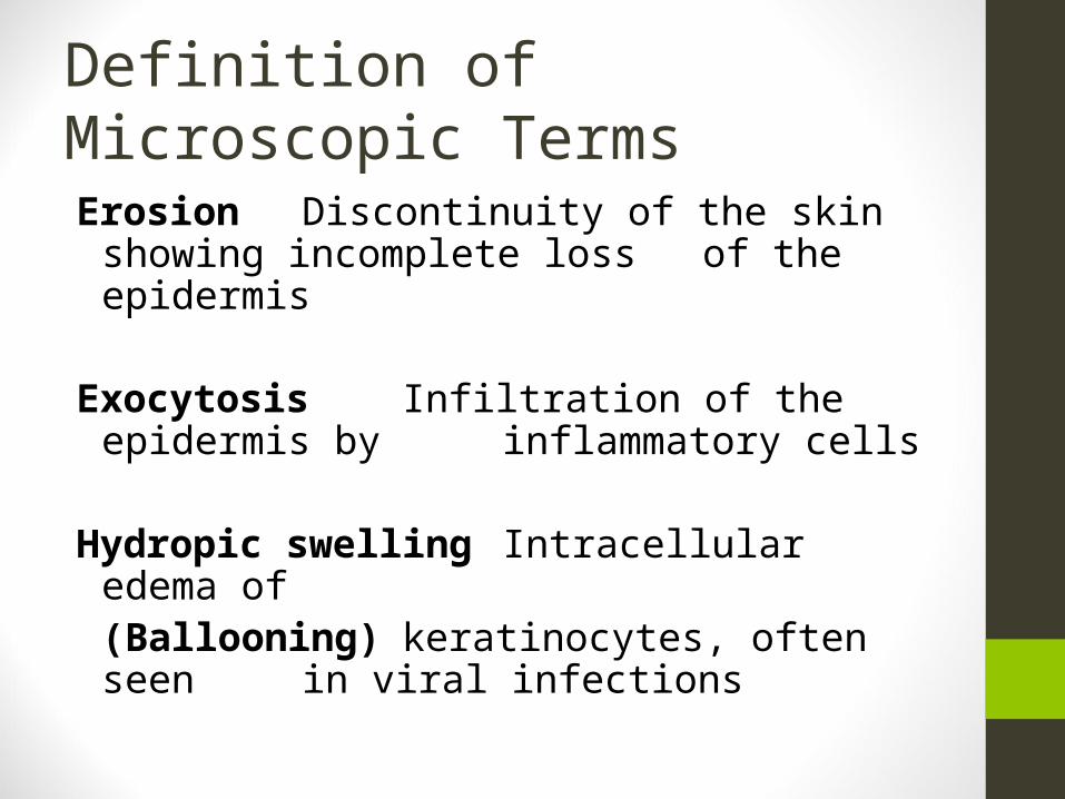
Definition of Microscopic Terms
Erosion Discontinuity of the skin showing incomplete loss of the epidermis
Exocytosis Infiltration of the epidermis by inflammatory cells
Hydropic swelling Intracellular edema of(Ballooning) keratinocytes, often seen
in viral infections
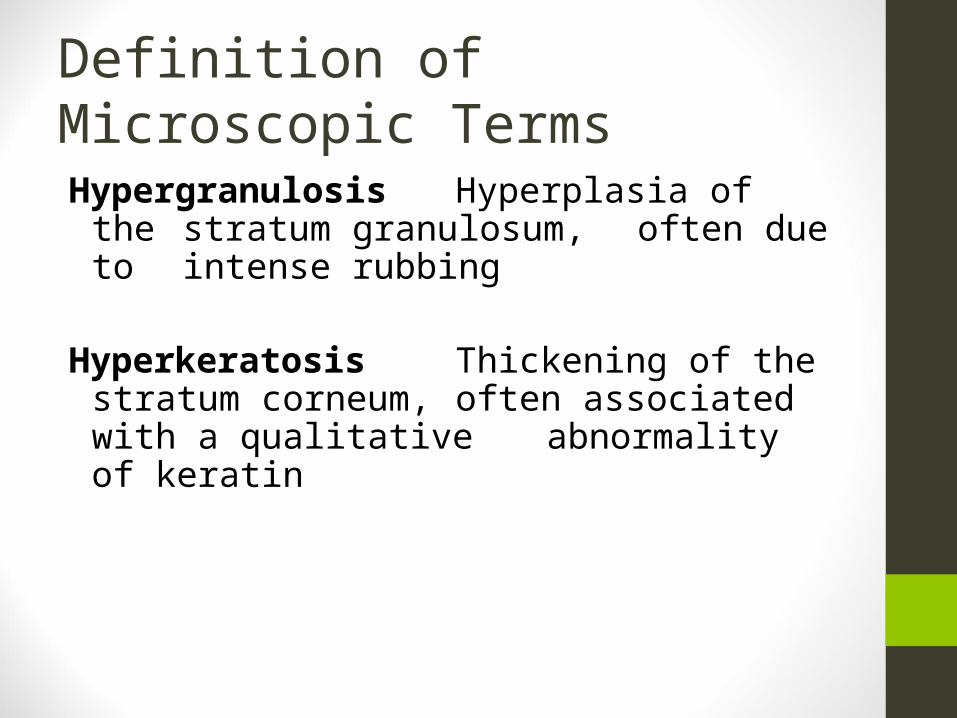
Definition of Microscopic Terms
Hypergranulosis Hyperplasia of the stratum
granulosum, often due to
intense rubbing
Hyperkeratosis Thickening of the stratum
corneum, often associated
with a qualitative abnormality of keratin
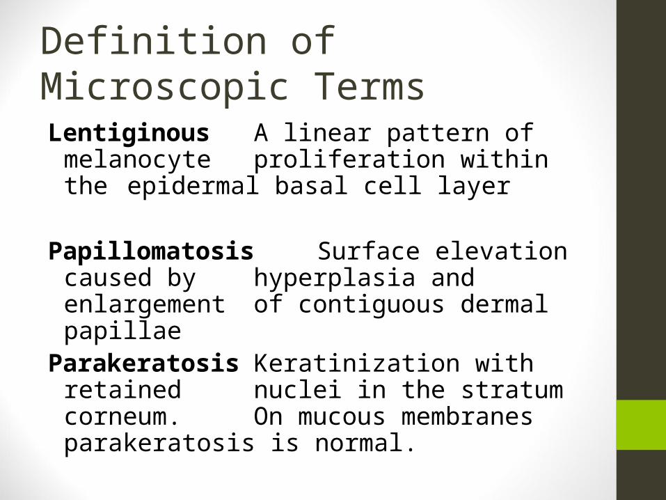
Definition of Microscopic Terms
Lentiginous A linear pattern of melanocyte proliferation within the epidermal basal cell layer
Papillomatosis Surface elevation caused by hyperplasia and enlargement of contiguous dermal papillae
Parakeratosis Keratinization with retained nuclei in the stratum corneum. On mucous membranes parakeratosis is normal.
Definition of Microscopic Terms
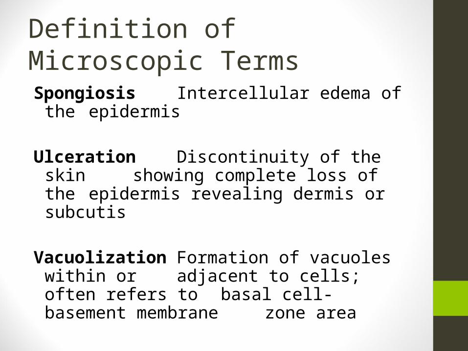
Spongiosis Intercellular edema of the epidermis
Ulceration Discontinuity of the skin showing complete loss of the epidermis revealing dermis or subcutis
Vacuolization Formation of vacuoles within or adjacent to cells; often refers to basal cell- basement membrane zone area
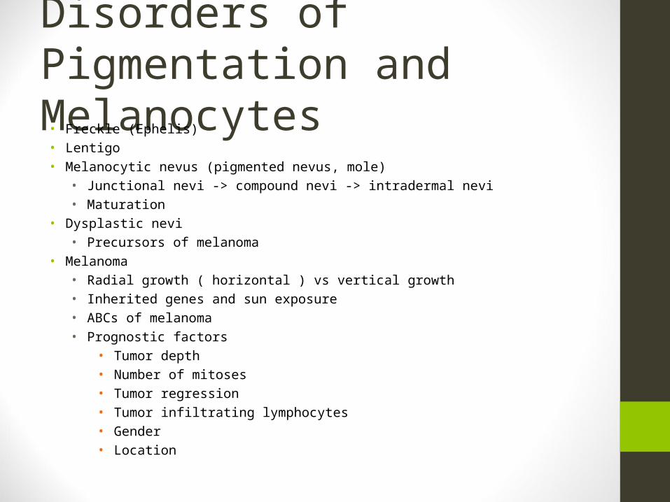
Disorders of Pigmentation and Melanocytes• Freckle (Ephelis)• Lentigo• Melanocytic nevus (pigmented nevus, mole)
• Junctional nevi -> compound nevi -> intradermal nevi• Maturation
• Dysplastic nevi• Precursors of melanoma
• Melanoma• Radial growth ( horizontal ) vs vertical growth• Inherited genes and sun exposure• ABCs of melanoma• Prognostic factors
• Tumor depth• Number of mitoses• Tumor regression• Tumor infiltrating lymphocytes• Gender• Location
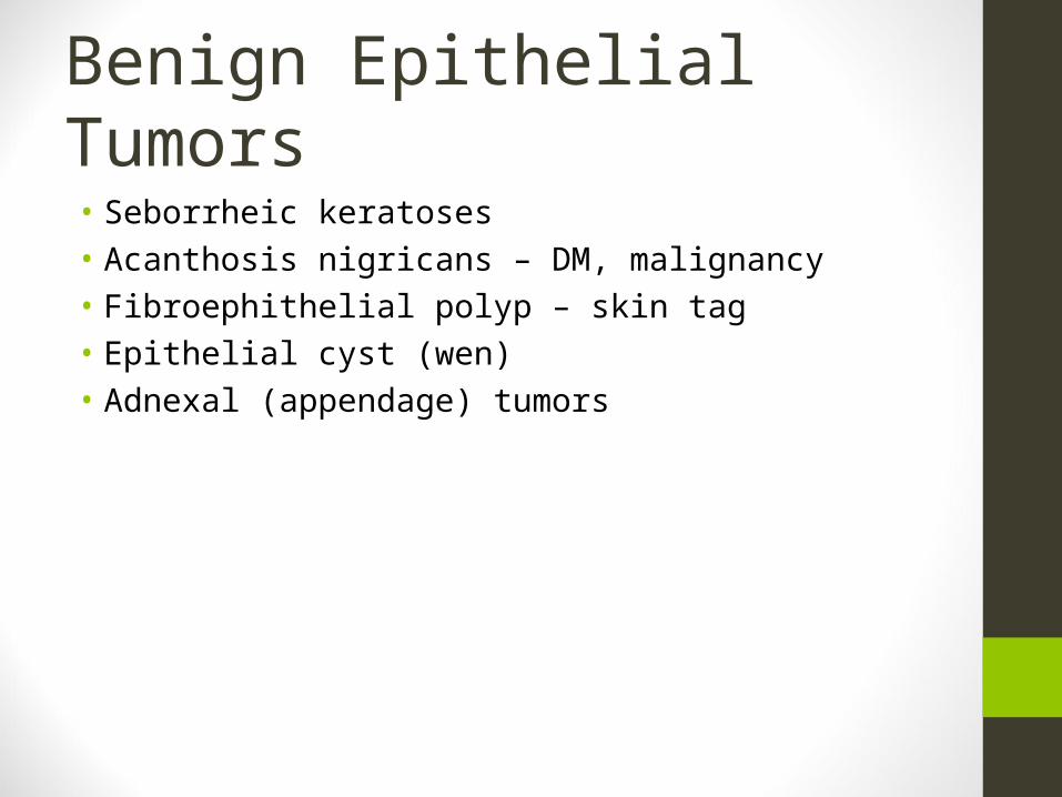
Benign Epithelial Tumors• Seborrheic keratoses• Acanthosis nigricans – DM, malignancy• Fibroephithelial polyp – skin tag• Epithelial cyst (wen)• Adnexal (appendage) tumors
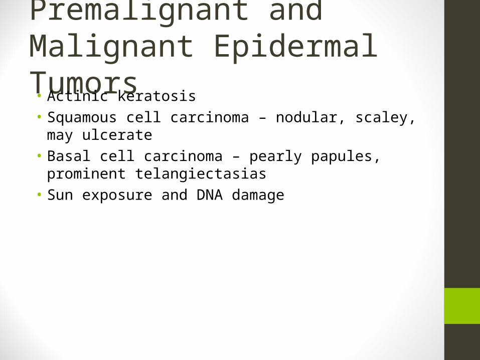
Premalignant and Malignant Epidermal Tumors• Actinic keratosis• Squamous cell carcinoma – nodular, scaley, may ulcerate• Basal cell carcinoma – pearly papules, prominent
telangiectasias• Sun exposure and DNA damage
Tumors of the Dermis• Benign fibrous histiocytoma (dermatofibroma)• Dermatofibrosarcoma proturberans
Tumors of Cellular Migrants to the Skin• Mycosis fungoides (cutaneous T cell lymphoma)• Sezary-Lutzner cells
• Mastocytosis• Mast cell degranulation• Darier sign , dermatographism
Disorders of Epidermal Maturation
• Icthyosis• Defective desquamation• Impaired epidermal maturation
Acute Inflammatory Dermatoses• Urticaria – hives• Wheal and flare• IgE mediated – Type I hypersensitivity
• Acute eczematous dermatitis• Allergic contact dermatitis• Atopic dermatitis• Drug-related eczematous dermatitis• Photoeczematous dermatitis• Primary irritant dermatitis
• Erythema multiforme• Infections, drugs, collagen vascular diseases
• Macules, papaules, vesicle, bullae, target lesions• Steven-Johnson syndrome – erosions of skin and mucous membranes,
toxic epidermal necrolysis
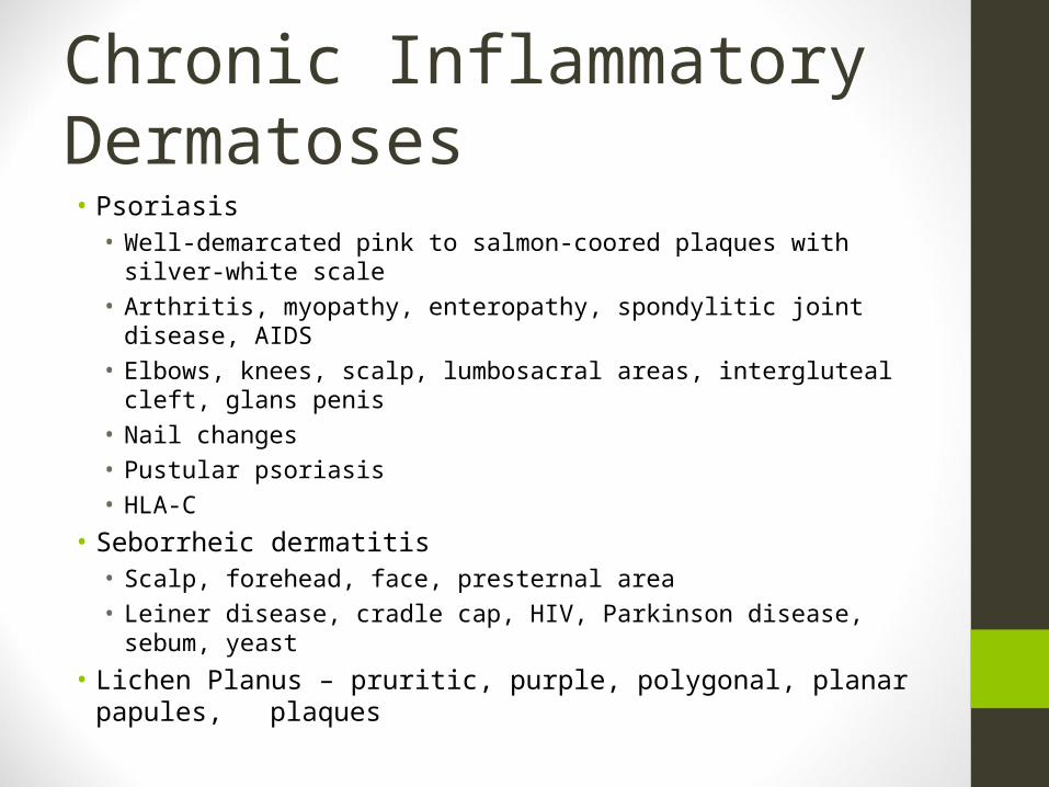
Chronic Inflammatory Dermatoses• Psoriasis• Well-demarcated pink to salmon-coored plaques with silver-white
scale• Arthritis, myopathy, enteropathy, spondylitic joint disease, AIDS• Elbows, knees, scalp, lumbosacral areas, intergluteal cleft, glans
penis• Nail changes• Pustular psoriasis• HLA-C
• Seborrheic dermatitis• Scalp, forehead, face, presternal area• Leiner disease, cradle cap, HIV, Parkinson disease, sebum, yeast
• Lichen Planus – pruritic, purple, polygonal, planar papules, plaques
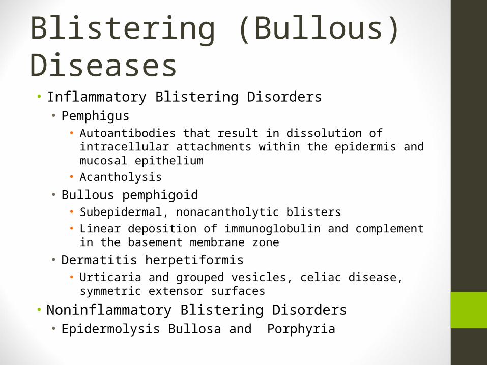
Blistering (Bullous) Diseases• Inflammatory Blistering Disorders• Pemphigus
• Autoantibodies that result in dissolution of intracellular attachments within the epidermis and mucosal epithelium
• Acantholysis
• Bullous pemphigoid• Subepidermal, nonacantholytic blisters• Linear deposition of immunoglobulin and complement in the
basement membrane zone
• Dermatitis herpetiformis• Urticaria and grouped vesicles, celiac disease, symmetric extensor
surfaces
• Noninflammatory Blistering Disorders• Epidermolysis Bullosa and Porphyria
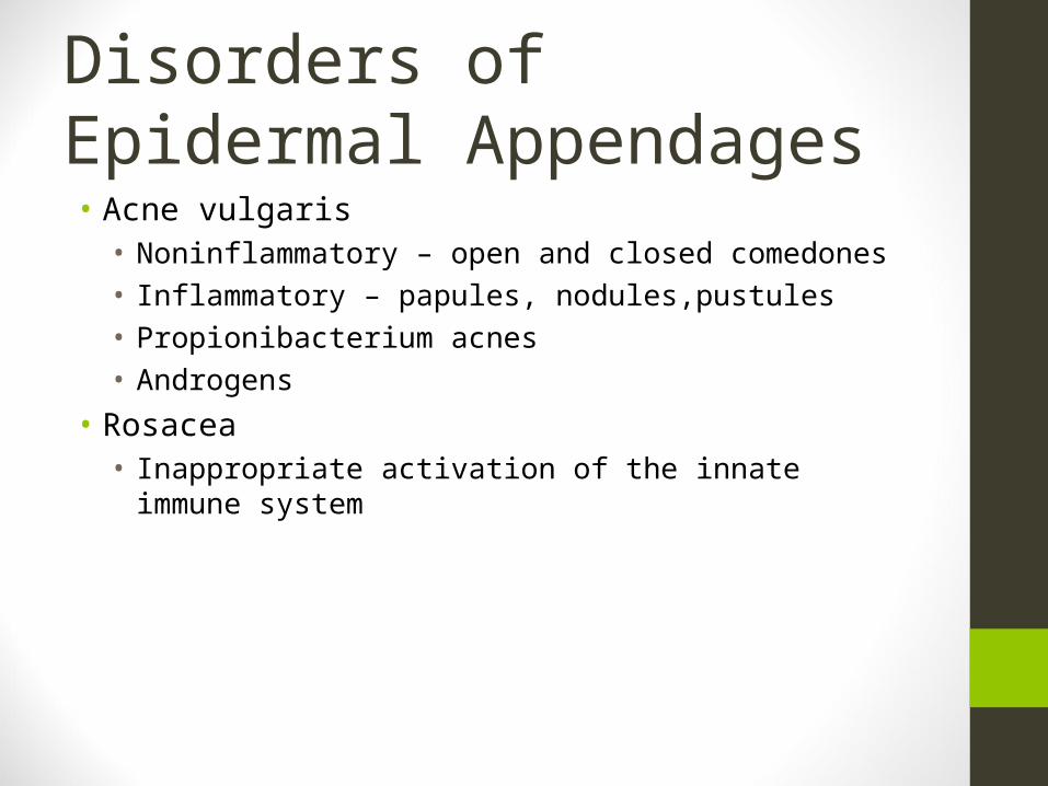
Disorders of Epidermal Appendages• Acne vulgaris• Noninflammatory – open and closed comedones• Inflammatory – papules, nodules,pustules• Propionibacterium acnes• Androgens
• Rosacea• Inappropriate activation of the innate immune system
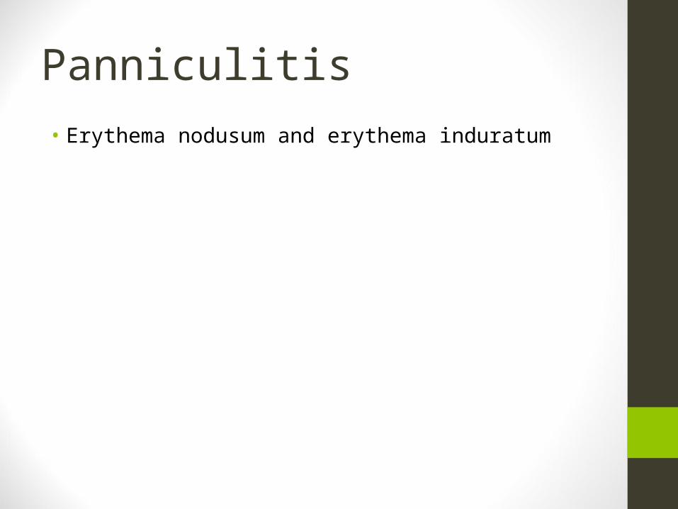
Panniculitis• Erythema nodusum and erythema induratum
Infection• Verrucae (warts) - HPV• Molluscum contagiosum – pox virus, umbilicated lesions• Impetigo – strep and staph, honey-colored crust• Superficial fungal infections – tinea capitis, barbae, corporis,
cruris, pedis, onychomycosis, dermatophytes; versicolor - yeast


![Toronto SCC epigenetics and aginginteresting skin lighteners on melanocytes looking atinteresting skin lighteners on melanocytes looking at Tyrosinase [TYR] and Ferritin [FTH1] gene](https://img.pdfslide.us/doc/110x75/602d4f8f53f48f1d883bdfdb/toronto-scc-epigenetics-and-aging-interesting-skin-lighteners-on-melanocytes-looking.jpg)
![arXiv:1806.04765v1 [cs.CV] 12 Jun 2018 · 2018. 6. 14. · of skin cancer originating in melanocytes which are the cells responsible for the pigmentation of skin, hair and eyes. Melanoma](https://img.pdfslide.us/doc/110x75/5fc71822141b566fcd3bf37b/arxiv180604765v1-cscv-12-jun-2018-2018-6-14-of-skin-cancer-originating.jpg)