Embed Size (px)
Citation preview
Medical Aspects of Harsh Environments, Volume 2
760
Chapter 24
ACUTE MOUNTAIN SICKNESS ANDHIGH-ALTITUDE CEREBRAL EDEMA
ROBERT ROACH, PHD*; JAN STEPANEK, MD†; AND PETER HACKETT, MD‡
INTRODUCTION
MODERN MILESTONES IN UNDERSTANDING THE SYNDROMES
DESCRIPTIONSymptoms and SignsIncidence, Severity, and Natural CoursePredisposing and Contributing FactorsScoring SystemsDifferential Diagnosis
PATHOPHYSIOLOGYVentilation and Gas ExchangeFluid Homeostasis and Permeability AbnormalitiesThe BrainClassification of Central Nervous System Edema
PROPHYLAXISAcclimatization and StagingDrugs
TREATMENTDescent, Both Real and SimulatedOxygen and Pharmacological Treatment
MILITARY OPERATIONS AT ALTITUDE
SUMMARY
*Scientist, New Mexico Resonance, PO Box 343, Montezuma, New Mexico 87731; Adjunct Assistant Professor, Department of Medicine,University of New Mexico School of Medicine, Albuquerque, New Mexico 87131, and Adjunct Assistant Professor, Department of Surgery,University of Colorado School of Medicine, Denver, Colorado 80220
†Senior Associate Consultant, Section of Aerospace Medicine, Department of Preventive and Occupational Medicine, Mayo Clinic, Rochester,Minnesota 55905
‡Affiliate Associate Professor of Medicine, Department of Medicine, University of Washington School of Medicine, Seattle, Washington 98195
761
Acute Mountain Sickness and High-Altitude Cerebral Edema
INTRODUCTION
just before the turn of the 20th century. EdwardWhymper12 described the symptoms of AMS atabout 3,500 m thus:
I found myself lying on my back, incapable of mak-ing the least exertion. We...had intense headaches,and were unable to satisfy our desire for air, ex-cept by breathing with open mouths. Headachewith all three of us was intense, and rendered usalmost frantic or crazy. ... Of course there was noinclination to eat.12(pp26–28)
An early and important description of HACEcomes from T. H. Ravenhill’s experiences as a min-ing camp physician in the Andes Mountains dur-ing the early part of the 20th century.13,14 He fre-quently observed AMS, called “puna” in the localdialect, and wrote a classic description of “nervouspuna” as a type of AMS characterized by predomi-nant neurological features, known today as HACE:
The most marked case I had was a young Chileno,aged 19. He arrived at the neighboring mine in theusual way; three days later I was called to see him.He was then unable to speak, there were violentspasmodic movements of the limbs, and he resistedexamination. The face was blanched, the lips almostwhite, the pupils slightly dilated. Temperature andrespiration were normal; the pulse 140. He wasunable to stand or to walk. I was told that he hadbeen in this condition almost since his arrival, andthat he had been delirious, talking all sorts of non-sense. I could find nothing organically wrong onphysical examination. He was sent down the sameday; three days later, ie, by the time he had justreached the coast, he had quite recovered.13(p315)
HACE remains rare and largely confined to alti-tudes over 4,000 m, although recent evidence15 frommagnetic resonance imaging (MRI) scans suggests thatHACE may also occur at lower altitudes (3,000–3,500m). In one survey of 1,925 soldiers at altitudes rang-ing from 3,350 to 5,000 m, only 23 men (1.2%) devel-oped the severe neurological signs of HACE16; simi-larly, only 5 (1.8%) of 278 trekkers were diagnosedwith HACE at 4,243 m.17 (Please see Exhibit 19-1 inChapter 19, Mountains and Military Medicine: AnOverview, for definitions of climbers, trekkers, andother categories of people who visit mountains.) In-creasingly, data from clinical studies support the no-tion that AMS is caused by cerebral edema.15,16,18 Whenthe degree of cerebral edema passes a critical thresh-old, the neurological signs are increasingly observedand diagnosis of HACE becomes clear.
Acute mountain sickness (AMS) and high-alti-tude cerebral edema (HACE) are syndromes thatprobably occur along a continuum of severity frommild, benign AMS to severe, life-threatening HACE.They strike people who travel too fast beyond alti-tudes to which they are adjusted. AMS and HACEcan destroy the effectiveness of even the fittestmountain troops on ascent to high altitude. In thischapter, we describe the clinical features of AMSand HACE, including what is known of their patho-physiology, and the best available approaches toprevention and treatment.
The scope of the chapter is limited to a criticalappraisal of the scientific literature concerning AMSand HACE to provide an up-to-date perspectivefocused on pathophysiology. Further exhaustiveinformation on history, incidence, treatment, andprevention are available,1–11 and only the essence ofthat material is covered here. This chapter providesthe information needed by military medical person-nel deploying to the mountain regions of the worldby (a) providing a thorough description of the clini-cal syndromes of AMS and HACE, including symp-toms, signs, and diagnosis; (b) using the latest ad-vances in research into the causes of AMS andHACE to thoroughly describe what is known (andhint at what is not yet known) about their patho-physiology; and (c) giving practical advice for theprevention and treatment of AMS and HACE basedon an explanation of the underlying physiology.
The major importance of AMS to the readers ofthis volume is that
• AMS can sharply reduce a military unit’seffectiveness in the field, especially in thefirst few days following insertion to highaltitude, and
• if AMS worsens and HACE develops, the riskof fatality is significant and the disruptionof planned activities to arrange rescue ortemporizing measures can be considerable.
Fortunately, most cases of AMS and HACE canbe prevented, personnel can be trained to identifythe syndromes early and reliably in the field with-out sophisticated instruments, and if AMS andHACE are recognized early, most cases respond rap-idly with complete recovery in a few hours (in thecase of AMS) to days (for HACE).
Enduring descriptions in English of AMS andHACE come from observations of European travel-ers to the Andes Mountains in South America at or
Medical Aspects of Harsh Environments, Volume 2
762
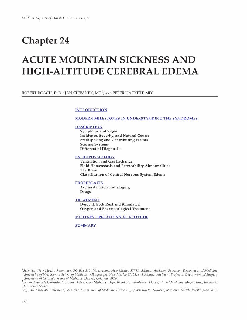
Fig. 24-1. The natural course of acute mountain sickness in 840soldiers who received no treatment for their symptoms at alti-tudes ranging from 3,350 to 5,000 m. Note that about one thirdwere free of symptoms after only 3 days, whereas the next thirdtook as long as 12 days to be symptom free. Data source: SinghI, Khanna PK, Srivastava MC, Lal M, Roy SB, SubramanyamCS. Acute mountain sickness. N Engl J Med. 1969;280:177.
Number of Da ys to Become S ymptom Free
3 4–7 8–1215–21
22–2829–35
36–4243–60
61–90
0.01%
91–150Never
40
30
20
10
0
Per
cent
age
of 8
40 U
ntre
ated
Sol
dier
s
MODERN MILESTONES IN UNDERSTANDING THE SYNDROMES
Paul Bert’s19 identification in late-19th-centuryFrance of hypoxia as the main environmental chal-lenge for balloonists (and mountaineers) was thebeginning of scientific investigation into the humanresponses to the stress of high altitude. By the late1950s, AMS and HACE had been described clini-cally, and acclimatization to high altitude was un-derstood to be a process not to be rushed. The In-dia–China–Pakistan border conflicts during the late1960s focused medical attention on practical prob-lems of military troops abruptly deployed to veryhigh altitudes without time for acclimatization. Theclassic study by Singh and colleagues16 (Figure 24-1) described the Indian Army’s experience in theconflict and laid the groundwork for future researchinto the pathogenesis of AMS and HACE.
Who is at risk for developing AMS is a questionthat has only recently been explored in detail witha large study population. Investigators in Coloradocompleted the largest epidemiological survey todate on 3,158 travelers visiting resorts in the RockyMountains of Colorado.20 Of those, 790 (about 25%)developed AMS, and most decreased their dailyactivity because of their symptoms. Tourists whosepermanent residence was below 3,000 m had a riskfor AMS that was 3.5-fold greater than those whopermanently resided above 3,000 m. Women, obesepersons, and those with underlying lung diseasealso had a slightly higher occurrence of AMS.20 Thenext step in this type of research is to couple large-scale epidemiological surveys with noninvasivephysiological measurements at sea level and at al-titude to better describe physiological characteris-tics that predispose to high-altitude illness.
Singh and colleagues16 mentioned results of sev-eral autopsies from HACE victims, but it was sev-eral more years before detailed postmortem reportsand case histories of HACE were published.20–25
Only recently have significant advances been madein the understanding of HACE. Numerous groupshave attempted to use noninvasive scanning tech-nologies (MRI and computed tomography [CT]scans) to investigate cerebral edema in mountain-eers, and to follow their recoveries.15,18,26 The recentMRI work of Hackett and colleagues15 shows that
AMS is a syndrome that occurs in susceptibleindividuals when ascent to high altitude outpacesthe ability to acclimatize. The symptoms, althoughoften incapacitating, are usually self-limited. The
incidence and severity of AMS depend on the rateof ascent and the altitude attained, the length oftime at altitude, the degree of physical exertion, andthe individual’s physiological susceptibility.35 The
DESCRIPTION
the edema is characterized by reversible white-mat-ter edema, which has important implications for un-derstanding the underlying pathophysiology.
Most major problems of prevention and treat-ment of AMS were solved within 2 decades afterSingh’s seminal paper16 had been published (ie, by1990). Acetazolamide was identified as effectiveprophylaxis17,27,28 and treatment for AMS29 and isnow considered the drug of choice for preventionof AMS.30,31 The study of Singh and colleagues16 alsosuggested that the steroid betamethasone was aneffective treatment of severe AMS. Researchers haveconfirmed and extended these original observationswith dexamethasone. Dexamethasone effectivelytreats severe AMS and HACE,27,32,33 and in personsintolerant of acetazolamide, it can also be used forprophylaxis of AMS.34 Its exact mechanism is un-clear. The next advances in AMS and HACE re-search will likely incorporate sophisticated imag-ing studies, pharmacological interventions, andperhaps biochemical and molecular markers of theonset or the resolution of brain edema.
763
Acute Mountain Sickness and High-Altitude Cerebral Edema
chief significance of AMS for the military is thatlarge numbers of troops rapidly deployed to highaltitude may be completely incapacitated in the firstfew days at a new altitude. Additionally, in a fewindividuals, AMS may progress to life-threateningHACE or to high-altitude pulmonary edema (HAPE),which is the subject of Chapter 25, High-AltitudePulmonary Edema.
Symptoms and Signs
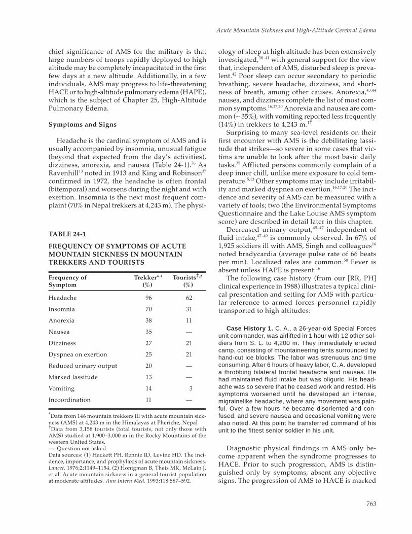
Headache is the cardinal symptom of AMS and isusually accompanied by insomnia, unusual fatigue(beyond that expected from the day’s activities),dizziness, anorexia, and nausea (Table 24-1).36 AsRavenhill13 noted in 1913 and King and Robinson37
confirmed in 1972, the headache is often frontal(bitemporal) and worsens during the night and withexertion. Insomnia is the next most frequent com-plaint (70% in Nepal trekkers at 4,243 m). The physi-
ology of sleep at high altitude has been extensivelyinvestigated,38–41 with general support for the viewthat, independent of AMS, disturbed sleep is preva-lent.42 Poor sleep can occur secondary to periodicbreathing, severe headache, dizziness, and short-ness of breath, among other causes. Anorexia,43,44
nausea, and dizziness complete the list of most com-mon symptoms.16,17,20 Anorexia and nausea are com-mon (~ 35%), with vomiting reported less frequently(14%) in trekkers to 4,243 m.17
Surprising to many sea-level residents on theirfirst encounter with AMS is the debilitating lassi-tude that strikes—so severe in some cases that vic-tims are unable to look after the most basic dailytasks.35 Afflicted persons commonly complain of adeep inner chill, unlike mere exposure to cold tem-perature.5,13 Other symptoms may include irritabil-ity and marked dyspnea on exertion.16,17,20 The inci-dence and severity of AMS can be measured with avariety of tools; two (the Environmental SymptomsQuestionnaire and the Lake Louise AMS symptomscore) are described in detail later in this chapter.
Decreased urinary output,45–47 independent offluid intake,47–49 is commonly observed. In 67% of1,925 soldiers ill with AMS, Singh and colleagues16
noted bradycardia (average pulse rate of 66 beatsper min). Localized rales are common.50 Fever isabsent unless HAPE is present.16
The following case history (from our [RR, PH]clinical experience in 1988) illustrates a typical clini-cal presentation and setting for AMS with particu-lar reference to armed forces personnel rapidlytransported to high altitudes:
Case History 1. C. A., a 26-year-old Special Forcesunit commander, was airlifted in 1 hour with 12 other sol-diers from S. L. to 4,200 m. They immediately erectedcamp, consisting of mountaineering tents surrounded byhand-cut ice blocks. The labor was strenuous and timeconsuming. After 6 hours of heavy labor, C. A. developeda throbbing bilateral frontal headache and nausea. Hehad maintained fluid intake but was oliguric. His head-ache was so severe that he ceased work and rested. Hissymptoms worsened until he developed an intense,migrainelike headache, where any movement was pain-ful. Over a few hours he became disoriented and con-fused, and severe nausea and occasional vomiting werealso noted. At this point he transferred command of hisunit to the fittest senior soldier in his unit.
Diagnostic physical findings in AMS only be-come apparent when the syndrome progresses toHACE. Prior to such progression, AMS is distin-guished only by symptoms, absent any objectivesigns. The progression of AMS to HACE is marked
TABLE 24-1
FREQUENCY OF SYMPTOMS OF ACUTEMOUNTAIN SICKNESS IN MOUNTAINTREKKERS AND TOURISTS
Frequency of Trekker*,1 Tourists†,2
Symptom (%) (%)
Headache 96 62
Insomnia 70 31
Anorexia 38 11
Nausea 35 —
Dizziness 27 21
Dyspnea on exertion 25 21
Reduced urinary output 20 —
Marked lassitude 13 —
Vomiting 14 3
Incoordination 11 —
*Data from 146 mountain trekkers ill with acute mountain sick-ness (AMS) at 4,243 m in the Himalayas at Pheriche, Nepal†Data from 3,158 tourists (total tourists, not only those withAMS) studied at 1,900–3,000 m in the Rocky Mountains of thewestern United States.—: Question not askedData sources: (1) Hackett PH, Rennie ID, Levine HD. The inci-dence, importance, and prophylaxis of acute mountain sickness.Lancet. 1976;2:1149–1154. (2) Honigman B, Theis MK, McLain J,et al. Acute mountain sickness in a general tourist populationat moderate altitudes. Ann Intern Med. 1993;118:587–592.
Medical Aspects of Harsh Environments, Volume 2
764
by development of truncal ataxia; severe lassitude;and altered mental status, including impaired men-tal capacity, drowsiness, and stupor.5,21,24 Coma maydevelop as soon as 24 hours after the onset of ataxiaor change in mental status.
The following case report describes the typicaldevelopment of HACE in the field setting:
Case History 2. H. E. was a 26-year-old German lum-berjack with extensive mountaineering experience. Heascended to 5,200 m from 2,000 m in 4 days, and at-tempted the summit (6,194 m) on the fifth day. At 5,800m he turned around owing to severe fatigue, headache,and malaise. He returned alone to 5,200 m, stumbling onthe way because of loss of coordination. He had no ap-petite and crawled into his sleeping bag too weak, tired,and disoriented to undress. He recalled no pulmonarysymptoms. In the morning, H. E. was unarousable, slightlycyanotic, and noted to have Cheyne–Stokes respirations.After 10 minutes on high-flow oxygen, H. E. began to re-gain consciousness although he was completely disori-ented and unable to move. A rescue team lowered himdown a steep slope, and on arrival at 4,400 m 4 hourslater, H. E. was conscious but still disoriented, able tomove his extremities but unable to stand. Respiratory ratewas 60 breaths per minute and heart rate was 112 beatsper minute. Papilledema and a few rales were present.The oxygen saturation of arterial blood (SaO2) was 54%on room air (normal is 85% to 90%). On a non-rebreather
oxygen mask with 14 L/min oxygen, the SaO2 increasedto 88% and his respiratory rate decreased to 40. Eightmg of dexamethasone was administered intramuscularlyat 1620 hours, and continued orally, 4 mg every 6 hours.At 1720 hours, H. E. began to respond to commands.The next morning H. E. was still ataxic, although able tostand, take fluids, and eat heartily. He was evacuated byair to Anchorage, Alaska (sea level), at 1200 hours, wherehe recovered fully within several days.5
This climber was fortunate to become ill on a highmountain where a sophisticated medical researchlaboratory was on site. Without the medical careprovided by the researchers there, HACE and HAPEwould have almost certainly been fatal. The ratio-nale for his treatment and strategies for preventingthe syndromes are discussed later in the chapter.
Incidence, Severity, and Natural Course
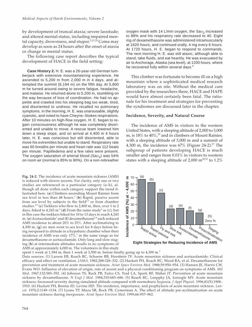
The incidence of AMS in visitors to the westernUnited States, with a sleeping altitude of 2,000 to 3,000m, is 18% to 40%,20 and in climbers of Mount Rainier,with a sleeping altitude of 3,000 m and a summit of4,300 m, the incidence was 67% (Figure 24-2).27 Thesubgroup of patients developing HACE is muchsmaller and ranges from 0.01% in visitors to westernstates with a sleeping altitude of 2,000 m20,51 to 1.2%
Fig. 24-2. The incidence of acute mountain sickness (AMS)is reduced with slower ascents. For clarity, only one or twostudies are referenced in a particular category (a–h), al-though all done within each category support the trend il-lustrated here. (a) Climbers ascending Mount Rainier fromsea level in less than 48 hours.1 (b) Rapid, passive ascentfrom sea level by subjects in the field2,3 or from chamberstudies.4,5 (c) Trekkers who flew to 2,800 m, then, over 1 to 2days, hiked to 4,243 m.6 (d) From the same study6 as (c), butin this case the trekkers hiked for 10 to 13 days to reach 4,243m. (e) Acetazolamide1 and (f) dexamethasone2,4 each reducedAMS incidence to about 20% to 25%. After acclimatizing to4,300 m, (g) six men went to sea level for 8 days before be-ing reexposed to altitude in a hypobaric chamber when theirincidence of AMS was only 17%,7 in the same range as fordexamethasone or acetazolamide. Only long and slow stag-ing (h) at intermediate altitudes results in no symptoms ofAMS at approximately 4,000 m. The volunteers in this studyspent 1 week at 1,584 m, then 1 week at 3,500 m, before finally going up to 4,300 m.3
Data sources: (1) Larson EB, Roach RC, Schoene RB, Hornbein TF. Acute mountain sickness and acetazolamide: Clinicalefficacy and effect on ventilation. JAMA. 1982;288:328–332. (2) Hackett PH, Roach RC, Wood RA, et al. Dexamethasone forprevention and treatment of acute mountain sickness. Aviat Space Environ Med. 1988;59:950–954. (3) Hansen JE, Harris CW,Evans WO. Influence of elevation of origin, rate of ascent and a physical conditioning program on symptoms of AMS. MilMed. 1967;132:585–592. (4) Johnson TS, Rock PB, Fulco CS, Trad LA, Spark RF, Maher JT. Prevention of acute mountainsickness by dexamethasone. N Engl J Med. 1984;310:683–686. (5) Roach RC, Loeppky JA, Icenogle MV. Acute mountainsickness: Increased severity during simulated altitude compared with normobaric hypoxia. J Appl Physiol. 1996;81(5):1908–1910. (6) Hackett PH, Rennie ID, Levine HD. The incidence, importance, and prophylaxis of acute mountain sickness. Lan-cet. 1976;2:1149–1154. (7) Lyons TP, Muza SR, Rock PB, Cymerman A. The effect of altitude pre-acclimatization on acutemountain sickness during reexposure. Aviat Space Environ Med. 1995;66:957–962.
Eight Strategies for Reducing Incidence of AMS
Inci
denc
e of
AM
S (
%)
at ~
4,0
00 m
70
60
50
40
30
20
10
0Zero %
Rapid active
Rapid passive
Fly and trek
Slow trek
Acetazolamide
Dexamethasone
Preacclimatization
Staging
(a)(b)
(c)
(d) (e)(f)
(g)
(h)
765
Acute Mountain Sickness and High-Altitude Cerebral Edema
in the Indian armed forces, where the sleeping alti-tude is up to 5,500 m,52,53 to 1.8% in trekkers at 4,300m on their way to Mount Everest base camp.17 Con-trolled studies have not yet determined whether menand women differ in their susceptibility to AMS. Lim-ited epidemiological studies suggest that women havethe same or slightly greater incidence of AMS but maybe less susceptible to HAPE. Honigman and col-leagues20 studied 3,158 adults visiting moderate alti-tude (1,900–3,000 m). Of 1,255 women included in thatstudy, 28% developed AMS, compared with 24% ofthe men (P < 0.01). In another survey conducted at ahigher altitude (4,243 m), Hackett and colleagues17
studied 278 unacclimatized trekkers in Nepal andnoted no gender differences in AMS susceptibility.
Prior physical condition has little influence onincidence.54 Older adults consistently report lessAMS than younger people with similar altitudeexposure. Whether this difference results from be-havioral adjustments or has a basis in physiologi-cal differences is not known. It is important to notethat older age, by itself, does not preclude travel tomoderate high altitude. In one study of 97 elderly(average age = 69.8 y) visitors to 2,500 m, only 16%reported AMS,55 compared with 20% to 25% personsaged about 44 years at a similar altitude.20
The natural course of AMS varies with the initialaltitude, rate of ascent, clinical severity, and indi-vidual susceptibility. Of 840 soldiers not treatedwith any drugs for their AMS symptoms at 3,300 to5,500 m, only 40% were symptom-free after 3 days16
(see Figure 24-1). After 3 weeks, 80% were symp-tom-free. The remaining 20% coped with symptomsfor up to 6 months; indeed, 9 soldiers were neverfree of AMS symptoms during the 6-month study.At lower altitudes more frequently visited by tour-ists, 99% of symptoms resolved within the first 36hours at altitude, and most individuals resumednormal activities shortly thereafter.20 Previous ex-perience is the best predictor of an individual’s re-sponse to altitude and of the natural course of AMS.
Predisposing and Contributing Factors
An understanding of predisposing and contrib-uting factors to the development of AMS and HACEis important to aid preventive action and early rec-ognition. Because HACE is on a continuum withAMS,5 risk factors for the development of AMS maybe viewed as risk factors for the development ofHACE in susceptible individuals. The lack of properacclimatization (ie, too rapid ascent), or any factorsthat impede acclimatization, clearly increases therisk of AMS and HACE.21 In their series of 1,925
soldiers exposed to high altitude, Singh and col-leagues16 reported that the exposure to cold envi-ronmental conditions and physical exertion seemedto aggravate the condition of individuals alreadyhaving symptoms of AMS or precipitate the condi-tion in persons who initially were well.
Ross56 hypothesized that the person who willtolerate hypoxic brain-swelling least well is the onewith small intracranial and intraspinal capacity andthus limited compliance. He based his deductionson pressure volume index measurements carriedout by Shapiro and colleagues,57 which indicate thatin a person under the age of 30, the intracranialspace not occupied by the brain is less than 1% andthat this value increases up to 5.9% by 70 to 80 yearsof age. More research is necessary to fit this inter-esting work to the known minimal effect of age (upto 60 y) on susceptibility to AMS.
Lack of acclimatization can certainly predisposeclimbers, hikers, and others to AMS. Mentioned ear-lier (see Case History 2) was the effect on SaO2 duringexercise, as affected by the degree of acclimatization:the better the acclimatization, the higher the SaO2during exercise. For example, among 104 climbersstudied at 4,200 m before attempting to climb to thesummit of Mount McKinley (6,200 m),58 those whoseSaO2 fell the furthest during exercise were the mostlikely to develop AMS during their climb.
In another attempt to predict subsequent AMS,Savourey and colleagues59 completed a number oftests at sea level in both normoxia and hypoxia. On asubsequent high-altitude trek, the AMS score did notrelate to body size, pulmonary function, hypoxic orhypercapnic ventilatory responses, or the cold pres-sor test. The end-tidal partial pressure of oxygen(PETO2) during submaximal exercise at sea level washighly correlated (r = 0.92, P < 0.001) with the subse-quent AMS score. Reeves and colleagues60 had earlierreported that SaO2 at altitude was best predicted bythe sea-level resting end-tidal partial pressure of car-bon dioxide (PETCO2), such that the lower the sea-levelPETCO2, the higher was the SaO2 on ascent to altitude.Further investigations into breathing pattern, hypoxia,oxygen and carbon dioxide chemosensitivity, andAMS are necessary to clarify these relationships.
Scoring Systems
Awareness of the scoring systems is of practicalimportance to the military medical officer to allowidentification and rapid quantification of the sever-ity of AMS and HACE. In the context of mountain-eering, the rule of thumb is that a severe headachewith nausea, vomiting, dizziness, or undue fatigue
Medical Aspects of Harsh Environments, Volume 2
766
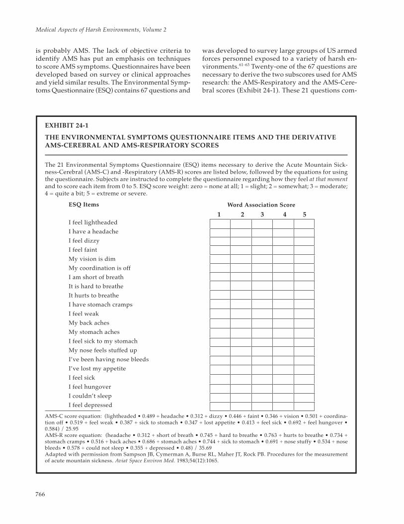
is probably AMS. The lack of objective criteria toidentify AMS has put an emphasis on techniquesto score AMS symptoms. Questionnaires have beendeveloped based on survey or clinical approachesand yield similar results. The Environmental Symp-toms Questionnaire (ESQ) contains 67 questions and
was developed to survey large groups of US armedforces personnel exposed to a variety of harsh en-vironments.61–63 Twenty-one of the 67 questions arenecessary to derive the two subscores used for AMSresearch: the AMS-Respiratory and the AMS-Cere-bral scores (Exhibit 24-1). These 21 questions com-
EXHIBIT 24-1
THE ENVIRONMENTAL SYMPTOMS QUESTIONNAIRE ITEMS AND THE DERIVATIVEAMS-CEREBRAL AND AMS-RESPIRATORY SCORES
The 21 Environmental Symptoms Questionnaire (ESQ) items necessary to derive the Acute Mountain Sick-ness-Cerebral (AMS-C) and -Respiratory (AMS-R) scores are listed below, followed by the equations for usingthe questionnaire. Subjects are instructed to complete the questionnaire regarding how they feel at that momentand to score each item from 0 to 5. ESQ score weight: zero = none at all; 1 = slight; 2 = somewhat; 3 = moderate;4 = quite a bit; 5 = extreme or severe.
ESQ Items
I feel lightheaded
I have a headache
I feel dizzy
I feel faint
My vision is dim
My coordination is off
I am short of breath
It is hard to breathe
It hurts to breathe
I have stomach cramps
I feel weak
My back aches
My stomach aches
I feel sick to my stomach
My nose feels stuffed up
I’ve been having nose bleeds
I’ve lost my appetite
I feel sick
I feel hungover
I couldn’t sleep
I feel depressed
AMS-C score equation: (lightheaded • 0.489 + headache • 0.312 + dizzy • 0.446 + faint • 0.346 + vision • 0.501 + coordina-tion off • 0.519 + feel weak • 0.387 + sick to stomach • 0.347 + lost appetite • 0.413 + feel sick • 0.692 + feel hungover •0.584) / 25.95AMS-R score equation: (headache • 0.312 + short of breath • 0.745 + hard to breathe • 0.763 + hurts to breathe • 0.734 +stomach cramps • 0.516 + back aches • 0.686 + stomach aches • 0.744 + sick to stomach • 0.691 + nose stuffy • 0.534 + nosebleeds • 0.578 + could not sleep • 0.355 + depressed • 0.48) / 35.69Adapted with permission from Sampson JB, Cymerman A, Burse RL, Maher JT, Rock PB. Procedures for the measurementof acute mountain sickness. Aviat Space Environ Med. 1983;54(12):1065.
Word Association Score
1 2 3 4 5
767
Acute Mountain Sickness and High-Altitude Cerebral Edema
prise a questionnaire of reasonable length andproven reliability, and a lengthy literature is avail-able for comparison. The major shortcoming of theESQ is that the entire system is based on the statisti-cal agreement between artificial clusters of symptomsused for the AMS-Cerebral and AMS-Respiratory
scores and the single response of “I feel sick.”Clinicians, favoring a more direct approach, have
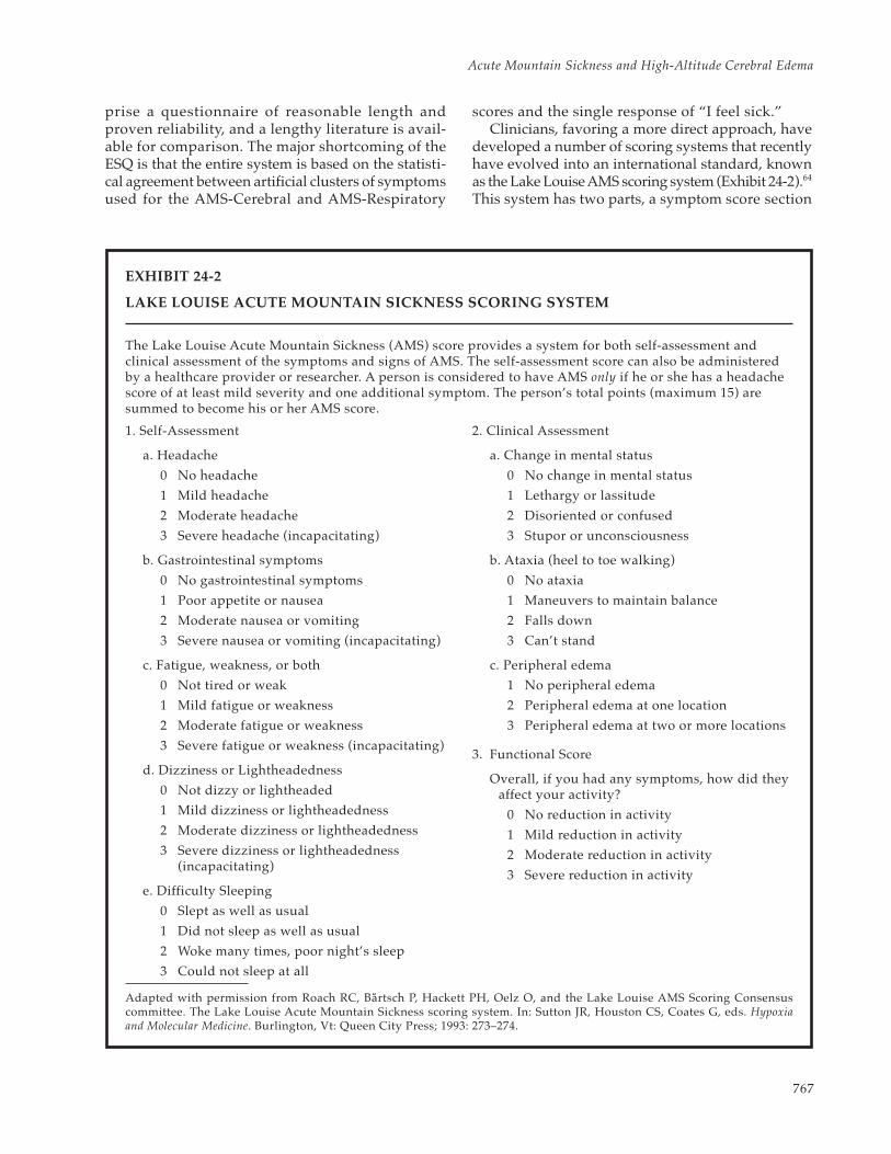
developed a number of scoring systems that recentlyhave evolved into an international standard, knownas the Lake Louise AMS scoring system (Exhibit 24-2).64
This system has two parts, a symptom score section
EXHIBIT 24-2
LAKE LOUISE ACUTE MOUNTAIN SICKNESS SCORING SYSTEM
The Lake Louise Acute Mountain Sickness (AMS) score provides a system for both self-assessment andclinical assessment of the symptoms and signs of AMS. The self-assessment score can also be administeredby a healthcare provider or researcher. A person is considered to have AMS only if he or she has a headachescore of at least mild severity and one additional symptom. The person’s total points (maximum 15) aresummed to become his or her AMS score.
1. Self-Assessment
a. Headache
0 No headache
1 Mild headache
2 Moderate headache
3 Severe headache (incapacitating)
b. Gastrointestinal symptoms
0 No gastrointestinal symptoms
1 Poor appetite or nausea
2 Moderate nausea or vomiting
3 Severe nausea or vomiting (incapacitating)
c. Fatigue, weakness, or both
0 Not tired or weak
1 Mild fatigue or weakness
2 Moderate fatigue or weakness
3 Severe fatigue or weakness (incapacitating)
d. Dizziness or Lightheadedness
0 Not dizzy or lightheaded
1 Mild dizziness or lightheadedness
2 Moderate dizziness or lightheadedness
3 Severe dizziness or lightheadedness(incapacitating)
e. Difficulty Sleeping
0 Slept as well as usual
1 Did not sleep as well as usual
2 Woke many times, poor night’s sleep
3 Could not sleep at all
2. Clinical Assessment
a. Change in mental status
0 No change in mental status
1 Lethargy or lassitude
2 Disoriented or confused
3 Stupor or unconsciousness
b. Ataxia (heel to toe walking)
0 No ataxia
1 Maneuvers to maintain balance
2 Falls down
3 Can’t stand
c. Peripheral edema
1 No peripheral edema
2 Peripheral edema at one location
3 Peripheral edema at two or more locations
3. Functional Score
Overall, if you had any symptoms, how did theyaffect your activity?
0 No reduction in activity
1 Mild reduction in activity
2 Moderate reduction in activity
3 Severe reduction in activity
Adapted with permission from Roach RC, Bartsch P, Hackett PH, Oelz O, and the Lake Louise AMS Scoring Consensuscommittee. The Lake Louise Acute Mountain Sickness scoring system. In: Sutton JR, Houston CS, Coates G, eds. Hypoxiaand Molecular Medicine. Burlington, Vt: Queen City Press; 1993: 273–274.
Medical Aspects of Harsh Environments, Volume 2
768
and a clinical examination section. The symptomscore may be completed by clinical interview or itcan be self-administered, like the ESQ can. An im-portant feature of this scoring system is that it em-phasizes the importance of headache in the defini-tion of AMS. To have AMS in this scoring system,the respondent must have a headache of at leastmild severity. The second part of the questionnaireis useful for identifying the progression of AMS toHACE because it asks about mental status, ataxia,and peripheral edema, and it includes a functionalscore that assesses the impact of any symptoms onnormal daily activity. An added advantage of theLake Louise AMS symptom score is that the scorecan be derived in a few seconds by hand (even at4,000 m!), whereas the ESQ requires considerablymore time for manual calculation.
In summary, both systems adequately quantifysubjective AMS symptom responses and are appro-priate for use in field or laboratory studies of AMS.
The scores could also be used by field medics fortriage or in making initial management decisions(eg, should the casualty be evacuated?)
Differential Diagnosis
Symptoms suggestive of AMS in a setting of re-cent ascent to a new altitude are probably due toaltitude sickness and should be treated as such un-til proven otherwise.35 It is common to misdiagnoseAMS as a viral flulike illness; and alcohol hangover,exhaustion, and dehydration are also invoked. Allmisdiagnoses must be eliminated by physical exam,history, or treatment. As noted previously, fever isusually absent in AMS, and alcohol or other druguse can be excluded by the history. Rest and rehy-dration can eliminate fatigue and dehydration inthe differential diagnosis of AMS. Mental confusionand ataxia, the hallmarks of HACE, are also presentwith hypothermia.
PATHOPHYSIOLOGY
Despite dozens of investigations, the basicmechanisms of AMS (and HACE) remain elusive.The extremely low incidence of HACE limits researchinto its pathophysiology largely to conclusionsdrawn from the similarity in the pathophysiologyof AMS and HACE. Available evidence suggeststhat the pathophysiology of AMS is brain swelling.This is aggravated by poor ventilatory response,fluid retention or overhydration, and cerebral vaso-dilation and leakage of the blood–brain barrier.
The pathophysiology of AMS and HACE includesmany common features, some well-understood andothers that remain obscure despite intense scien-tific scrutiny. Singh and colleagues16 proposed in1969 that the high-altitude syndromes are second-ary to the body’s responses to hypobaric hypoxia,not due simply to hypoxemia. They based this con-clusion on two observations: (1) there is a delaybetween the onset of hypoxia and the onset of symp-toms after ascent (from hours to days) and (2) notall symptoms are immediately reversed with oxy-gen. Scientists have long assumed that AMS andHACE are due solely to the hypoxia of high alti-tude, based largely on two reports: the pioneeringexperiments of Paul Bert19 and the Glass House ex-periment of Barcroft.65 Until recently, no studieshave challenged the assumption that hypoxia alonewas responsible for the symptoms of AMS. A com-parison of symptom responses to simulated altitude,hypoxia alone, and hypobaric normoxia revealedmore AMS with simulated altitude, compared with
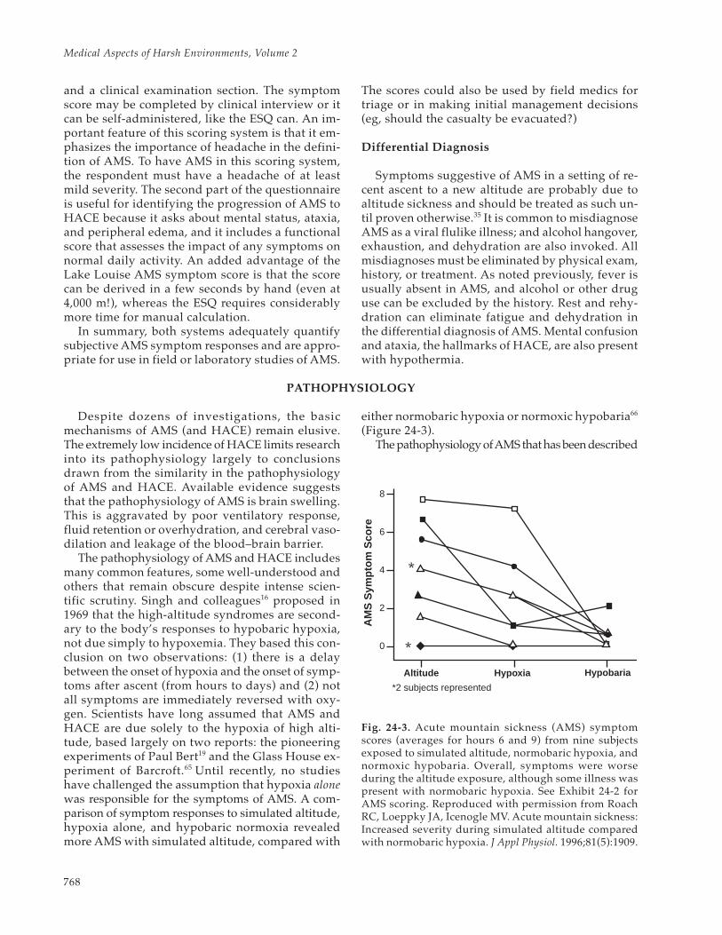
either normobaric hypoxia or normoxic hypobaria66
(Figure 24-3).The pathophysiology of AMS that has been described
Fig. 24-3. Acute mountain sickness (AMS) symptomscores (averages for hours 6 and 9) from nine subjectsexposed to simulated altitude, normobaric hypoxia, andnormoxic hypobaria. Overall, symptoms were worseduring the altitude exposure, although some illness waspresent with normobaric hypoxia. See Exhibit 24-2 forAMS scoring. Reproduced with permission from RoachRC, Loeppky JA, Icenogle MV. Acute mountain sickness:Increased severity during simulated altitude comparedwith normobaric hypoxia. J Appl Physiol. 1996;81(5):1909.
AM
S S
ympt
om S
core
Altitude Hypoxia Hypobaria
8�
6�
4�
2�
0
*
*
*2 subjects represented
769
Acute Mountain Sickness and High-Altitude Cerebral Edema
includes relative hypoventilation,46,67 a widened alveo-lar–arterial oxygen tension difference (PAO2 – PaO2),29,68
and decreased vital capacity and peak expiratoryflow16,27,69; subclinical pulmonary edema may be com-mon.70 Fluid retention,46,47,71–73 proteinuria,74,75 weightgain,46 increased cerebrospinal fluid (CSF) pres-sure,16,76,77 and cerebral edema are also noted.16,18,26 Wequickly recognize pulmonary, fluid-balance, and ce-rebral components in the pathophysiology of AMS.What determines how AMS will progress is not pres-ently known. The findings documented in HACE re-lated to pathophysiology include elevated CSF pres-sures,16 evidence of cerebral edema on CT scan18 andMRI,15 and gross cerebral edema on postmortem ex-amination.23–25 Well-documented cases of HACE of-ten include pulmonary edema.
Writing in 1924, Barcroft65 elegantly argued thatthe brain’s response to hypoxia was central to un-derstanding the pathophysiology of mountain sick-ness. He wrote:
Taking it, therefore, as settled that mountain sick-ness is due to oxygen want, the question arises,“oxygen want of what?” And the answer is, “of thebrain.” Such evidence as is at our disposal goes toshow that the brain wants but little oxygen; thatlittle, however, it wants very badly indeed.65(p91)
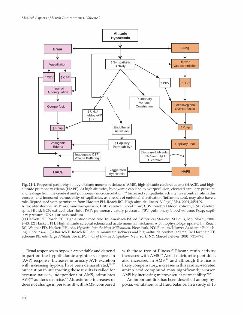
By 1970, enough had been learned of the basicpathophysiology of AMS and HACE to allowHansen and Evans78 to develop an elegant hypoth-esis of the pathogenesis of these illnesses. Theirtheory is that compression of the brain, either byincreased cerebral venous volume, reduced absorp-tion of CSF, or increased brain-tissue hydration,initiates the development of the symptoms andsigns of AMS and HACE. This approach is increas-ingly supported by studies of brain edema in thesyndromes (Figure 24-4). That the pulmonary andfluid-balance abnormalities of AMS and HACE aresecondary to central nervous system (CNS) re-sponses to sustained hypoxia may help explain thevaried results from the large number of studies doneon these topics since about 1950. Consequently, thefollowing discussion of the pathophysiology ofAMS and HACE primarily deals with the responseof the brain to sustained hypoxia. But first we shallbriefly review the known factors associated withAMS and HACE.
Ventilation and Gas Exchange
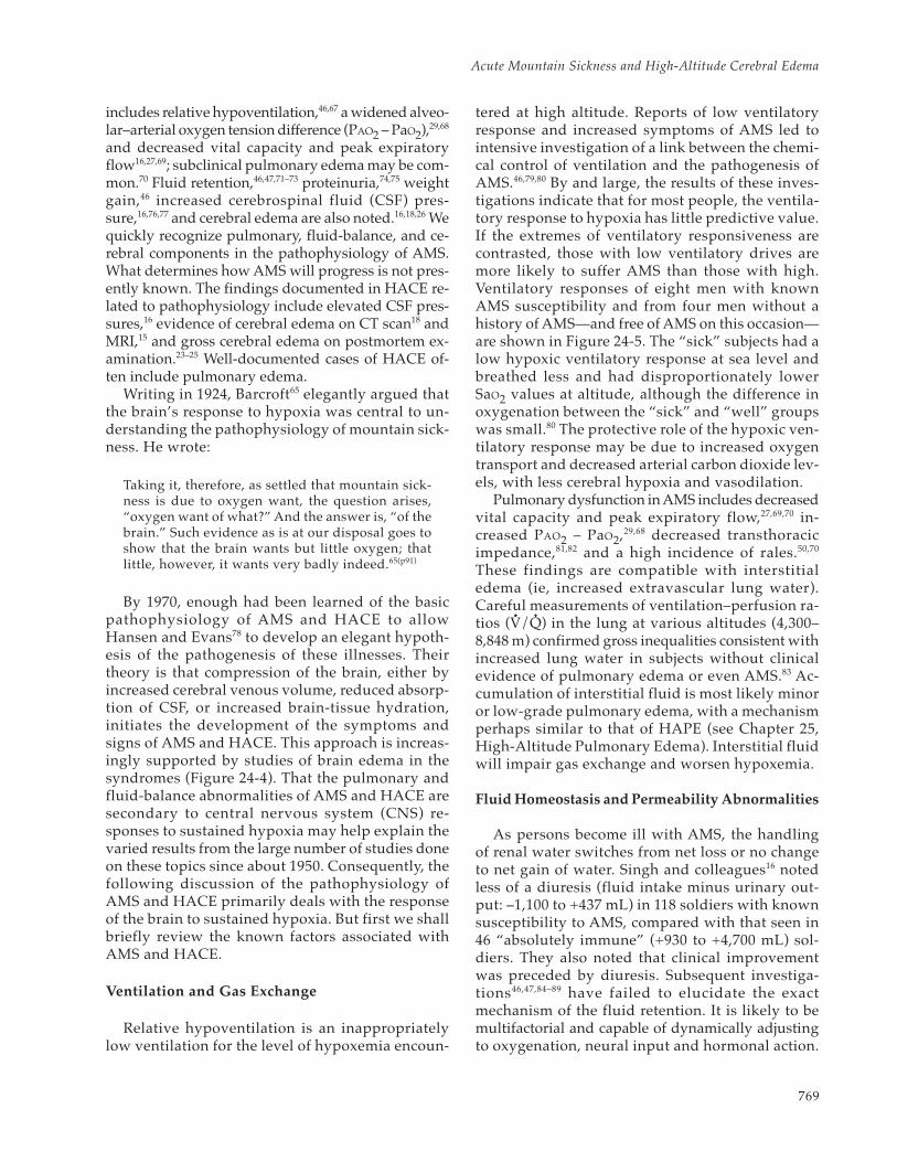
Relative hypoventilation is an inappropriatelylow ventilation for the level of hypoxemia encoun-
tered at high altitude. Reports of low ventilatoryresponse and increased symptoms of AMS led tointensive investigation of a link between the chemi-cal control of ventilation and the pathogenesis ofAMS.46,79,80 By and large, the results of these inves-tigations indicate that for most people, the ventila-tory response to hypoxia has little predictive value.If the extremes of ventilatory responsiveness arecontrasted, those with low ventilatory drives aremore likely to suffer AMS than those with high.Ventilatory responses of eight men with knownAMS susceptibility and from four men without ahistory of AMS—and free of AMS on this occasion—are shown in Figure 24-5. The “sick” subjects had alow hypoxic ventilatory response at sea level andbreathed less and had disproportionately lowerSaO2 values at altitude, although the difference inoxygenation between the “sick” and “well” groupswas small.80 The protective role of the hypoxic ven-tilatory response may be due to increased oxygentransport and decreased arterial carbon dioxide lev-els, with less cerebral hypoxia and vasodilation.
Pulmonary dysfunction in AMS includes decreasedvital capacity and peak expiratory flow,27,69,70 in-creased PAO2 – PaO2,29,68 decreased transthoracicimpedance,81,82 and a high incidence of rales.50,70
These findings are compatible with interstitialedema (ie, increased extravascular lung water).Careful measurements of ventilation–perfusion ra-tios (V/Q) in the lung at various altitudes (4,300–8,848 m) confirmed gross inequalities consistent withincreased lung water in subjects without clinicalevidence of pulmonary edema or even AMS.83 Ac-cumulation of interstitial fluid is most likely minoror low-grade pulmonary edema, with a mechanismperhaps similar to that of HAPE (see Chapter 25,High-Altitude Pulmonary Edema). Interstitial fluidwill impair gas exchange and worsen hypoxemia.
Fluid Homeostasis and Permeability Abnormalities
As persons become ill with AMS, the handlingof renal water switches from net loss or no changeto net gain of water. Singh and colleagues16 notedless of a diuresis (fluid intake minus urinary out-put: –1,100 to +437 mL) in 118 soldiers with knownsusceptibility to AMS, compared with that seen in46 “absolutely immune” (+930 to +4,700 mL) sol-diers. They also noted that clinical improvementwas preceded by diuresis. Subsequent investiga-tions46,47,84–89 have failed to elucidate the exactmechanism of the fluid retention. It is likely to bemultifactorial and capable of dynamically adjustingto oxygenation, neural input and hormonal action.
Medical Aspects of Harsh Environments, Volume 2
770
Renal responses to hypoxia are variable and dependin part on the hypothalamic arginine vasopressin(AVP) response. Increases in urinary AVP excretionwith increasing hypoxia have been demonstrated,90–93
but caution in interpreting these results is called forbecause nausea, independent of AMS, stimulatesAVP,94 as does exercise.95 Aldosterone increases ordoes not change in persons ill with AMS, compared
with those free of illness.90 Plasma renin activityincreases with AMS.86 Atrial natriuretic peptide isalso increased in AMS,90 and although the rise islikely compensatory, increases in this cardiac-secretedamino acid compound may significantly worsenAMS by increasing microvascular permeability.96,97
An important link has been described among hy-poxia, ventilation, and fluid balance. In a study of 15
Fig. 24-4. Proposed pathophysiology of acute mountain sickness (AMS), high-altitude cerebral edema (HACE), and high-altitude pulmonary edema (HAPE). At high altitudes, hypoxemia can lead to overperfusion, elevated capillary pressure,and leakage from the cerebral and pulmonary microcirculation.1–3 Increased sympathetic activity has a central role in thisprocess, and increased permeability of capillaries, as a result of endothelial activation (inflammation), may also have arole. Reproduced with permission from Hackett PH, Roach RC. High-altitude illness. N Engl J Med. 2001;345:109.Aldo: aldosterone; AVP: arginine vasopressin; CBF: cerebral blood flow; CBV: cerebral blood volume; CSF: cerebralspinal fluid; ECF: extracellular fluid; PAP: pulmonary artery pressure; PBV: pulmonary blood volume; Pcap: capil-lary pressure; UNa+: urinary sodium(1) Hackett PH, Roach RC. High-altitude medicine. In: Auerbach PA, ed. Wilderness Medicine. St Louis, Mo: Mosby; 2001:2–43. (2) Hackett PH. High altitude cerebral edema and acute mountain sickness: A pathophysiology update. In: RoachRC, Wagner PD, Hackett PH, eds. Hypoxia: Into the Next Millennium. New York, NY: Plenum/Kluwer Academic Publish-ing; 1999: 23–46. (3) Bartsch P, Roach RC. Acute mountain sickness and high-altitude cerebral edema. In: Hornbein TF,Schoene RB, eds. High Altitude: An Exploration of Human Adaptation. New York, NY: Marcel Dekker; 2001: 731–776.
Brain Lung
HACE HAPE
Altitude�Hypoxemia
↑ Sympathetic�Activity
↑ PBV
↑ Capillary Permeability?
↑ Pcap
↑ CBV ↑ CBF
VasodilationUneven�
Vasoconstriction
Capillary Leak
Impaired�Autoregulation
Overperfusion
Vasogenic�Edema
Pulmonary�Venous
Constriction
Exaggerated�Hypoxemia
↓ UNa+�↑ Aldo/AVP
↑ ECF
?
Endothelial�Activation
Focal/Regional�Overperfusion
Inadequate CSFVolume Buffering
Decreased AlveolarNa+ and H O
Clearance2
�
�
↑
↑ PAP
Pcap
771
Acute Mountain Sickness and High-Altitude Cerebral Edema
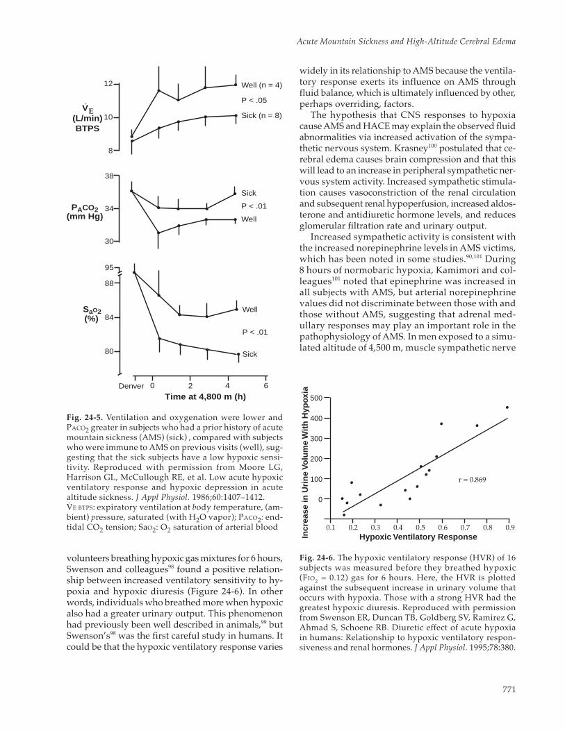
volunteers breathing hypoxic gas mixtures for 6 hours,Swenson and colleagues98 found a positive relation-ship between increased ventilatory sensitivity to hy-poxia and hypoxic diuresis (Figure 24-6). In otherwords, individuals who breathed more when hypoxicalso had a greater urinary output. This phenomenonhad previously been well described in animals,99 butSwenson’s98 was the first careful study in humans. Itcould be that the hypoxic ventilatory response varies
widely in its relationship to AMS because the ventila-tory response exerts its influence on AMS throughfluid balance, which is ultimately influenced by other,perhaps overriding, factors.
The hypothesis that CNS responses to hypoxiacause AMS and HACE may explain the observed fluidabnormalities via increased activation of the sympa-thetic nervous system. Krasney100 postulated that ce-rebral edema causes brain compression and that thiswill lead to an increase in peripheral sympathetic ner-vous system activity. Increased sympathetic stimula-tion causes vasoconstriction of the renal circulationand subsequent renal hypoperfusion, increased aldos-terone and antidiuretic hormone levels, and reducesglomerular filtration rate and urinary output.
Increased sympathetic activity is consistent withthe increased norepinephrine levels in AMS victims,which has been noted in some studies.90,101 During8 hours of normobaric hypoxia, Kamimori and col-leagues101 noted that epinephrine was increased inall subjects with AMS, but arterial norepinephrinevalues did not discriminate between those with andthose without AMS, suggesting that adrenal med-ullary responses may play an important role in thepathophysiology of AMS. In men exposed to a simu-lated altitude of 4,500 m, muscle sympathetic nerve
PACO2
Time at 4,800 m (h)Denver
12
10
8
38
34
30
95
88
84
80
(mm Hg)
SaO2(%)
EV(L/min)BTPS
0 2 4 6
Sick
Well
P < .01
Sick
Well
P < .01
Sick (n = 8)
Well (n = 4)
P < .05
Fig. 24-5. Ventilation and oxygenation were lower andPACO2 greater in subjects who had a prior history of acutemountain sickness (AMS) (sick) , compared with subjectswho were immune to AMS on previous visits (well), sug-gesting that the sick subjects have a low hypoxic sensi-tivity. Reproduced with permission from Moore LG,Harrison GL, McCullough RE, et al. Low acute hypoxicventilatory response and hypoxic depression in acutealtitude sickness. J Appl Physiol. 1986;60:1407–1412.VE BTPS: expiratory ventilation at body temperature, (am-bient) pressure, saturated (with H2O vapor); PACO2: end-tidal CO2 tension; SaO2: O2 saturation of arterial blood
Incr
ease
in U
rine
Volu
me
With
Hyp
oxia 500�
400�
300�
200�
100�
0
0.1 0.2 0.3 0.4 0.5 0.7 0.9Hypoxic Ventilatory Response
r = 0.869
0.6 0.8
Fig. 24-6. The hypoxic ventilatory response (HVR) of 16subjects was measured before they breathed hypoxic(FIO2 = 0.12) gas for 6 hours. Here, the HVR is plottedagainst the subsequent increase in urinary volume thatoccurs with hypoxia. Those with a strong HVR had thegreatest hypoxic diuresis. Reproduced with permissionfrom Swenson ER, Duncan TB, Goldberg SV, Ramirez G,Ahmad S, Schoene RB. Diuretic effect of acute hypoxiain humans: Relationship to hypoxic ventilatory respon-siveness and renal hormones. J Appl Physiol. 1995;78:380.
Medical Aspects of Harsh Environments, Volume 2
772
activity increased 2-fold after 1 hour at simulatedaltitude and remained elevated after 24 hours.102 Toofew subjects experienced AMS in this study to drawconclusions about a relationship between elevatedsympathetic tone and AMS; the important point isthat direct measurements of sympathetic activityindicate a high degree of activation by hypoxia thatis maintained during at least the first 24 hours ofaltitude exposure. Whether differences in the inten-sity of this response may be related to who gets sickand who remains free of AMS remains to be deter-mined. Additionally, α-adrenergic blockade hasbeen shown to be effective for the treatment ofHAPE,103 acting presumably by decreasing sympa-thetically mediated pulmonary hypertension (seeChapter 25, High-Altitude Pulmonary Edema).
Additional support for a role of sympathetic acti-vation in the pathogenesis of AMS comes from thework by Fulco and colleagues,104 showing that sub-jects with β-adrenergic blockade had less AMS thansubjects taking placebo. More complete adrenergicblockade may result in even greater decrease in AMSresponses if the hypothesis is correct that sympatheticactivation plays a central role in the pathogenesis ofAMS. Taken together, the evidence points to the pos-sibility that the sympathetic nervous system has a rolein the early development of AMS and HACE.
A generalized permeability defect has been hy-pothesized as an early defect common to the ede-mas of altitude.105 With the discovery of many va-soactive mediators of endothelial permeability,several studies briefly explored this important area.Urinary leukotriene E4 levels were measured in 8healthy men at sea level and after 36 hours at 4,300m.106 Leukotriene E4 levels were nearly doubled ataltitude, and the concentration was related to theseverity of AMS symptoms. In another study in 10subjects exposed to 4,350 m for 8 days, leukotrieneB4 levels mirrored the increase in AMS symptomscore from sea level to high altitude, and decreasedas symptoms resolved over time at altitude.107 Fur-ther studies are needed to establish cause andeffect for these modulators of endothelial perme-ability in the pathogenesis of AMS and HACE.
The Brain
Factors that determine the brain’s responses tosustained hypoxia include changes in cerebral bloodflow (CBF) and metabolism, CSF pressure, and cere-brovascular hemodynamics. Each response is nowconsidered and examined for how they comprisethe brain’s responses to sustained hypoxia.
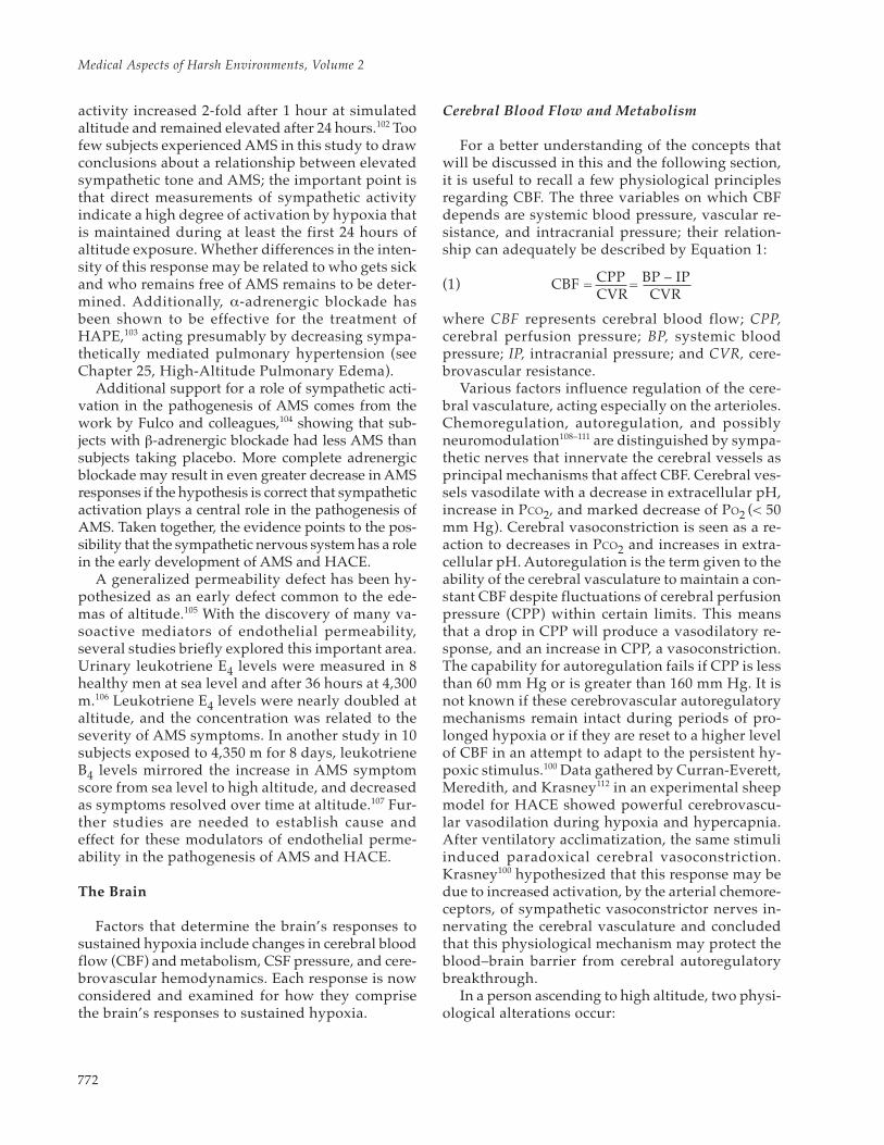
Cerebral Blood Flow and Metabolism
For a better understanding of the concepts thatwill be discussed in this and the following section,it is useful to recall a few physiological principlesregarding CBF. The three variables on which CBFdepends are systemic blood pressure, vascular re-sistance, and intracranial pressure; their relation-ship can adequately be described by Equation 1:
(1) CBF = CPP = BP – IP
where CBF represents cerebral blood flow; CPP,cerebral perfusion pressure; BP, systemic bloodpressure; IP, intracranial pressure; and CVR, cere-brovascular resistance.
Various factors influence regulation of the cere-bral vasculature, acting especially on the arterioles.Chemoregulation, autoregulation, and possiblyneuromodulation108–111 are distinguished by sympa-thetic nerves that innervate the cerebral vessels asprincipal mechanisms that affect CBF. Cerebral ves-sels vasodilate with a decrease in extracellular pH,increase in PCO2, and marked decrease of PO2 (< 50mm Hg). Cerebral vasoconstriction is seen as a re-action to decreases in PCO2 and increases in extra-cellular pH. Autoregulation is the term given to theability of the cerebral vasculature to maintain a con-stant CBF despite fluctuations of cerebral perfusionpressure (CPP) within certain limits. This meansthat a drop in CPP will produce a vasodilatory re-sponse, and an increase in CPP, a vasoconstriction.The capability for autoregulation fails if CPP is lessthan 60 mm Hg or is greater than 160 mm Hg. It isnot known if these cerebrovascular autoregulatorymechanisms remain intact during periods of pro-longed hypoxia or if they are reset to a higher levelof CBF in an attempt to adapt to the persistent hy-poxic stimulus.100 Data gathered by Curran-Everett,Meredith, and Krasney112 in an experimental sheepmodel for HACE showed powerful cerebrovascu-lar vasodilation during hypoxia and hypercapnia.After ventilatory acclimatization, the same stimuliinduced paradoxical cerebral vasoconstriction.Krasney100 hypothesized that this response may bedue to increased activation, by the arterial chemore-ceptors, of sympathetic vasoconstrictor nerves in-nervating the cerebral vasculature and concludedthat this physiological mechanism may protect theblood–brain barrier from cerebral autoregulatorybreakthrough.
In a person ascending to high altitude, two physi-ological alterations occur:
CVR CVR
773
Acute Mountain Sickness and High-Altitude Cerebral Edema
1. the reduced partial pressure of inspired oxy-gen (PIO2) leads to progressive hypoxia, and
2. the ventilatory acclimatization induces hy-perventilation with subsequent hypocapnia.
The former (reduced PIO2) leads to a decrease inCVR; the latter usually induces cerebrovascularconstriction, but its effects are offset by the hypoxicstimulus. The net effect is an increase in CBF; thisfact has been documented in animal models113–115
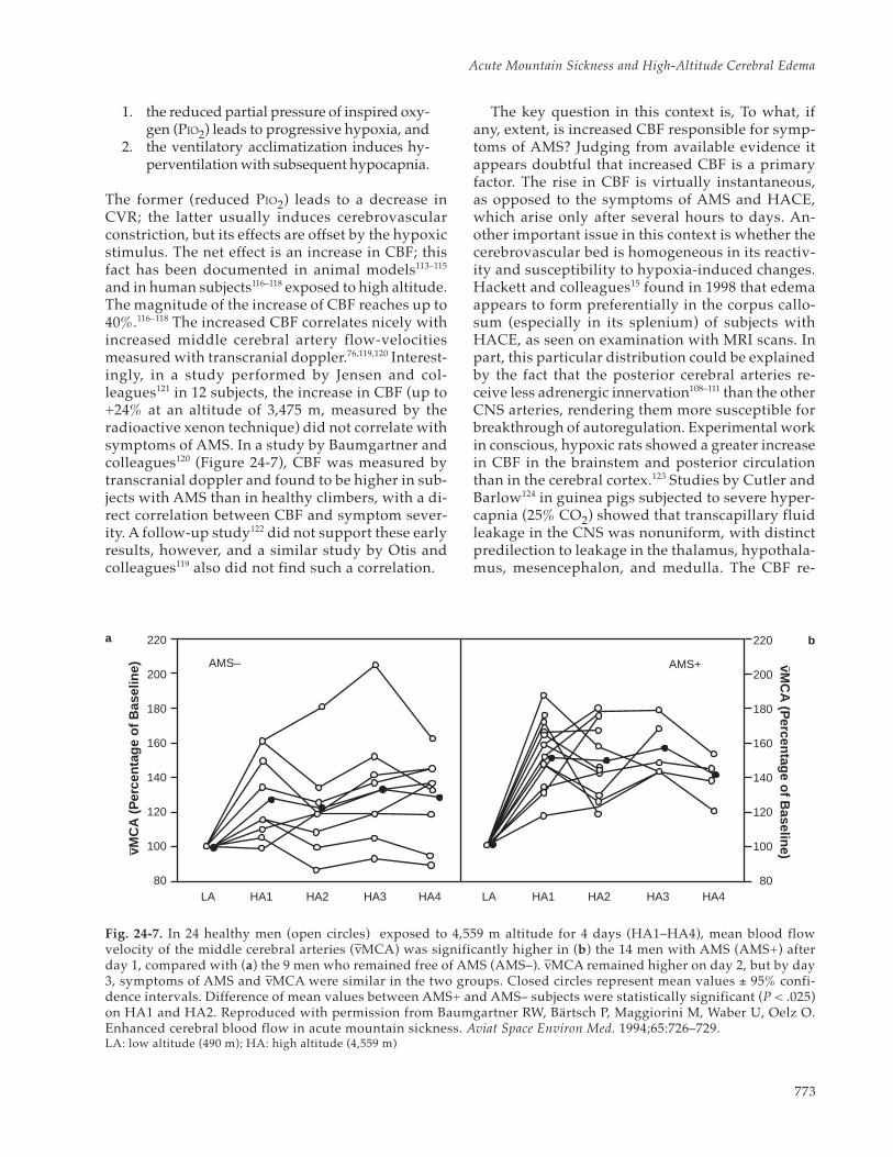
and in human subjects116–118 exposed to high altitude.The magnitude of the increase of CBF reaches up to40%.116–118 The increased CBF correlates nicely withincreased middle cerebral artery flow-velocitiesmeasured with transcranial doppler.76,119,120 Interest-ingly, in a study performed by Jensen and col-leagues121 in 12 subjects, the increase in CBF (up to+24% at an altitude of 3,475 m, measured by theradioactive xenon technique) did not correlate withsymptoms of AMS. In a study by Baumgartner andcolleagues120 (Figure 24-7), CBF was measured bytranscranial doppler and found to be higher in sub-jects with AMS than in healthy climbers, with a di-rect correlation between CBF and symptom sever-ity. A follow-up study122 did not support these earlyresults, however, and a similar study by Otis andcolleagues119 also did not find such a correlation.
The key question in this context is, To what, ifany, extent, is increased CBF responsible for symp-toms of AMS? Judging from available evidence itappears doubtful that increased CBF is a primaryfactor. The rise in CBF is virtually instantaneous,as opposed to the symptoms of AMS and HACE,which arise only after several hours to days. An-other important issue in this context is whether thecerebrovascular bed is homogeneous in its reactiv-ity and susceptibility to hypoxia-induced changes.Hackett and colleagues15 found in 1998 that edemaappears to form preferentially in the corpus callo-sum (especially in its splenium) of subjects withHACE, as seen on examination with MRI scans. Inpart, this particular distribution could be explainedby the fact that the posterior cerebral arteries re-ceive less adrenergic innervation108–111 than the otherCNS arteries, rendering them more susceptible forbreakthrough of autoregulation. Experimental workin conscious, hypoxic rats showed a greater increasein CBF in the brainstem and posterior circulationthan in the cerebral cortex.123 Studies by Cutler andBarlow124 in guinea pigs subjected to severe hyper-capnia (25% CO2) showed that transcapillary fluidleakage in the CNS was nonuniform, with distinctpredilection to leakage in the thalamus, hypothala-mus, mesencephalon, and medulla. The CBF re-
220
200
180
160
140
120
100
80LA HA1 HA2 HA3 HA4 LA HA1 HA2 HA3 HA4
vMC
A (P
ercentage of B
aseline)
220
200
180
160
140
120
100
80
AMS– AMS+
vMC
A (
Per
cent
age
of B
asel
ine)
Fig. 24-7. In 24 healthy men (open circles) exposed to 4,559 m altitude for 4 days (HA1–HA4), mean blood flowvelocity of the middle cerebral arteries (v–MCA) was significantly higher in (b) the 14 men with AMS (AMS+) afterday 1, compared with (a) the 9 men who remained free of AMS (AMS–). v–MCA remained higher on day 2, but by day3, symptoms of AMS and v–MCA were similar in the two groups. Closed circles represent mean values ± 95% confi-dence intervals. Difference of mean values between AMS+ and AMS– subjects were statistically significant (P < .025)on HA1 and HA2. Reproduced with permission from Baumgartner RW, Bärtsch P, Maggiorini M, Waber U, Oelz O.Enhanced cerebral blood flow in acute mountain sickness. Aviat Space Environ Med. 1994;65:726–729.LA: low altitude (490 m); HA: high altitude (4,559 m)
a b

Medical Aspects of Harsh Environments, Volume 2
774
mained constant or even decreased in these areas,as opposed to in the cerebral hemispheres, whichexperienced the most significant increase in CBFwithout any edema formation. Certainly, manyquestions remain open in this regard.
Early hypotheses of the pathogenesis of HACEpostulated that hypoxia may cause a dysfunctionin cellular Na+/K+ adenosine triphosphatase (ATP-ase), producing an influx of sodium into cells andthus causing cytotoxic CNS edema.125 Metabolicstudies focusing on cerebral oxygen and glucoseuptake revealed that these parameters are main-tained even during prolonged hypoxic exposure of5 days.112 In fact, the CNS optimizes oxygen uptakeduring hypoxemia, resulting in increased oxygenextraction. Interestingly, there appears to be a rela-tionship between oxygen extraction capacity andthe propensity to develop the AMS–HACE con-tinuum in sheep, as described by Curran-Everettand colleagues.126 They found that animals suscep-tible to AMS and HACE showed a lower oxygenextraction and higher CBF during normoxic condi-tions, and hypothesized that this predisposed theanimals to high cerebral capillary pressures duringhypoxia, leading to a transcapillary fluid shift. Asimilar investigation needs to be done in humans.
Changes in Cerebrospinal Fluid Pressure andCerebral and Intracranial Hemodynamics
In the following section, hemodynamic and CSFpressure changes observed during the developmentof AMS and HACE are discussed. To that end, it isuseful to recall a few pertinent physiological con-cepts and classifications.
The blood–brain barrier is the physiologicalmechanism that prevents free transition of sub-stances from the bloodstream into the extracellularCNS compartment. The anatomical structure re-sponsible for the barrier function is at the tight junc-tions between adjacent endothelial cells, and in theCNS parenchyma astrocytic foot-processes that areclosely associated with the endothelial cells andtheir basement membrane. The blood–brain barrieris not uniform, and in select areas of the CNS it isvirtually absent. In other areas (eg, eminentiamediana hypothalami, glandula pinealis), it is di-minished by the presence of fenestrated capillaries.
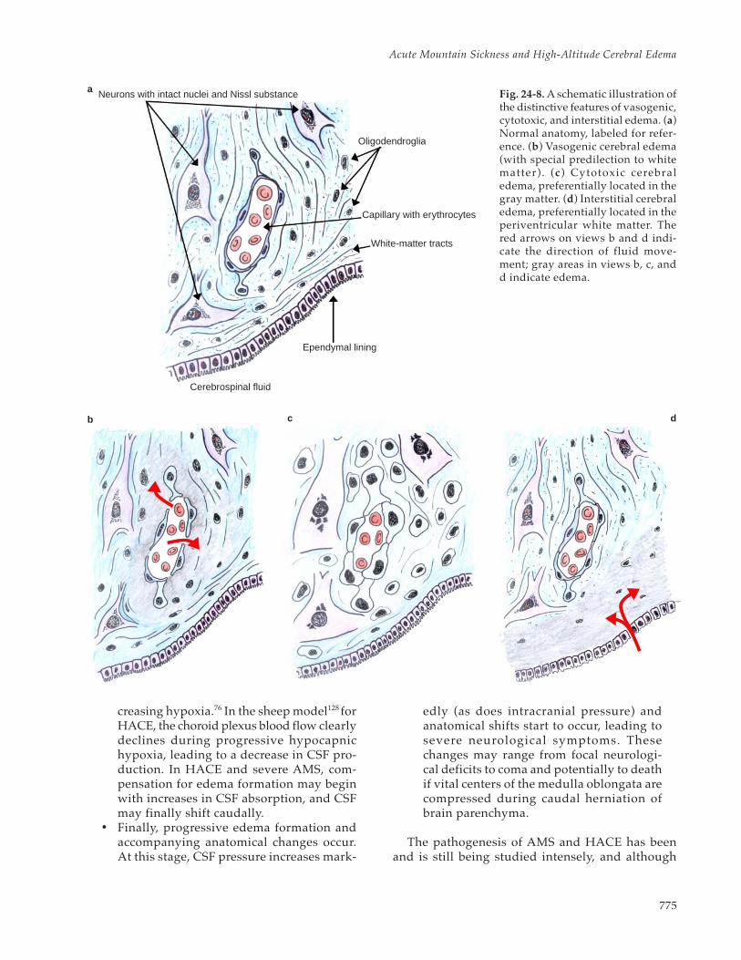
Classification of Central Nervous System Edema
In his classic description in 1967, Klatzo127 di-vided the pathological entity of brain edema intotwo distinct categories:
1. Vasogenic edema: protein-rich fluid spreadsinto the extracellular space through a com-promised blood–brain barrier. The prefer-ential site for this type of edema is thewhite matter.
2. Cytotoxic edema: intracellular fluid accu-mulation in neurons and neuroglia occursafter a severe insult (eg, toxins, ischemia).This type of edema is preferentially locatedin the gray matter.
A third type of brain edema was later added to theclassification to include the edema associated withderangements of CSF125 (Figure 24-8):
3. Interstitial edema: block of CSF absorptionresults in increased cerebral fluid locatedin the cellular interstitium. This type of ce-rebral edema is preferentially located in theperiventricular white matter.
Clear distinctions among these subtypes are oftennot possible, because a mixed picture may be seenwith the progression of the causative pathology. Forexample, severe anoxia may compromise cellularmetabolism and lead to cytotoxic edema, but theconcomitant vascular damage will produce progres-sive vasogenic edema as well. In his original de-scription, Klatzo127 commented on the particularpredilection of white matter in the setting ofvasogenic edema and hypothesized that the regulararrangement of extracellular channels offers lessresistance to the invasion of edema fluid than doesthe dense meshwork of the cerebral gray matter. Thefollowing overview synopsizes the events that occur:
• During the initial stages of AMS and HACE,responses related to decreased PIO2 and thehypobaric environment predispose to, oract as risk factors for the development of,the ensuing pathophysiological cascade.
• Next, hypobaric hypoxia generates a com-pensatory increase in CBF and ventilation.The concomitant hypocapnia (caused by theincreased ventilation) causes a respiratoryalkalosis, which in turn acts as a slowingmechanism on the central respiratory cen-ter, preventing excessive increases in ven-tilation.5 The kidneys excrete bicarbonate tocompensate for the respiratory alkalosis.The cerebrum also compensates for the res-piratory alkalosis by reducing the bicarbon-ate content of the CSF. A progressive, mild,increase in CSF pressure is seen with in-
775
Acute Mountain Sickness and High-Altitude Cerebral Edema
creasing hypoxia.76 In the sheep model128 forHACE, the choroid plexus blood flow clearlydeclines during progressive hypocapnichypoxia, leading to a decrease in CSF pro-duction. In HACE and severe AMS, com-pensation for edema formation may beginwith increases in CSF absorption, and CSFmay finally shift caudally.
• Finally, progressive edema formation andaccompanying anatomical changes occur.At this stage, CSF pressure increases mark-
edly (as does intracranial pressure) andanatomical shifts start to occur, leading tosevere neurological symptoms. Thesechanges may range from focal neurologi-cal deficits to coma and potentially to deathif vital centers of the medulla oblongata arecompressed during caudal herniation ofbrain parenchyma.
The pathogenesis of AMS and HACE has beenand is still being studied intensely, and although
a
b c d
Neurons with intact nuclei and Nissl substance
Oligodendroglia
Capillary with erythrocytes
White-matter tracts
Ependymal lining
Cerebrospinal fluid
Fig. 24-8. A schematic illustration ofthe distinctive features of vasogenic,cytotoxic, and interstitial edema. (a)Normal anatomy, labeled for refer-ence. (b) Vasogenic cerebral edema(with special predilection to whitematter). (c) Cytotoxic cerebraledema, preferentially located in thegray matter. (d) Interstitial cerebraledema, preferentially located in theperiventricular white matter. Thered arrows on views b and d indi-cate the direction of fluid move-ment; gray areas in views b, c, andd indicate edema.
Medical Aspects of Harsh Environments, Volume 2
776
many advances have been made in the understand-ing of the pathophysiology, many questions stillremain open and require further study. With a fo-cus on the blood–brain barrier and the role of the
endothelium and its mediators, as well as the roleof the neuroglial component, and with the use ofthe available animal models, further advancesshould be forthcoming.
PROPHYLAXIS
The fundamentals of prevention are similar forAMS and HACE. They are based on slow ascent,time for acclimatization, a low sleeping altitude,and avoidance of any factors that increase hypox-emia, such as sedative hypnotics and alcohol.
Acclimatization and Staging
Gradual ascent with appropriate time to adjustto new altitudes above 2,500 m is the safest pro-phylaxis for AMS. How slow is best determined byprevious experience. A general guideline is to avoidrapid ascent from sea level to a sleeping altitudeabove 3,000 m, and to spend 2 to 3 days at this alti-tude for initial acclimatization. As Heber and Bristolwrote in 1921, the “altitude of happiness” varies foreach individual, noting that they felt good at3,300 m but “a good deal less so” at 4,100 m.129(p1148)
The concept of “sleeping altitude” is important tounderstanding the practical aspects of the acclima-tization process. As is mentioned in Chapter 25,High-Altitude Pulmonary Edema, respiration canvary widely among individuals at high altitude,with the two extreme conditions being respirationduring exercise and during sleep. By sleeping at toohigh an altitude too soon after arrival, severe peri-odic breathing during sleep can occur, causingmarked arterial oxygen desaturation and poor sleepand thus hindering acclimatization. After acclima-tization to approximately 3,000 m, stopping for anextra night is recommend every time the sleepingaltitude increases 500 to 1,000 m. Above 3,000 m,climbing or hiking to higher altitudes during theday aids acclimatization. Abrupt increases of morethan 500 to 1,000 m in sleeping altitudes should beavoided without prior daytime exposures to thehigher sleeping altitudes. Spending several nightsat 3,000 m and climbing to 4,000 m in the interven-ing days will allow the sleeping altitude to be in-creased to 4,000 m with few problems; in contrast,the increase to 4,000 m after several days and nightsspent at only at 3,000 m would likely result in diffi-cult acclimatization and perhaps AMS. Staged ac-climatization is time consuming but very effective(see Figure 24-1). By having subjects spend 1 weekat 1,584 m followed by 1 week at 3,500 m before
driving them to 4,300 m, Hansen, Harris, andEvans54 were able to completely prevent AMS intheir volunteers.
Overexertion should be avoided while ascend-ing.130 Early in their altitude exposure, six of sevenvolunteers experienced significantly worse AMSwhen they exercised compared with when theywere resting. During exercise, SaO2 levels dropped8%, but at rest after exercise, ventilation and fluidbalance were not significantly different than thesame variables measured during rest without exer-cise.130
After acclimatization, in contrast, SaO2 is betterdefended during exercise than on sudden exposureto high altitude.58,131 Diets high in carbohydrate con-tent (> 70% of caloric intake as carbohydrate) mayinfluence acclimatization by increasing SaO2 lev-els.132 High-carbohydrate diets decreased AMSsymptoms 30%,133 presumably by increasing therespiratory quotient (V•CO2/V•O2) and thereby stimu-lating ventilation and increasing oxygenation.134,135
These findings were not confirmed in subjects whobreathed hypoxic gas at sea-level pressure for 12hours.135
Climbers maintain that recent acclimatization isprotective. Scientific support for this argumentcomes from Lyons and colleagues136 who took sixyoung, healthy men to the US Army Pikes PeakLaboratory Facility (a part of the US Army ResearchInstitute of Environmental Medicine, Natick, Mass),located at 4,300 m, for 16 days, and measured AMSsymptoms and SaO2. On day 1, four of the six (67%)had AMS, and their average SaO2 was 77%. After 14days at 4,300 m, as expected, their AMS scores haddecreased to near zero and their SaO2 levels had in-creased about 10%. The new finding from this studycame a week later when the same subjects, who hadbeen at sea level for the intervening week, werereexposed to altitude in a hypobaric chamber. Theirsymptoms were much less pronounced than theyhad been during their initial exposure (only onesubject of six was ill with AMS) and their SaO2 lev-els were 83%—higher than on day 1 on Pikes Peak(77%) but somewhat lower than on day 14 (87%).This suggests that factors that protect against AMSare activated during acclimatization, and they con-
777
Acute Mountain Sickness and High-Altitude Cerebral Edema
tinue to function after 1 week (or more?) at sea level.
Drugs
When rapid ascent is unavoidable, or known sus-ceptibility to AMS and HACE exists, several drugsare helpful for prevention of the conditions (Exhibit24-3; also see Figure 24-1). Acetazolamide is thedrug of choice for prophylaxis of AMS.6,30 As a car-bonic anhydrase inhibitor, acetazolamide reducesreabsorption of bicarbonate and sodium in the kid-ney, thus causing a bicarbonate diuresis and a meta-bolic acidosis. These effects start within 1 hour afteroral ingestion and rapidly enhance ventilatory andrenal acclimatization to high altitude. Arterial oxy-genation is improved. Acetazolamide’s diuretic ac-tion counteracts the fluid retention characteristic ofAMS, and perhaps more importantly, decreases CSFproduction and volume and possibly CSF pressure.93
Indications for acetazolamide prophylaxis in-clude rapid ascent (≤ 1 d) to altitudes higher than3,000 m, a rapid gain in sleeping altitude, a historyof recurrent AMS, and troublesome periodic breath-ing. Doses of 125 to 250 mg twice daily, starting 24hours prior to ascent, are as effective as higher doses
started earlier.5,6,30 A 500-mg sustained-action cap-sule of acetazolamide given once every 24 hours isprobably equally effective.5,137 Once acclimatizationis established, acetazolamide can be safely discon-tinued or reserved solely for prevention of persis-tent periodic breathing during sleep.6,138 Spirono-lactone and other diuretics have shown equivocalresults for AMS prevention.139–141
Another drug, dexamethasone, has also proveneffective for AMS prophylaxis. In eight healthy, youngmen rapidly exposed to 4,570 m in a hypobaric cham-ber, 4 mg every 6 hours decreased AMS symptoms75%, compared with placebo treatment.34 A field studyon Pikes Peak (4,300 m) reported a 21% reduction inAMS symptoms.142 Various dexamethasone dose regi-mens have been tested, from 4 mg every 6 hours to0.25 mg every 12 hours. Based on the findings of Rockand colleagues,143 the lowest effective dose for AMSprophylaxis is 4 mg given every 12 hours, which re-duced symptoms by 52%. In one study, combined ac-etazolamide and dexamethasone proved superior tothe use of either agent alone.144 Unlike acetazolamide,however, dexamethasone does not aid acclimatization.Once dexamethasone is discontinued, rebound ofAMS is likely.32,143 Dexamethasone, therefore, should
EXHIBIT 24-3
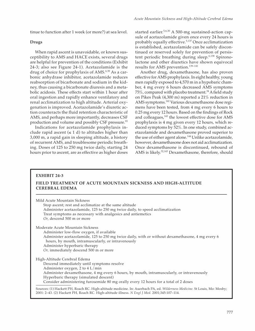
FIELD TREATMENT OF ACUTE MOUNTAIN SICKNESS AND HIGH-ALTITUDECEREBRAL EDEMA
Mild Acute Mountain SicknessStop ascent; rest and acclimatize at the same altitudeAdminister acetazolamide, 125 to 250 mg twice daily, to speed acclimatizationTreat symptoms as necessary with analgesics and antiemeticsOr, descend 500 m or more
Moderate Acute Mountain SicknessAdminister low-flow oxygen, if availableAdminister acetazolamide, 125 to 250 mg twice daily, with or without dexamethasone, 4 mg every 6
hours, by mouth, intramuscularly, or intravenouslyAdminister hyperbaric therapyOr, immediately descend 500 m or more
High-Altitude Cerebral EdemaDescend immediately until symptoms resolveAdminister oxygen, 2 to 4 L/minAdminister dexamethasone, 4 mg every 6 hours, by mouth, intramuscularly, or intravenouslyHyperbaric therapy (simulated descent)Consider administering furosemide 80 mg orally every 12 hours for a total of 2 doses
Sources: (1) Hackett PH, Roach RC. High-altitude medicine. In: Auerbach PA, ed. Wilderness Medicine. St Louis, Mo: Mosby;2001: 2–43. (2) Hackett PH, Roach RC. High-altitude illness. N Engl J Med. 2001;345:107–114.
Medical Aspects of Harsh Environments, Volume 2
778
be reserved for treatment of AMS and HACE, ratherthan prevention, except when necessary in persons in-tolerant of acetazolamide.
Two studies have found that a herbal remedymade from the plant Ginkgo biloba (a) preventedAMS during a gradual ascent to 5,000 m145 and (b)reduced both the symptoms and the incidence of
AMS by 50% during an abrupt ascent to 4,100 m.146
Extracts from the plant G biloba are potent antioxi-dants, and it may be this property that is respon-sible for the herbal remedy’s effectiveness in AMS.147
With respect to high-altitude headache, prophylac-tic aspirin (325 mg every 4 hours for a total of threedoses) reduced the incidence from 50% to 7%.148
TREATMENT
Treatment of mild AMS can include additionaltime to acclimatize at the present altitude, symp-tomatic therapy for headache and nausea, or both.Symptoms often resolve in 1 to 2 days (see Figure24-2). Moderate-to-severe AMS and HACE shouldbe treated with immediate descent when possible.In a patient with mild AMS, any deterioration ofneurological status or signs of pulmonary edemademand immediate descent. In the following de-scriptions of simulated descent, oxygen therapy andpharmacological approaches to the treatment ofAMS and HACE are expected, in moderate-to-severe illness, to serve only as temporizing measuresuntil real descent can be accomplished. Treatmentoptions are summarized in Exhibit 24-3.
Descent, Both Real and Simulated
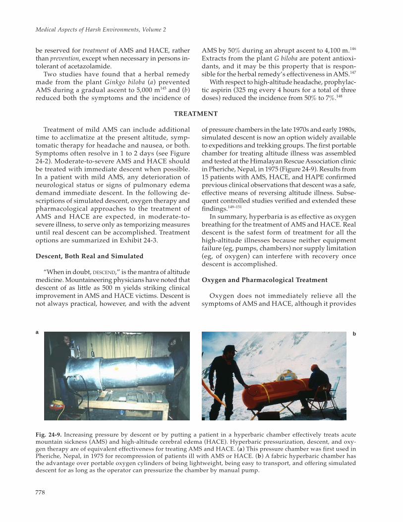
“When in doubt, DESCEND,” is the mantra of altitudemedicine. Mountaineering physicians have noted thatdescent of as little as 500 m yields striking clinicalimprovement in AMS and HACE victims. Descent isnot always practical, however, and with the advent
of pressure chambers in the late 1970s and early 1980s,simulated descent is now an option widely availableto expeditions and trekking groups. The first portablechamber for treating altitude illness was assembledand tested at the Himalayan Rescue Association clinicin Pheriche, Nepal, in 1975 (Figure 24-9). Results from15 patients with AMS, HACE, and HAPE confirmedprevious clinical observations that descent was a safe,effective means of reversing altitude illness. Subse-quent controlled studies verified and extended thesefindings.149–151
In summary, hyperbaria is as effective as oxygenbreathing for the treatment of AMS and HACE. Realdescent is the safest form of treatment for all thehigh-altitude illnesses because neither equipmentfailure (eg, pumps, chambers) nor supply limitation(eg, of oxygen) can interfere with recovery oncedescent is accomplished.
Oxygen and Pharmacological Treatment
Oxygen does not immediately relieve all thesymptoms of AMS and HACE, although it provides
Fig. 24-9. Increasing pressure by descent or by putting a patient in a hyperbaric chamber effectively treats acutemountain sickness (AMS) and high-altitude cerebral edema (HACE). Hyperbaric pressurization, descent, and oxy-gen therapy are of equivalent effectiveness for treating AMS and HACE. (a) This pressure chamber was first used inPheriche, Nepal, in 1975 for recompression of patients ill with AMS or HACE. (b) A fabric hyperbaric chamber hasthe advantage over portable oxygen cylinders of being lightweight, being easy to transport, and offering simulateddescent for as long as the operator can pressurize the chamber by manual pump.
a b
779
Acute Mountain Sickness and High-Altitude Cerebral Edema
significant symptomatic relief in mild-to-moderatecases. Oxygen can be a life-saving temporizingmeasure in cases of severe AMS and HACE.
In mild AMS, acetazolamide (125–250 mg, ad-ministered orally twice daily) will speed acclimati-zation and alleviate illness. Headache can be treatedwith analgesics6 such as aspirin (650 mg),acetaminophen (650–1,000 mg),148,152 or ibuprofen (≥200 mg).153 Nausea and vomiting respond well toprochlorperazine (5–10 mg, administered intramus-cularly). Alcohol and sedative hypnotics should beavoided because of their depressive effect on respi-ration, especially during sleep. In moderate AMS,acetazolamide (250 mg, given three times daily) waseffective in relieving symptoms and improvingpulmonary gas exchange and SaO2%.29 Dexametha-sone is effective for treatment of moderate AMS.Using a dose of either 4 mg every 6 hours32 or an8-mg initial dose followed by 4 mg every 6 hours,33
symptoms were notably minimized, with no signifi-cant side effects. However, dexamethasone does notaid acclimatization, and symptom rebound has re-peatedly been observed.32,33,142 Therefore, dexa-methasone could be used to relieve symptoms andacetazolamide to speed acclimatization.
Successful treatment of HACE requires early rec-ognition. At the first sign of ataxia or change in con-
sciousness, descent should be started, dexamethasone(initially 4–8 mg intravenously, intramuscularly, or bymouth, followed by 4 mg every 6 h) administered, andoxygen (2–4 L/min by mask or nasal cannula) applied,if available. Oxygen can be titrated to maintain SaO2at or higher than 90% if oximetry is available. Coma-tose patients require additional airway managementand bladder drainage. Attempting to decrease intrac-ranial pressure by intubation and hyperventilationis a reasonable approach, although these patientsare already alkalotic and over-hyperventilationcould result in cerebral ischemia. Loop diureticssuch as furosemide (40–80 mg) or bumetanide (1–2mg) may help reduce brain hydration, but an ad-equate intravascular volume to maintain perfusionpressure is critical. Hypertonic solutions of saline,mannitol, and urea have been suggested7 but areused rarely in the field. Controlled studies are lack-ing, but empirically, the response to steroids andoxygen seems to be excellent if given early in thecourse of the illness and disappointing if not starteduntil the patient is unconscious. Coma may persistfor days, even after evacuation to low altitude, inwhich case other causes of coma must be consid-ered and ruled out by appropriate evaluation. Se-quelae lasting weeks are common; longer-term fol-low-up has been limited.
MILITARY OPERATIONS AT ALTITUDE
The rapid deployment of troops to high altitude(ie, > 2,500 m) may pose significant logistical, tech-nical, environmental, tactical, and medical prob-lems.16,154,155 The routes for supply in difficult moun-tainous terrain may be restricted to air-drop andmay be hampered by unfavorable weather condi-tions—necessitating autonomy in regard to food,clothing, and equipment. The environment mayrequire special attention to dangers of rockfall; highultraviolet light exposures; difficult technical as-cents requiring special gear; avalanches; and severe,prolonged periods of cold weather in the winterseason. The human factors that have to be taken intoconsideration are more likely to be crucial in thissetting. The lack of experience in a new environ-ment and insufficient physical and psychologicalpreparedness are factors that can be overcome bycareful planning and training for the specific op-eration. The environmental conditions (eg, low tem-peratures, difficult ascents) can produce a state ofphysical and psychological exhaustion that mayaffect decision making and result in faulty assess-ments of terrain and the feasibility of tasks at hand.The medical issues that arise during a military op-
eration at high altitude are multiple and complex;the following discussion will focus on the potentialdangers of AMS and HACE.
The symptomatology of the two syndromes isunfortunately not specific in its initial presentationand thus requires constant attention from the medi-cal officer for early detection. Headache, lassitude,and mild ataxia are early warning signs that shouldbe carefully watched and acted on as necessary. Theobservations by Singh and colleagues16 are the mostcomprehensive series of published observationsmade in troops exposed to a high-altitude environ-ment. Singh studied 1,925 soldiers, aged 18 to 53years, air-lifted to the Indian Himalaya at altitudesof 3,350 to 5,500 m. A variety of neurological mani-festations were observed, including headaches (themost prevalent and most persistent symptom), de-creased ability to concentrate, blurred vision, and,in 24 soldiers, more-severe signs and symptomsincluding papilledema, stupor, coma, seizures, andfocal neurological deficits. In Singh’s series, threepatients died from neurological complications andtwo had marked cerebral edema on postmortemexamination.
Medical Aspects of Harsh Environments, Volume 2
780
The neurological changes seen in AMS after rapidexposure to high altitude may thus have significantimpact on the physical and mental functioning ofsusceptible individuals. These changes in conjunc-tion with the hardships of the environment canmake troop leadership difficult. Constant attentionto the physical and mental state of the troops is nec-essary to detect early changes that can be appro-priately treated. Lack of attention to these early is-sues will entail secondary dangers to the militaryoperation, such as misjudgments of tactical situa-tions and mistakes made during the operation ofvehicles, weapons, or complex instruments. Thecapability for rapid deployment is a hallmark oftoday’s modern armies and confers obvious strate-gic advantages. However, rapid deployment itselfexposes the troops to a higher risk of altitude-related illnesses in the mountainous environment.To avoid disability and death of the troops, the fol-
lowing actions must be taken by the commandingofficer, in collaboration with the medical officer:
1. Allow time for acclimatization throughgraded ascent to high altitude.
2. Educate troops about signs and symptomsof AMS and HACE to ensure early report-ing (make use of the “buddy-system,” inwhich individuals observe each other foroccurrence of symptoms).
3. Evaluate the need for pharmacological pro-phylaxis, in view of the nature of the mis-sion and the available time.
4. Minimize severe exertion and exposure tocold as possible precipitating factors.
5. Plan ahead for rapid evacuation (to loweraltitude) for severe cases.
6. Plan for treatment of AMS to keep soldiersfunctional and to avoid evacuations.
SUMMARY
Altitude-related illnesses such as AMS are usu-ally a nuisance (eg, headaches, anorexia, nausea)rather than a threat to life and are most commonlyself-limited. However, they may have a significantimpact on an individual’s ability to function. Mili-tary commanders and medical officers can preventor reduce altitude-related illnesses by following thesix actions enumerated just above.
To avoid fatal outcomes, severe complicationssuch as HACE in susceptible individuals may re-quire rapid recognition and efficient treatment, suchas evacuation to lower altitude, simulated descentin a hyperbaric bag, and pharmacological therapy.
If descent is not an option, then acetazolamide anddexamethasone effectively reverse AMS, and HACEis reversed with dexamethasone. Aspirin success-fully treats high-altitude headache.
Pharmacological prevention can include acetazola-mide, dexamethasone, and a herbal remedy madefrom the plant Ginko biloba. Recent studies support theeffectiveness of G biloba extract in small study groups;larger trials are underway. Prevention can largely beachieved by slow, staged ascent. All travelers to highaltitude should be mindful that awareness of the earlysymptoms of AMS and HACE can be the most effec-tive strategy for preventing fatal outcomes.
REFERENCES
1. Sutton JR, Lassen NA. Pathophysiology of acute mountain sickness and high altitude pulmonary edema: Anhypothesis. In: Brendel W, Link RA, eds. High Altitude Physiology and Medicine. New York, NY: Springer-Verlag;1982: 266–267.
2. Malconian MK, Rock PB. Medical problems related to altitude. In: Pandolf KB, Sawka MN, Gonzalez RR, eds.Human Performance Physiology and Environmental Medicine at Terrestrial Extremes. Dubuque, Iowa: Brown & Bench-mark; 1986: 545–564.
3. Johnson TS, Rock PB. Acute mountain sickness. N Engl J Med. 1988;319:841–845.
4. Houston CS. Mountain sickness. Sci Am. 1992;267:58–66.
5. Hackett PH, Roach RC. High-altitude medicine. In: Auerbach PA, ed. Wilderness Medicine. St Louis, Mo: Mosby;2001: 2–43.
6. Hackett PH, Roach RC. Medical therapy of altitude illness. Ann Emerg Med. 1987;16:980–986.
781
Acute Mountain Sickness and High-Altitude Cerebral Edema
7. Hackett PH, Roach RC. High-altitude illness. N Engl J Med. 2001;345:107–114.
8. Roach RC, Hackett PH. Frontiers of hypoxia research: Acute mountain sickness. J Exp Biol. 2001;204:3161–3170.
9. Hackett PH. High altitude cerebral edema and acute mountain sickness: A pathophysiology update. Adv ExpMed Biol. 1999;474:23–45.
10. Hackett PH. The cerebral etiology of high-altitude cerebral edema and acute mountain sickness. WildernessEnviron Med. 1999;10:97–109.
11. Bartsch P, Roach RC. Acute mountain sickness and high-altitude cerebral edema. In: Hornbein TF, Schoene RB,eds. High Altitude: An Exploration of Human Adaptation. New York, NY: Marcel Dekker; 2001: 731–776.
12. Whymper E; Shipton E, ed. Travels Amongst the Great Andes of the Equator. London, England: Charles Knight;1972. Republished; originally published in London, England: Murray; 1891–1892.
13. Ravenhill TH. Some experiences of mountain sickness in the Andes. J Trop Med Hygiene. 1913;1620:313–320.
14. West JB. T. H. Ravenhill and his contributions to mountain sickness. J Appl Physiol. 1996;80:715–724.
15. Hackett PH, Yarnell PR, Hill R, Reynard K, Heit J, McCormick J. High altitude cerebral edema evaluated withmagnetic resonance imaging: Clinical correlation and pathophysiology. JAMA. 1998;280(22):1920–1925.
16. Singh I, Khanna PK, Srivastava MC, Lal M, Roy SB, Subramanyam CS. Acute mountain sickness. N Engl J Med.1969;280:175–218.
17. Hackett PH, Rennie ID, Levine HD. The incidence, importance, and prophylaxis of acute mountain sickness.Lancet. 1976;2:1149–1154.
18. Matsuzawa Y, Kobayashi T, Fujimoto K, Shinozaki S, Yoshikawa S. Cerebral edema in acute mountain sickness.In: Ueda G, Reeves JT, Sekiguchi M, eds. High Altitude Medicine. Matsumoto, Japan: Shinshu University Press;1992: 300–304.
19. Bert P. La Pression Barométrique: Recherches de Physiologie Expérimentale. Paris, France: G. Masson; 1878. Reprintedas Barometric Pressure. Bethesda, Md: Undersea Medical Society; 1978.
20. Honigman B, Theis MK, McLain J, et al. Acute mountain sickness in a general tourist population at moderatealtitudes. Ann Intern Med. 1993;118:587–592.
21. Houston CS, Dickinson JG. Cerebral form of high altitude illness. Lancet. 1975;2:758–761.
22. Wilson R, Mills WJ Jr, Rogers DR, Propst MT. Death on Denali: Fatalities among climbers in Mount McKinleyNational Park 1903–1976: Analysis of injuries, illnesses and rescues in 1976. West J Med. 1976;128:471–476.
23. Wilson R. Acute high altitude illness in mountaineers and problems of rescue. Ann Intern Med. 1973;78:421–427.
24. Dickinson JG. High altitude cerebral edema: Cerebral acute mountain sickness. Semin Respir Med. 1983;5:151–158.
25. Dickinson JG, Heath J, Gosney J, Williams D. Altitude related deaths in seven trekkers in the Himalayas. Tho-rax. 1983;38:646–656.
26. Levine BD, Yoshimura K, Kobayashi T, Fukushima M, Shibamoto T, Ueda G. Dexamethasone in the treatmentof acute mountain sickness. N Engl J Med. 1989;321:1707–1713.
27. Larson EB, Roach RC, Schoene RB, Hornbein TF. Acute mountain sickness and acetazolamide: Clinical efficacyand effect on ventilation. JAMA. 1982;288:328–332.
Medical Aspects of Harsh Environments, Volume 2
782
28. Forwand SA, Landowne M, Folansbee IN, Hansen JE. Effect of acetazolamide on acute mountain sickness. NEngl J Med. 1968;279:839–845.
29. Grissom CK, Roach RC, Sarnquist FH, Hackett PH. Acetazolamide in the treatment of acute mountain sickness:Clinical efficacy and effect on gas exchange. Ann Intern Med. 1992;116:461–465.
30. Editorial. Acetazolamide for acute mountain sickness. FDA Drug Bull. 1983;13:27.
31. Editorial. High altitude sickness. Med Lett Drugs Ther. 1992;34:84–86.
32. Hackett PH, Roach RC, Wood RA, et al. Dexamethasone for prevention and treatment of acute mountain sick-ness. Aviat Space Environ Med. 1988;59:950–954.
33. Ferrazzini G, Maggiorini M, Kriemler S, Bartsch P, Oelz O. Successful treatment of acute mountain sicknesswith dexamethasone. Br Med J. 1987;294:1380–1382.
34. Johnson TS, Rock PB, Fulco CS, Trad LA, Spark RF, Maher JT. Prevention of acute mountain sickness by dexam-ethasone. N Engl J Med. 1984;310:683–686.
35. Hackett PH. Mountain Sickness: Prevention, Recognition and Treatment. New York, NY: American Alpine Club; 1980.
36. Sanchez del Rio M, Moskowitz MA. High altitude headache: Lessons from headaches at sea level. Adv Exp MedBiol. 1999;474:145–153.
37. King AB, Robinson SM. Vascular headache of acute mountain sickness. Aerospace Med. 1972;43:849–851.
38. Weil JC, Kryger MH, Scoggin CH. Sleep and breathing at high altitude. In: Guilleminault C, Demant WC, eds.Sleep Apnea Syndromes. New York, NY: AR Liss; 1978: 119–136.
39. Reite M, Jackson D, Cahoon RL, Weil JV. Sleep physiology at high altitude. Electroencephalogr Clin Neurophysiol.1975;38:463–471.
40. Powles AP, Sutton JR. Sleep at altitude. Semin Respir Med. 1983;5:175–180.
41. Anholm JD, Powles AC, Downey R, et al. Operation Everest II: Arterial oxygen saturation and sleep at extremesimulated altitude. Am Rev Respir Dis. 1992;145:817–826.
42. Powles AP, Sutton JR, Gray GW, Mansell AL, McFadden M, Houston CS. Sleep hypoxemia at altitude: Its rela-tionship to acute mountain sickness and ventilatory responsiveness to hypoxia and hypercapnia. In: FolinsbeeLJ, Wagner JA, Borgia JF, Drinkwater BL, Gliner JA, Bedi JF, eds. Environmental Stress: Individual Human Adap-tations. New York, NY: Academic Press; 1978: 317–324.
43. Westerterp-Plantenga MS, Westerterp KR, Rubbens M, Verwegen CR, Richelet JP, Gardette B. Appetite at “highaltitude” (Operation Everest III [Comex-’97]): A simulated ascent of Mount Everest. J Appl Physiol. 1999;87:391–399.
44. Bailey DM, Davies B, Milledge JS, et al. Elevated plasma cholecystokinin at high altitude: Metabolic implica-tions for the anorexia of acute mountain sickness. High Alt Med Biol. 2000;1:9–23.
45. Singh MV, Rawal SB, Tyagi AK. Body fluid status on induction, reinduction and prolonged stay at high altitudeof human volunteers. Int J Biometeorol. 1990;34:93–97.
46. Hackett PH, Rennie ID, Hofmeister SE, Grover RF, Grover EB, Reeves JT. Fluid retention and relativehypoventilation in acute mountain sickness. Respiration. 1982;43:321–329.
47. Bartsch P, Shaw S, Wiedmann P, Franciolli M, Maggiorini M, Oelz O. Aldosterone, antidiuretic hormone andatrial natriuretic peptide in acute mountain sickness. In: Sutton JR, Coates G, Houston CS, eds. Hypoxia andMountain Medicine. Burlington, Vt: Queen City Press; 1992: Chap 8.
783
Acute Mountain Sickness and High-Altitude Cerebral Edema
48. Aoki VS, Robinson SM. Body hydration and the incidence and severity of acute mountain sickness. J ApplPhysiol. 1971;31:363–367.
49. Heyes MP, Sutton JR. High altitude ills: A malady of water, electrolyte, and hormonal imbalance? Semin RespirMed. 1983;5:207–212.
50. Hackett PH, Rennie ID. Rales, peripheral edema, retinal hemorrhage and acute mountain sickness. Am J Med.1979;67:214–218.
51. Montgomery AB, Mills J, Luce JM. Incidence of acute mountain sickness at intermediate altitude. JAMA.1989;261:732–734.
52. Singh I, Roy SB. High altitude pulmonary edema: Clinical, hemodynamic, and pathologic studies. In: HegnauerA, ed. Biomedical Problems of High Terrestrial Elevations. Springfield, Va: Federal Science and Technical Informa-tion Service; 1962: 108–120.
53. Singh I, Kapila CC, Khanna PK, Nanda RB, Rao BD. High altitude pulmonary oedema. Lancet. 1965;1:229–234.
54. Hansen JE, Harris CW, Evans WO. Influence of elevation of origin, rate of ascent and a physical conditioningprogram on symptoms of AMS. Mil Med. 1967;132:585–592.
55. Roach RC, Houston CS, Honigman B, et al. How well do older persons tolerate moderate altitude? West J Med.1995;162:32–36.
56. Ross RT. The random nature of cerebral mountain sickness. Lancet. 1985;1:990–991.
57. Shapiro K, Marmarou A, Shulman K. Characterization of clinical CSF dynamics and neural axis complianceusing the pressure volume index, I: The normal pressure–volume index. Ann Neurol. 1980;7:508–514.
58. Roach RC, Lium D, Hackett PH. Arterial oxygen desaturation may determine subsequent acute mountain sick-ness [abstract]. Med Sci Sport Exerc. 1994;26:S22.
59. Savourey G, Moirant C, Eterradossi J, Bittel J. Acute mountain sickness relates to sea-level partial pressure ofoxygen. Eur J Appl Physiol. 1995;70:469–476.
60. Reeves JT, Moore LG, Cymerman A, Weil JV. Sea-Level PCO2 relates to ventilatory acclimatization at 4,300 m. JAppl Physiol. 1993;75:1117–1122.
61. Kobrick JL, Sampson JB. New inventory for the assessment of symptom occurrence and severity at high alti-tude. Aviat Space Environ Med. 1979;50:925–929.
62. Sampson JB, Kobrick JL. The environmental symptoms questionnaire: Revisions and new field data. Aviat SpaceEnviron Med. 1980;51:872–877.
63. Sampson JB, Cymerman A, Burse RL, Maher JT, Rock PB. Procedures for the measurement of acute mountainsickness. Aviat Space Environ Med. 1983;54:1063–1073.
64. Roach RC, Bartsch P, Oelz O, Hackett PH. The Lake Louise Acute Mountain Sickness scoring system. In: Sutton JR,Houston CS, Coates G, eds. Hypoxia and Molecular Medicine. Burlington, Vt: Queen City Press; 1993: Chap 26.
65. Barcroft J. Mountain sickness. Nature. 1924;2855:90–92.
66. Roach RC, Loeppky JA, Icenogle MV. Acute mountain sickness: Increased severity during simulated altitudecompared with normobaric hypoxia. J Appl Physiol. 1996;81(5):1908–1910.
67. Matsuzawa Y, Fujimoto K, Kobayashi T, et al. Blunted hypoxic ventilatory drive in subjects susceptible to high-altitude pulmonary edema. J Appl Physiol. 1989;66:1152–1157.
Medical Aspects of Harsh Environments, Volume 2
784
68. Sutton JR, Bryan AC, Gray GW, et al. Pulmonary gas exchange in acute mountain sickness. Aviat Space EnvironMed. 1976;47:1032–1037.
69. Anholm JD, Houston CS, Hyers TM. The relationship between acute mountain sickness and pulmonary venti-lation at 2,835 meters (9,300 feet). Chest. 1979;75:33–36.
70. Cremona G, Asnaghi R, Baderna P, et al. Pulmonary extravascular fluid accumulation in recreational climbers:A prospective study. Lancet. 2002;359:303–309.
71. Bartsch P, Pfluger N, Audetat M, et al. Effects of slow ascent to 4559m on fluid homeostasis. Aviat Space EnvironMed. 1991;62:105–110.
72. Claybaugh JR, Brooks DP, Cymerman A. Hormonal control of fluid and electrolyte balance at high altitude innormal subjects. In: Sutton JR, Coates G, Houston CS, eds. Hypoxia and Mountain Medicine. Burlington, Vt:Queen City Press; 1992: Chap 7.
73. Singh MV, Rawal SB, Tyagi AK, Bhagat JK, Parshad R, Divekar HM. Changes in body fluid compartments onre-induction to high altitude and effects of diuretics. Int J Biometeorol. 1988;32:36–40.
74. Editorial. Proteinuria at high altitude. Br Med J. 1979;1:508–509.
75. Winterborn MH, Bradwell AR, Chesner IM, Jones GT. The origin of proteinuria at high altitude. Postgrad Med J.1987;63:179–181.
76. Hartig GS, Hackett PH. Cerebral spinal fluid pressure and cerebral blood velocity in acute mountain sickness.In: Sutton JR, Coates G, Houston CS, eds. Hypoxia and Mountain Medicine. Burlington, Vt: Queen City Press;1992: Chap 24.
77. Schaltenbrand G. Atmospheric pressure, circulation, respiration and cerebrospinal fluid pressure. ActaAerophysiol. 1933;1:65–78.
78. Hansen JE, Evans WO. A hypothesis regarding the pathophysiology of acute mountain sickness. Arch EnvironHealth. 1970;21:666–669.
79. King AB, Robinson SM. Ventilation response to hypoxia and acute mountain sickness. Aerospace Med. 1972;43:419–421.
80. Moore LG, Harrison GL, McCullough RE, et al. Low acute hypoxic ventilatory response and hypoxic depres-sion in acute altitude sickness. J Appl Physiol. 1986;60:1407–1412.
81. Hoon RS, Balasubramanian V, Tiwari SC, Mathew OP, Behl A. Changes in transthoracic electrical impedance athigh altitude. Br Heart J. 1977;39:61–66.
82. Roy SB, Balasubramanian V, Khan MR, Kaushik VS, Manchanda SC, Guha SK. Transthoracic electrical imped-ance in cases of high-altitude hypoxia. Br Med J. 1974;3:771–775.
83. Wagner PD, Sutton JR, Reeves JT, Cymerman A, Groves BM, Malconian MK. Operation Everest II: Pulmonarygas exchange during a simulated ascent of Mt Everest. J Appl Physiol. 1987;63:2348–2359.
84. Bartsch P, Shaw S, Francioli M, Gnadinger MP, Weidmann P. Atrial natriuretic peptide in acute mountain sick-ness. J Appl Physiol. 1988;65:1929–1937.
85. Hannon JP, Chinn KS, Shields JL. Effects of acute high altitude exposure on body fluids. Fed Proc. 1969;28:1178–1184.
86. Claybaugh JR, Hansen JE, Wozniak DB. Response of antidiuretic hormone to acute exposure to mild and severehypoxia in man. J Endocrinol. 1978;77:157–160.
785
Acute Mountain Sickness and High-Altitude Cerebral Edema
87. Claybaugh JR, Sato AK, Eichinger MR. Blood pressure, AVP, ACTH, and PRA responses to IV and IVT angio-tensin II during hypoxia [abstract]. In: Sutton JR, Coates G, Houston CS, eds. Hypoxia and Mountain Medicine.Burlington, Vt: Queen City Press; 1993: 296.
88. Olsen NV, Kanstrup IL, Richalet JP, Hansen JM, Plazen G, Galen FX. Effects of acute hypoxia on renal andendocrine function at rest and during graded exercise in hydrated subjects. J Appl Physiol. 1992;73:2036–2043.
89. Westerterp KR, Robach P, Wouters L, Richalet JP. Water balance and acute mountain sickness before and afterarrival at high altitude of 4,350 m. J Appl Physiol. 1996;80:1968–1972.
90. Bartsch P, Maggiorini M, Schobersberger W, et al. Enhanced exercise-induced rise of aldosterone and vaso-pressin preceding mountain sickness. J Appl Physiol. 1991; 711:136–143.
91. Hackett PH, Forsling ML, Milledge JS, Rennie ID. Release of vasopressin in man at altitude. Horm Metab Res.1978;10(6):571.
92. de Souich P, Saunier C, Hartemann D, et al. Effect of moderate hypoxemia on atrial natriuretic factor andarginine vasopressin in normal man. Biochem Biophys Res Commun. 1987;148:906–912.
93. Senay LC, Tolbert DL. Effect of arginine vasopressin, acetazolamide and angiotensin II on CSF pressure atsimulated altitude. Aviat Space Environ Med. 1984;55:370–376.
94. Convertino VA, Brock PJ, Keil LC, Bemauer EM, Greenleaf JE. Exercise training induced hypervolemia: Role ofplasma albumin, renin and vasopressin. J Appl Physiol. 1980;48:665–669.
95. Robertson GL. Regulation of vasopressin in health and disease. Recent Progress in Hormone Research. 1987;33:333–385.
96. Westendorp RG, Roos ANA, Tjiong MY, et al. Atrial natriuretic peptide improves pulmonary gas exchange insubjects exposed to hypoxia. Am Rev Respir Dis. 1993;148(2):304–309.
97. Lockette W, Brennaman B. Atrial natriuretic factor increases vascular permeability. Aviat Space Environ Med.1990;61:1121–1124.
98. Swenson ER, Duncan TB, Goldberg SV, Ramirez G, Ahmad S, Schoene RB. Diuretic effect of acute hypoxia in hu-mans: Relationship to hypoxic ventilatory responsiveness and renal hormones. J Appl Physiol. 1995;78:377–383.
99. Honig A. Peripheral arterial chemoreceptors and reflex control of sodium and water homeostasis. Am J Physiol.1989;257:R1282–R1302.
100. Krasney JA. A neurogenic basis for acute altitude illness. Med Sci Sport Exerc. 1994;26:195–208.
101. Kamimori GH, Davis HO, Ruocco M, Balkin TJ, Lawless N, Robles H. Effects of hypoxia (12% O2) on cerebraledema and acute mountain sickness in men and women [abstract]. Med Sci Sport Exerc. 1995;28:S203.
102. Roach RC, Vissing SF, Calbet JAL, Savard GK, Saltin B. Peak exercise heart rate after 24 hours at high altitude.FASEB J. 1996;10:A811.
103. Hackett PH, Roach RC, Hartig GS, Greene ER, Levine BD. The effect of vasodilators on pulmonary hemody-namics in high altitude pulmonary edema: A comparison. Int J Sports Med. 1992;13:S68–S70.
104. Fulco CS, Rock PB, Reeves JT, Trad LA, Young PM, Cymerman A. Effects of propranolol on acute mountainsickness (AMS) and well-being at 4,300 meters of altitude. Aviat Space Environ Med. 1989;60:679–683.
105. Hackett PH, Rennie ID, Grover RF, Reeves JT. Acute mountain sickness and the edemas of high altitude: Acommon pathogenesis? Respir Physiol. 1981;46:383–390.
Medical Aspects of Harsh Environments, Volume 2
786
106. Roach JM, Muza SR, Rock PB, Lyons TP, Cymerman A. Urinary leukotriene levels are elevated upon exposureto high altitude and correlate with symptoms of acute mountain sickness [abstract]. In: Sutton JR, Coates G,Houston CS, eds. Hypoxia and Mountain Medicine. Burlington, Vt: Queen City Press; 1995: 337.
107. Richalet JP, Hornych A, Rathat C, Aumont J, Larmignant P, Remy P. Plasma prostaglandins, leukotrienes andthromboxane in acute high altitude hypoxia. Respir Physiol. 1991;85:205–215.
108. Vatner SF, Priano LL, Rutherford JD, Manders WT. Sympathetic regulation of the cerebral circulation by thecarotid chemoreceptor reflex. Am J Physiol. 1980;238:H594–H598.
109. Busija DW. Sympathetic nerves reduce blood flow during hypoxia in awake rabbits. Am J Physiol. 1984;247:H446–H451.
110. Kissen I, Weiss HR. Cervical sympathectomy and cerebral microvascular and blood flow responses to hypocapnichypoxia. Am J Physiol. 1989;256:H460–H467.
111. Kissen I, Weiss HR. Effect of peripheral and central alpha adrenoceptor blockade on cerebral microvascularand blood flow responses to hypoxia. Life Sci. 1991;48:1351–1363.
112. Curran-Everett DC, Meredith MP, Krasney JA. Acclimatization to hypoxia alters cerebral convective and diffu-sive oxygen delivery. Respir Physiol. 1992;88:355–371.
113. Krasney J, McDonald BW, Matalon S. Regional circulatory responses to 96 hours of hypoxia in conscious sheep.Respir Physiol. 1984;57:73–88.
114. Krasney J, Miki K, McAndrews K, Hajduczok G, Curran-Everett DC. Peripheral circulatory responses to 96hours of eucapnic hypoxia in conscious sheep. Respir Physiol. 1985;59:197–211.
115. Yang Y, Sun B, Yang Z, Wang J, Pong Y. Effects of acute hypoxia on intracranial dynamics in unanesthetizedgoats. J Appl Physiol. 1993;74:2067–2071.
116. Jensen JB, Sperling B, Severinghaus JW, Lassen NA. Augmented hypoxic cerebral vasodilation in men during 5days at 3,810 m altitude. J Appl Physiol. 1996;80:1214–1218.
117. Severinghaus JW, Chiodi H, Eger El, Brandstater B, Hornbein TF. Cerebral blood flow in man at high altitude:Role of cerebrospinal fluid pH in normalization of flow in chronic hypoxia. Circ Res. 1966;19:274–282.
118. Roy SB, Guleria JS, Khanna PK, et al. Immediate circulatory response to high altitude hypoxia in man. Nature.1968;217:1177–1178.
119. Otis SM, Rossman ME, Schneider PA, Rush MP, Ringelstein EB. Relationship of cerebral blood flow regulationto acute mountain sickness. J Ultrasound Med. 1989;8:143–148.
120. Baumgartner RW, Bartsch P, Maggiorini M, Waber U, Oelz O. Enhanced cerebral blood flow in acute mountainsickness. Aviat Space Environ Med. 1994;65:726–729.
121. Jensen JB, Wright AD, Lassen NA, et al. Cerebral blood flow in acute mountain sickness. J Appl Physiol.1990;69:430–433.
122. Baumgartner RW, Spyridopoulos I, Bartsch P, Maggiorini M, Oelz O. Acute mountain sickness is not related tocerebral blood flow: A decompression chamber study. J Appl Physiol. 1999;86:1578–1582.
123. Edelman NH, Santiago TV, Neubauer JA. Hypoxia and brain blood flow. In: West JB, Lahiri S, eds. High Altitudeand Man. Bethesda, Md: American Physiological Society; 1984: 101–114.
124. Cutler RW, Barlow CF. The effect of hypercapnia on brain permeability to protein. Arch Neurol. 1966;14:54–63.
125. Fishman RA. Brain edema. N Engl J Med. 1975;293:706–711.
787
Acute Mountain Sickness and High-Altitude Cerebral Edema
126. Curran-Everett DC, Iwamoto J, Meredith MP, Krasney JA. Intracranial pressures and O2 extraction in con-scious sheep during 72 h of hypoxia. Am J Physiol. 1991;261:H103–H109.
127. Klatzo I. Presidential address: Neuropathological aspects of brain edema. J Neuropathol Exp Neurol. 1967;26(1):1–14.
128. Iwamoto J, Curran-Everett DC, Krasney E, Krasney JA. Cerebral metabolic and pressure-flow responses dur-ing sustained hypoxia in awake sheep. J Appl Physiol. 1991;71:1447–1453.
129. Heber AR, Bristol CB. Some effects of altitude on the human body. Lancet. 1921;i:1148–1150.
130. Roach RC, Maes D, Sandoval D, et al. Exercise exacerbates acute mountain sickness at simulated high altitude.J Appl Physiol. 2000;88:581–585.
131. West JB, Lahiri S, Gill MB, Milledge JS, Pugh LG, Ward MP. Arterial oxygen saturation during exercise at highaltitude. J Appl Physiol. 1962;17:617–621.
132. Lawless NP, Dillard TA, Torrington KG, Davis Ha, Kamimori G. Improvement in hypoxemia at 4600 meters ofsimulated altitude with carbohydrate ingestion. Aviat Space Environ Med. 1999;70:874–878.
133. Consolazio CF, Matoush LO, Johnson HL, Krzywicki HJ, Daws TA, Isaac GJ. Effects of a high-carbohydrate dieton performance and clinical symptomology after rapid ascent to high altitude. Fed Proc. 1969;28:937–943.
134. Hansen JE, Hartley LH, Hogan RP. Arterial oxygen increased by high-carbohydrate diet at altitude. J ApplPhysiol. 1972;33:441–445.
135. Swenson ER, MacDonald A, Treadwell A, Allen R, Vatheuer M, Schoene RB. Effect of increased dietary carbo-hydrate on symptoms of acute mountain sickness and circulating cytokines [abstract]. In: Sutton JR, Coates G,Houston CS, eds. Hypoxia and Mountain Medicine. Burlington, Vt: Queen City Press; 1995: 341.
136. Lyons TP, Muza SR, Rock PB, Cymerman A. The effect of altitude pre-acclimatization on acute mountain sick-ness during reexposure. Aviat Space Environ Med. 1995;66:957–962.
137. Hackett P. Pharmacological prevention of acute mountain sickness: Many climbers and trekkers find acetazola-mide 500 mg/day to be useful. BMJ. 2001;322:48. Discussion 49.
138. Sutton JR, Houston CS, Marsell AL, McFadden MD, Hackett PH. Effect of acetazolamide on hypoxemia duringsleep at high altitude. N Engl J Med. 1979;301:1329–1331.
139. Larsen RF, Rock PB, Fulco CS, Edelman B, Young AJ, Cymerman A. Effect of spironolactone on acute mountainsickness. Aviat Space Environ Med. 1986;57:543–547.
140. Singh MV, Jain SC, Rawal SB, et al. Comparative study of acetazolamide and spironolactone on body fluidcompartments on induction to high altitude. Int J Biometeorol. 1986;30:33–41.
141. Jain SC, Singh MV, Rawal SB. The effects of acetazolamide and spironolactone on the body water distributionof rabbits during acute exposure to simulated altitude. Int J Biometeorol. 1984;28:101–107.
142. Rock PB, Johnson TS, Cymerman A, Burse RL, Falk LJ, Fulco CS. Effect of dexamethasone on symptoms ofacute mountain sickness at Pikes Peak, Colorado (4,300 m). Aviat Space Environ Med. 1987;58(7):668–672.
143. Rock PB, Johnson TS, Larsen RF, Fulco CS, Trad LA, Cymerman A. Dexamethasone as prophylaxis for acutemountain sickness: Effect of dose level. Chest. 1989;95:568–573.
144. Zell SC, Goodman PH. Acetazolamide and dexamethasone in the prevention of acute mountain sickness. WestJ Med. 1988;148:541–545.
145. Roncin JP, Schwartz F, D’Arbigny P. EGb 761 in control of acute mountain sickness and vascular reactivity tocold exposure. Aviat Space Environ Med. 1996;67:445–452.
Medical Aspects of Harsh Environments, Volume 2
788
146. Leadbetter G, Maakestad K, Olson S, Hackett PH. Ginkgo biloba reduces incidence and severity of acute moun-tain sickness. High Altitude Medicine and Biology. 2001;2(12):110.
147. Bailey DM, Davies B. Acute mountain sickness: Prophylactic benefits of antioxidant vitamin supplementationat high altitude. High Alt Med Biol. 2001;2:21–29.
148. Burtscher M, Likar R, Nachbauer W, Philadelphy M. Aspirin for prophylaxis against headache at high alti-tudes: Randomised, double blind, placebo controlled trial. BMJ. 1998;316:1057–1058.
149. Kasic JF, Yaron M, Nicholas RA, Lickteig JA, Roach RC. Treatment of acute mountain sickness: Hyperbaricversus oxygen therapy. Ann Emerg Med. 1991;20:1109–1112.
150. Bartsch P, Merki B, Hofstetter D, Maggiorini M, Kayser B, Oelz O. Treatment of acute mountain sickness bysimulated descent: A randomised controlled trial. Br Med J. 1993;306:1098–1101.
151. Kayser B, Jean D, Herry JP, Bärtsch P. Pressurization and acute mountain sickness. Aviat Space Environ Med.1993;64:928–931.
152. Burtscher M, Likar R, Nachbauer W, Philadelphy M, Puhringer R, Lammle T. Effects of aspirin during exerciseon the incidence of high-altitude headache: A randomized, double-blind, placebo-controlled trial. Headache.2001;41:542–545.
153. Broome JR, Stoneham MD, Beeley JM, Milledge JS, Hughes AS. High altitude headache: Treatment withibuprofen. Aviat Space Environ Med. 1994;65:19–20.
154. Dusek ER, Hansen JE. Biomedical study of military performance at high terrestrial elevation. Mil Med.1969;134:1497–1507.
155. Pigman EC, Karakla DW. Acute mountain sickness at intermediate altitude: Military mountainous training.Am J Emerg Med. 1990;8:7–10.