Embed Size (px)
Citation preview

Copyright © 2009 Pearson Education, Inc.
PowerPoint Lectures for
Biology: Concepts & Connections, Sixth Edition
Campbell, Reece, Taylor, Simon, and Dickey
Chapter 23 Circulation
Lecture by Edward J. Zalisko
Copyright © 2009 Pearson Education, Inc.
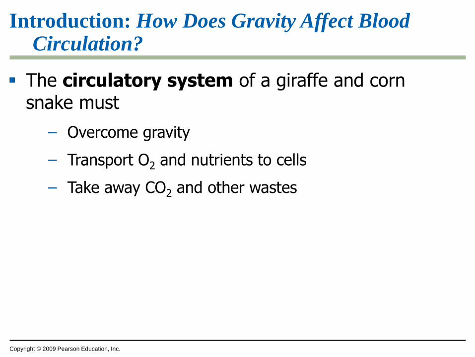
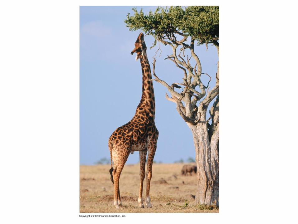
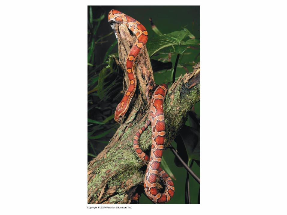
Introduction: How Does Gravity Affect Blood Circulation?
The circulatory system of a giraffe and corn snake must
– Overcome gravity
– Transport O2 and nutrients to cells
– Take away CO2 and other wastes
Copyright © 2009 Pearson Education, Inc.
Introduction: How Does Gravity Affect Blood Circulation?
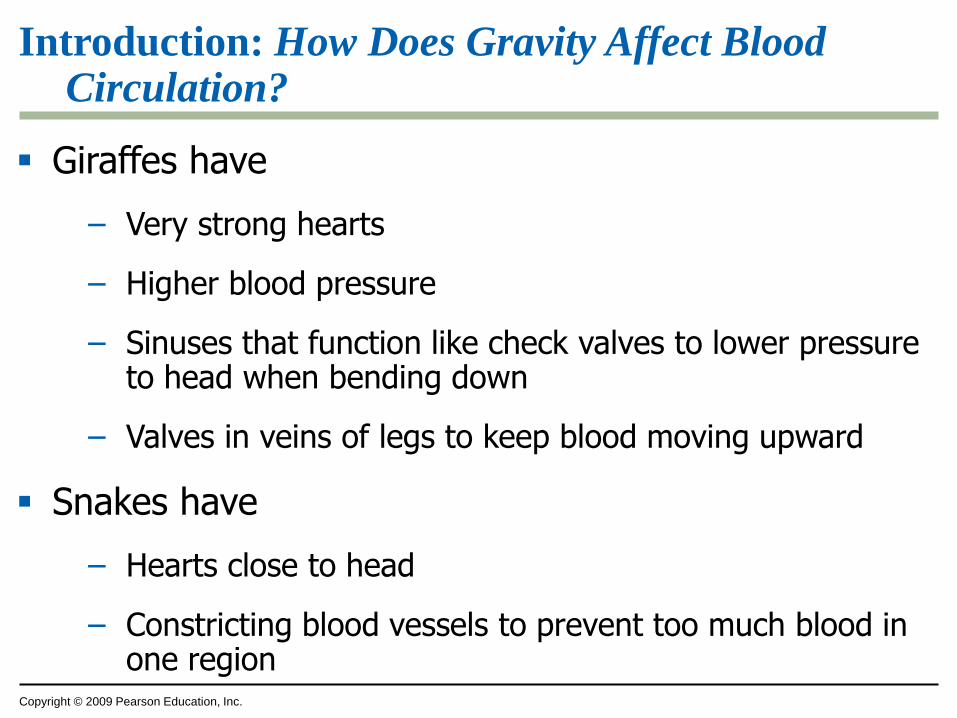
Giraffes have
– Very strong hearts
– Higher blood pressure
– Sinuses that function like check valves to lower pressure to head when bending down
– Valves in veins of legs to keep blood moving upward
Snakes have
– Hearts close to head
– Constricting blood vessels to prevent too much blood in one region
Copyright © 2009 Pearson Education, Inc.
MECHANISMS OF INTERNAL TRANSPORT
Copyright © 2009 Pearson Education, Inc.
23.1 Circulatory systems facilitate exchange with all body tissues
All cells need
– Nutrients
– Gas exchange
– Removal of wastes
Diffusion alone is inadequate for large and complex bodies
Copyright © 2009 Pearson Education, Inc.
23.1 Circulatory systems facilitate exchange with all body tissues
An internal transport system assists diffusion by moving materials between
– Surfaces of the body
– Internal tissues
Copyright © 2009 Pearson Education, Inc.
23.1 Circulatory systems facilitate exchange with all body tissues
A gastrovascular cavity in cnidarians and flatworms serves
– Digestion
– Distribution of substances
Most animals use a circulatory system
– Blood
– Heart
– Blood vessels
Copyright © 2009 Pearson Education, Inc.
23.1 Circulatory systems facilitate exchange with all body tissues
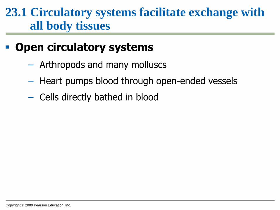
Open circulatory systems
– Arthropods and many molluscs
– Heart pumps blood through open-ended vessels
– Cells directly bathed in blood
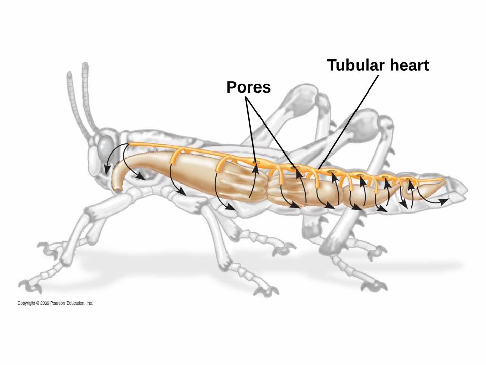
Pores
Tubular heart
Copyright © 2009 Pearson Education, Inc.
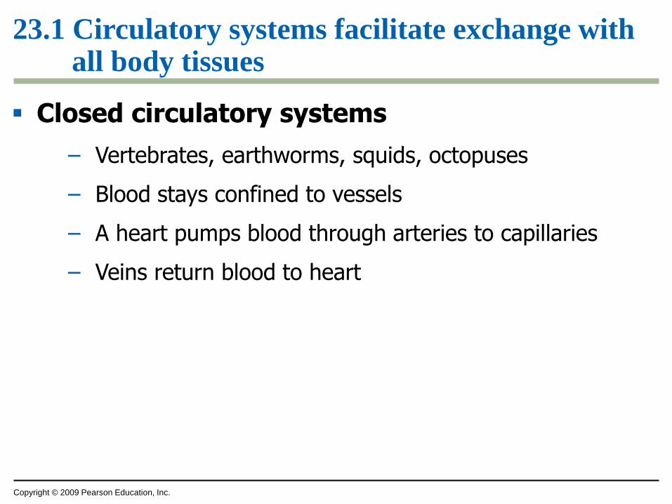
23.1 Circulatory systems facilitate exchange with all body tissues
Closed circulatory systems
– Vertebrates, earthworms, squids, octopuses
– Blood stays confined to vessels
– A heart pumps blood through arteries to capillaries
– Veins return blood to heart
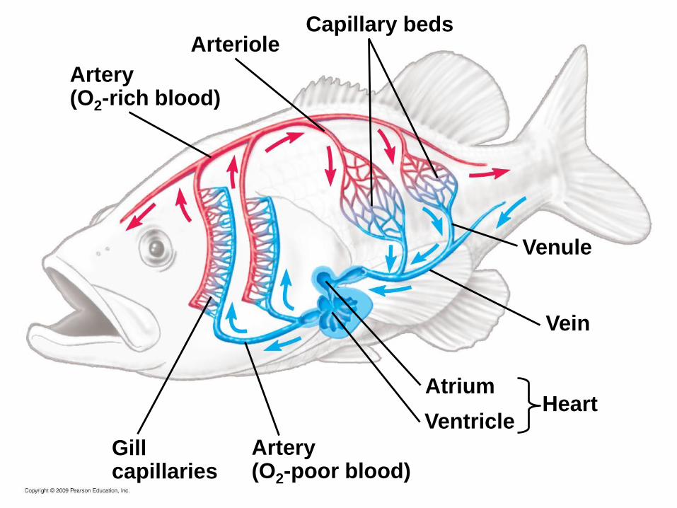
Capillary beds
Artery(O2-rich blood)
Arteriole
Artery(O2-poor blood)
HeartVentricle
Atrium
Vein
Venule
Gillcapillaries
Copyright © 2009 Pearson Education, Inc.
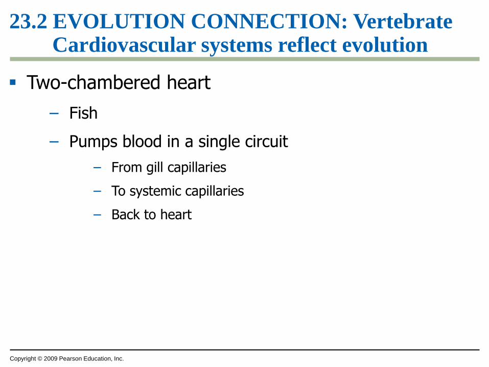
23.2 EVOLUTION CONNECTION: Vertebrate Cardiovascular systems reflect evolution
Two-chambered heart
– Fish
– Pumps blood in a single circuit
– From gill capillaries
– To systemic capillaries
– Back to heart
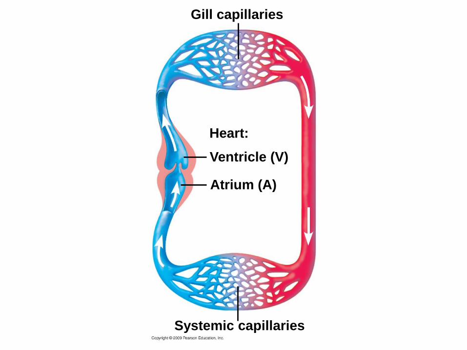
Gill capillaries
Heart:
Ventricle (V)
Atrium (A)
Systemic capillaries
Copyright © 2009 Pearson Education, Inc.
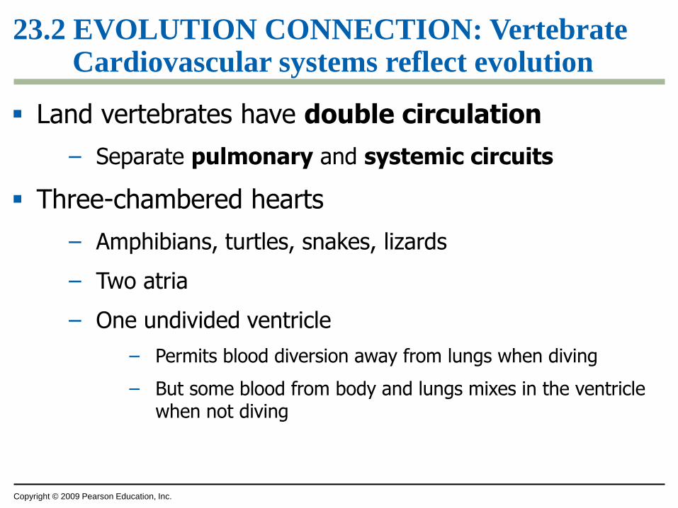
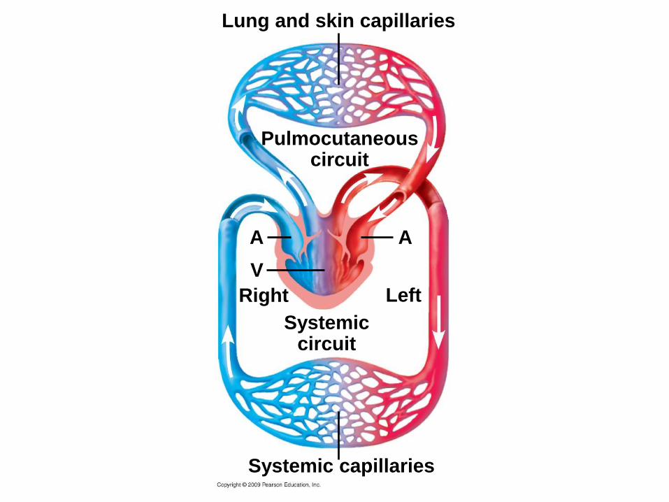
23.2 EVOLUTION CONNECTION: Vertebrate Cardiovascular systems reflect evolution
Land vertebrates have double circulation
– Separate pulmonary and systemic circuits
Three-chambered hearts
– Amphibians, turtles, snakes, lizards
– Two atria
– One undivided ventricle
– Permits blood diversion away from lungs when diving
– But some blood from body and lungs mixes in the ventricle when not diving
Lung and skin capillaries
Pulmocutaneouscircuit
V
Right
Systemic capillaries
A A
Left
Systemiccircuit
Copyright © 2009 Pearson Education, Inc.
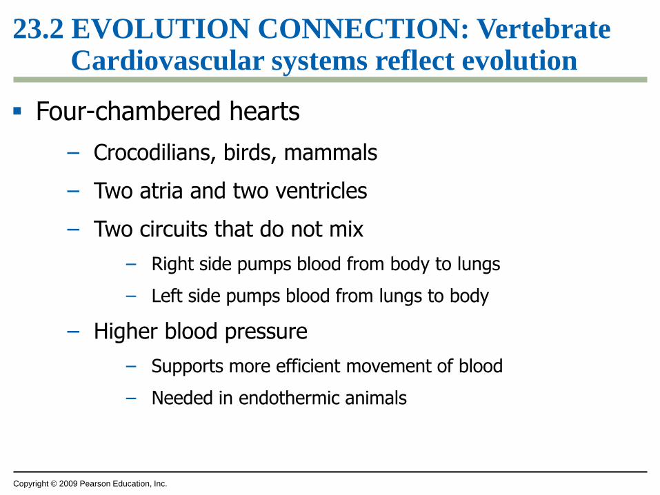
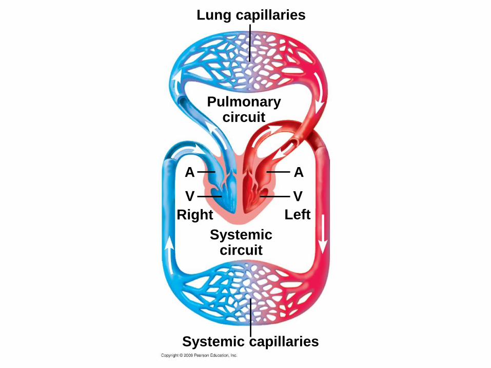
23.2 EVOLUTION CONNECTION: Vertebrate Cardiovascular systems reflect evolution
Four-chambered hearts
– Crocodilians, birds, mammals
– Two atria and two ventricles
– Two circuits that do not mix
– Right side pumps blood from body to lungs
– Left side pumps blood from lungs to body
– Higher blood pressure
– Supports more efficient movement of blood
– Needed in endothermic animals
Lung capillaries
Pulmonarycircuit
V
Right
Systemic capillaries
A A
Left
Systemiccircuit
V
Copyright © 2009 Pearson Education, Inc.
THE HUMAN CARDIOVASCULAR
SYSTEM
Copyright © 2009 Pearson Education, Inc.
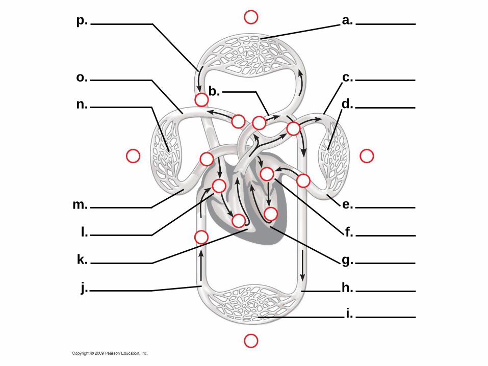
23.3 The human cardiovascular system illustrates the double circulation of mammals
Blood flow through the double circulatory system of humans
Animation: Path of Blood Flow in Mammals
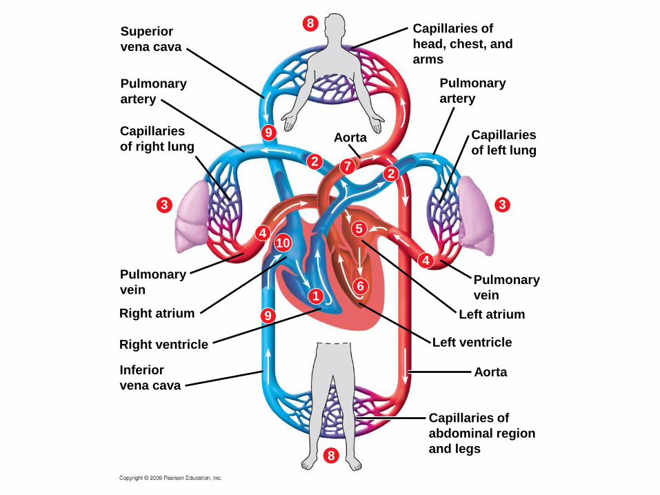
Superior
vena cava
Pulmonary
artery
Capillaries
of right lung
8
9
2
3
Aorta
4 510
16
Pulmonary
vein
9Right atrium
Inferior
vena cava
Right ventricle
4
8
3
Pulmonary
artery
Capillaries
of left lung
Aorta
Pulmonary
vein
Left atrium
Left ventricle
27
Capillaries of
head, chest, and
arms
Capillaries of
abdominal region
and legs
Copyright © 2009 Pearson Education, Inc.
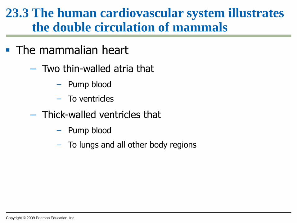
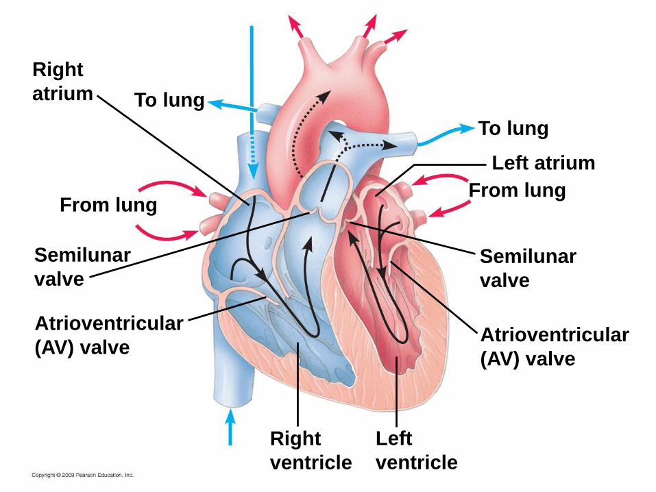
23.3 The human cardiovascular system illustrates the double circulation of mammals
The mammalian heart
– Two thin-walled atria that
– Pump blood
– To ventricles
– Thick-walled ventricles that
– Pump blood
– To lungs and all other body regions
Right
atrium To lung
From lung
Semilunar
valve
Atrioventricular
(AV) valve
Left atrium
To lung
From lung
Semilunar
valve
Atrioventricular
(AV) valve
Right
ventricle
Left
ventricle
Copyright © 2009 Pearson Education, Inc.
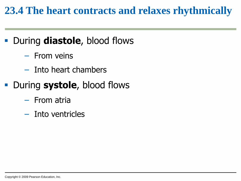
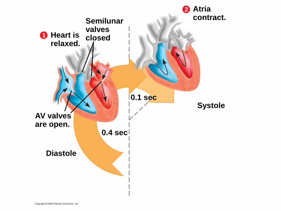
23.4 The heart contracts and relaxes rhythmically
During diastole, blood flows
– From veins
– Into heart chambers
During systole, blood flows
– From atria
– Into ventricles
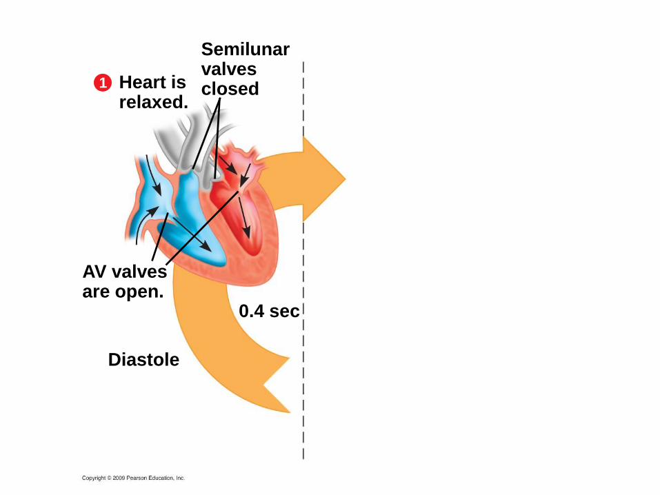
Semilunarvalvesclosed1 Heart is
relaxed.
AV valvesare open.
Diastole
0.4 sec
Semilunarvalvesclosed1 Heart is
relaxed.
AV valvesare open.
Diastole
0.4 sec
2 Atriacontract.
Systole0.1 sec
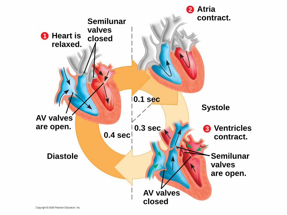
Semilunarvalvesclosed1 Heart is
relaxed.
AV valvesare open.
Diastole
0.4 sec
2 Atriacontract.
Systole0.1 sec
Semilunarvalvesare open.
3 Ventriclescontract.
AV valvesclosed
0.3 sec
Copyright © 2009 Pearson Education, Inc.
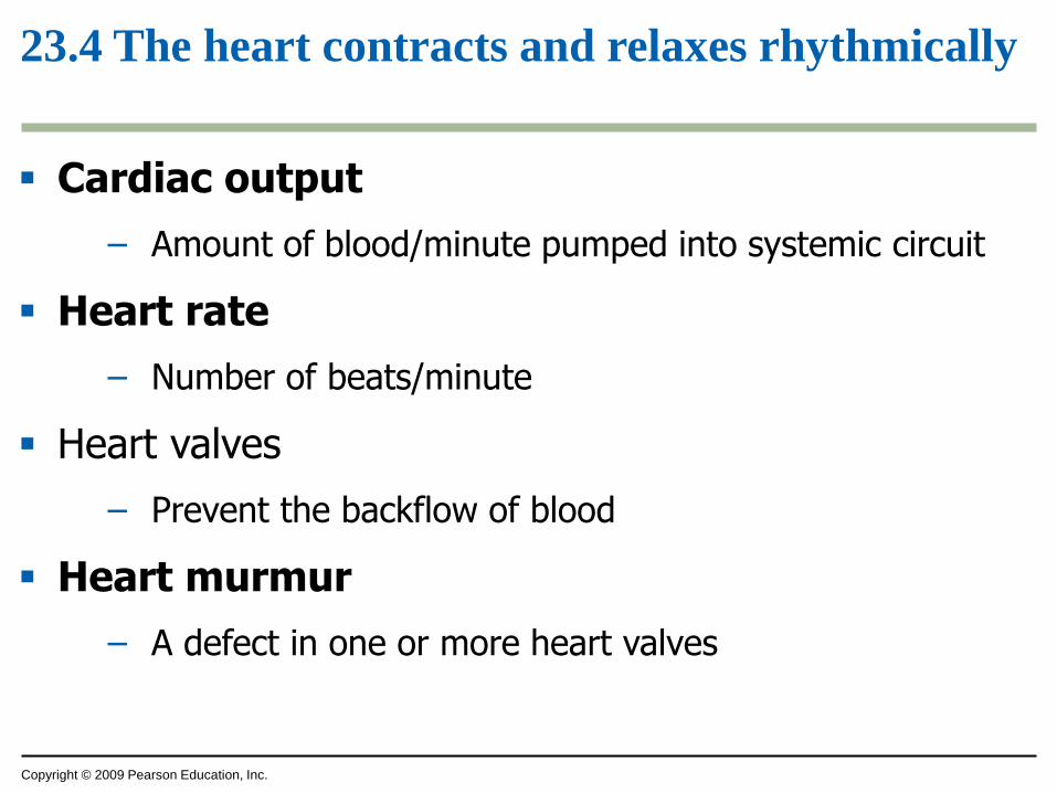
23.4 The heart contracts and relaxes rhythmically
Cardiac output
– Amount of blood/minute pumped into systemic circuit
Heart rate
– Number of beats/minute
Heart valves
– Prevent the backflow of blood
Heart murmur
– A defect in one or more heart valves
Copyright © 2009 Pearson Education, Inc.
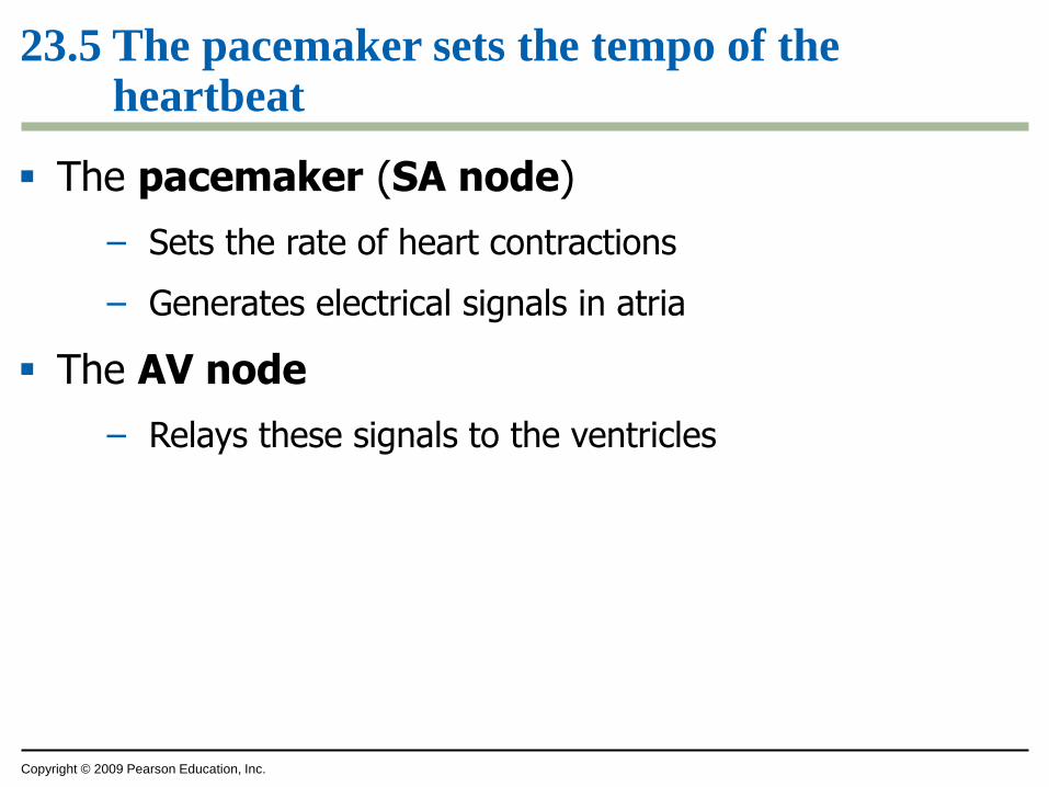
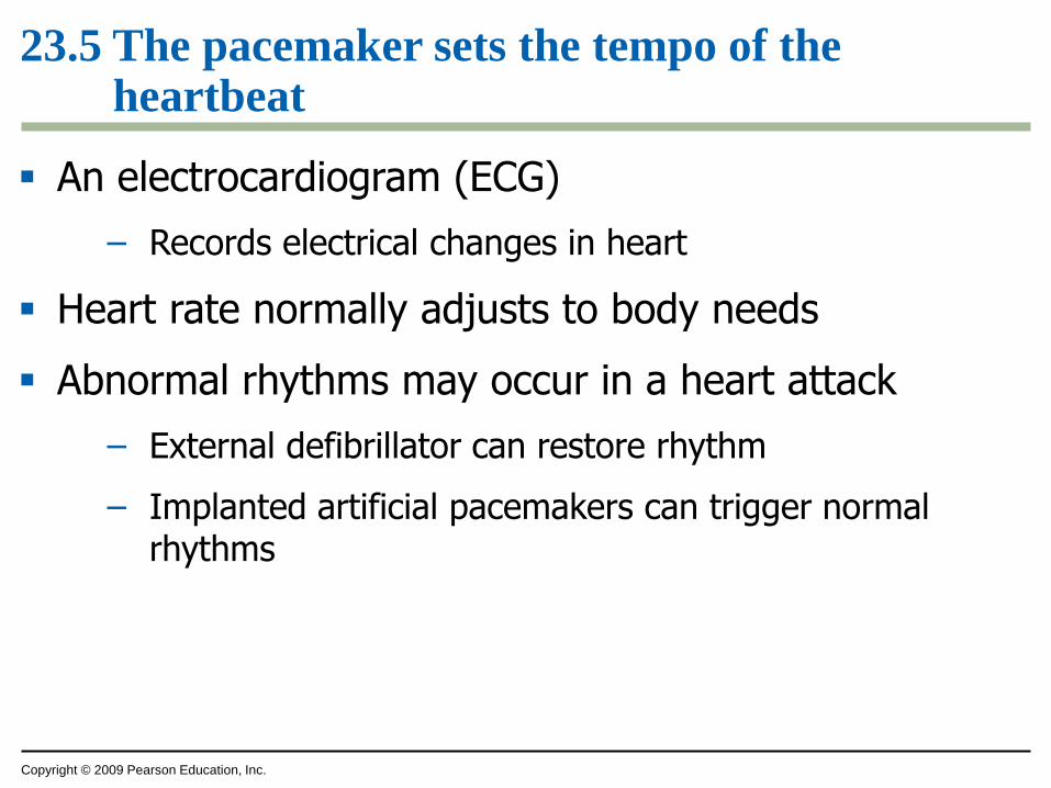
23.5 The pacemaker sets the tempo of the heartbeat
The pacemaker (SA node)
– Sets the rate of heart contractions
– Generates electrical signals in atria
The AV node
– Relays these signals to the ventricles
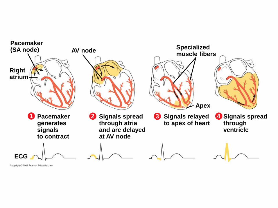
Pacemaker(SA node) AV node
Rightatrium
1 Pacemakergeneratessignalsto contract
2 Signals spreadthrough atriaand are delayedat AV node
ECG
3 Signals relayedto apex of heart
4 Signals spreadthroughventricle
Apex
Specializedmuscle fibers
Copyright © 2009 Pearson Education, Inc.
23.5 The pacemaker sets the tempo of the heartbeat
An electrocardiogram (ECG)
– Records electrical changes in heart
Heart rate normally adjusts to body needs
Abnormal rhythms may occur in a heart attack
– External defibrillator can restore rhythm
– Implanted artificial pacemakers can trigger normal rhythms
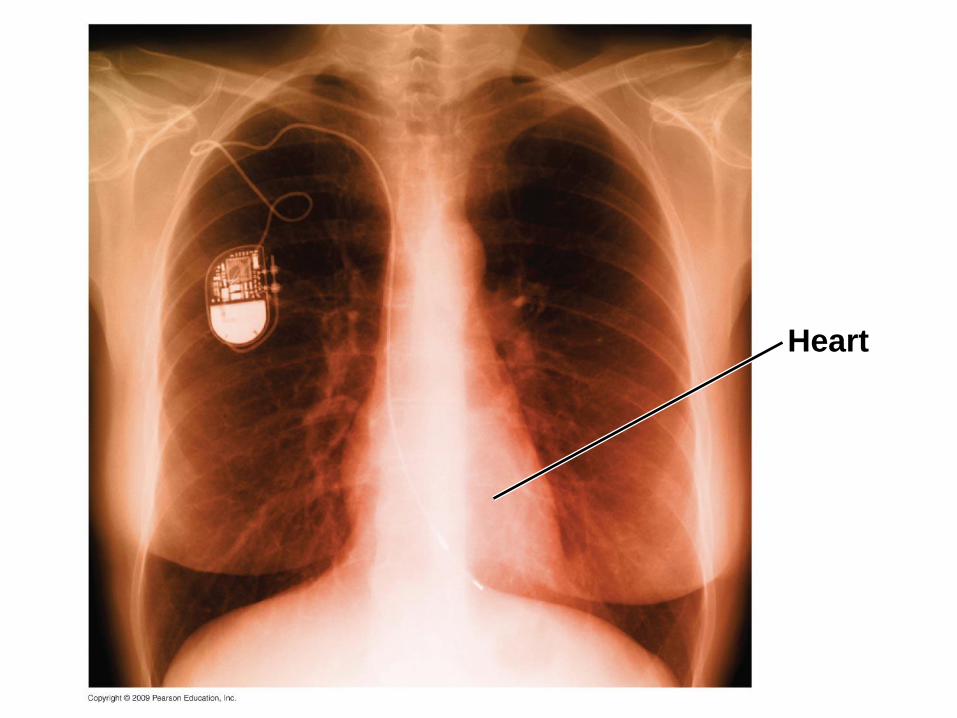
Heart
Copyright © 2009 Pearson Education, Inc.
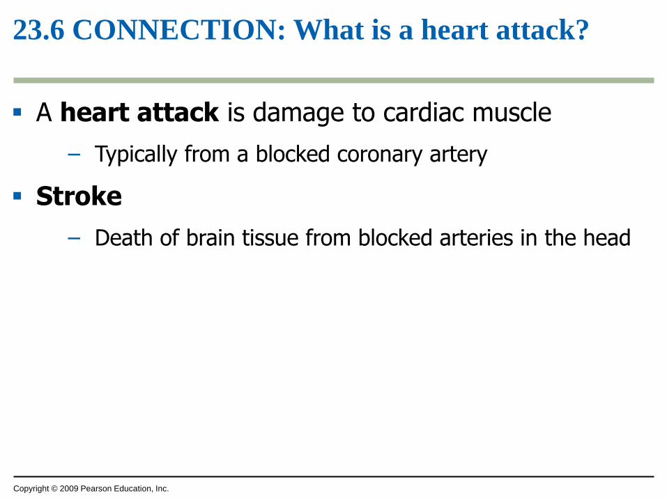
23.6 CONNECTION: What is a heart attack?
A heart attack is damage to cardiac muscle
– Typically from a blocked coronary artery
Stroke
– Death of brain tissue from blocked arteries in the head
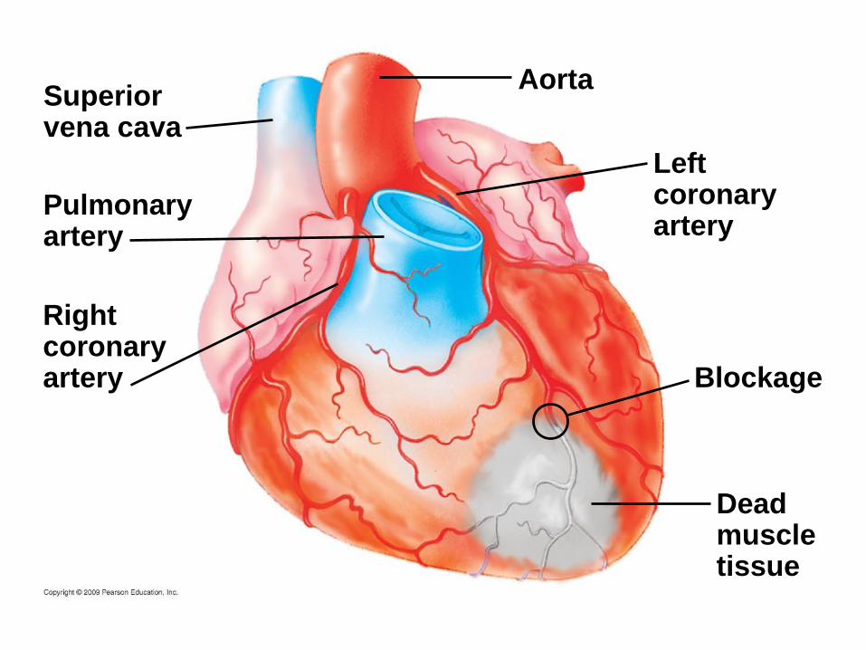
Blockage
Deadmuscletissue
Rightcoronaryartery
Superiorvena cava
Pulmonaryartery
Aorta
Leftcoronaryartery
Copyright © 2009 Pearson Education, Inc.
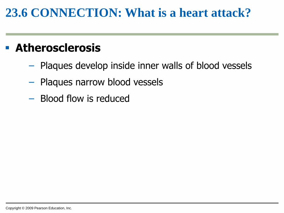
23.6 CONNECTION: What is a heart attack?
Atherosclerosis
– Plaques develop inside inner walls of blood vessels
– Plaques narrow blood vessels
– Blood flow is reduced
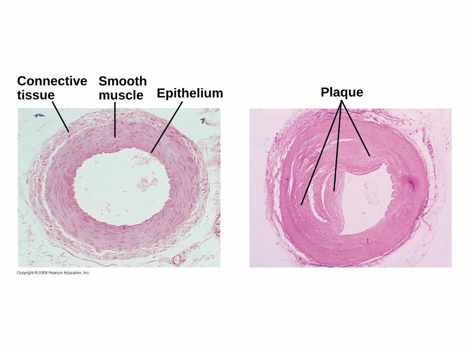
PlaqueEpitheliumConnectivetissue
Smoothmuscle
Copyright © 2009 Pearson Education, Inc.
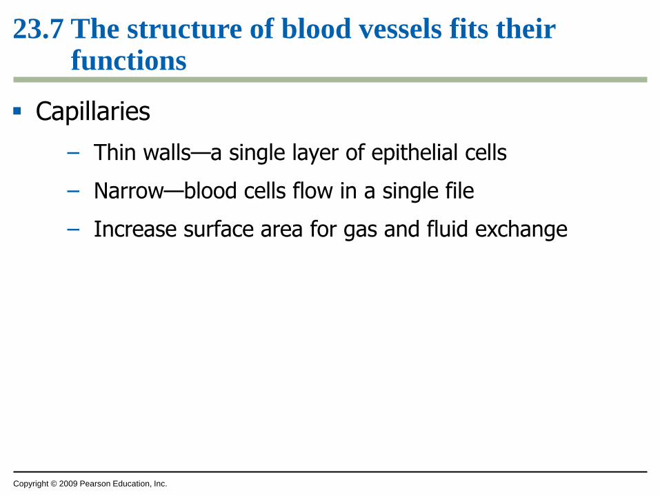
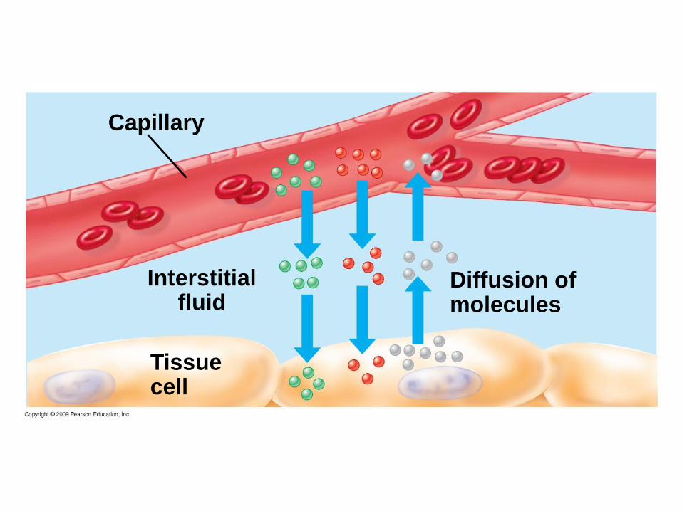
23.7 The structure of blood vessels fits their functions
Capillaries
– Thin walls—a single layer of epithelial cells
– Narrow—blood cells flow in a single file
– Increase surface area for gas and fluid exchange
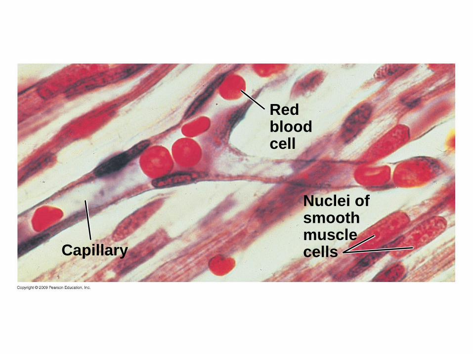
Nuclei ofsmoothmusclecells
Redbloodcell
Capillary
Diffusion ofmolecules
Capillary
Interstitialfluid
Tissuecell
Copyright © 2009 Pearson Education, Inc.
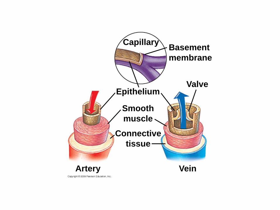
23.7 The structure of blood vessels fits their functions
Arteries and veins
– Lined by single layer of epithelial cells
– Smooth muscle in walls can reduce blood flow
– Elastic fibers permit recoil after stretching
– Veins have one-way valves that restrict backward flow
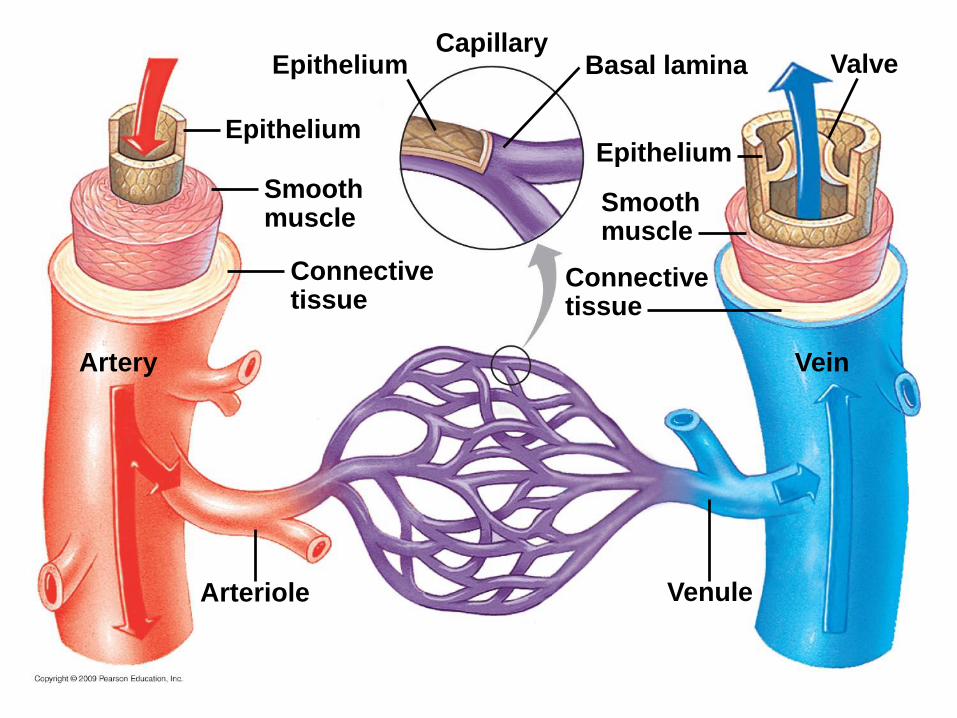
Connectivetissue
Capillary
Venule
Smoothmuscle
Arteriole
Artery Vein
Valve
Epithelium
Basal lamina
Epithelium
Smoothmuscle
Epithelium
Connectivetissue
Copyright © 2009 Pearson Education, Inc.
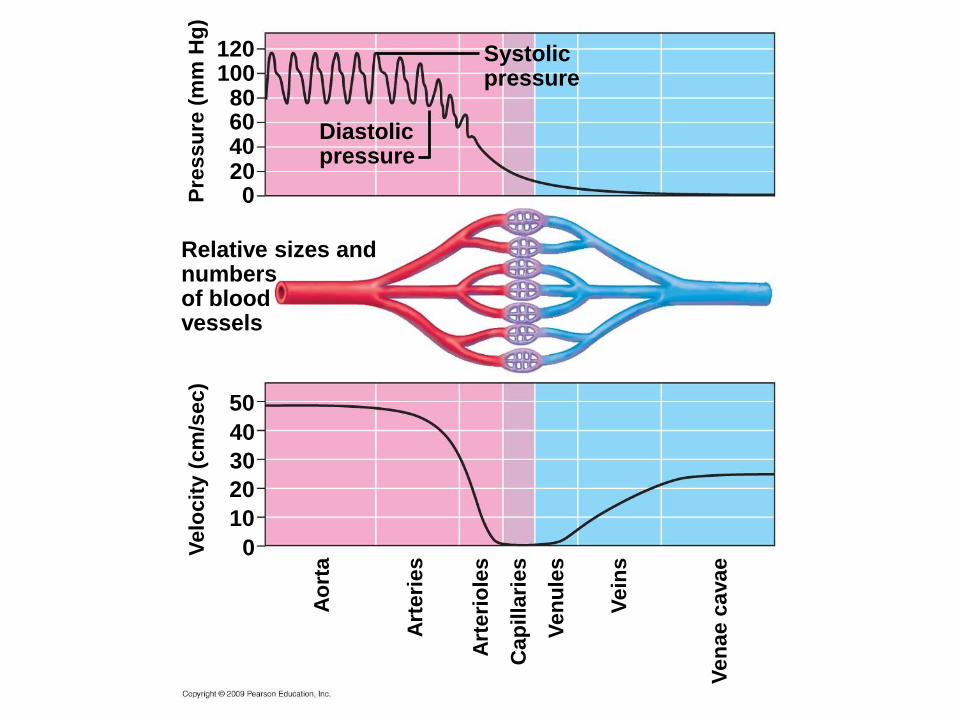
23.8 Blood pressure and velocity reflect the structure and arrangement of blood vessels
Blood pressure
– The force blood exerts on vessel walls
– Depends on
– Cardiac output
– Resistance of vessels
– Decreases as blood moves away from heart
Systolicpressure
Diastolicpressure
120100
806040200
Relative sizes andnumbersof bloodvessels
Pre
ssu
re (
mm
Hg
)V
elo
cit
y (
cm
/sec)
50
40
30
20
10
0
Ao
rta
Ven
ae c
avae
Art
eri
es
Cap
illa
ries
Ven
ule
s
Vein
s
Art
eri
ole
s
Copyright © 2009 Pearson Education, Inc.
23.8 Blood pressure and velocity reflect the structure and arrangement of blood vessels
Blood pressure is
– Highest in arteries
– Lowest in veins
Blood pressure is measured as
– Systolic pressure—caused by ventricular contraction
– Diastolic pressure—low pressure between contractions
Copyright © 2009 Pearson Education, Inc.
23.8 Blood pressure and velocity reflect the structure and arrangement of blood vessels
How does blood travel against gravity, up legs?
– Pressure from muscle contractions
– Between two muscles
– Between muscles and bone or skin
– Squeezes veins
– One-way valves limit blood flow to one direction, towards heart
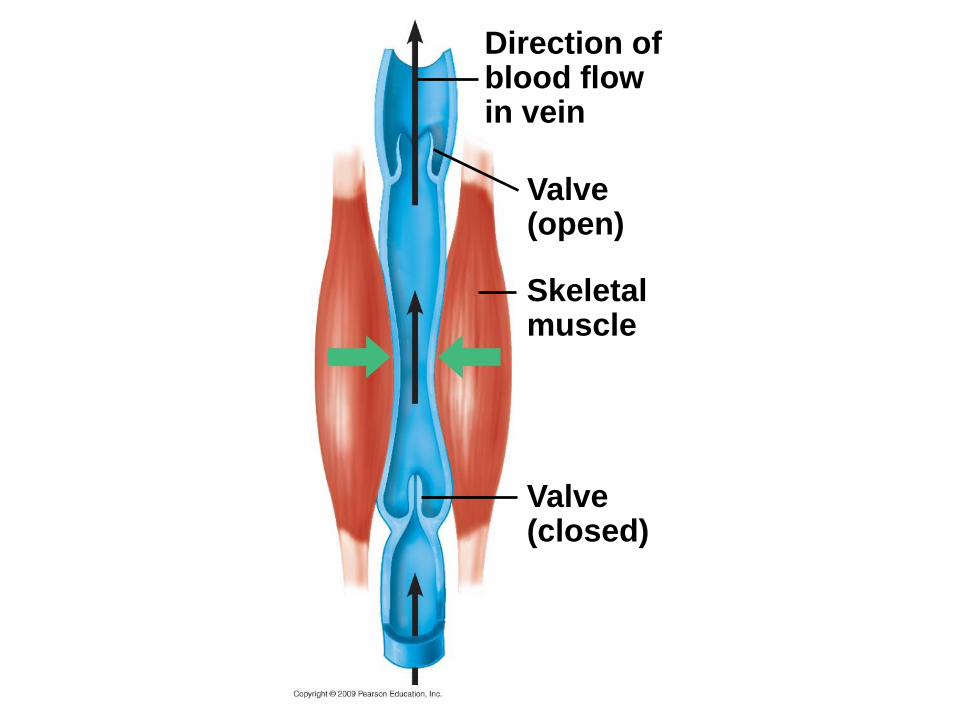
Direction ofblood flowin vein
Valve(open)
Skeletalmuscle
Valve(closed)
Copyright © 2009 Pearson Education, Inc.
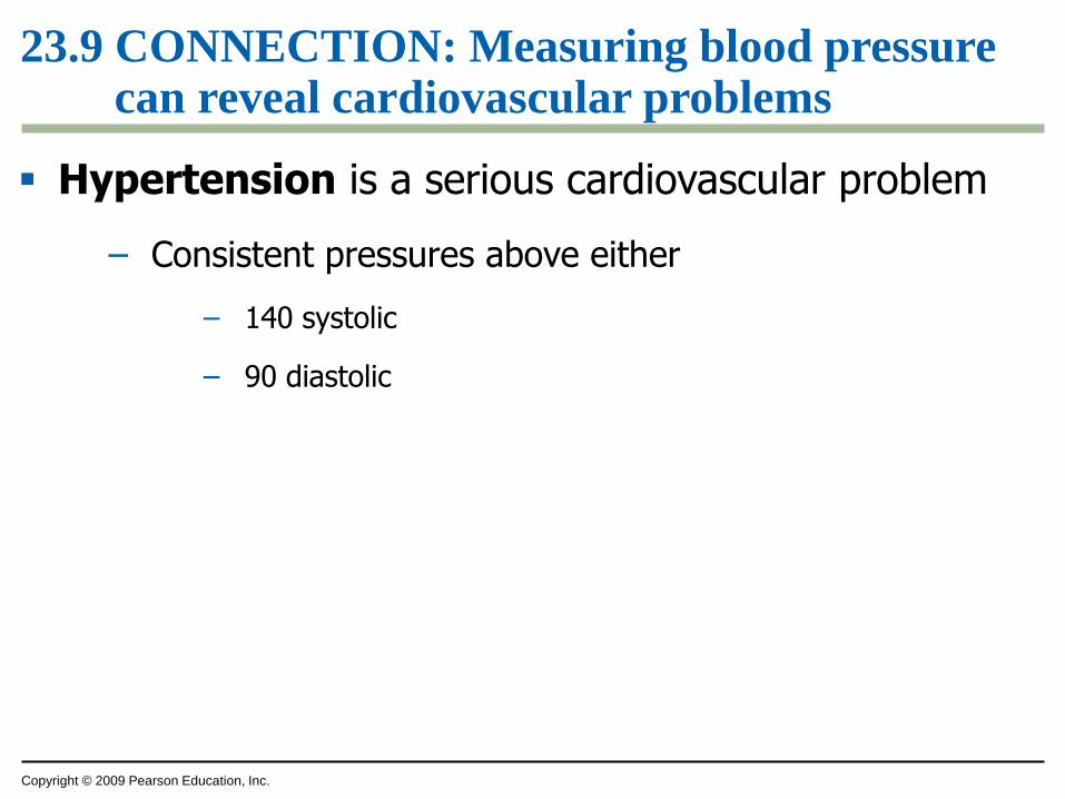
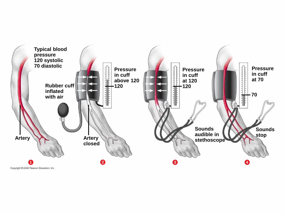
23.9 CONNECTION: Measuring blood pressure can reveal cardiovascular problems
Hypertension is a serious cardiovascular problem
– Consistent pressures above either
– 140 systolic
– 90 diastolic
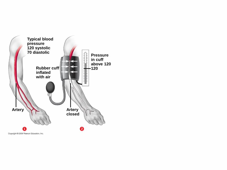
Typical bloodpressure120 systolic70 diastolic
Pressurein cuffabove 120120Rubber cuff
inflatedwith air
Arteryclosed
Artery
1 2
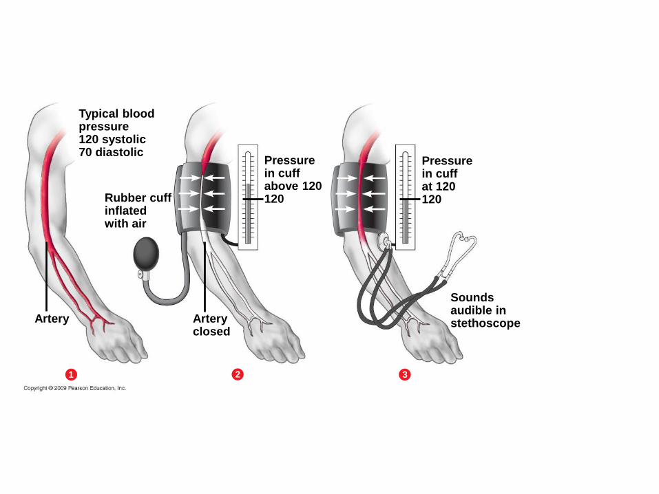
Typical bloodpressure120 systolic70 diastolic
Pressurein cuffabove 120120Rubber cuff
inflatedwith air
Arteryclosed
Artery
1 2
Pressurein cuffat 120120
Soundsaudible instethoscope
3
Typical bloodpressure120 systolic70 diastolic
Pressurein cuffabove 120120Rubber cuff
inflatedwith air
Arteryclosed
Artery
1 2
Pressurein cuffat 120120
Soundsaudible instethoscope
3
70
Soundsstop
4
Pressurein cuffat 70
Copyright © 2009 Pearson Education, Inc.
23.9 CONNECTION: Measuring blood pressure can reveal cardiovascular problems
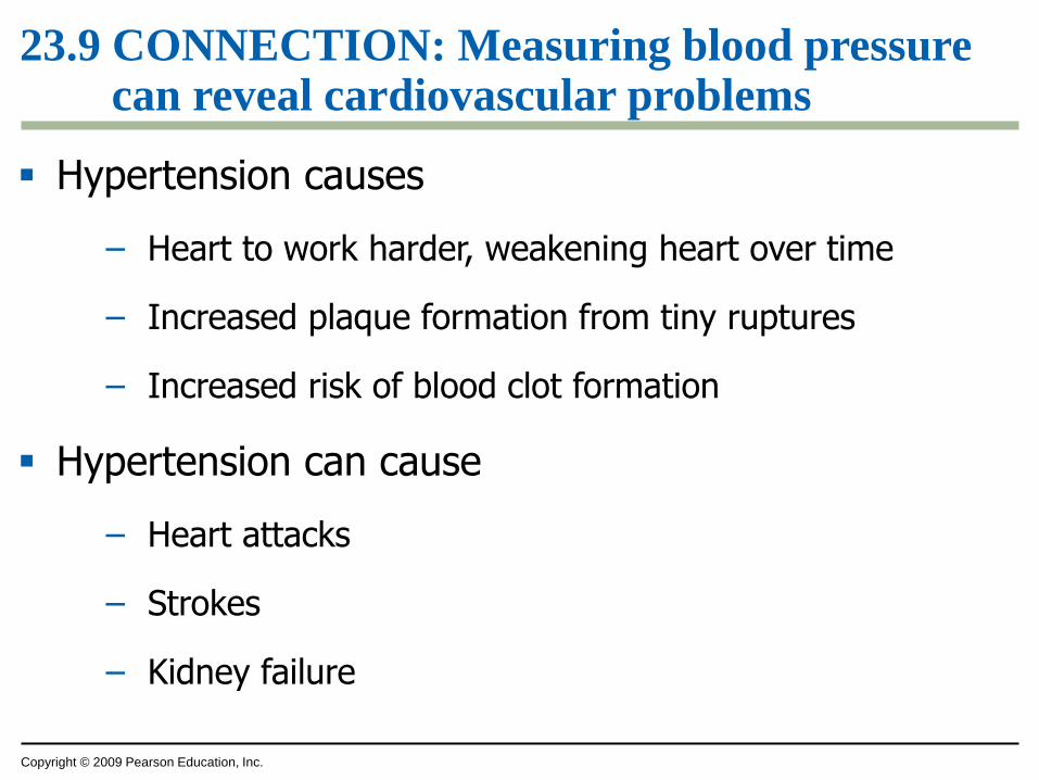
Hypertension causes
– Heart to work harder, weakening heart over time
– Increased plaque formation from tiny ruptures
– Increased risk of blood clot formation
Hypertension can cause
– Heart attacks
– Strokes
– Kidney failure
Copyright © 2009 Pearson Education, Inc.
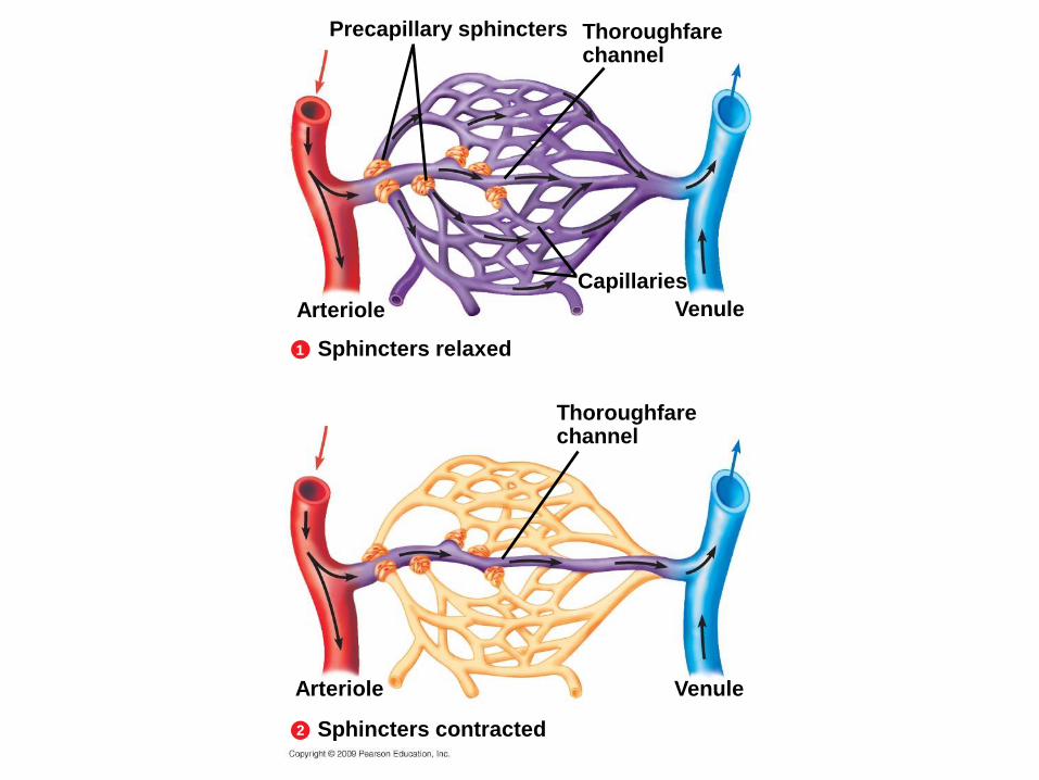
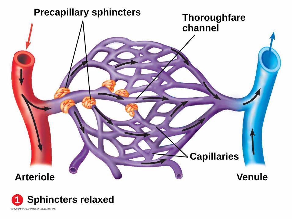
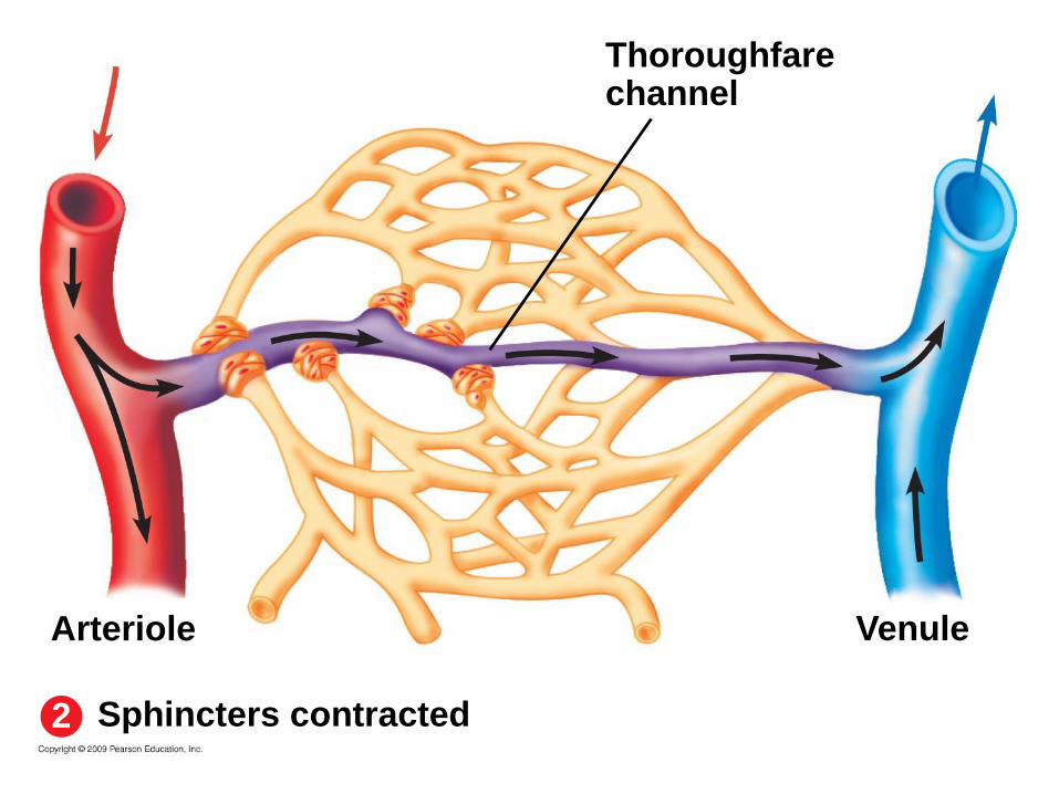
23.10 Smooth muscle controls the distribution of blood
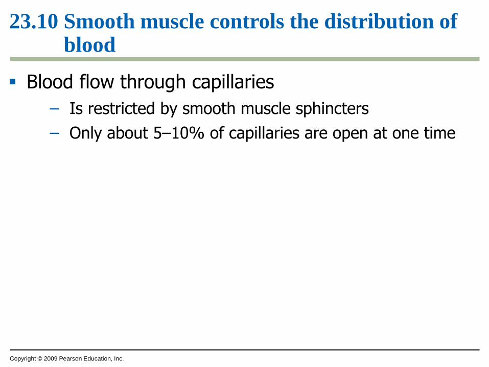
Blood flow through capillaries
– Is restricted by smooth muscle sphincters
– Only about 5–10% of capillaries are open at one time
Capillaries
Thoroughfarechannel
Precapillary sphincters
Venule
Sphincters relaxed
Thoroughfarechannel
VenuleArteriole
2
1
Sphincters contracted
Arteriole
Capillaries
Thoroughfarechannel
Precapillary sphincters
Venule
Sphincters relaxed
Arteriole
1
Thoroughfarechannel
VenuleArteriole
Sphincters contracted2
Copyright © 2009 Pearson Education, Inc.
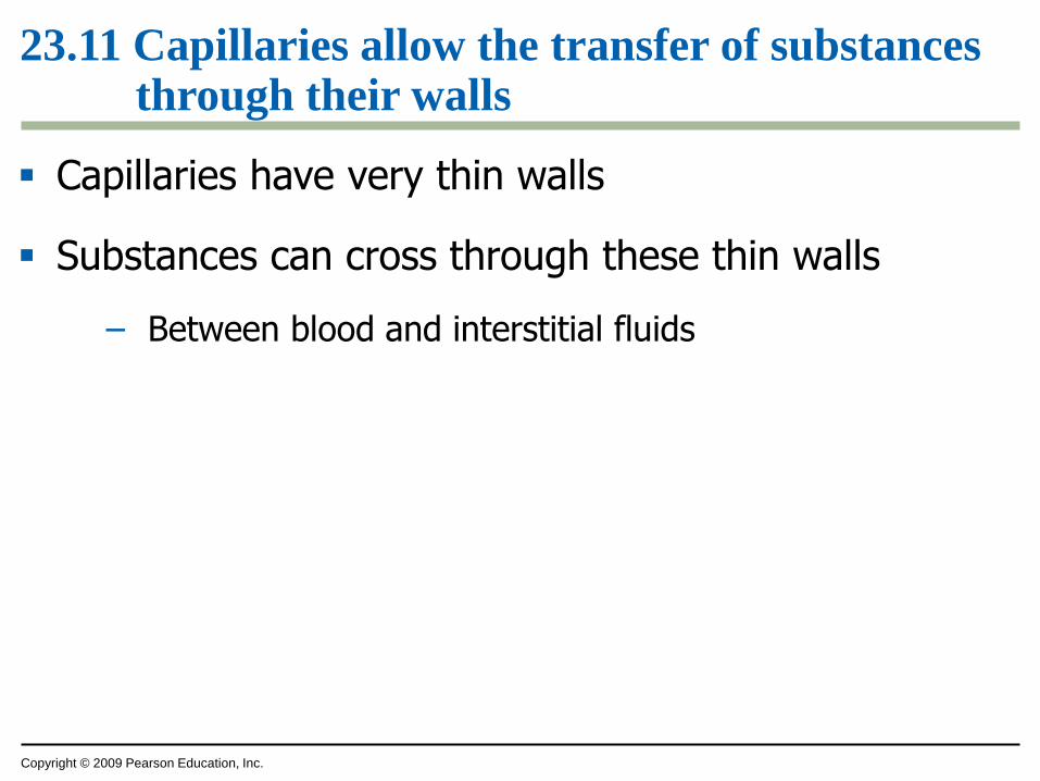
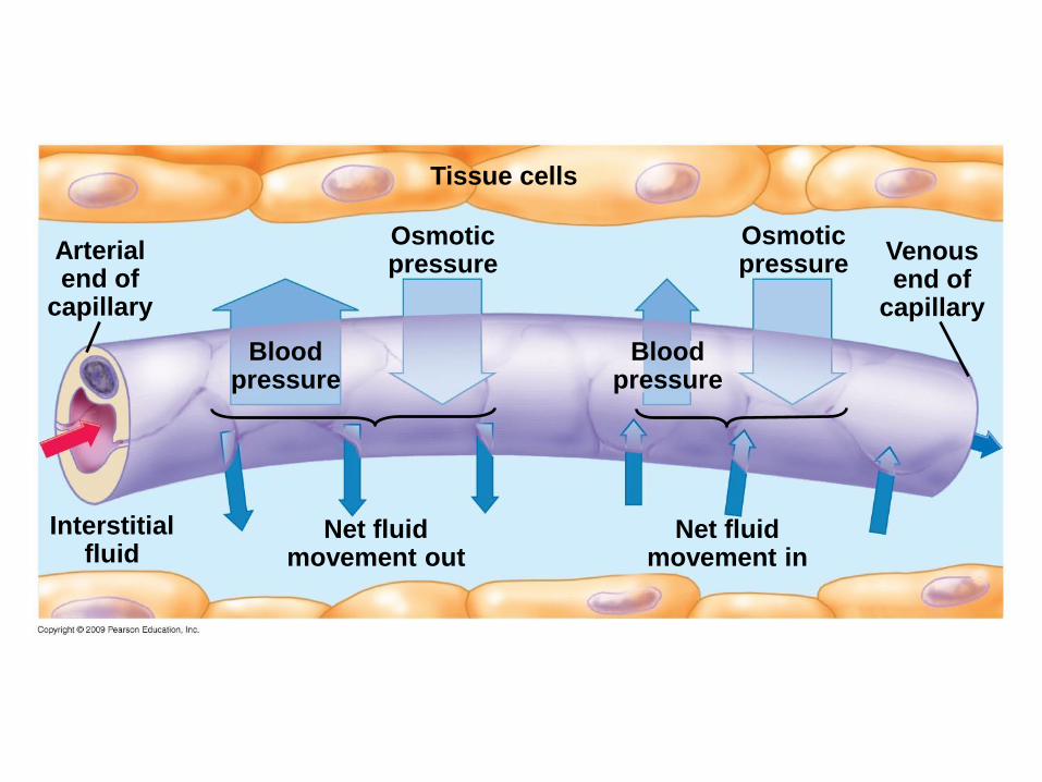
23.11 Capillaries allow the transfer of substances through their walls
Capillaries have very thin walls
Substances can cross through these thin walls
– Between blood and interstitial fluids
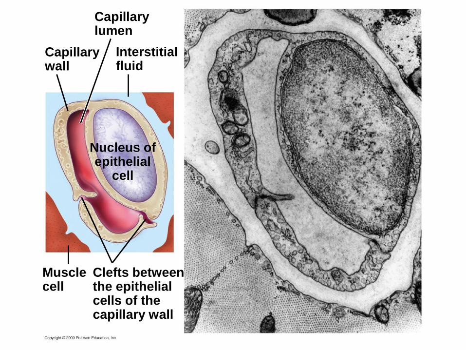
Nucleus ofepithelial
cell
Capillarylumen
Interstitialfluid
Capillarywall
Musclecell
Clefts betweenthe epithelialcells of thecapillary wall
Copyright © 2009 Pearson Education, Inc.
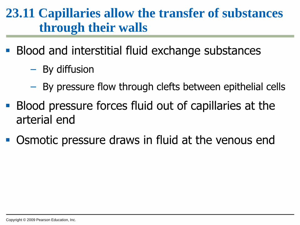
23.11 Capillaries allow the transfer of substances through their walls
Blood and interstitial fluid exchange substances
– By diffusion
– By pressure flow through clefts between epithelial cells
Blood pressure forces fluid out of capillaries at the arterial end
Osmotic pressure draws in fluid at the venous end
Tissue cells
Osmoticpressure
Interstitialfluid
Net fluidmovement in
Bloodpressure
Osmoticpressure
Venousend of
capillary
Arterialend of
capillary
Bloodpressure
Net fluidmovement out
Copyright © 2009 Pearson Education, Inc.
STRUCTURE AND FUNCTION
OF BLOOD
Copyright © 2009 Pearson Education, Inc.
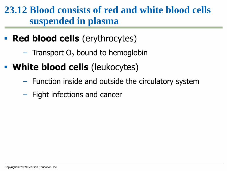
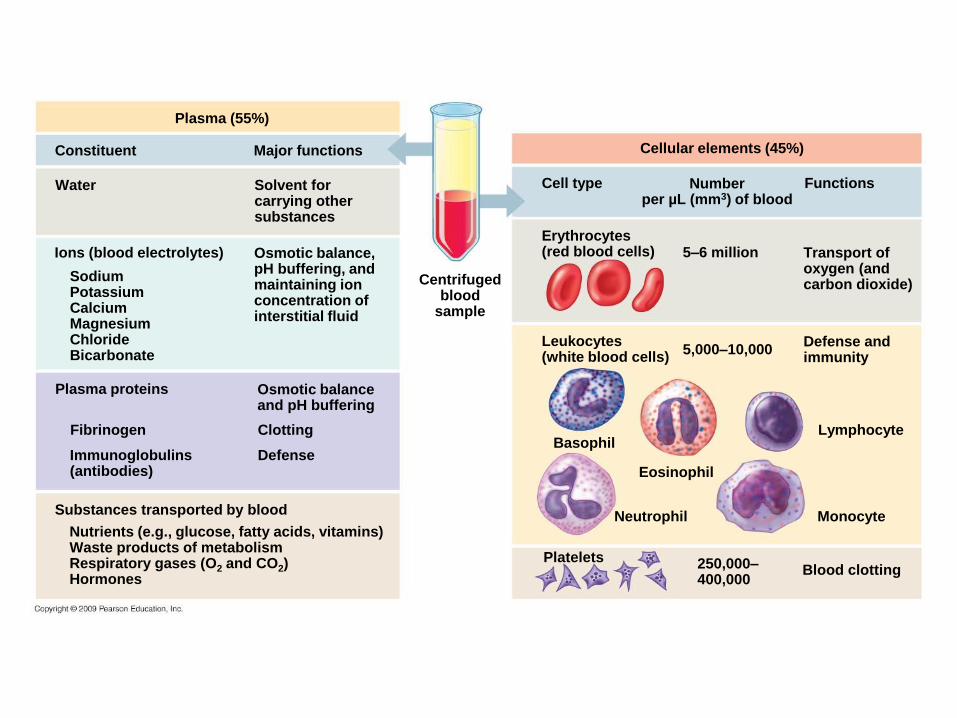
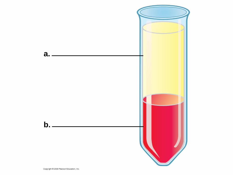
23.12 Blood consists of red and white blood cells suspended in plasma
Plasma is about 90% water
Plasma contains
– Various inorganic ions
– Proteins, nutrients
– Wastes, gases
– Hormones
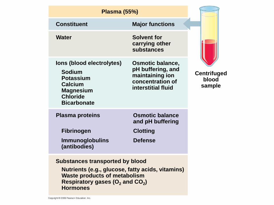
Plasma (55%)
Constituent
Osmotic balance,pH buffering, andmaintaining ionconcentration ofinterstitial fluid
Solvent forcarrying othersubstances
Water
Ions (blood electrolytes)
Major functions
SodiumPotassiumCalciumMagnesiumChlorideBicarbonate
Plasma proteins
ClottingFibrinogen
Osmotic balanceand pH buffering
Defense Immunoglobulins(antibodies)
Substances transported by blood
Nutrients (e.g., glucose, fatty acids, vitamins)Waste products of metabolismRespiratory gases (O2 and CO2)Hormones
Centrifugedblood
sample
Copyright © 2009 Pearson Education, Inc.
23.12 Blood consists of red and white blood cells suspended in plasma
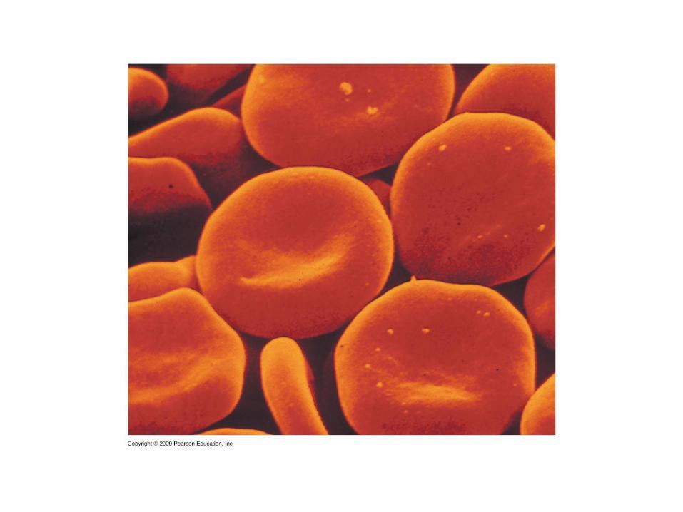
Red blood cells (erythrocytes)
– Transport O2 bound to hemoglobin
White blood cells (leukocytes)
– Function inside and outside the circulatory system
– Fight infections and cancer
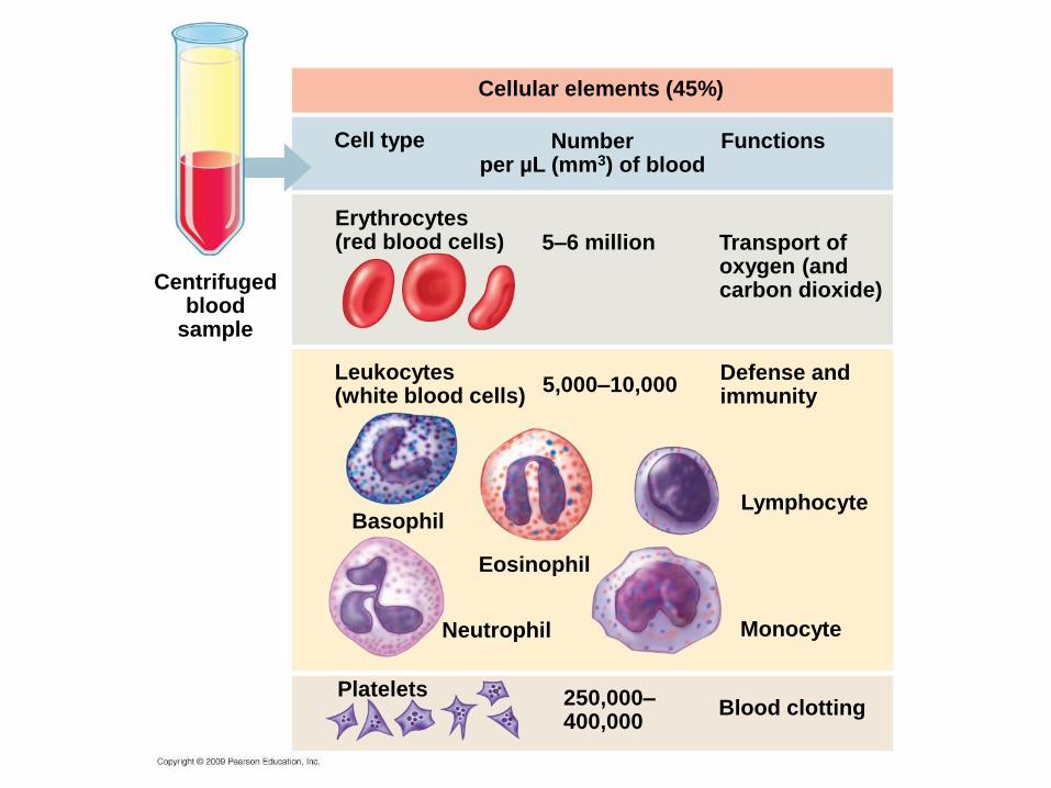
Cellular elements (45%)
Centrifugedblood
sample
Numberper µL (mm3) of blood
Cell type Functions
Erythrocytes(red blood cells) 5–6 million Transport of
oxygen (andcarbon dioxide)
Leukocytes(white blood cells)
BasophilLymphocyte
Defense andimmunity
Eosinophil
5,000–10,000
250,000–400,000
Neutrophil Monocyte
Blood clottingPlatelets
Plasma (55%)
Constituent
Osmotic balance,pH buffering, andmaintaining ionconcentration ofinterstitial fluid
Solvent forcarrying othersubstances
Water
Ions (blood electrolytes)
Major functions
SodiumPotassiumCalciumMagnesiumChlorideBicarbonate
Plasma proteins
ClottingFibrinogen
Osmotic balanceand pH buffering
Defense Immunoglobulins(antibodies)
Substances transported by blood
Nutrients (e.g., glucose, fatty acids, vitamins)Waste products of metabolismRespiratory gases (O2 and CO2)Hormones
Cellular elements (45%)
Centrifugedblood
sample
Numberper µL (mm3) of blood
Cell type Functions
Erythrocytes(red blood cells) 5–6 million Transport of
oxygen (andcarbon dioxide)
Leukocytes(white blood cells)
BasophilLymphocyte
Defense andimmunity
Eosinophil
5,000–10,000
250,000–400,000
Neutrophil Monocyte
Blood clottingPlatelets
Copyright © 2009 Pearson Education, Inc.
23.13 CONNECTION: Too few or too many red blood cells can be unhealthy
Anemia
– Abnormally low amounts of hemoglobin or red blood cells
– Causes fatigue due to lack of oxygen in tissues
Copyright © 2009 Pearson Education, Inc.
23.13 CONNECTION: Too few or too many red blood cells can be unhealthy
Hormone erythropoietin (EPO)
– Regulates red blood cell production
Some athletes artificially increase red blood cell production by injecting erythropoietin
– Can lead to
– Clotting
– Stroke
– Heart failure
– Death
Copyright © 2009 Pearson Education, Inc.
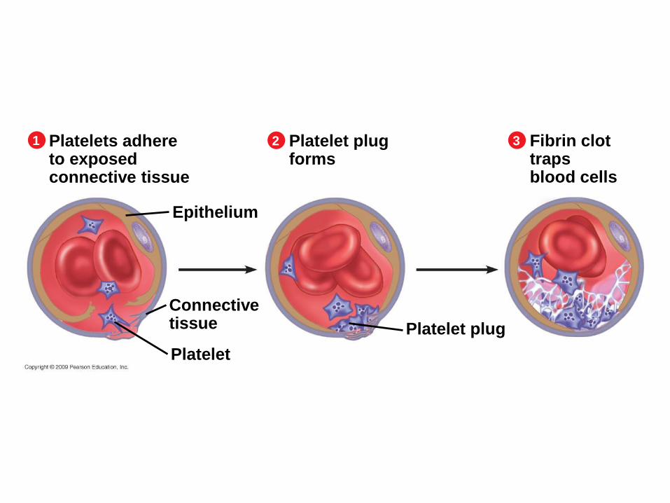
23.14 Blood clots plug leaks when blood vessels are injured
When a blood vessel is damaged
– Platelets help trigger the conversion of fibrinogen to fibrin
– Which forms a clot that plugs the leak
Copyright © 2009 Pearson Education, Inc.
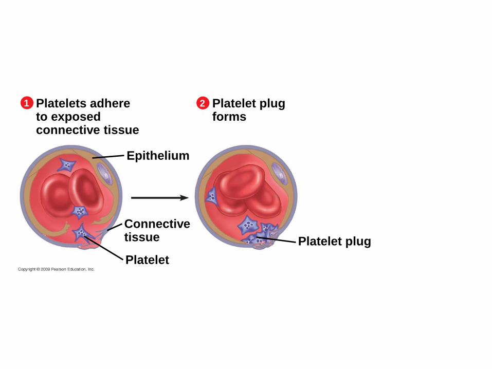
23.14 Blood clots plug leaks when blood vessels are injured
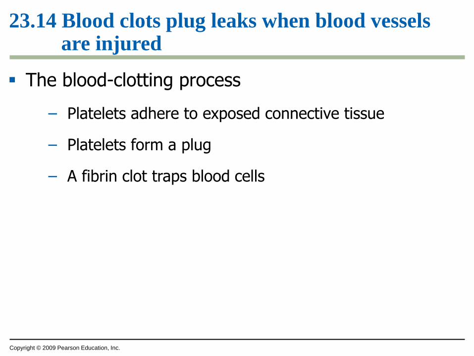
The blood-clotting process
– Platelets adhere to exposed connective tissue
– Platelets form a plug
– A fibrin clot traps blood cells
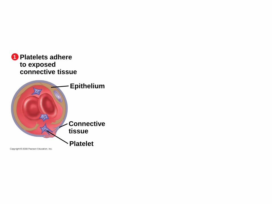
Platelets adhereto exposedconnective tissue
1
Epithelium
Connective tissue
Platelet
Platelets adhereto exposedconnective tissue
1
Epithelium
Connective tissue
Platelet
Platelet plugforms
2
Platelet plug
Platelets adhereto exposedconnective tissue
1
Epithelium
Connective tissue
Platelet
Platelet plugforms
2
Platelet plug
Fibrin clottrapsblood cells
3
Copyright © 2009 Pearson Education, Inc.
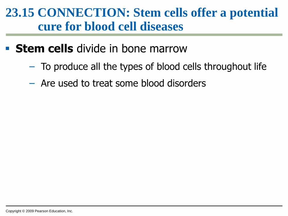
23.15 CONNECTION: Stem cells offer a potential cure for blood cell diseases
Stem cells divide in bone marrow
– To produce all the types of blood cells throughout life
– Are used to treat some blood disorders
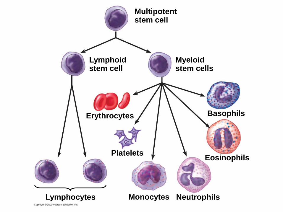
Multipotentstem cell
Lymphoidstem cell
Myeloidstem cells
Erythrocytes
PlateletsEosinophils
Basophils
NeutrophilsMonocytesLymphocytes
Copyright © 2009 Pearson Education, Inc.
23.15 CONNECTION: Stem cells offer a potential cure for blood cell diseases
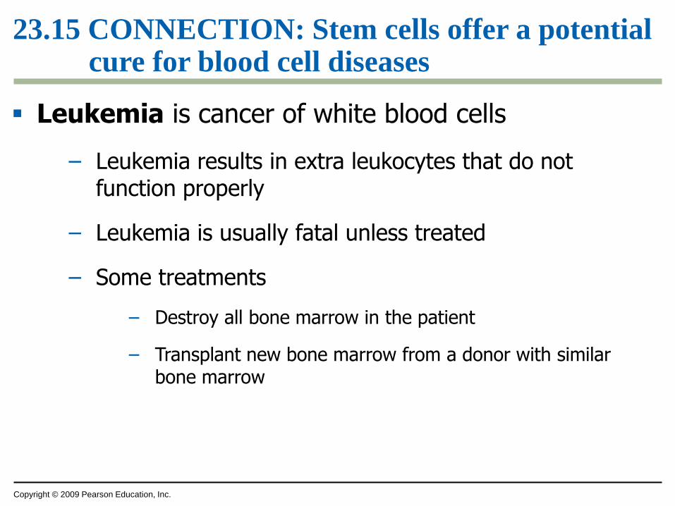
Leukemia is cancer of white blood cells
– Leukemia results in extra leukocytes that do not function properly
– Leukemia is usually fatal unless treated
– Some treatments
– Destroy all bone marrow in the patient
– Transplant new bone marrow from a donor with similar bone marrow
Capillary
EpitheliumValve
Basement
membrane
Connective
tissue
Smooth
muscle
VeinArtery
a.
c.
d.
e.
f.
g.
h.
i.
b.
p.
o.
n.
m.
l.
k.
j.
a.
b.
Copyright © 2009 Pearson Education, Inc.
You should now be able to
1. Explain how the circulatory systems of a giraffe and snake resist gravity
2. Describe the general need for and functions of a circulatory system
3. Compare the structures and functions of gastrovascular cavities, open circulatory systems, and closed circulatory systems
4. Compare the circulatory systems of a fish, frog, and mammal
Copyright © 2009 Pearson Education, Inc.
You should now be able to
5. Explain how heartbeats are controlled
6. Describe the causes and consequences of a heart attack and cardiovascular disease
7. Relate the structure of blood vessels to their functions
8. Describe the components of blood and their functions
Copyright © 2009 Pearson Education, Inc.
You should now be able to
9. Describe the process of blood clotting
10. Describe the causes and treatments for leukemia