Embed Size (px)
Citation preview
© 2012 Pearson Education, Inc. Lecture by Edward J. Zalisko
PowerPoint Lectures for
Campbell Biology: Concepts & Connections, Seventh Edition
Reece, Taylor, Simon, and Dickey
Chapter 20 Unifying Concepts of Animal
Structure and Function
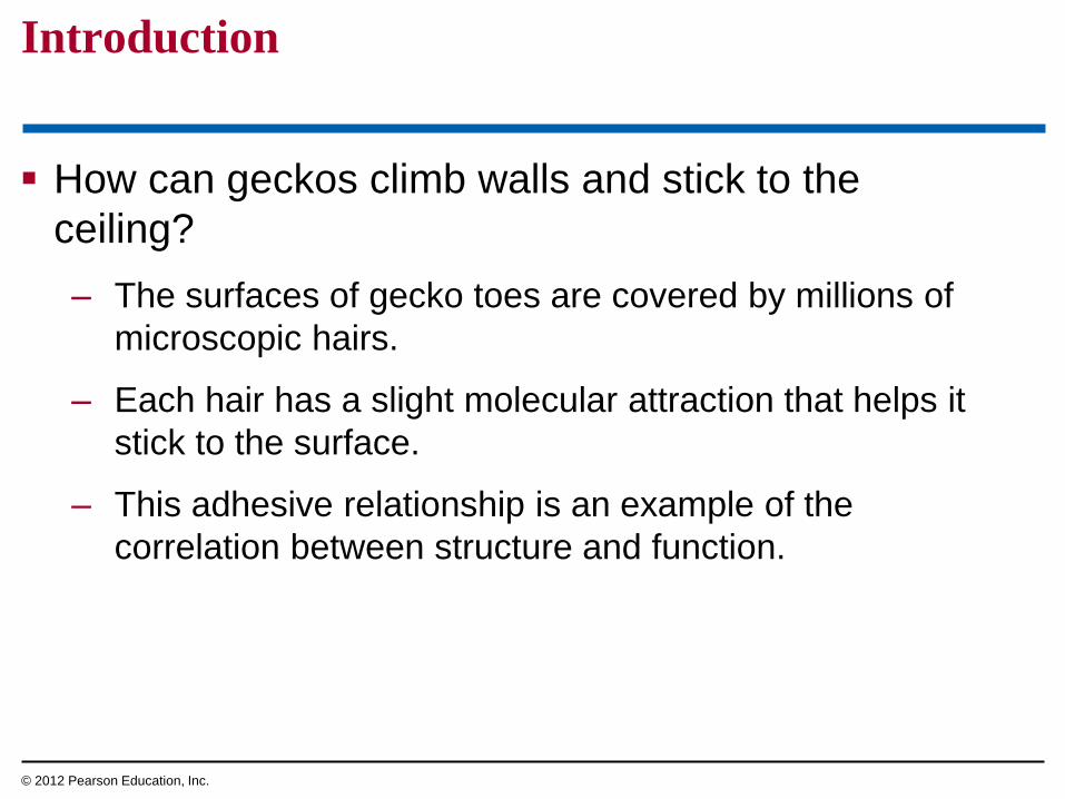
How can geckos climb walls and stick to the
ceiling?
– The surfaces of gecko toes are covered by millions of
microscopic hairs.
– Each hair has a slight molecular attraction that helps it
stick to the surface.
– This adhesive relationship is an example of the
correlation between structure and function.
Introduction
© 2012 Pearson Education, Inc.

Figure 20.0_2
Chapter 20: Big Ideas
Structure and Function
in Animal Tissues
Organs and Organ
Systems
External Exchange and
Internal Regulation
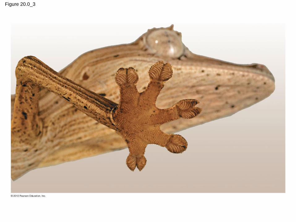
Figure 20.0_3
STRUCTURE AND FUNCTION
IN ANIMAL TISSUES
© 2012 Pearson Education, Inc.
20.1 Structure fits function at all levels of organization in the animal body
Anatomy is the study of structure.
Physiology is the study of function.
Animals consist of a hierarchy of levels or
organization.
– Tissues are an integrated group of similar cells that
perform a common function.
– Organs perform a specific task and consist of two or
more tissues.
– Organ systems consist of multiple organs that together
perform a vital body function.
© 2012 Pearson Education, Inc.
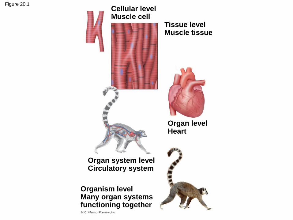
Figure 20.1
Tissue level Muscle tissue
Cellular level Muscle cell
Organ level Heart
Organ system level Circulatory system
Organism level Many organ systems functioning together

20.2 EVOLUTION CONNECTION: An animal’s form reflects natural selection
The body plan or design of an organism
– reflects the relationship between form and function,
– results from natural selection, and
– does not imply a process of conscious invention.
Streamlined and tapered bodies
– increase swimming speeds and
– have similarly evolved in fish, sharks, and aquatic birds
and mammals, representing convergent evolution.
© 2012 Pearson Education, Inc.
Video: Shark Eating a Seal Video: Galápagos Sea Lion
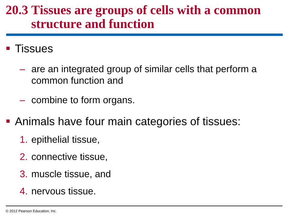
20.3 Tissues are groups of cells with a common structure and function
Tissues
– are an integrated group of similar cells that perform a
common function and
– combine to form organs.
Animals have four main categories of tissues:
1. epithelial tissue,
2. connective tissue,
3. muscle tissue, and
4. nervous tissue.
© 2012 Pearson Education, Inc.

20.4 Epithelial tissue covers the body and lines its organs and cavities
Epithelial tissues, or epithelia, are sheets of
closely packed cells that
– cover body surfaces and
– line internal organs and cavities.
Epithelial cells come in three shapes:
1. squamous—like a fried egg,
2. cuboidal—as tall as they are wide, and
3. columnar—taller than they are wide.
© 2012 Pearson Education, Inc.

20.4 Epithelial tissue covers the body and lines its organs and cavities
Epithelial tissues are named according to the
– number of cell layers they have and
– shape of the cells on their apical surface.
© 2012 Pearson Education, Inc.
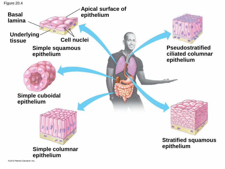
Figure 20.4
Stratified squamous epithelium
Pseudostratified ciliated columnar epithelium
Simple columnar epithelium
Simple cuboidal epithelium
Simple squamous epithelium
Basal lamina
Underlying tissue
Apical surface of epithelium
Cell nuclei

20.5 Connective tissue binds and supports other tissues
Connective tissue can be grouped into six major
types.
1. Loose connective tissue
– is the most widespread,
– consists of ropelike collagen and elastic fibers that are strong
and resilient, and
– helps to join skin to underlying tissues.
2. Fibrous connective tissue
– has densely packed collagen fibers and
– forms tendons that attach muscle to bone.
© 2012 Pearson Education, Inc.

20.5 Connective tissue binds and supports other tissues
3. Adipose tissue stores fat in large, closely packed cells
held in a matrix of fibers.
4. Cartilage
– is a strong and flexible skeletal material and
– commonly surrounds the ends of bones.
5. Bone
– has a matrix of collagen fibers
– embedded in a hard mineral substance containing calcium,
magnesium, and phosphate.
6. Blood transports substances throughout the body.
© 2012 Pearson Education, Inc.
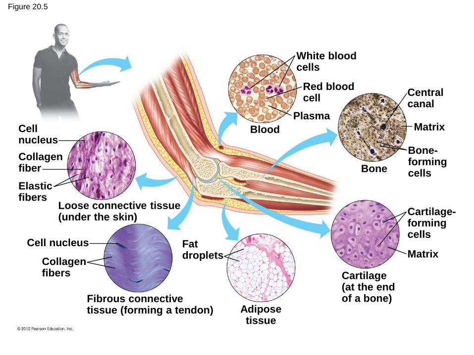
Figure 20.5
Cell nucleus
Collagen fiber
Elastic fibers
Loose connective tissue (under the skin)
Cell nucleus
Collagen fibers
Fibrous connective tissue (forming a tendon)
Fat droplets
Adipose tissue
White blood cells
Red blood cell
Plasma
Blood
Central canal
Matrix
Bone
Bone- forming cells
Cartilage- forming cells
Matrix
Cartilage (at the end of a bone)
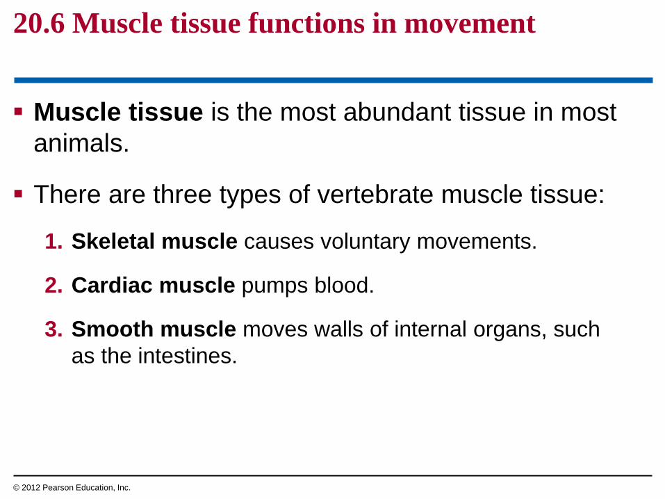
20.6 Muscle tissue functions in movement
Muscle tissue is the most abundant tissue in most
animals.
There are three types of vertebrate muscle tissue:
1. Skeletal muscle causes voluntary movements.
2. Cardiac muscle pumps blood.
3. Smooth muscle moves walls of internal organs, such
as the intestines.
© 2012 Pearson Education, Inc.
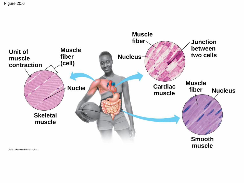
Figure 20.6
Unit of muscle contraction
Muscle fiber (cell)
Nuclei
Skeletal muscle
Muscle fiber
Nucleus
Junction between two cells
Cardiac muscle
Muscle fiber
Smooth muscle
Nucleus
20.7 Nervous tissue forms a communication network
Nervous tissue
– senses stimuli and
– rapidly transmits information.
Neurons carry signals by conducting electrical
impulses.
Other cells in nervous tissue
– insulate axons,
– nourish neurons, and
– regulate the fluid around neurons.
© 2012 Pearson Education, Inc.

Figure 20.7
Dendrites
Cell body
Axon
ORGANS AND
ORGAN SYSTEMS
© 2012 Pearson Education, Inc.
20.8 Organs are made up of tissues
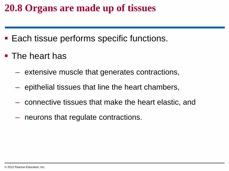
Each tissue performs specific functions.
The heart has
– extensive muscle that generates contractions,
– epithelial tissues that line the heart chambers,
– connective tissues that make the heart elastic, and
– neurons that regulate contractions.
© 2012 Pearson Education, Inc.
20.8 Organs are made up of tissues
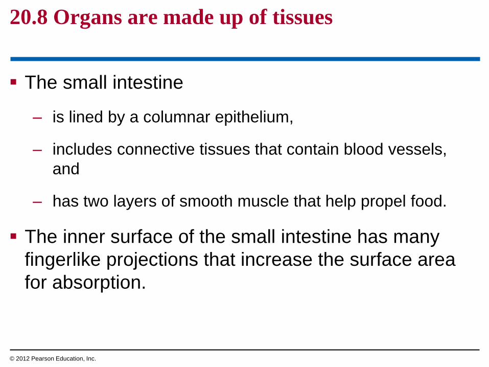
The small intestine
– is lined by a columnar epithelium,
– includes connective tissues that contain blood vessels,
and
– has two layers of smooth muscle that help propel food.
The inner surface of the small intestine has many
fingerlike projections that increase the surface area
for absorption.
© 2012 Pearson Education, Inc.
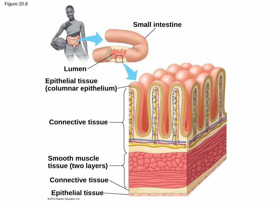
Figure 20.8
Small intestine
Lumen
Epithelial tissue (columnar epithelium)
Connective tissue
Smooth muscle tissue (two layers)
Connective tissue
Epithelial tissue
Bioengineering is seeking ways to repair or replace
damaged tissues and organs.
New tissues and organs are being grown using a
patient’s own cells.
These techniques
– remove the risk of tissue rejection and
– may someday reduce the shortage of organs available
for transplants.
20.9 CONNECTION: Bioengineers are learning to produce tissues and organs for transplants
© 2012 Pearson Education, Inc.
20.10 Organ systems work together to perform life’s functions
Each organ system
– typically consists of many organs,
– has one or more functions, and
– works with other organ systems to create a functional
organism.
© 2012 Pearson Education, Inc.
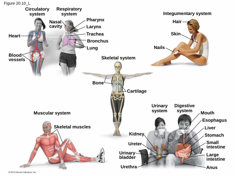
Figure 20.10_L
Blood vessels
Heart
Circulatory system
Respiratory system
Nasal cavity
Pharynx
Larynx
Trachea
Bronchus
Lung
Bone
Cartilage
Skeletal system
Muscular system
Skeletal muscles
Integumentary system
Hair
Skin
Nails
Urinary system
Digestive system
Urinary bladder
Small intestine
Large intestine
Kidney
Ureter
Urethra
Mouth
Esophagus
Liver
Stomach
Anus
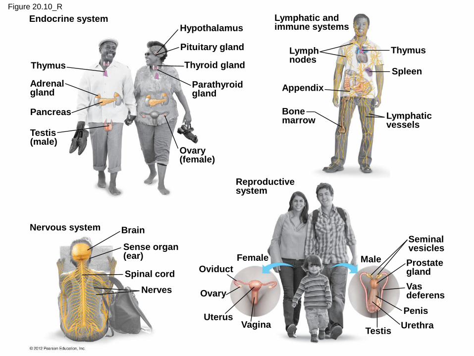
Figure 20.10_R
Endocrine system
Thymus
Adrenal gland
Pancreas
Testis (male)
Hypothalamus
Pituitary gland
Thyroid gland
Parathyroid gland
Ovary (female)
Lymphatic and immune systems
Lymph nodes
Appendix
Bone marrow
Thymus
Spleen
Lymphatic vessels
Reproductive system
Female
Oviduct
Ovary
Uterus Vagina
Male
Seminal vesicles
Prostate gland
Vas deferens
Penis
Urethra Testis
Nervous system Brain
Sense organ (ear)
Spinal cord
Nerves
The skeletal and muscular systems support and
move the body.
The digestive and respiratory systems obtain
food and oxygen.
The circulatory system transports these
materials.
The urinary system disposes of wastes.
The integumentary system covers the body.
20.10 Organ systems work together to perform life’s functions
© 2012 Pearson Education, Inc.

Figure 20.10_1
Circulatory system
Respiratory system
Nasal cavity
Blood vessels
Heart
Pharynx
Larynx
Trachea
Bronchus
Lung
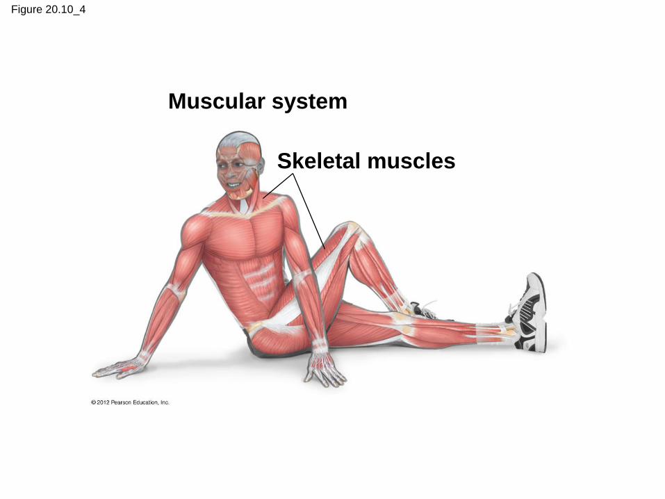
Figure 20.10_4
Muscular system
Skeletal muscles

Figure 20.10_5
Urinary system
Digestive system
Urinary bladder
Small intestine
Large intestine
Kidney
Ureter
Urethra
Mouth
Esophagus
Liver
Stomach
Anus
The lymphatic and immune systems protect the
body from infection.
The nervous and endocrine systems control and
coordinate body functions.
The reproductive system produces offspring.
20.10 Organ systems work together to perform life’s functions
© 2012 Pearson Education, Inc.

Figure 20.10_6
Endocrine system
Thymus
Adrenal gland
Pancreas
Testis (male)
Hypothalamus
Pituitary gland
Thyroid gland
Parathyroid gland
Ovary (female)
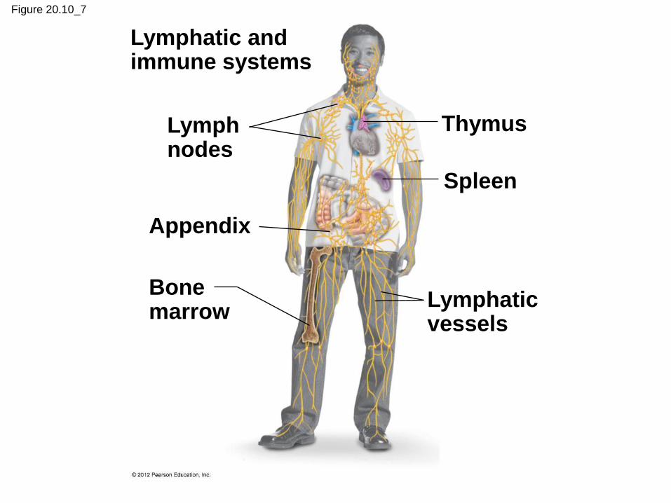
Figure 20.10_7
Lymphatic and immune systems
Lymph nodes
Appendix
Bone marrow
Thymus
Spleen
Lymphatic vessels
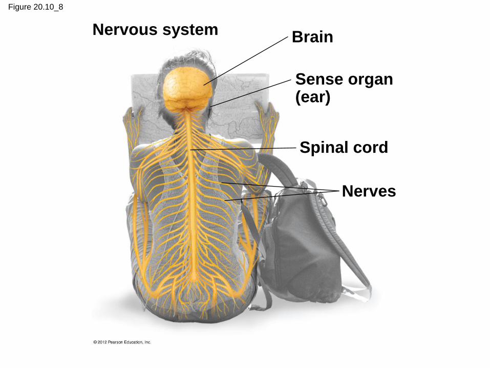
Figure 20.10_8
Nervous system Brain
Sense organ (ear)
Spinal cord
Nerves

Figure 20.10_9
Reproductive system
Female
Oviduct
Ovary
Uterus Vagina
Male
Seminal vesicles
Prostate gland
Vas deferens
Penis
Urethra Testis
20.11 CONNECTION: New imaging technology reveals the inner body
New technologies
– are used in medical diagnosis and research and
– allow physicians to examine organ systems without surgery.
X-rays help create images of hard structures such as bones and teeth.
Magnetic resonance imaging (MRI)
– takes advantage of the behavior of the hydrogen atoms in water molecules and
– provides three-dimensional images of very small structures.
© 2012 Pearson Education, Inc.

Figure 20.11A
Femur
(thigh bone)
Torn meniscus
Tibia (shin bone)

A newer X-ray technology called computed tomography (CT)
– produces high-resolution images of cross sections of the body and
– can detect small differences between normal and abnormal tissues in many organs.
Positron-emission tomography (PET) helps identify metabolic processes at specific body locations.
CT and PET images can be combined for an even more informative image.
20.11 CONNECTION: New imaging technology reveals the inner body
© 2012 Pearson Education, Inc.
20.12 The integumentary system protects the body
The skin consists of two layers:
1. The epidermis
– is a stratified squamous epithelium and
– forms the surface of the skin.
2. The dermis
– forms a deeper skin layer and
– is composed of dense connective tissue with many resilient elastic fibers and strong collagen fibers.
– The dermis contains hair follicles, oil and sweat glands, muscle cells, nerves, sensory receptors, and blood vessels.
© 2012 Pearson Education, Inc.

Figure 20.12
Epidermis
Dermis
Hypodermis
(under the skin)
Adipose tissue
Blood vessels
Hair follicle
Oil gland
Sweat
gland
Nerve
Muscle
Sweat
pore
Hair

Skin has many functions.
– The epidermis
– resists physical damage,
– decreases water loss, and
– prevents penetration by microbes.
– The dermis
– collects sensory information,
– synthesizes vitamin D, and
– helps regulate body temperature.
20.12 The integumentary system protects the body
© 2012 Pearson Education, Inc.
Exposure of the skin to ultraviolet light
– causes skin cells to release melanin, which contributes
to a visible tan, and
– damages DNA of skin cells and can lead to
– premature aging of the skin,
– cataracts, and
– skin cancers.
20.12 The integumentary system protects the body
© 2012 Pearson Education, Inc.
Hair
– is an important component of the integumentary system of mammals,
– helps to insulate their bodies, and
– consists of a shaft of keratin-filled dead cells.
Oil glands release oils that
– are associated with hair follicles,
– lubricate hair,
– condition surrounding skin, and
– inhibit the growth of bacteria.
20.12 The integumentary system protects the body
© 2012 Pearson Education, Inc.
EXTERNAL EXCHANGE AND
INTERNAL REGULATION
© 2012 Pearson Education, Inc.
20.13 Structural adaptations enhance exchange with the environment
Every organism is an open system that must
exchange matter and energy with its surroundings.
Cells in small and flat animals can exchange
materials directly with the environment.
© 2012 Pearson Education, Inc.
20.13 Structural adaptations enhance exchange with the environment
However, as organisms increase in size, the surface
area
– is too small for the corresponding volume and
– too far away from the deepest cells of the body.
– In these organisms, evolutionary adaptations
– consist of extensively branched or folded surfaces, which
increase the area of these surfaces and
– provide for sufficient environmental exchange.
© 2012 Pearson Education, Inc.
20.13 Structural adaptations enhance exchange with the environment
The respiratory system exchanges gases between
the external environment and blood.
The digestive system acquires food and eliminates
wastes.
The excretory system eliminates metabolic waste.
The circulatory system
– distributes gases, nutrients, and wastes throughout the
body and
– exchanges materials between blood and body cells
through the interstitial fluid that bathes body cells.
© 2012 Pearson Education, Inc.
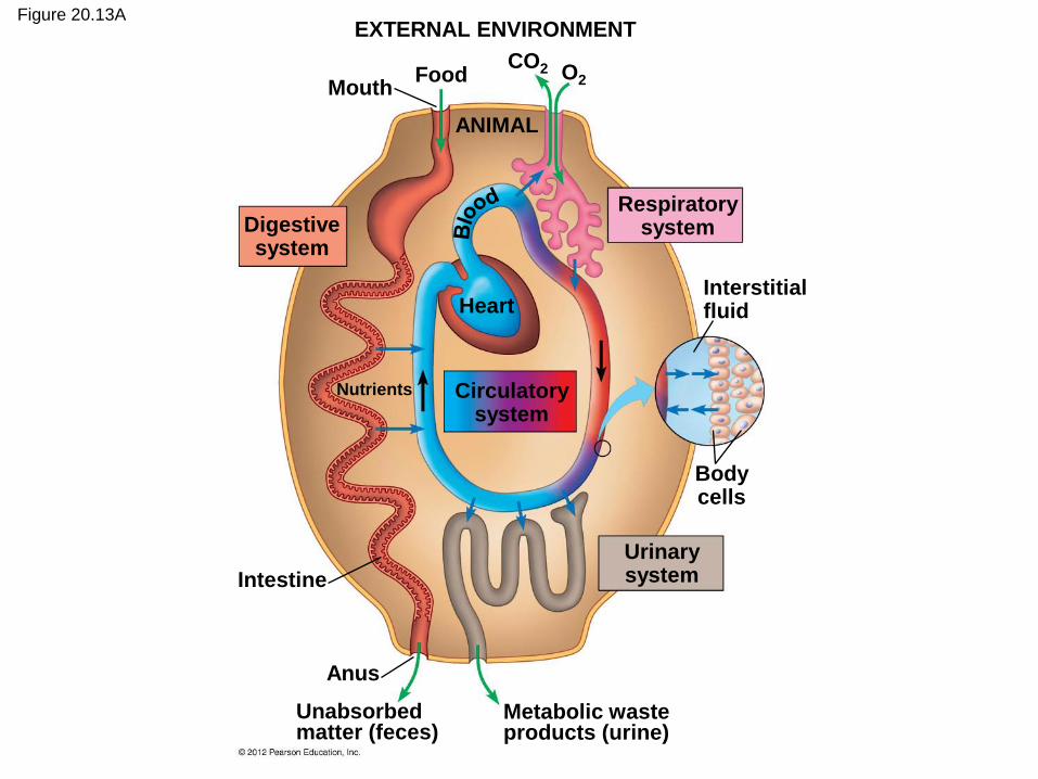
Figure 20.13A EXTERNAL ENVIRONMENT
Mouth Food
CO2 O2
ANIMAL
Digestive system
Respiratory system
Circulatory system
Urinary system
Heart Interstitial fluid
Body cells
Intestine
Anus
Unabsorbed matter (feces)
Metabolic waste products (urine)
Nutrients

Homeostasis is the active maintenance of a
steady state within the body.
– External environmental conditions may fluctuate wildly.
– Homeostatic mechanisms regulate internal conditions.
20.14 Animals regulate their internal environment
© 2012 Pearson Education, Inc.

Figure 20.14
Homeostatic mechanisms
External environment
Large fluctuations
Internal environment
Small fluctuations
Control systems
– detect change and
– direct responses.
Negative-feedback mechanisms
– keep internal variables steady and
– permit only small fluctuations around set points.
20.15 Homeostasis depends on negative feedback
© 2012 Pearson Education, Inc.
Animation: Negative Feedback
Animation: Positive Feedback
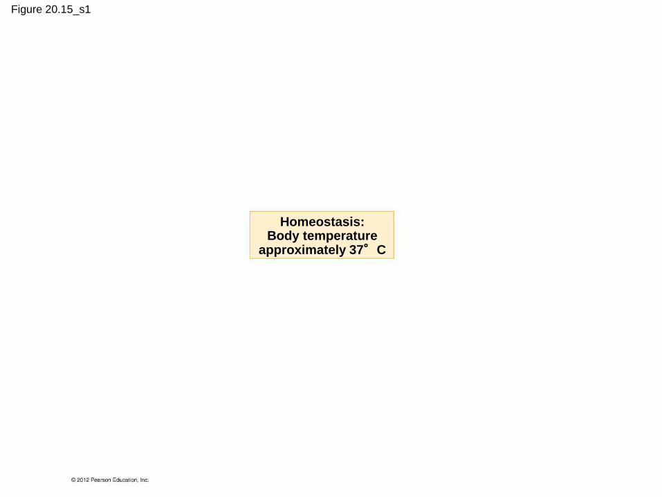
Figure 20.15_s1
Homeostasis: Body temperature
approximately 37°C
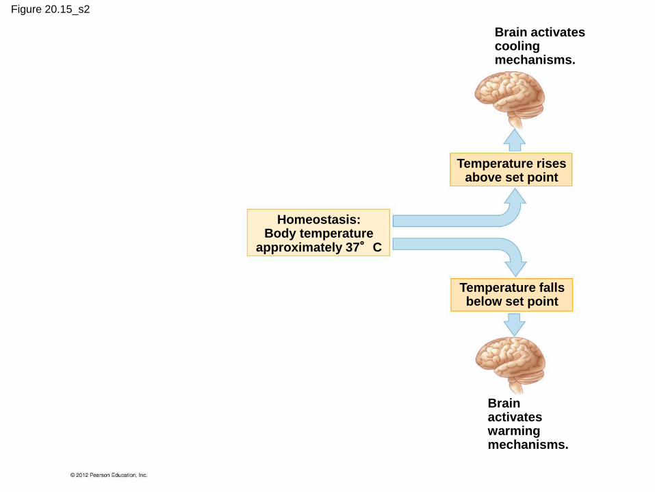
Figure 20.15_s2
Brain activates cooling mechanisms.
Brain activates warming mechanisms.
Temperature rises above set point
Temperature falls below set point
Homeostasis: Body temperature
approximately 37°C
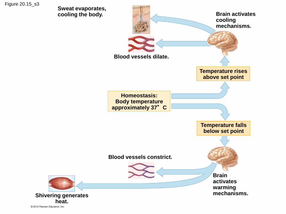
Figure 20.15_s3 Sweat evaporates, cooling the body.
Blood vessels dilate.
Brain activates cooling mechanisms.
Blood vessels constrict.
Brain activates warming mechanisms. Shivering generates
heat.
Temperature rises above set point
Temperature falls below set point
Homeostasis: Body temperature
approximately 37°C
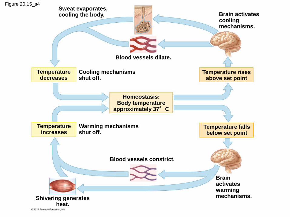
Figure 20.15_s4 Sweat evaporates, cooling the body.
Blood vessels dilate.
Brain activates cooling mechanisms.
Cooling mechanisms shut off.
Warming mechanisms shut off.
Blood vessels constrict.
Brain activates warming mechanisms. Shivering generates
heat.
Temperature decreases
Temperature increases
Temperature rises above set point
Temperature falls below set point
Homeostasis: Body temperature
approximately 37°C

Figure 20.15_5
Homeostasis: Body temperature
approximately 37°C
Temperature decreases
Temperature rises above set point
The thermostat shuts off the cooling mechanisms.
Blood vessels in the skin dilate, increasing heat loss.
Sweat glands secrete sweat that evaporates, cooling the body. The thermostat
in the brain activates cooling mechanisms.
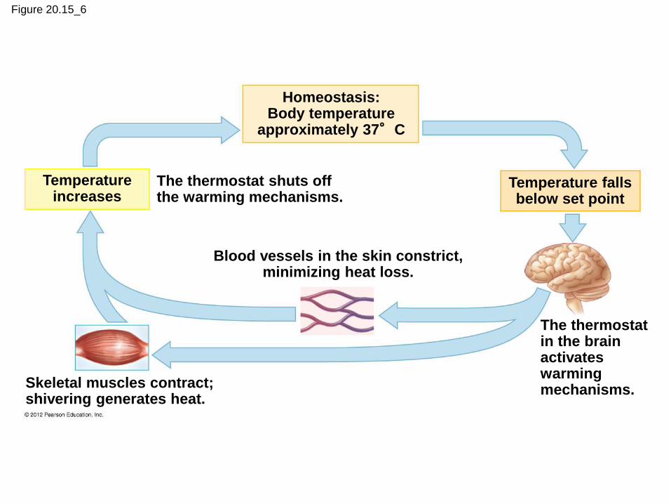
Figure 20.15_6
Homeostasis: Body temperature
approximately 37°C
Temperature increases
Temperature falls below set point
The thermostat shuts off the warming mechanisms.
Blood vessels in the skin constrict, minimizing heat loss.
Skeletal muscles contract; shivering generates heat.
The thermostat in the brain activates warming mechanisms.
You should now be able to
1. Describe the levels of organization in an animal’s
body.
2. Explain how size and shape can influence the
structure of an animal.
3. Define a tissue, describe the four main types of
animal tissue, and note their structures and their
functions.
4. Explain how the structure of organs is based on
the cooperative interactions of tissues.
© 2012 Pearson Education, Inc.
5. Explain how artificial tissues are created and
used.
6. Describe the general structures and functions of
the 12 major vertebrate organ systems.
7. Describe and compare X-ray, CT, MRI, and PET
imaging technologies.
8. Relate the structure of the skin to its functions.
You should now be able to
© 2012 Pearson Education, Inc.

9. Describe the systems that help an animal
exchange materials with its environment.
10. Describe examples of adaptations to increase
the surface-to-volume ratio.
11. Define the concept of homeostasis and illustrate
it with examples.
12. Explain how negative feedback is used to
regulate internal body temperature.
You should now be able to
© 2012 Pearson Education, Inc.
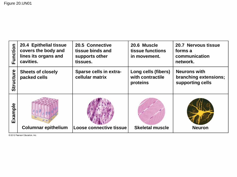
Figure 20.UN01
20.4 Epithelial tissue
covers the body and
lines its organs and
cavities.
20.5 Connective
tissue binds and
supports other
tissues.
20.6 Muscle
tissue functions
in movement.
20.7 Nervous tissue
forms a
communication
network.
Sheets of closely
packed cells
Sparse cells in extra-
cellular matrix
Long cells (fibers)
with contractile
proteins
Neurons with
branching extensions;
supporting cells
Columnar epithelium Loose connective tissue Skeletal muscle Neuron
Ex
am
ple
S
tru
ctu
re
Fu
nc
tio
n
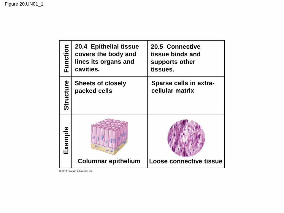
Figure 20.UN01_1
20.4 Epithelial tissue
covers the body and
lines its organs and
cavities.
20.5 Connective
tissue binds and
supports other
tissues.
Sheets of closely
packed cells
Sparse cells in extra-
cellular matrix
Columnar epithelium Loose connective tissue
Ex
am
ple
S
tru
ctu
re
Fu
nc
tio
n
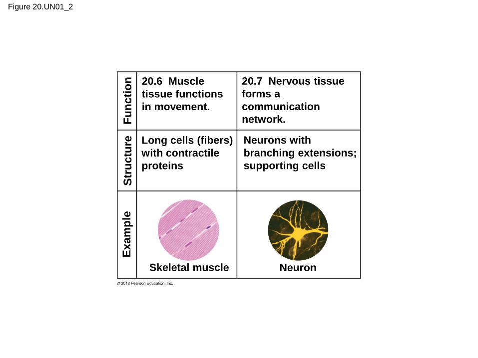
Figure 20.UN01_2
Ex
am
ple
S
tru
ctu
re
Fu
nc
tio
n
20.6 Muscle
tissue functions
in movement.
20.7 Nervous tissue
forms a
communication
network.
Long cells (fibers)
with contractile
proteins
Neurons with
branching extensions;
supporting cells
Skeletal muscle Neuron

Figure 20.UN02
a.
b.
c.
d. e.