Embed Size (px)
Citation preview
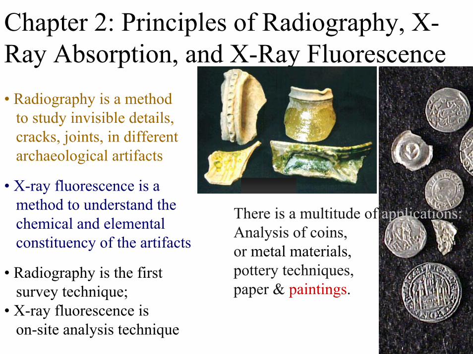
Chapter 2: Principles of Radiography, X-Ray Absorption, and X-Ray Fluorescence
• X-ray fluorescence is amethod to understand thechemical and elementalconstituency of the artifacts
There is a multitude of applications:Analysis of coins, or metal materials,pottery techniques,paper & paintings.
• Radiography is a method to study invisible details, cracks, joints, in different archaeological artifacts
• Radiography is the firstsurvey technique;
• X-ray fluorescence ison-site analysis technique

The Value of Art and Paintings

Investment in Analytical Techniques
X-ray facilities as quick testing tool
Electron Beam
Tungsten ormolybdenum
Nature & Origin of X-Rays – a Reminder
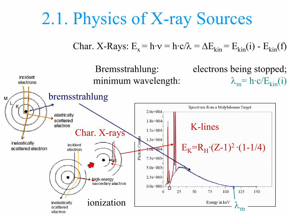
2.1. Physics of X-ray Sources
bremsstrahlung
Char. X-rays K-lines
ionization λm
Char. X-Rays: Ex = h·ν = h·c/λ = ∆Ekin = Ekin(i) - Ekin(f)
Bremsstrahlung: electrons being stopped; minimum wavelength: λm= h·c/Ekin(i)
EK=RH·(Z-1)2 ·(1-1/4)
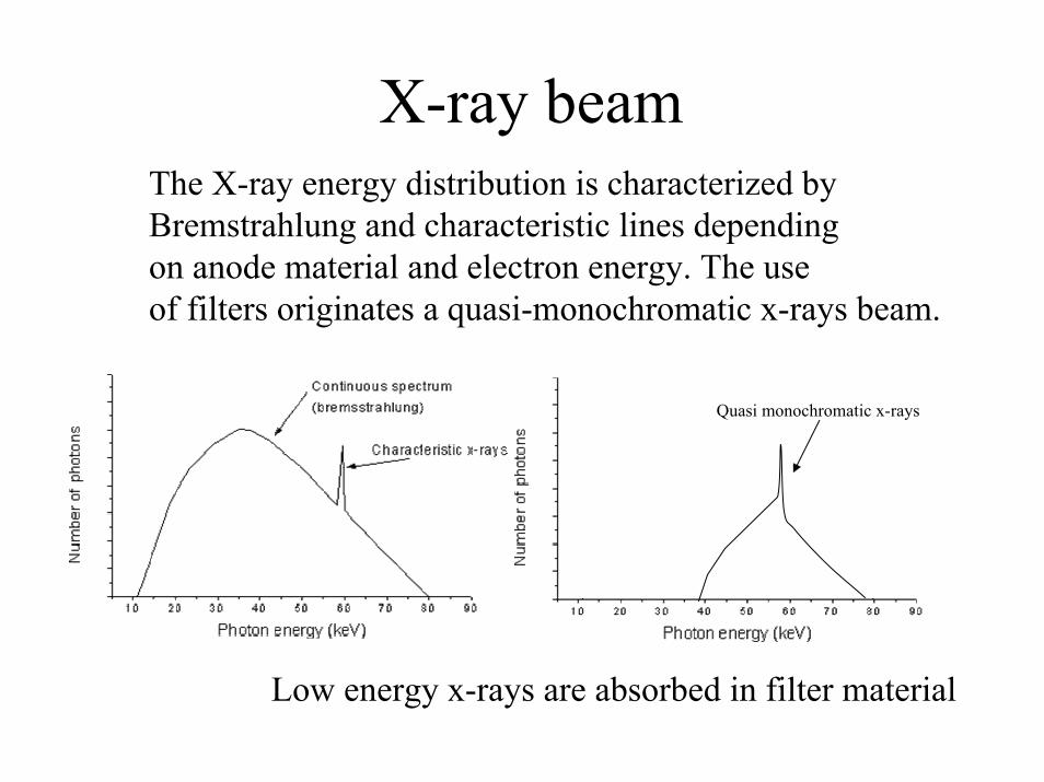
X-ray beamThe X-ray energy distribution is characterized byBremstrahlung and characteristic lines dependingon anode material and electron energy. The use of filters originates a quasi-monochromatic x-rays beam.
Quasi monochromatic x-rays
Low energy x-rays are absorbed in filter material
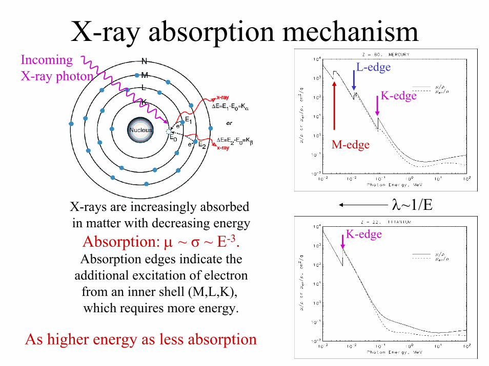
X-ray absorption mechanism
λ~1/EX-rays are increasingly absorbed in matter with decreasing energy
Absorption: µ ~ σ ~ E-3.Absorption edges indicate the
additional excitation of electronfrom an inner shell (M,L,K), which requires more energy.
M-edge
L-edge
K-edge
K-edge
IncomingX-ray photon
As higher energy as less absorption
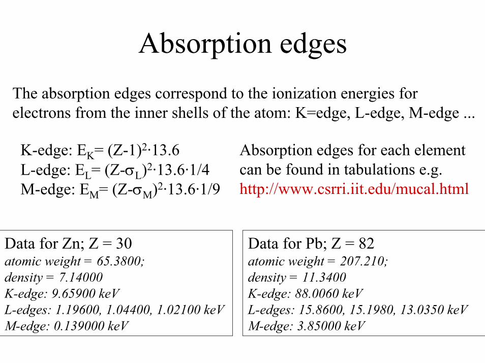
Absorption edgesThe absorption edges correspond to the ionization energies for electrons from the inner shells of the atom: K=edge, L-edge, M-edge ...
K-edge: EK= (Z-1)2·13.6L-edge: EL= (Z-σL)2·13.6·1/4M-edge: EM= (Z-σM)2·13.6·1/9
Absorption edges for each element can be found in tabulations e.g.http://www.csrri.iit.edu/mucal.html
Data for Zn; Z = 30 atomic weight = 65.3800; density = 7.14000 K-edge: 9.65900 keV L-edges: 1.19600, 1.04400, 1.02100 keV M-edge: 0.139000 keV
Data for Pb; Z = 82 atomic weight = 207.210; density = 11.3400 K-edge: 88.0060 keV L-edges: 15.8600, 15.1980, 13.0350 keV M-edge: 3.85000 keV
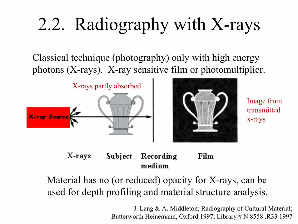
2.2. Radiography with X-rays
Classical technique (photography) only with high energyphotons (X-rays). X-ray sensitive film or photomultiplier.
Material has no (or reduced) opacity for X-rays, can beused for depth profiling and material structure analysis.
X-rays partly absorbed
Image fromtransmittedx-rays
J. Lang & A. Middleton; Radiography of Cultural Material; Butterworth Heinemann, Oxford 1997; Library # N 8558 .R33 1997
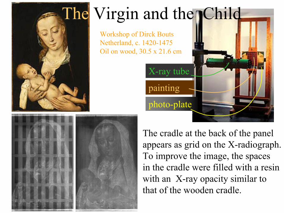
X-ray tube
painting
photo-plate
The Virgin and the ChildWorkshop of Dirck BoutsNetherland, c. 1420-1475Oil on wood, 30.5 x 21.6 cm
The cradle at the back of the panelappears as grid on the X-radiograph. To improve the image, the spaces in the cradle were filled with a resin with an X-ray opacity similar to that of the wooden cradle.
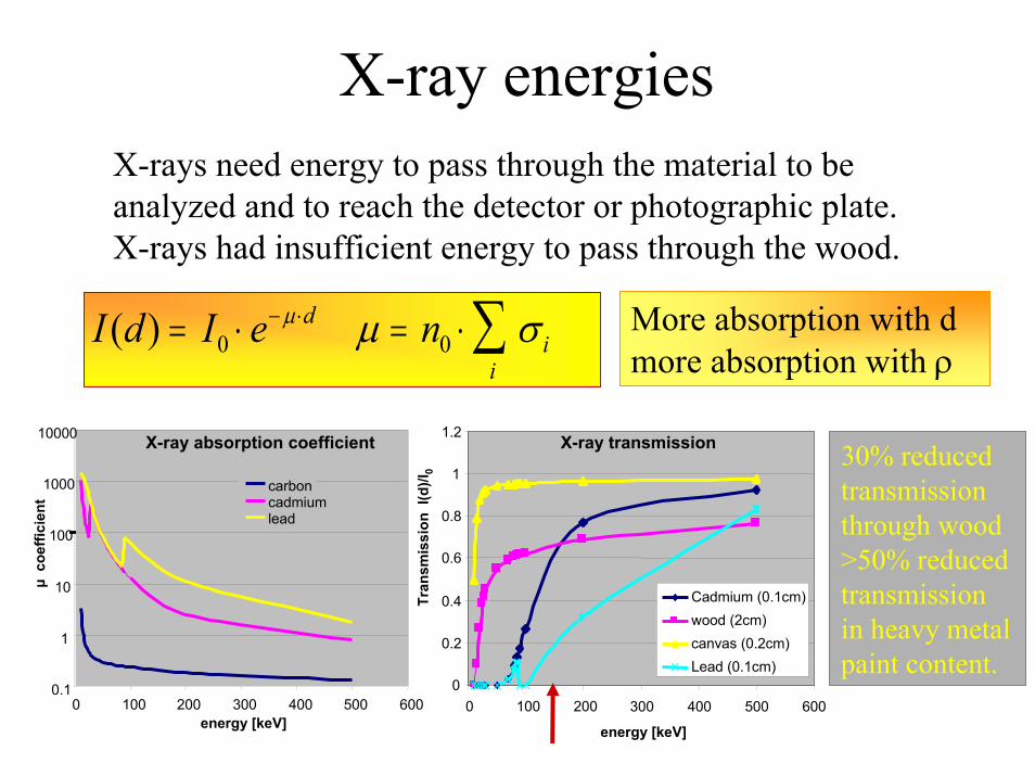
X-ray energiesX-rays need energy to pass through the material to be analyzed and to reach the detector or photographic plate. X-rays had insufficient energy to pass through the wood.
I d I e ndi
i( ) = ⋅ = ⋅− ⋅ ∑0 0
µ µ σ More absorption with dmore absorption with ρ
energy [keV]
0.1
1
10
100
1000
10000
0 100 200 300 400 500 600
µ c
oeffi
cien
t
carboncadmiumlead
X-ray absorption coefficient
0
0.2
0.4
0.6
0.8
1
1.2
0 100 200 300 400 500 600
energy [keV]
Tran
smis
sion
I(d
)/I0
Cadmium (0.1cm)wood (2cm)canvas (0.2cm)Lead (0.1cm)
X-ray transmission 30% reducedtransmission through wood>50% reducedtransmissionin heavy metalpaint content.
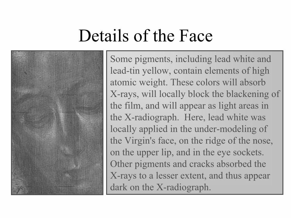
Details of the FaceSome pigments, including lead white and lead-tin yellow, contain elements of highatomic weight. These colors will absorbX-rays, will locally block the blackening of the film, and will appear as light areas in the X-radiograph. Here, lead white was locally applied in the under-modeling of the Virgin's face, on the ridge of the nose, on the upper lip, and in the eye sockets. Other pigments and cracks absorbed the X-rays to a lesser extent, and thus appear dark on the X-radiograph.
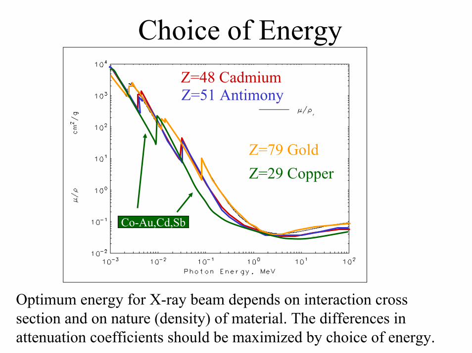
Choice of EnergyZ=48 CadmiumZ=51 Antimony
Z=79 GoldZ=29 Copper
Co-Au,Cd,Sb
Optimum energy for X-ray beam depends on interaction cross section and on nature (density) of material. The differences in attenuation coefficients should be maximized by choice of energy.
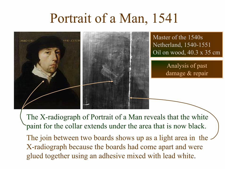
Portrait of a Man, 1541
The X-radiograph of Portrait of a Man reveals that the white paint for the collar extends under the area that is now black.The join between two boards shows up as a light area in the X-radiograph because the boards had come apart and were glued together using an adhesive mixed with lead white.
Master of the 1540sNetherland, 1540-1551Oil on wood, 40.3 x 35 cm
Analysis of pastdamage & repair
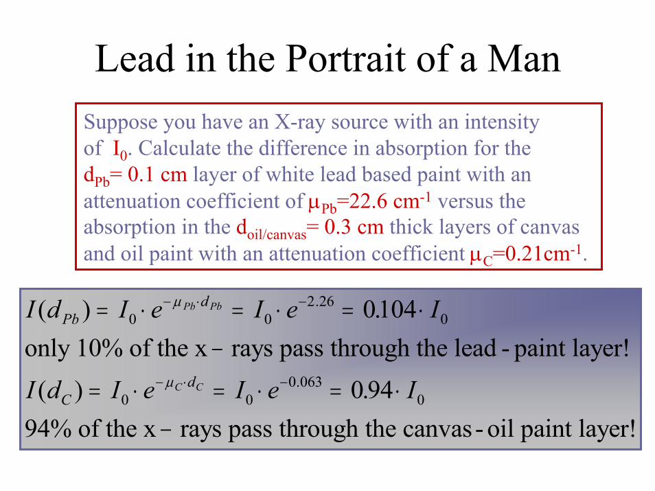
Lead in the Portrait of a Man Suppose you have an X-ray source with an intensityof I0. Calculate the difference in absorption for the dPb= 0.1 cm layer of white lead based paint with an attenuation coefficient of µPb=22.6 cm-1 versus the absorption in the doil/canvas= 0.3 cm thick layers of canvas and oil paint with an attenuation coefficient µC=0.21cm-1.
I d I e I e I
I d I e I e I
Pbd
Cd
Pb Pb
C C
( ) .
( ) .
.
.
= ⋅ = ⋅ = ⋅−
= ⋅ = ⋅ = ⋅−
− ⋅ −
− ⋅ −
0 02 26
0
0 00 063
0
0104
0 94
µ
µ
only 10% of the x rays pass through the lead - paint layer!
94% of the x rays pass through the canvas- oil paint layer!
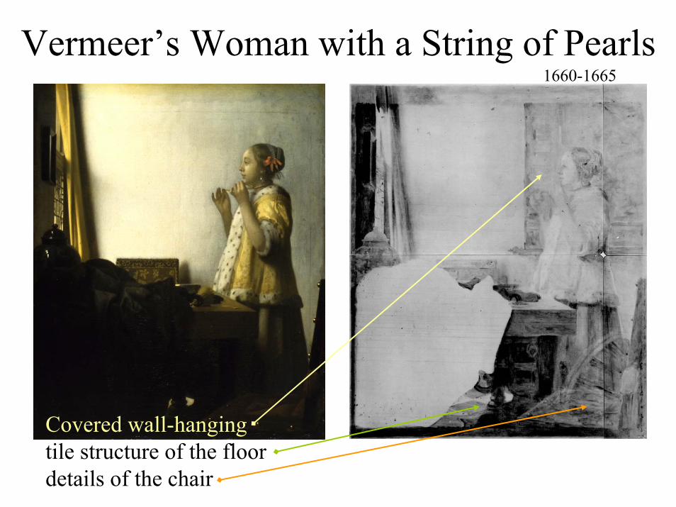
Vermeer’s Woman with a String of Pearls
Covered wall-hangingtile structure of the floordetails of the chair
1660-1665

Another Vermeer
Vermeer van Delft; The woman with the balance
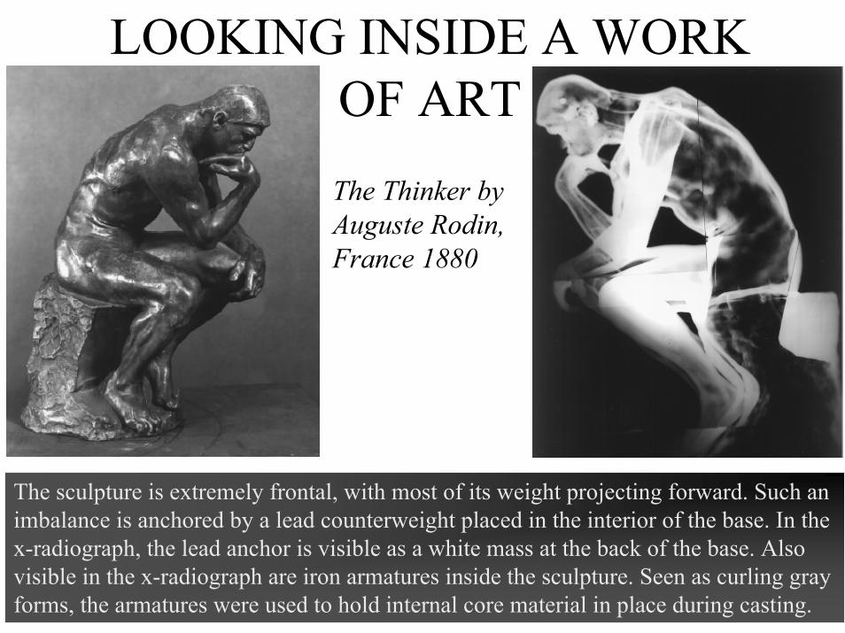
LOOKING INSIDE A WORK OF ARTThe Thinker by Auguste Rodin,France 1880
The sculpture is extremely frontal, with most of its weight projecting forward. Such an imbalance is anchored by a lead counterweight placed in the interior of the base. In the x-radiograph, the lead anchor is visible as a white mass at the back of the base. Also visible in the x-radiograph are iron armatures inside the sculpture. Seen as curling gray forms, the armatures were used to hold internal core material in place during casting.
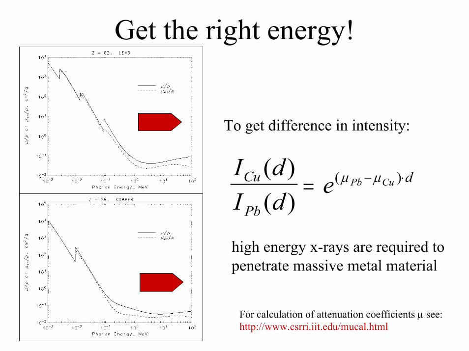
Get the right energy!
To get difference in intensity:
I dI d
eCu
Pb
dPb Cu( )( )
( )= − ⋅µ µ
high energy x-rays are required to penetrate massive metal material
For calculation of attenuation coefficients µ see:http://www.csrri.iit.edu/mucal.html
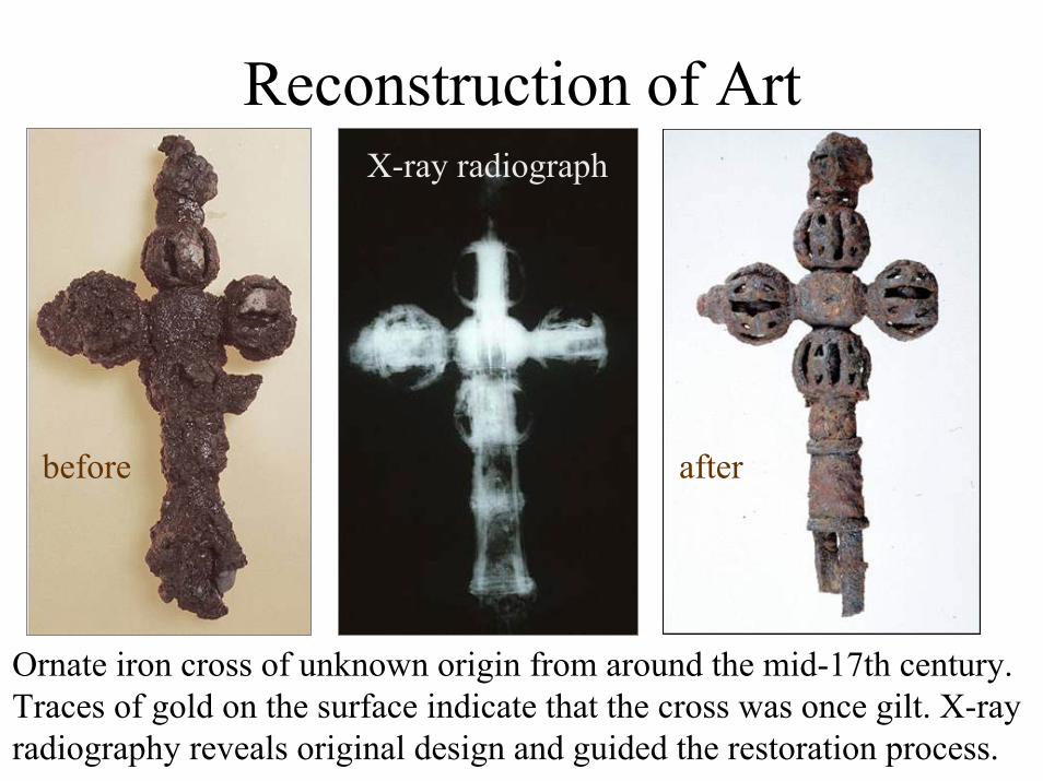
Reconstruction of Art
before
X-ray radiograph
after
Ornate iron cross of unknown origin from around the mid-17th century. Traces of gold on the surface indicate that the cross was once gilt. X-ray radiography reveals original design and guided the restoration process.

Back to the value of paintings
Summary X-ray radiography
Radiography is a powerful tool with a wide rangeof applications. Its usefulness is mainly based onthe differences in material densities which affects
the x-ray attenuation coefficients. This determines the x-ray opacity for heavy metal or high density
material compared to low density material like paper. The method gives only qualitative differences on photo-screen, it is a tool for first investigation, a
detailed analysis requires more sophisticated studies.