Embed Size (px)
DESCRIPTION
Presentation on Cell Structure & Function
Citation preview
© 2012 Pearson Education Inc.
Lecture prepared by Mindy Miller-KittrellNorth Carolina State University
Chapter 3
Cell Structure and Function

Processes of Life
• Growth• Reproduction• Responsiveness• Metabolism
© 2012 Pearson Education Inc.

Figure 3.1 Examples of types of cells-overview

• Prokaryotes– Lack nucleus
– Lack various internal structures bound with phospholipid membranes
– Are small (~1.0 µm in diameter)
– Have a simple structure
– Include bacteria and archaea
© 2012 Pearson Education Inc.
Prokaryotic and Eukaryotic Cells: An Overview

Figure 3.2 Typical prokaryotic cell
Ribosome
Cytoplasm
Nucleoid
GlycocalyxCell wall Cytoplasmic membrane
Inclusions
Flagellum
• Eukaryotes– Have nucleus
– Have internal membrane-bound organelles
– Are larger (10–100 µm in diameter)
– Have more complex structure
– Include algae, protozoa, fungi, animals, and plants
© 2012 Pearson Education Inc.
Prokaryotic and Eukaryotic Cells: An Overview

Figure 3.3 Typical eukaryotic cell
Nucleolus
Cilium
Ribosomes
Nuclear envelope
Nuclear pore
Lysosome
Mitochondrion
Centriole
Secretory vesicle
Golgi body
Transport vesicles
Rough endoplasmicreticulum
Smooth endoplasmicreticulum
Cytoplasmicmembrane
Cytoskeleton

Figure 3.4 Approximate size of various types of cells
Chicken egg4.7 cm diameter
(47,000 m)*
VirusOrthopoxvirus
0.3 m diameter
BacteriumStaphylococcus1 m diameter
Parasitic protozoanGiardia
14 m length
*Actually, the inset box on the egg would be too small to be visible. (Width of box would be about 0.002 mm.)

External Structures of Bacterial Cells
• Glycocalyces
– Gelatinous, sticky substance surrounding the outside of the cell
– Composed of polysaccharides, polypeptides, or both
© 2012 Pearson Education Inc.

External Structures of Bacterial Cells
• Two Types of Glycocalyces– Capsule
– Composed of organized repeating units of organic chemicals
– Firmly attached to cell surface– May prevent bacteria from being recognized by host
– Slime layer– Loosely attached to cell surface– Water soluble– Sticky layer allows prokaryotes to attach to surfaces
© 2012 Pearson Education Inc.

Figure 3.5 Glycocalyces-overview
Glycocalyx(capsule)
Glycocalyx(slime layer)

External Structures of Bacterial Cells
ANIMATION Motility
© 2012 Pearson Education Inc.
External Structures of Bacterial Cells
• Flagella– Are responsible for movement
– Have long structures that extend beyond cell surface
– Are not present on all bacteria
© 2012 Pearson Education Inc.
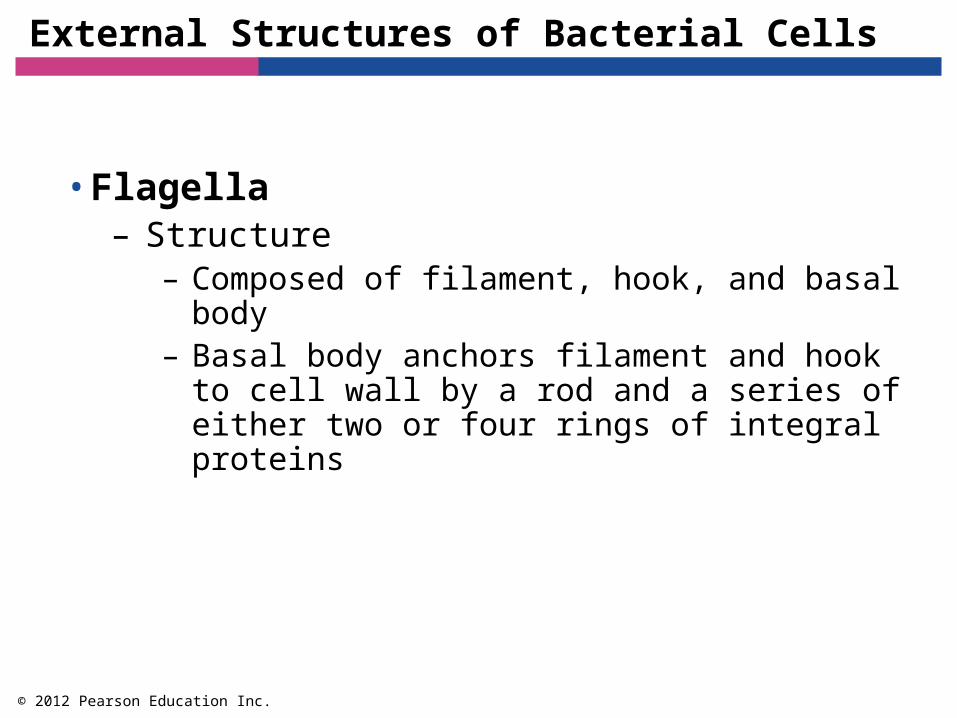
External Structures of Bacterial Cells
• Flagella– Structure
– Composed of filament, hook, and basal body– Basal body anchors filament and hook to cell wall
by a rod and a series of either two or four rings of integral proteins
© 2012 Pearson Education Inc.

External Structures of Bacterial Cells
© 2012 Pearson Education Inc.
ANIMATION Flagella: Structure

Figure 3.6 Proximal structure of bacterial flagella-overview
Filament
Directionof rotationduring run
Peptidoglycanlayer (cell wall)
Cytoplasmicmembrane
Cytoplasm
Rod
Protein rings
Gram Gram
Filament
Outerproteinrings
Rod
Integral protein
Innerproteinrings
Integralprotein
Basalbody
Cytoplasm
Outer membrane
Peptidoglycanlayer
Cytoplasmicmembrane
Cellwall
Ho
o
k
Ho
o
k

Figure 3.7 Micrographs of basic arrangements of bacterial flagella-overview

External Structures of Bacterial Cells
© 2012 Pearson Education Inc.
ANIMATION Flagella: Arrangement
ANIMATION Flagella: Spirochetes

Figure 3.8 Axial filament-overview
Axial filament
Endoflagellarotate Axial filament
rotates aroundcell
Outermembrane
Cytoplasmicmembrane
Axial filamentSpirochetecorkscrewsand movesforward

External Structures of Bacterial Cells
• Flagella– Function
– Rotation propels bacterium through environment– Rotation reversible; can be counterclockwise or
clockwise– Bacteria move in response to stimuli (taxis)
– Runs – Tumbles
© 2012 Pearson Education Inc.

Figure 3.9 Motion of a peritrichous bacterium
Tumble
Run
Tumble
Attractant
Run

External Structures of Bacterial Cells
© 2012 Pearson Education Inc.
ANIMATION Flagella: Movement

External Structures of Bacterial Cells
• Fimbriae and Pili– Rodlike proteinaceous extensions
© 2012 Pearson Education Inc.

External Structures of Bacterial Cells
© 2012 Pearson Education Inc.
• Fimbriae– Sticky, bristlelike projections– Used by bacteria to adhere to one another, to
hosts, and to substances in environment– Shorter than flagella– Serve an important function in biofilms

Figure 3.10 Fimbriae
Flagellum Fimbria

External Structures of Bacterial Cells
• Pili– Special type of fimbria
– Also known as conjugation pili
– Longer than other fimbriae but shorter than flagella
– Bacteria typically have only one or two per cell
– Mediate the transfer of DNA from one cell to another (conjugation)
© 2012 Pearson Education Inc.

Figure 3.11 Pili
Conjugation pilus

Bacterial Cell Walls
• Bacterial Cell Walls– Provide structure and shape and protect cell from
osmotic forces– Assist some cells in attaching to other cells or in
resisting antimicrobial drugs– Can target cell wall of bacteria with antibiotics– Give bacterial cells characteristic shapes– Composed of peptidoglycan– Scientists describe two basic types of bacterial cell
walls, Gram-positive and Gram-negative
© 2012 Pearson Education Inc.

Figure 3.12 Bacterial shapes and arrangements-overview

Figure 3.13 Comparison of the structures of glucose, NAG, and NAM-overview
Glucose N-acetylglucosamineNAG
N-acetylmuramic acidNAM

Bacterial Cell Walls
• Gram-Positive Bacterial Cell Walls– Relatively thick layer of peptidoglycan
– Contain unique polyalcohols called teichoic acids
– Appear purple following Gram staining procedure
– Up to 60% mycolic acid in acid-fast bacteria helps cells survive desiccation
© 2012 Pearson Education Inc.

Figure 3.15a Comparison of cell walls of Gram-positive and Gram-negative bacteria
Gram-positive cell wall
Peptidoglycan layer(cell wall)
Cytoplasmic membrane
Teichoic acid
Lipoteichoic acid
Integralprotein

Bacterial Cell Walls
• Gram-Negative Bacterial Cell Walls– Have only a thin layer of peptidoglycan
– Bilayer membrane outside the peptidoglycan contains phospholipids, proteins, and lipopolysaccharide (LPS)
– May be impediment to the treatment of disease
– Appear pink following Gram staining procedure
© 2012 Pearson Education Inc.

Figure 3.15b Comparison of cell walls of Gram-positive and Gram-negative bacteria
Gram-negative cell wall
Outermembraneof cell wall
Peptidoglycanlayer of cell wall
Cytoplasmicmembrane
Lipopolysaccharide(LPS)
Porin
Porin(sectioned)
Periplasmic space
Phospholipid layers
Integralproteins
n
Corepolysaccharide
O side chain(varies Inlength andcomposition)
Lipid A(embeddedin outermembrane)
Fatty acid

Bacterial Cytoplasmic Membranes
• Structure– Referred to as phospholipid bilayer
– Composed of lipids and associated proteins– Fluid mosaic model describes current
understanding of membrane structure
© 2012 Pearson Education Inc.

Figure 3.16 The structure of a prokaryotic cytoplasmic membrane: a phospholipid bilayer
Head, whichcontains phosphate(hydrophilic)
Tail(hydrophobic)
Phospholipid
Phospholipidbilayer
Integral proteinPeripheral protein
Integralprotein
Cytoplasm
Integralproteins
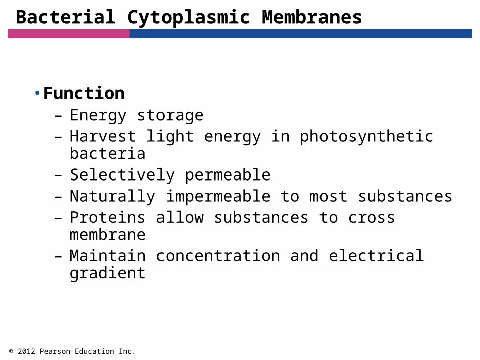
Bacterial Cytoplasmic Membranes
• Function– Energy storage– Harvest light energy in photosynthetic bacteria– Selectively permeable– Naturally impermeable to most substances– Proteins allow substances to cross membrane– Maintain concentration and electrical gradient
© 2012 Pearson Education Inc.

Figure 3.17 Electrical potential of a cytoplasmic membrane
Cell exterior (extracellular fluid)
Cell interior (cytoplasm)
Cytoplasmic membrane
Integralprotein
Protein
Protein
DNA

Bacterial Cytoplasmic Membranes
• Function– Passive processes
– Diffusion– Facilitated diffusion – Osmosis
© 2012 Pearson Education Inc.

Figure 3.18 Passive processes of movement across a cytoplasmic membrane-overview
Extracellular fluid
Cytoplasm
Diffusionthrough thephospholipidbilayer
Facilitateddiffusionthrough anonspecificchannelprotein
Facilitated diffusionthrough a permeasespecific for one chemical;binding of substratecauses shape change inthe channel protein
Osmosis,the diffusion ofwater through aspecific channelprotein or throughthe phospholipidbilayer
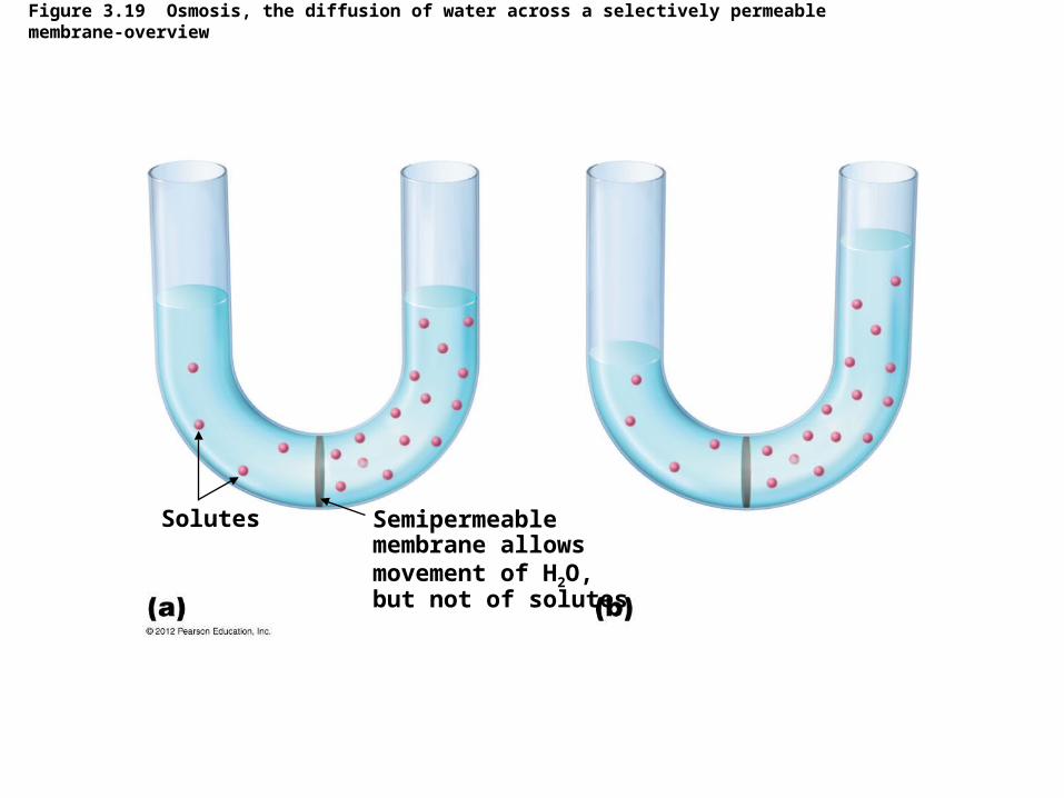
Figure 3.19 Osmosis, the diffusion of water across a selectively permeable membrane-overview
Solutes Semipermeablemembrane allowsmovement of H2O,but not of solutes

Figure 3.20 Effects of isotonic, hypertonic, and hypotonic solutions on cells-overview
Isotonic solution Hypertonic solution Hypotonic solution
Cells without a wall(e.g., mycoplasmas,animal cells)
Cells with a wall(e.g., plants, fungaland bacterial cells)
Cell wall Cell wall
Cell membrane Cell membrane
H2O H2O
H2O
H2O H2O H2O

Bacterial Cytoplasmic Membranes
• Function– Active processes
– Active transport
– Group translocation– Substance chemically modified during transport
© 2012 Pearson Education Inc.
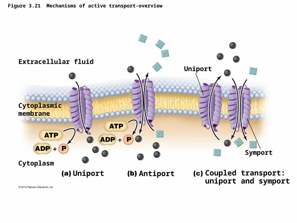
Figure 3.21 Mechanisms of active transport-overview
Extracellular fluidUniport
Cytoplasmicmembrane
Symport
Cytoplasm
Uniport Antiport Coupled transport:uniport and symport

Bacterial Cytoplasmic Membranes
© 2012 Pearson Education Inc.
ANIMATION Active Transport: Overview
ANIMATION Active Transport: Types

Figure 3.22 Group translocation
Extracellular fluid
Glucose
Cytoplasm
Glucose 6-PO4
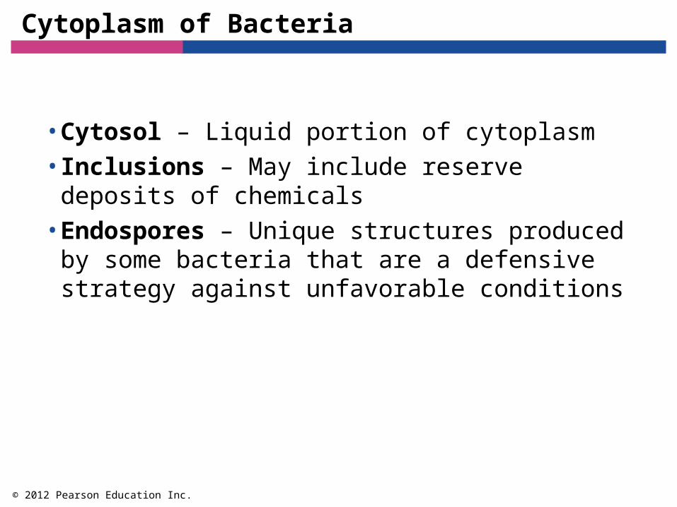
Cytoplasm of Bacteria
• Cytosol – Liquid portion of cytoplasm• Inclusions – May include reserve deposits of
chemicals• Endospores – Unique structures produced by
some bacteria that are a defensive strategy against unfavorable conditions
© 2012 Pearson Education Inc.

Figure 3.23 Granules of PHB in the bacterium Azotobacter chroococcum
Polyhydroxybutyrate

Figure 3.24 The formation of an endospore-overview
DNA is replicated.
DNA aligns alongthe cell’s long axis.
Cytoplasmic membraneinvaginates to formforespore.
Cytoplasmic membranegrows and engulfsforespore within a second membrane.Vegetative cell’s DNAdisintegrates.
Cell wallCytoplasmicmembrane
DNA
Vegetative cell
Forespore
Firstmembrane
Second membrane
A cortex of calcium anddipicolinic acid isdeposited betweenthe membranes.
Spore coat formsaround endospore.
Endospore matures:completion of spore coatand increase in resistanceto heat and chemicals byunknown process.
Endospore is released fromoriginal cell.
Cortex
Spore coat
Outerspore coat
Endospore
Outerspore coat

Cytoplasm of Bacteria
• Nonmembranous Organelles
– Ribosomes – Sites of protein synthesis
– Cytoskeleton – Plays a role in forming the cell’s basic shape
© 2012 Pearson Education Inc.
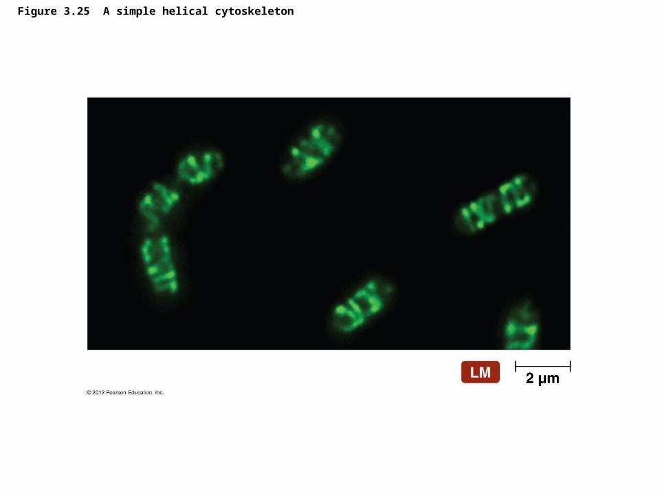
Figure 3.25 A simple helical cytoskeleton

External Structures of Archaea
• Glycocalyces– Function in the formation of biofilms– Adhere cells to one another and inanimate objects
• Flagella– Consist of basal body, hook, and filament – Numerous differences with bacterial flagella
• Fimbriae and Hami– Many archaea have fimbriae– Some make fimbriae-like structures called hami
– Function to attach archaea to surfaces
© 2012 Pearson Education Inc.
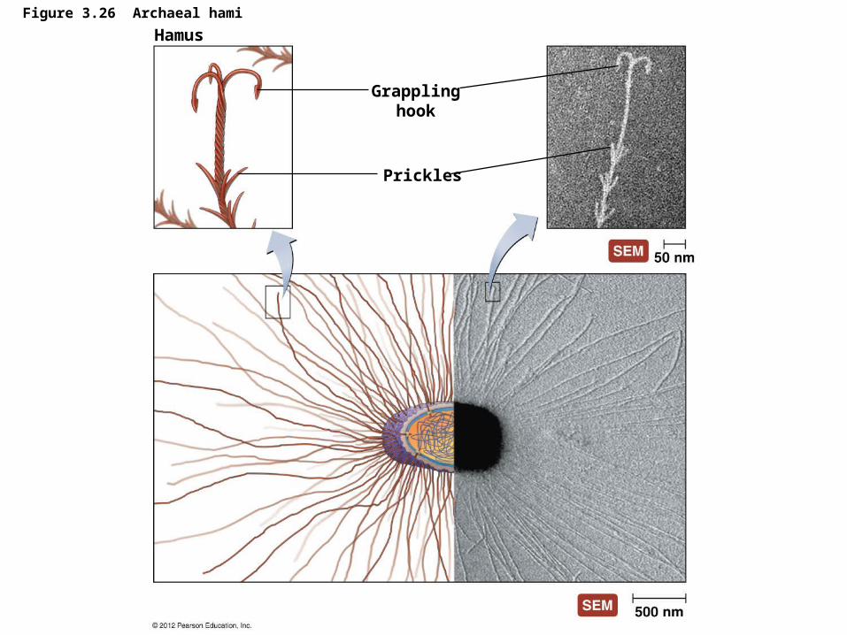
Figure 3.26 Archaeal hami
Hamus
Grapplinghook
Prickles

Archaeal Cell Walls and Cytoplasmic Membrane
– Most archaea have cell walls– Do not have peptidoglycan – Contain variety of specialized polysaccharides
and proteins
– All archaea have cytoplasmic membranes– Maintain electrical and chemical gradients– Control import and export of substances from the
cell
© 2012 Pearson Education Inc.
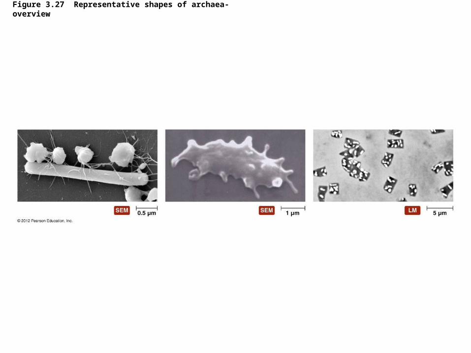
Figure 3.27 Representative shapes of archaea-overview

Cytoplasm of Archaea
– Archaeal cytoplasm similar to bacterial cytoplasm– Have 70S ribosomes – Fibrous cytoskeleton– Circular DNA
– Archaeal cytoplasm also differs from bacterial cytoplasm– Different ribosomal proteins– Different metabolic enzymes to make RNA– Genetic code more similar to eukaryotes
© 2012 Pearson Education Inc.

External Structure of Eukaryotic Cells
• Glycocalyces – Never as organized as prokaryotic capsules
– Help anchor animal cells to each other
– Strengthen cell surface
– Provide protection against dehydration
– Function in cell-to-cell recognition and communication
© 2012 Pearson Education Inc.

Eukaryotic Cell Walls
– Fungi, algae, plants, and some protozoa have cell walls
– Composed of various polysaccharides– Plant cell walls composed of cellulose– Fungal cell walls composed of cellulose, chitin,
and/or glucomannan– Algal cell walls composed of a variety of
polysaccharides
© 2012 Pearson Education Inc.

Figure 3.28 A eukaryotic cell wall
Cell wall Cytoplasmic membrane

Eukaryotic Cytoplasmic Membranes
– All eukaryotic cells have cytoplasmic membrane– Are a fluid mosaic of phospholipids and proteins– Contain steroid lipids to help maintain fluidity– Contain regions of lipids and proteins called
membrane rafts– Control movement into and out of cell
© 2012 Pearson Education Inc.

Figure 3.29 Eukaryotic cytoplasmic membrane
Cytoplasmicmembrane
Intercellularmatrix
Cytoplasmicmembrane

Figure 3.30 Endocytosis-overview
Pseudopodium

Cytoplasm of Eukaryotes
• Flagella– Structure and arrangement
– Differ structurally and functionally from prokaryotic flagella
– Within the cytoplasmic membrane– Shaft composed of tubulin arranged to form
microtubules– Filaments anchored to cell by basal body– May be single or multiple
– Function– Do not rotate but undulate rhythmically
© 2012 Pearson Education Inc.

Figure 3.31a Eukaryotic flagella and cilia
Flagellum
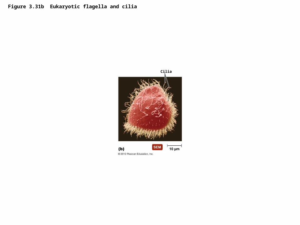
Figure 3.31b Eukaryotic flagella and cilia
Cilia

Figure 3.32 Movement of eukaryotic flagella and cilia-overview
Direction of motion
Direction of motion
Flagella
Cilia
Cytoplasm of Eukaryotes
• Cilia– Shorter and more numerous than flagella
– Coordinated beating propels cells through their environment
– Also used to move substances past the surface of the cell
© 2012 Pearson Education Inc.

Figure 3.31c Eukaryotic flagella and cilia
Cytoplasmic membrane
Cytosol
Central pairmicrotubules
Microtubules(doublet)
“9 2”arrangement
Cytoplasmicmembrane
Basal body
Microtubules(triplet) “9 0”
arrangement
Portioncut away to showtransition areafrom doubletsto triplets andthe end ofcentralmicrotubules. ..

Cytoplasm of Eukaryotes
• Other Nonmembranous Organelles– Ribosomes
– Larger than prokaryotic ribosomes (80S versus 70S)– Composed of 60S and 40S subunits
– Cytoskeleton– Extensive network of fibers and tubules– Anchors organelles– Produces basic shape of the cell– Made up of tubulin microtubules, actin
microfilaments, and intermediate filaments
© 2012 Pearson Education Inc.

Figure 3.33 Eukaryotic cytoskeleton-overview
Microtubule Microfilament
Intermediate filament
Tubulin
Actinsubunit
Proteinsubunits

Cytoplasm of Eukaryotes
• Other Nonmembranous Organelles– Centrioles and centrosome
– Centrioles play a role in mitosis, cytokinesis, and formation of flagella and cilia
– Centrosome is region of cytoplasm where centrioles are found
© 2012 Pearson Education Inc.

Figure 3.34 Centrosome-overview Centrosome (made up of two centrioles)
Microtubules
Triplet

Cytoplasm of Eukaryotes
• Membranous Organelles– Nucleus
– Often largest organelle in cell– Contains most of the cell’s DNA– Nucleoplasm
– Contains chromatin– Nucleoli present in nucleoplasm; RNA
synthesized in nucleoli– Surrounded by nuclear envelope
© 2012 Pearson Education Inc.

Figure 3.35 Eukaryotic nucleus
Nucleolus
Nucleoplasm
Chromatin
Nuclear envelope
Two phospholipidbilayers
Nuclear pores
Rough ER

Cytoplasm of Eukaryotes
• Membranous Organelles– Endoplasmic reticulum
– Netlike arrangement of flattened, hollow tubules continuous with nuclear envelope
– Functions as transport system– Two forms
– Smooth endoplasmic reticulum (SER) – Rough endoplasmic reticulum (RER)
© 2012 Pearson Education Inc.

Figure 3.36 Endoplasmic reticulum
Membrane-boundribosomes
Mitochondrion
Free ribosome
Smooth endoplasmic reticulum (SER)
Rough endoplasmicreticulum (RER)

Cytoplasm of Eukaryotes
• Membranous Organelles– Golgi body
– Flattened hollow sacs surrounded by phospholipid bilayer
– Receives, processes, and packages large molecules for export from cell
– Packages molecules in secretory vesicles– Not in all eukaryotic cells
© 2012 Pearson Education Inc.

Secretory vesicles
Vesiclesarrivingfrom ER
Figure 3.37 Golgi body

Cytoplasm of Eukaryotes
• Membranous Organelles– Lysosomes, peroxisomes, vacuoles, and vesicles
– Store and transfer chemicals within cells– May store nutrients in cell– Lysosomes contain catabolic enzymes – Peroxisomes contain enzymes that degrade
poisonous wastes
© 2012 Pearson Education Inc.

Figure 3.38 Vacuole
Cell wall
Nucleus
Central vacuole
Cytoplasm

Bacterium
Phagosome(food vesicle)
Vesicle fuses with a lysosome
Smoothendoplasmicreticulum(SER)
Transportvesicle
Lysosome
Phagolysosome
Golgi body
Secretoryvesicle
Endocytosis(phagocytosis)
Exocytosis(elimination, secretion)
Figure 3.39 The roles of vesicles in the destruction of a phagocytized pathogen within a white blood cell

Cytoplasm of Eukaryotes
• Membranous Organelles– Mitochondria
– Have two membranes composed of phospholipid bilayer
– Produce most of cell’s ATP– Interior matrix contains 70S ribosomes and
molecule of DNA
© 2012 Pearson Education Inc.

Figure 3.40 Mitochondrion
Outer membrane
Inner membrane
Crista
Matrix
Ribosomes

Cytoplasm of Eukaryotes
• Membranous Organelles– Chloroplasts
– Light-harvesting structures found in photosynthetic eukaryotes
– Have two phospholipid bilayer membranes and DNA
– Have 70S ribosomes
© 2012 Pearson Education Inc.
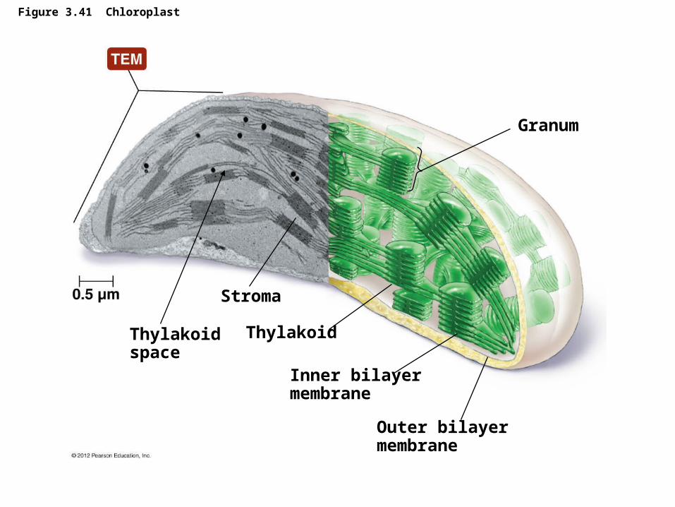
Figure 3.41 Chloroplast
Granum
Thylakoidspace
Stroma
Thylakoid
Inner bilayermembrane
Outer bilayermembrane

Cytoplasm of Eukaryotes
• Endosymbiotic Theory– Eukaryotes formed from union of small aerobic
prokaryotes with larger anaerobic prokaryotes– Smaller prokaryotes became internal parasites
– Parasites lost ability to exist independently– Larger cell became dependent on parasites for
aerobic ATP production– Aerobic prokaryotes evolved into mitochondria– Similar scenario for origin of chloroplasts
– Not universally accepted
© 2012 Pearson Education Inc.