Embed Size (px)
Citation preview
Chapter 19
Organization and Control of Eukaryotic Genomes
(here are at least 6 different modes of eukaryotic gene control…)
(Remember: the example of operons is only seen in prokaryotic cells)
Use this as a note taking guide…
Copy this and add to it with the following slides
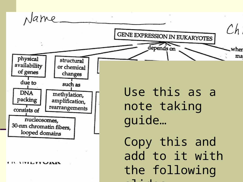
Eukaryotes have many different modes of control over gene expressionAT ALL STEPS INTHE DIAGRAM
Until the last step… degradation
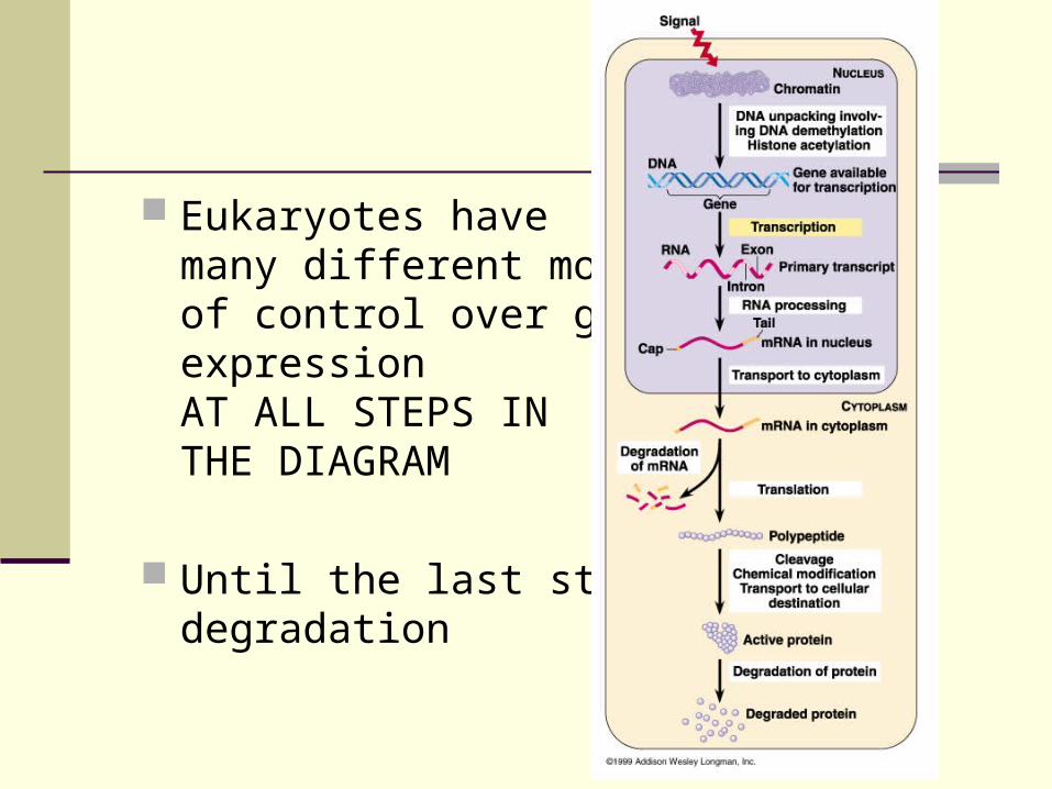
Packing of chromatin
Areas of tightly coiled DNA are not easily transcribed. Histones, nucleosomes
Each specialized cell only translates a small fraction of its genes, for example, the pancreas makes insulin and glucagon where as the liver,brain,skin etc. do not.
(use your CD to view an animation on these)
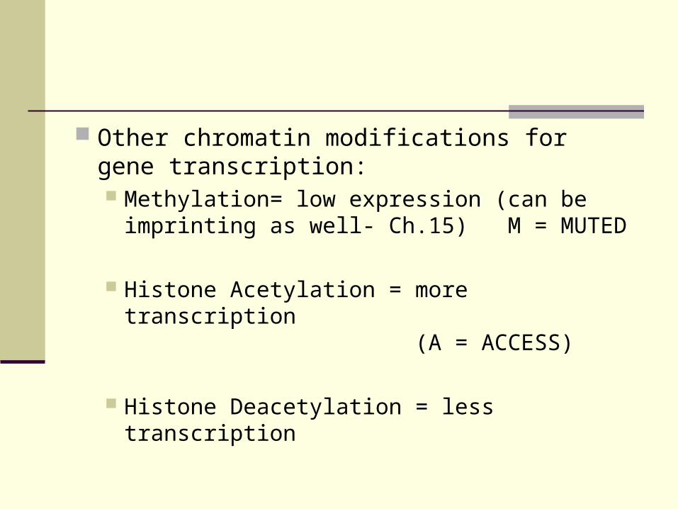
Other chromatin modifications for gene transcription: Methylation= low expression (can be
imprinting as well- Ch.15) M = MUTED
Histone Acetylation = more transcription (A = ACCESS)
Histone Deacetylation = less transcription
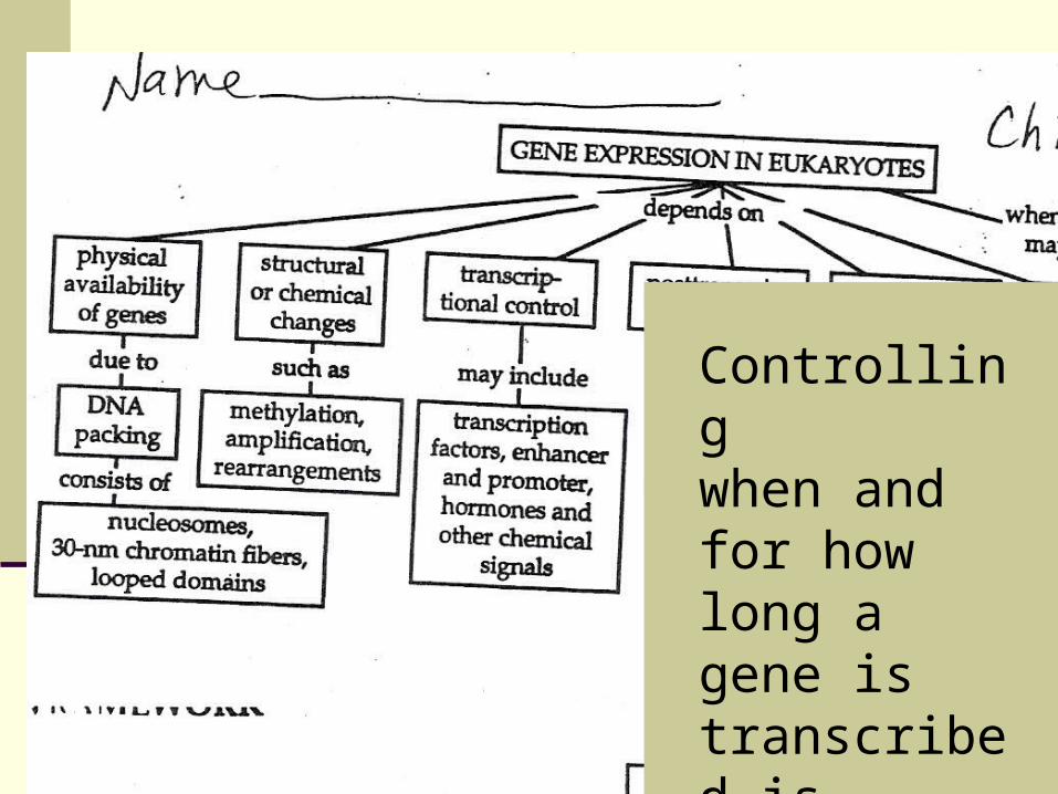
Controlling when and for how long a gene is transcribed is “transcriptional control”.
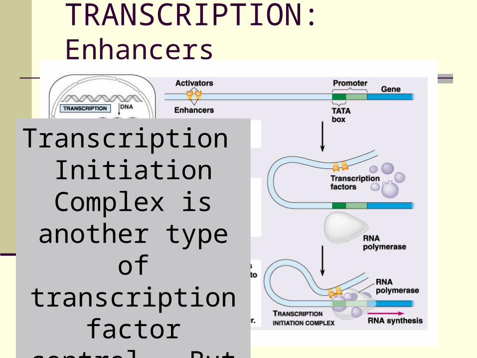
TRANSCRIPTION: Enhancers
Transcription Initiation Complex is another type of
transcription factor control. But still the same idea as the last
chapters 16-17)
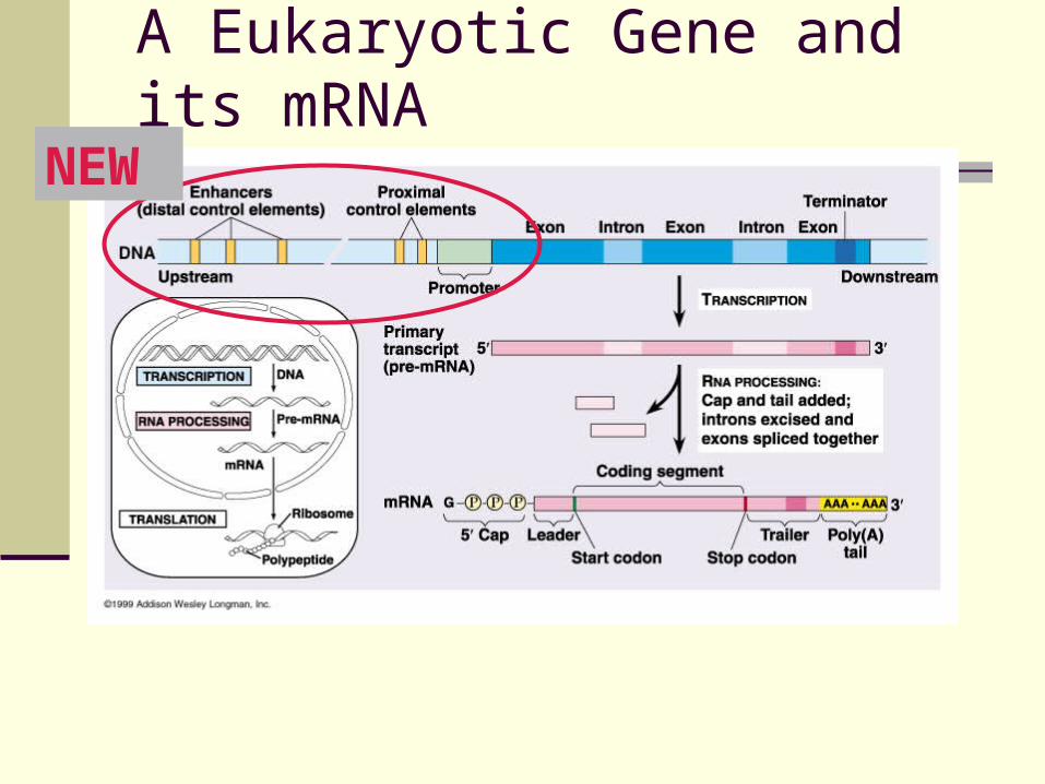
A Eukaryotic Gene and its mRNA
NEW
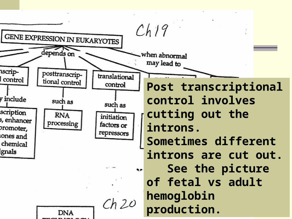
Post transcriptional control involves cutting out the introns. Sometimes different introns are cut out. See the picture of fetal vs adult hemoglobin production.
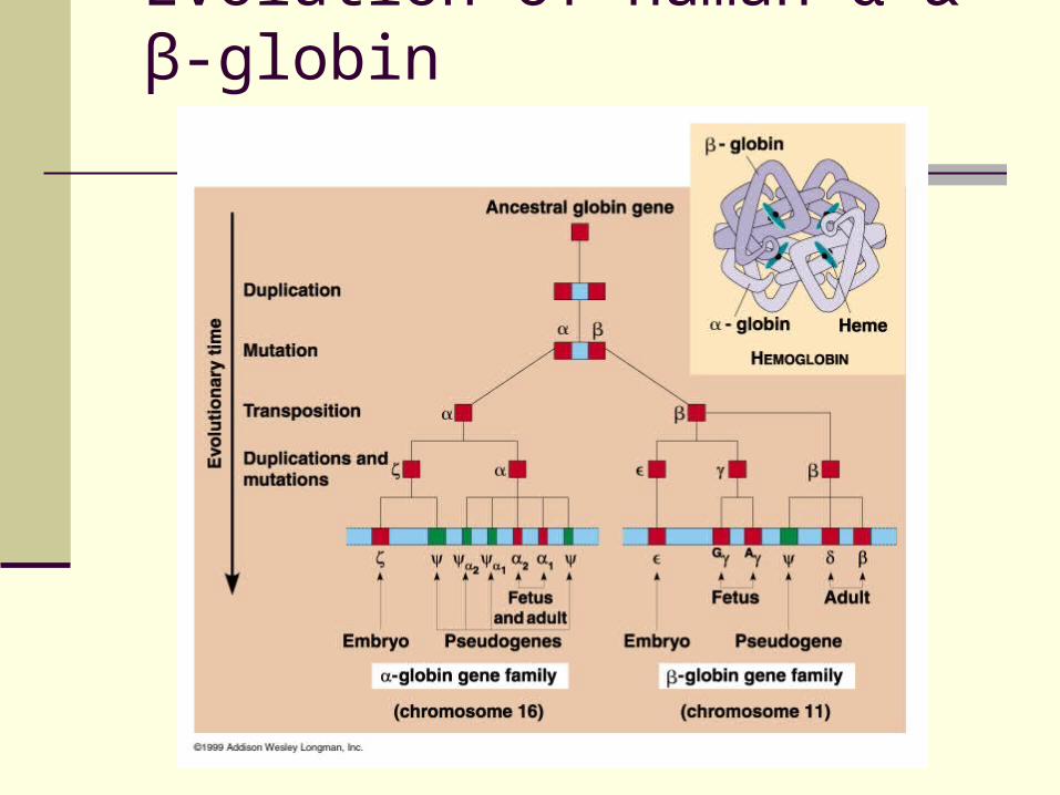
Evolution of human α & β-globin
~ 97 % of human DNA does not encode for proteins or RNA
Regions of telomeres and centromeres have many tandemly repeating sequences / satellite DNA … WHY?
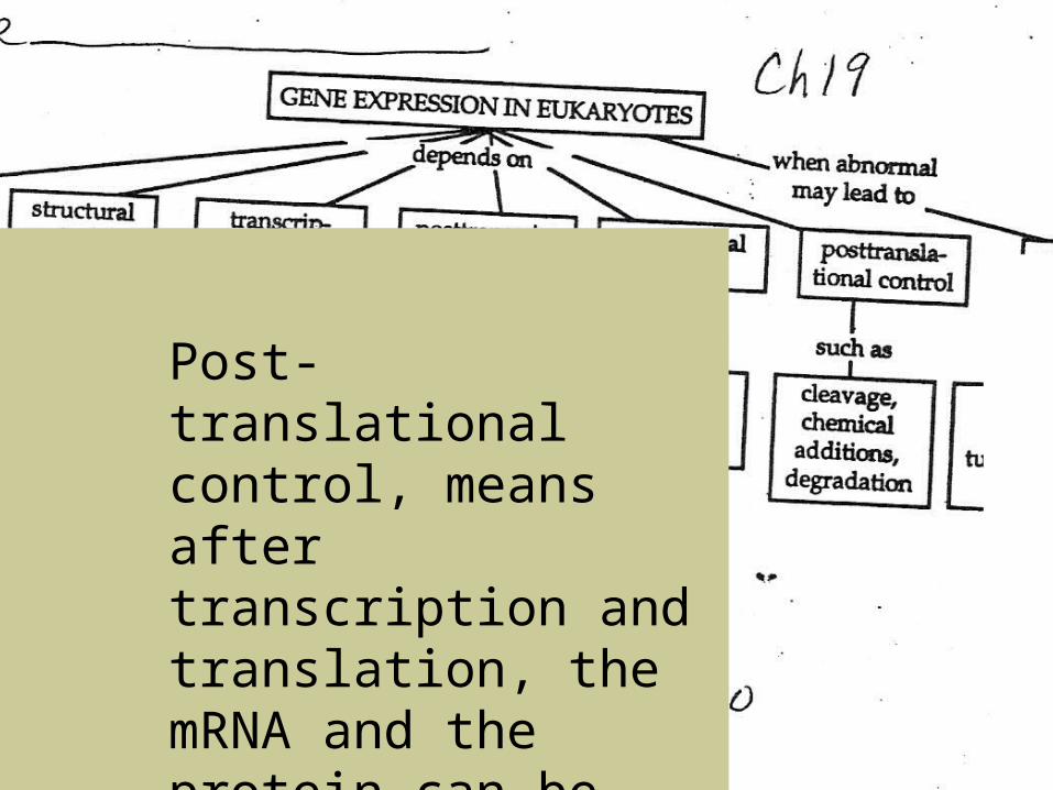
Post-translational control, means after transcription and translation, the mRNA and the protein can be degraded/stopped.
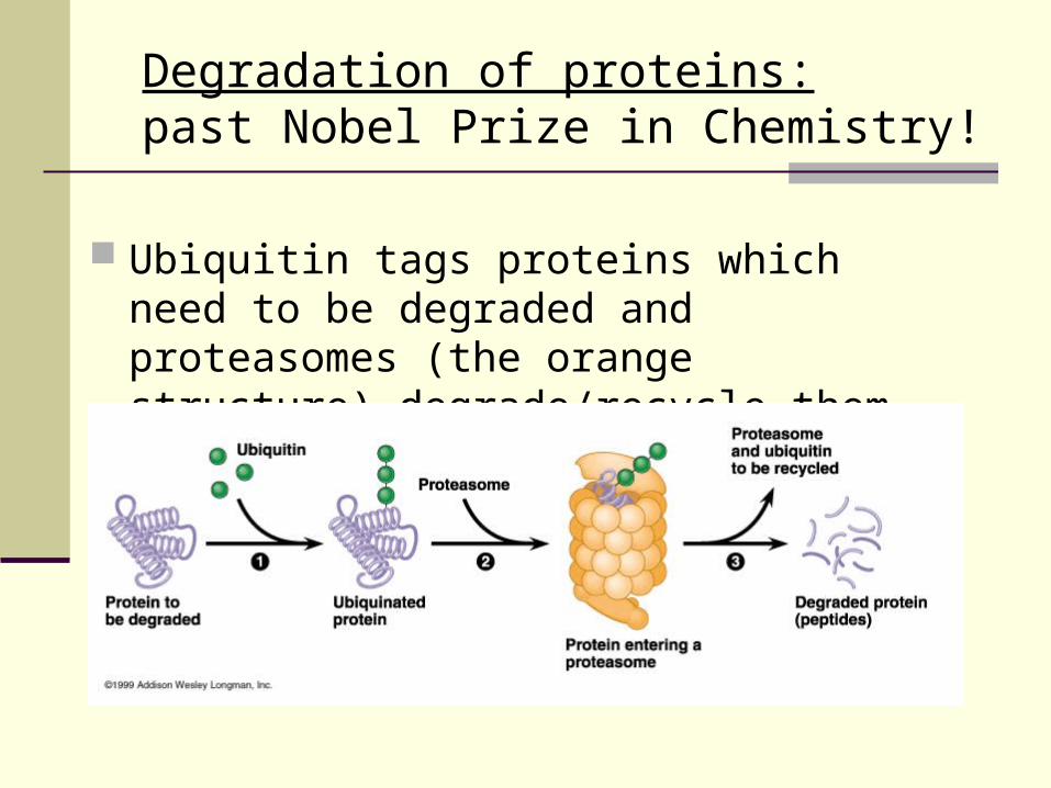
Ubiquitin tags proteins which need to be degraded and proteasomes (the orange structure) degrade/recycle them.
Degradation of proteins: past Nobel Prize in Chemistry!
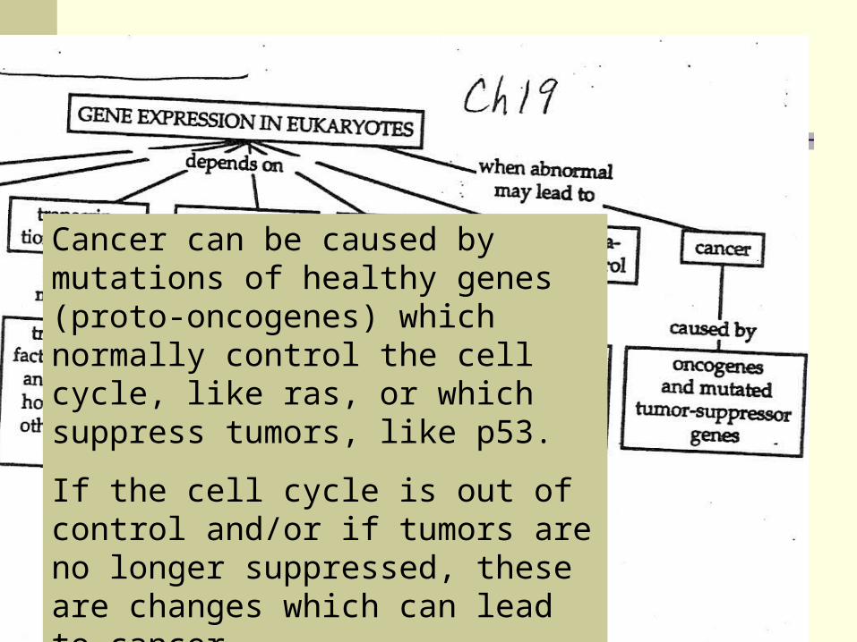
Cancer can be caused by mutations of healthy genes (proto-oncogenes) which normally control the cell cycle, like ras, or which suppress tumors, like p53.
If the cell cycle is out of control and/or if tumors are no longer suppressed, these are changes which can lead to cancer.
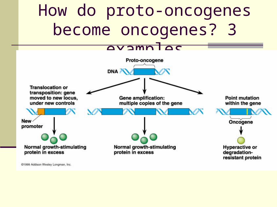
How do proto-oncogenes become oncogenes? 3 examples
What is a proto-oncogene? A normal gene, that usually is involved in
some control of cell growth and division. These genes are not cancerous, but if mutated, could lead to cancer.
What is an oncogene? A mutated proto-oncogene which causes too
much growth or loss of control over the cell cycle in some way.
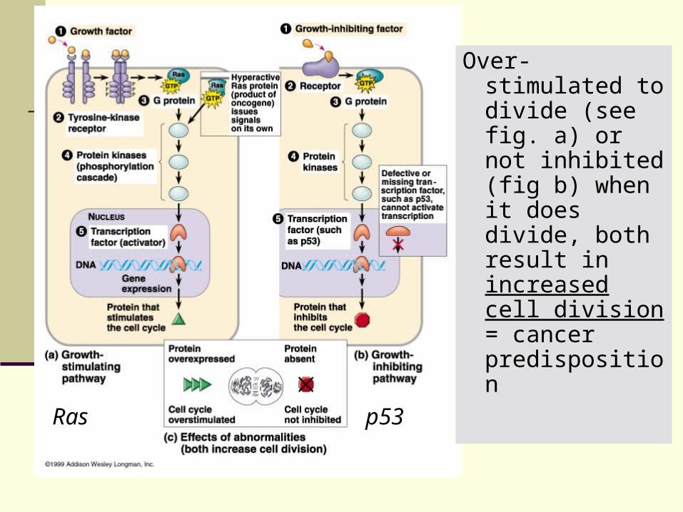
Over-stimulated to divide (see fig. a) or not inhibited (fig b) when it does divide, both result in increased cell division = cancer predisposition
Ras p53
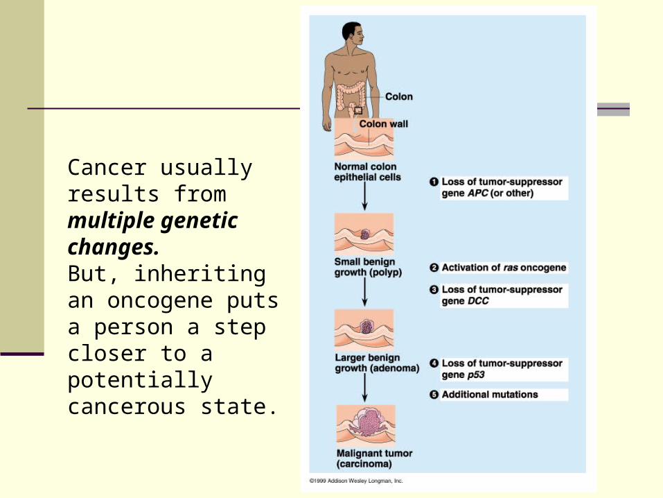
Cancer usually results from multiple genetic changes. But, inheriting an oncogene puts a person a step closer to a potentially cancerous state.
Chapter 20Genetic/DNA technologyDifferent uses and
techniques
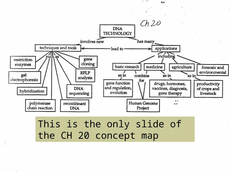
This is the only slide of the CH 20 concept map
Ch 20 cDNA
This means Complementary DNA Can be used to make DNA from a finished
(introns already cut out) piece of mRNA. This would be important for the insertion of
eukaryotic genes into bacteria (as in lab 6A, the pGLO gene)
Since prokaryotes do not recognize introns, the DNA which is complementary to the mRNA must be used.
Ch 20 Nucleic acid probes
Used to identify which colonies or samples of DNA contain a desired gene.
A radioactive DNA Hybird is made (a single strand of a portion of DNA that is the desired gene, or part of it)
If it binds to a sample of denatured (untwisted and unwound) DNA then you know the gene is in that sample. (fig 20.4)
Ch 20 PCR, polymerase chain reaction
Used to make more DNA if only a small sample is obtained. (many times this technique is used for crime scene DNA evidence)
The DNA is carefully heated, to make it separate into single strands, then it is cooled
Special DNA Primers are added to the solution and the corresponding bases align with the help of DNA polymerase = copying of the DNA. Done over & over to get a larger sample.
Ch 20 Restriction Enzymes
Are used to cut DNA at specific sites (at palindromes like MOM or A MAN, A PLAN, A CANAL, PANAMA or RACECAR)
These are used in Gel Electrophoresis and also in cloning, inserting genes into plasmids or bacterial chromosomes—since you get “sticky ends”
These were first discovered in bacteria, bac. used them to cut up foreign DNA from viruses or other bacteria.
Ch 20 Gel Electrophoresis
A solution of DNA pieces (which were cut by restriction enzymes) is carefully pipetted into a thick gel layer.
An electric current is passed through the gel. DNA is negatively charged (because of the phosphate
groups), it is attracted to the + electrode. The fragments separate based upon their size. Small fragments move further than the long fragments. RFLP: restriction fragment length polymorphisms, the
analysis of the various lengths of DNA in a gel, this is the DNA fingerprint = (RFLP).