Embed Size (px)
Citation preview

Chapter 19:
Anthropometric Measurementsand Vital Signs

Chapter Objectives
Cognitive Domain
Note: AAMA/CAAHEP 2008 Standards are italicized.
1. Spell and define key terms 2. Explain the procedures for measuring a patient’s height and
weight 3. Identify and describe the types of thermometers 4. Compare the procedures for measuring a patient’s
temperature using the oral, rectal, axillary, and tympanic methods
5. List the fever process, including the stages of fever

Chapter Objectives (cont’d.)
6. Describe the procedure for measuring a patient’s pulse and respiratory rates
7. Identify the various sites on the body used for palpating a pulse
8. Define Korotkoff sounds and the five phases of blood pressure
9. Identify factors that may influence the blood pressure 10. Explain the factors to consider when choosing the correct
blood pressure cuff size 11. Discuss implications for disease and disability when
homeostasis is not maintained

Chapter Objectives (cont’d.)
Psychomotor Domain
Note: AAMA/CAAHEP 2008 Standards are italicized.
1. Measure and record a patient’s weight 2. Measure and record a patient’s height 3. Measure and record a patient’s oral temperature using a
glass mercury thermometer 4. Measure and record a patient’s rectal temperature 5. Measure and record a patient’s axillary temperature 6. Measure and record a patient’s temperature using an
electronic thermometer
Chapter Objectives (cont’d.)
7. Measure and record a patient’s temperature using a tympanic thermometer
8. Measure and record a patient’s temperature using a temporal artery thermometer
9. Measure and record a patient’s radial pulse 10. Measure and record a patient’s respirations 11. Measure and record a patient’s blood pressure 12. Obtain vital signs 13. Practice standard precautions 14. Document accurately in the patient record

Chapter Objectives (cont’d.)
Affective Domain
Note: AAMA/CAAHEP 2008 Standards are italicized.
1. Apply critical thinking skills in performing patient assessment and care
2. Demonstrate respect for diversity in approaching patients and families
3. Explain rationale for performance of a procedure to the patient
4. Apply active listening skills 5. Demonstrate empathy in communicating with patients,
family, and staff

Chapter Objectives (cont’d.)
6. Use appropriate body language and other nonverbal skills in communicating with patients, family, and staff
7. Demonstrate awareness of the territorial boundaries of the person with whom you are communicating
8. Demonstrate sensitivity appropriate to the message being delivered
9. Demonstrate recognition of the patient’s level of understanding communications
10. Recognize and protect personal boundaries in communicating with others
11. Demonstrate respect for individual diversity, incorporating awareness of one’s own biases in areas including gender, race, religion, age, and economic status

Chapter Objectives (cont’d)
ABHES Competencies
1. Take vital signs 2. Document accurately

Vital signs (cardinal signs) measured and recorded by the medical assistant include the temperature, pulse rate, respiratory rate, and blood pressure.
Anthropometric measurements include height and weight.
Measurements taken at the first visit are recorded as baseline data and are used as reference points for comparison during subsequent visits.
anthropometric: pertaining to measurements of the human body
Back to chapter objectives
baseline: original or initial measure with which other measurements will be compared
cardinal signs: usually, vital signs; signifies their importance in assessment
Introduction

Anthropometric Measurements
Weight Taken every visit — prenatal,
infants/children, older adults Types of scales:
o Balance beam, digital, dial
o Pounds or kilograms
Figure 19-1 The three types of scales used in medical offices include the digital, dial, and balance beam scale.
Back to chapter objectives
Weight may be measured in pounds or kilograms, depending upon the preference of the physician and the type of scale in the medical office.
Anthropometric Measurements (cont’d.)
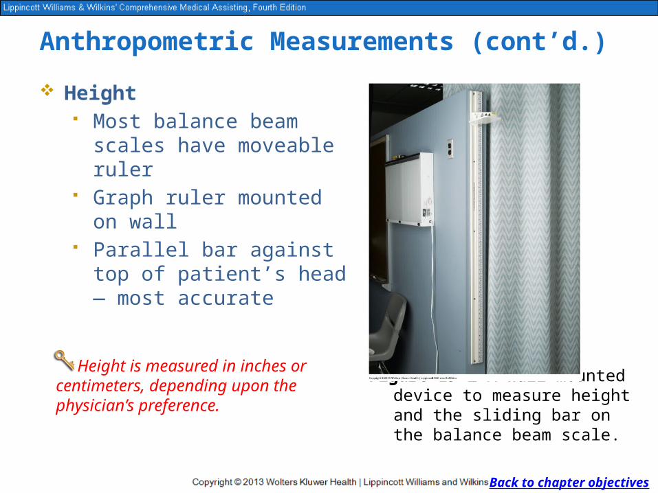
Height Most balance beam scales
have moveable ruler Graph ruler mounted on wall Parallel bar against top of
patient’s head — most accurate
Figure 19-2 A wall-mounted device to measure height and the sliding bar on the balance beam scale.
Back to chapter objectives
Height is measured in inches or centimeters, depending upon thephysician’s preference.

Checkpoint Question
Why is it important to accurately measure vital signs at every patient visit?
Back to chapter objectives

Checkpoint Question
Answer: Accurately measuring vital signs assists the physician in diagnosing and treating various disorders.
Back to chapter objectives

Vital Signs
Temperature Produced through metabolism and
muscle movement Heat lost through: Respiration Elimination Conduction through skin Normal = 98.6° Fahrenheit or 37°
Celsius Normal = afebrile Above normal = febrile
Back to chapter objectives
Body temperature reflects a balance between heat produced and heat lost by the body.
afebrile: body temperature not elevated above normal
febrile: having an above-normal body temperature

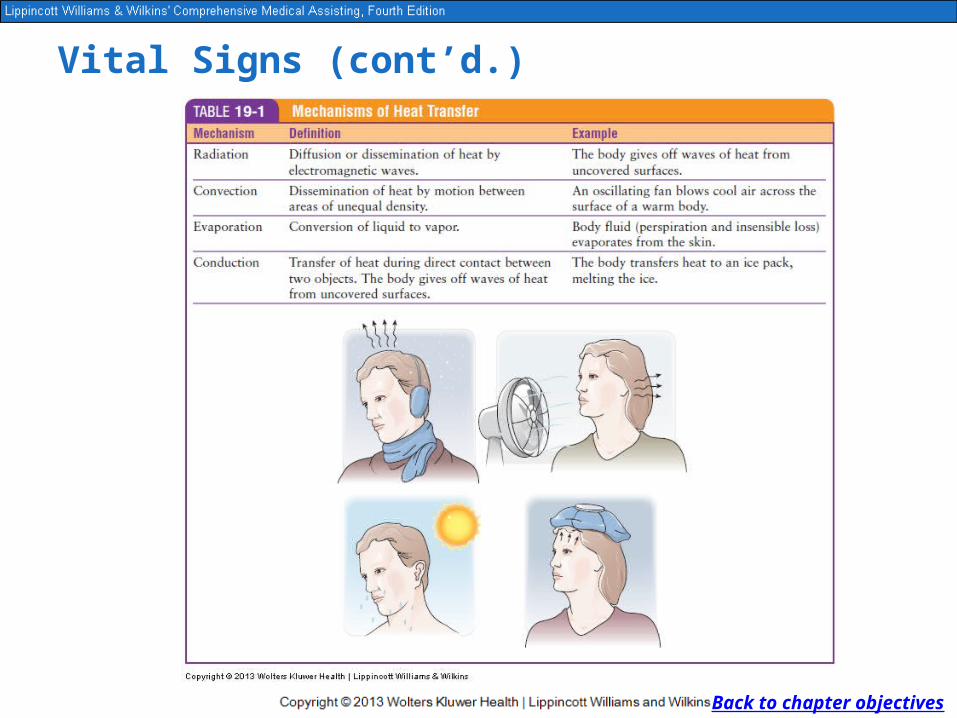
Figure 19-3 Factors affecting the balance between heat loss and heat production.
Vital Signs (cont’d.)
Back to chapter objectives
Vital Signs (cont’d.)
Back to chapter objectives

Vital Signs (cont’d.)
Back to chapter objectives
Temperature can be measured by oral, rectal, axillary, or tympanic method
Oral most common Tympanic prevalent in pediatric
offices New type—temporal artery
thermometer
Thermometers are used to measure body temperature using either the Fahrenheit or Celsius scale.
Figure 19-4 A temporal artery scanning thermometer.
Vital Signs (cont’d.)
Back to chapter objectives
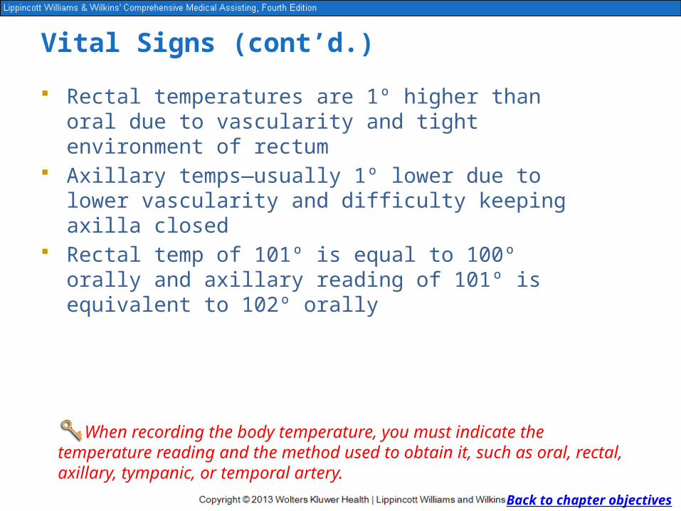
Rectal temperatures are 1º higher than oral due to vascularity and tight environment of rectum
Axillary temps—usually 1º lower due to lower vascularity and difficulty keeping axilla closed
Rectal temp of 101º is equal to 100º orally and axillary reading of 101º is equivalent to 102º orally
When recording the body temperature, you must indicate the temperature reading and the method used to obtain it, such as oral, rectal, axillary, tympanic, or temporal artery.
Checkpoint Question
How does an oral temperature measurement differ from a rectal measurement? Why?
Back to chapter objectives
Checkpoint Question
Answer: Rectal temperature measurements are usually 1° higher than oral measurements because of the vascularity and tightly closed environment of the rectum.
Back to chapter objectives

Vital Signs (cont’d.)
Fever Processes Temperature regulated by hypothalamus Balance between heat produced and heat lost Factors affecting temperature
o Age — children higher, older adults loweroGender — women higher o Exercise — highero Time of day — early morning lowero Emotion — stress higher, depression lowero Illness — elevation can be a sign of illness
Back to chapter objectives
Temperature elevations and variations are often a sign of disease but are not diseases in themselves.

Vital Signs (cont’d.)
Stages of Fever Often related to bacterial or viral infection Types Pyrexia: 101°F+ oral or 102°F+ rectal Hyperpyrexia: 105°F–106°F
Back to chapter objectives
An elevated temperature, or fever, usually results from a disease process, such as a bacterial or viral infection.
pyrexia: body temperature of 102°F or higher rectally or 101°F or higher orally
hyperpyrexia: dangerously high temperature, 105° to 106°F

Vital Signs (cont’d.)
Onset: rapid or gradual Course:
o Sustainedo Remittento Intermittento Relapsing
Resolution: o Crisis — abrupto Lysis — gradual
Back to chapter objectives
remittent fever: fluctuating
intermittent fever: occurring at intervals
sustained fever: fever that is constant or not fluctuating
relapsing fever: fever that returns after extended periods of being within normal limits

Vital Signs (cont’d.)
Back to chapter objectives

Checkpoint Question
Explain why the body temperature of a young child may be different from that of an adult.
Back to chapter objectives

Checkpoint Question
Answer: A child’s body temperature may be slightly higher than an adult’s because of the faster metabolism in a child.
Back to chapter objectives
Vital Signs (cont’d.)
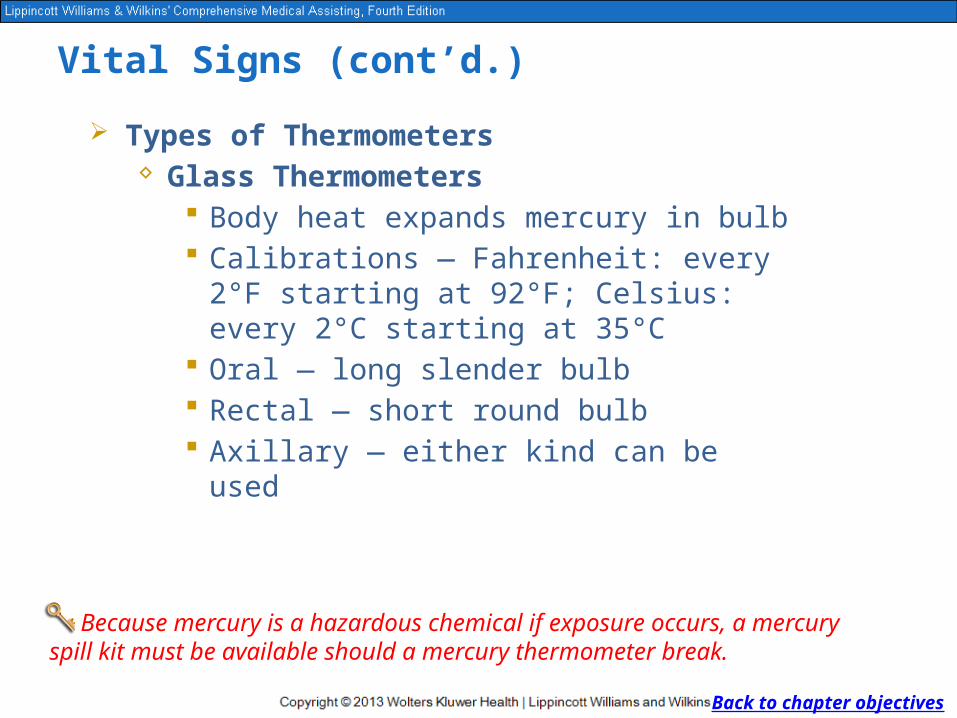
Types of Thermometers Glass Thermometers
Body heat expands mercury in bulb Calibrations — Fahrenheit: every 2°F starting
at 92°F; Celsius: every 2°C starting at 35°C Oral — long slender bulb Rectal — short round bulb Axillary — either kind can be used
Back to chapter objectives
Because mercury is a hazardous chemical if exposure occurs, a mercury spill kit must be available should a mercury thermometer break.

Vital Signs (cont’d.)
Back to chapter objectives
Figure 19-5 Glass mercury thermometers. Front: Slender bulb, oral. Center: Rounded bulb, red tip, rectal. Back: Blue tip, oral.

Vital Signs (cont’d.)
Before using glass thermometer, place in disposable, clear plastic sheath
Remove thermometer from patient, remove sheath by pulling thermometer out — turns sheath inside out
Traps saliva inside Dispose of sheath in biohazard container Sanitize and disinfect thermometer Typically washing in warm soapy water and soaking in
70% isopropyl alcohol
Back to chapter objectives
Glass thermometers may be reused if properly disinfected between patients.

Vital Signs (cont’d.)
Back to chapter objectives
Figure 19-6 The two glass thermometers on the top are calibrated in the Celsius (centigrade) scale, and the two on the bottom use the Fahrenheit scale. Note the blunt bulb on the rectal thermometers and the long thin bulb on the oral thermometers.
Vital Signs (cont’d.)
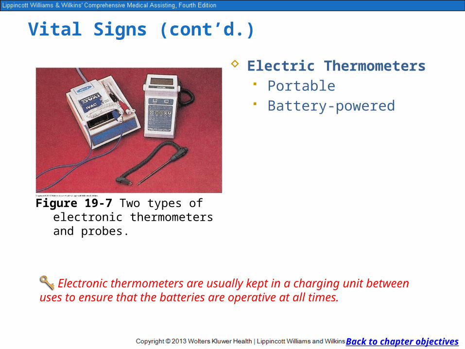
Electric Thermometers Portable Battery-powered
Back to chapter objectives
Electronic thermometers are usually kept in a charging unit between uses to ensure that the batteries are operative at all times.
Figure 19-7 Two types of electronic thermometers and probes.

Vital Signs (cont’d.)
Tympanic Thermometers For ear — relies on infrared
light bounced off tympanic membrane
Use increasing — accuracy like oral but less invasive
Back to chapter objectives
When correctly positioned in the ear, the sensor in the thermometer determines the temperature of the blood in the tympanic membrane.
Figure 19-8 The tympanic thermometer in use.

Vital Signs (cont’d.)
Temporal Artery Thermometers Upon release of on-off button temperature
immediately recorded Read manufacturer’s instructions carefully
Back to chapter objectives
Depending on the brand and type of temporal artery thermometer purchased, you should read the manufacturer’s instructions carefully for proper use and care of the unit.

Vital Signs (cont’d.)
Disposable Thermometers Single use Not as reliable
Back to chapter objectives
These thermometers are not reliable for definitive measurement, but they are acceptable for screening in settings such as day care centers and schools.
Figure 19-9 Disposable paper thermometer. The dots change color to indicate the body temperature.

Checkpoint Question
How is the reading displayed on an electronic, tympanic, and temporal artery thermometer?
Back to chapter objectives

Checkpoint Question
Answer: The electronic, tympanic, and temporal artery thermometers have digital display screens that show the obtained temperature.
Back to chapter objectives

Vital Signs (cont’d.)
Pulse Pumping of blood causes
expansion and contraction of arteries — heart beat
Techniques:o Feel — palpateo Hear — auscultateo Doppler
Back to chapter objectives
The heartbeat can be palpated (felt) or auscultated (heard) at several pulse points.
palpation: technique in which the examiner feels the texture, size, consistency, and location of parts of the body with the hands
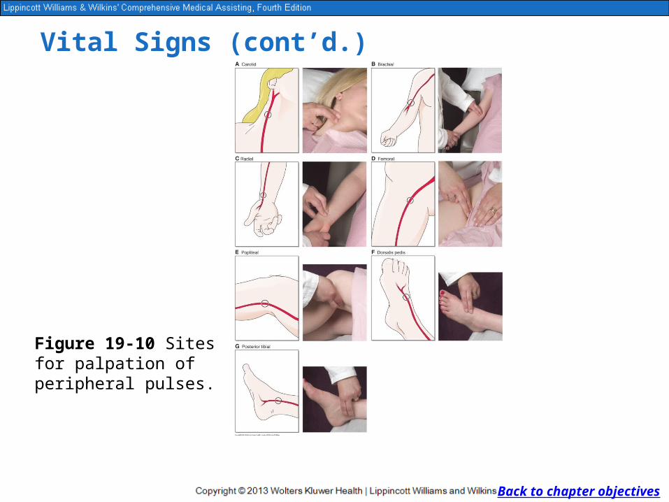
Figure 19-10 Sites for palpation of peripheral pulses.
Vital Signs (cont’d.)
Back to chapter objectives

Vital Signs (cont’d.)
Palpation techniqueo Place middle and index finger,
middle and ring, or all three against pulse point
o Do not use thumbo Radial artery most used
Back to chapter objectives
Figure 19-11 Measuring a radial pulse.

Vital Signs (cont’d.)
Auscultation techniqueo Place bell of stethoscope over
apex of hearto Alternative for pulse rate if
radial artery hard to palpate
Back to chapter objectives
Figure 19-12 Measuring an apical pulse.
Vital Signs (cont’d.)
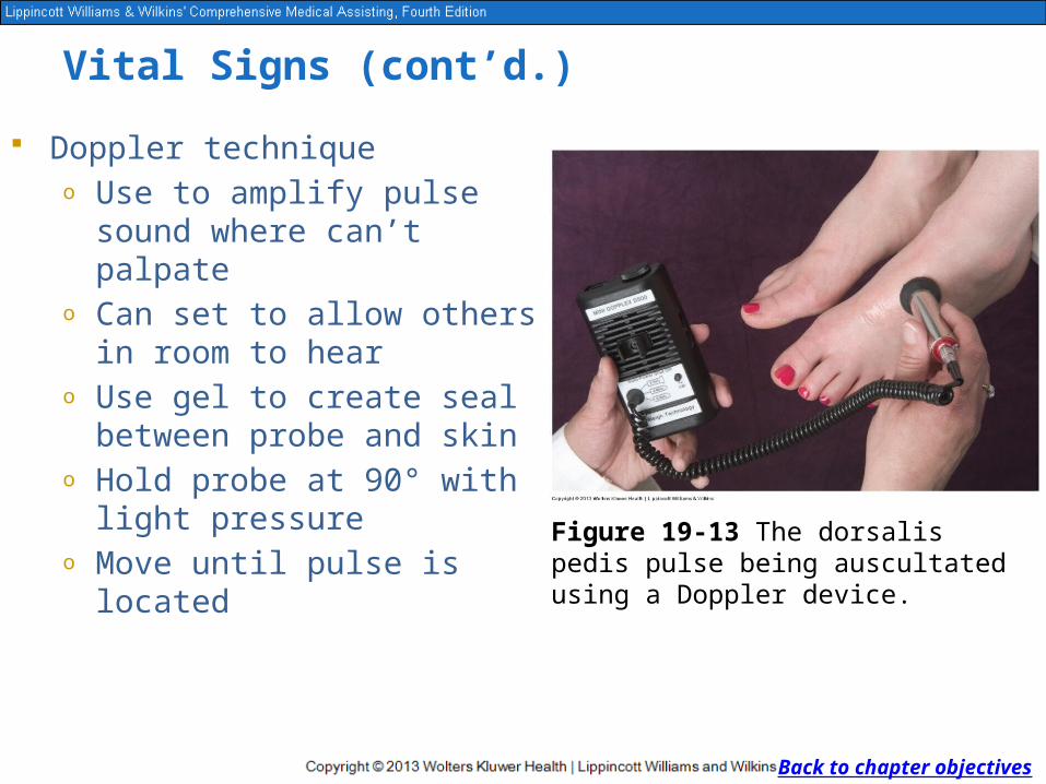
Doppler techniqueo Use to amplify pulse sound
where can’t palpateo Can set to allow others in room
to hearo Use gel to create seal between
probe and skino Hold probe at 90° with light
pressureo Move until pulse is located
Back to chapter objectives
Figure 19-13 The dorsalis pedis pulse being auscultated using a Doppler device.

Vital Signs (cont’d.)
Pulse Characteristics Rate — can vary with
age or other factors Rhythm — normal is
even = consistent time between pulses
Volume — strength/force of heartbeat
Back to chapter objectives
Volume, the strength or force of the heartbeat, can be described as soft, bounding, weak, thready, strong, or full.
The rhythm is the interval between each heartbeat or the pattern of beats.
The rate is the number of heartbeats in 1 minute. In healthy adults, the average pulse rate is 60 to 100 beats per minute.
Vital Signs (cont’d.)
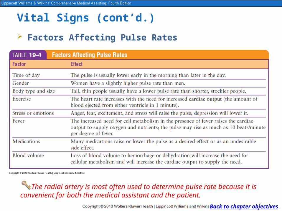
Factors Affecting Pulse Rates
Back to chapter objectives
The radial artery is most often used to determine pulse rate because it is convenient for both the medical assistant and the patient.

Checkpoint Question
What characteristics of a patient’s pulse should be assessed, and how should they be recorded in the medical record?
Back to chapter objectives

Checkpoint Question
Answer: Measuring a patient’s pulse entails assessing and recording the rate (number of heartbeats in 1 minute), rhythm (regular or irregular), and volume (thready, bounding).
Back to chapter objectives

Vital Signs (cont’d.)
Respiration Inspiration — contract diaphragm, breathe oxygen in Expiration — relax diaphragm, breathe carbon
dioxide out Respiration — one full inspiration and expiration
o Count for 1 minuteo During pulse measuremento Count without patient knowledge; rate can be
changed voluntarily
Back to chapter objectives
Respiration is the exchange of gases between the atmosphere and the blood in the body.
Observing the rise and fall of the chest to count respirations is usually performed as a part of the pulse measurement.

Vital Signs (cont’d.)
Back to chapter objectives
Figure 19-14 The apical pulse is found at the 5th intercostal space at the midclavicular line.
Vital Signs (cont’d.)
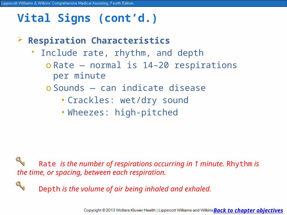
Respiration Characteristics Include rate, rhythm, and depth
o Rate — normal is 14–20 respirations per minuteo Sounds — can indicate disease
• Crackles: wet/dry sound• Wheezes: high-pitched
Back to chapter objectives
Rate is the number of respirations occurring in 1 minute. Rhythm is the time, or spacing, between each respiration.
Depth is the volume of air being inhaled and exhaled.

Vital Signs (cont’d.) Factors Affecting Respiration
Factorso Ageo Elevated body temperature
Abnormal respirationso Tachypnea: faster rateo Bradypnea: slower rateo Dyspnea: difficulty breathingo Apnea: no respirationso Hyperpnea: deeper/gaspingo Hypopnea: shalloweroOrthopnea: unable to breathe lying downo Hyperventilation: rate exceeds oxygen demand
Back to chapter objectives
In healthy adults, the average respiratory rate is 14 to 20 breaths per minute.

Checkpoint Question
What happens within the chest cavity when the diaphragm contracts?
Back to chapter objectives
Checkpoint Question
Answer: Contraction of the diaphragm causes negative pressure in the lungs, which respond by filling with inhaled air.
Back to chapter objectives

Vital Signs (cont’d.)
Blood Pressure Blood pressure recorded as
systolic/diastolic Measurements in millimeters
of mercury (mm Hg) Average adult = 120/80 Athletes can be lower
Back to chapter objectives
Blood pressure is a measurement of the pressure of the blood in an artery as it is forced against the arterial walls.
cardiac cycle: period from the beginning of one heartbeat to the beginning of the next; includes systole and diastole
systole: contraction phase of the cardiac cycle
diastole: relaxation phase of the cardiac cycle

Vital Signs (cont’d.)
Measured with sphygmomanometer — blood pressure cuffo Aneroid: dialo Mercury: column
Back to chapter objectives
Although only one type of cuff actually contains mercury, both types are calibrated and measure blood pressure in millimeters of mercury (mm Hg).
sphygmomanometer: device used to measure blood pressure
postural hypotension: sudden drop in blood pressure upon standing

Vital Signs (cont’d.)
Back to chapter objectives
Figure 19-15 A mercury column sphygmomanometer and an aneroid sphygmomanometer.
Vital Signs (cont’d.)
Back to chapter objectives

Checkpoint Question
What is happening to the heart during systole? During diastole?
Back to chapter objectives

Checkpoint Question
Answer: During systole, the heart contracts and forces blood out and through the arteries. In diastole, the heart relaxes and fills with blood.
Back to chapter objectives

Vital Signs (cont’d.)
Korotkoff Sounds Only sounds heard during
phase I (first sound heard) and phase V (last sound heard) are recorded as blood pressure
Not necessary to record other Korotkoff sounds
Back to chapter objectives
Korotkoff sounds can be classified into five phases of sounds heard while auscultating the blood pressure as described by the Russian neurologist Nicolai Korotkoff.

Vital Signs (cont’d.)
Pulse Pressure Average adult blood pressure = 120/80; 120 − 80 = 40 Average normal range for pulse pressure = 30 to 50
mm Hg Pulse pressure should be no more than one-third of
the systolic reading
Back to chapter objectives
The difference between the systolic and diastolic readings is known as the pulse pressure.
Vital Signs (cont’d.)
Auscultatory Gap Heard during phase II in
hypertensive patients Loss of sounds or drop of
pressure 30 mm Hg or more while cuff deflates
Can cause errors in blood pressure readings so must watch dial/column
Back to chapter objectives
An auscultatory gap is the loss of any sounds for a drop of up to 30 mm Hg (sometimes more) during the release of air from the blood pressure cuff after the first sound is heard.
hypertension: morbidly high blood pressure

Vital Signs (cont’d.)
Factors Influencing Blood Pressure General health
o Diet, alcohol, tobacco use, exercise, family history, previous cardiac conditions
o Atherosclerosis and arteriosclerosis — affect size and elasticity of arteries
Back to chapter objectives
Atherosclerosis and arteriosclerosis are two disease processes that greatly influence blood pressure.
Vital Signs (cont’d.)
o Other:• Age: Older — higher• Activity: Exercise — higher• Stress: Fight or flight — higher• Body position: Supine — lower• Medications • Hypertension• Errors in blood pressure readings
Back to chapter objectives
Vital Signs (cont’d.)
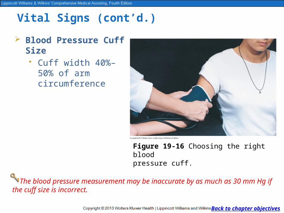
Blood Pressure Cuff Size Cuff width 40%–50%
of arm circumference
Back to chapter objectives
The blood pressure measurement may be inaccurate by as much as 30 mm Hg if the cuff size is incorrect.
Figure 19-16 Choosing the right blood pressure cuff.
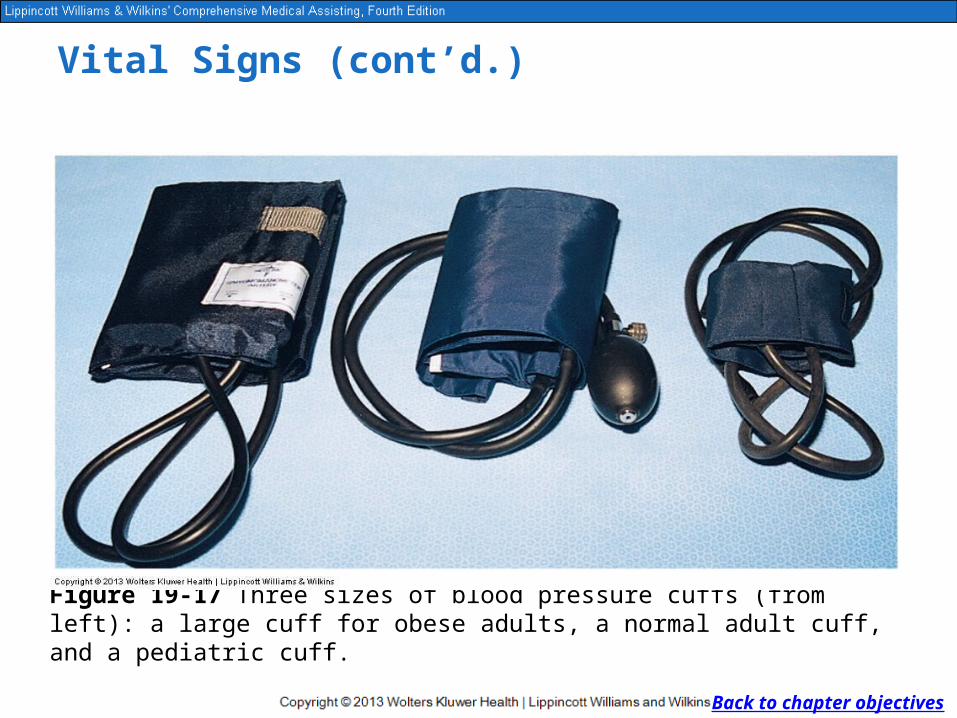
Figure 19-17 Three sizes of blood pressure cuffs (from left): a large cuff for obese adults, a normal adult cuff, and a pediatric cuff.
Vital Signs (cont’d.)
Back to chapter objectives

Checkpoint Question
How are the pulse pressure and the auscultatory gap different?
Back to chapter objectives
Checkpoint Question
Answer: The pulse pressure is the difference between the systolic and diastolic blood pressures, and the auscultatory gap is an abrupt, but temporary, end to the tapping sound heard when auscultating the blood pressure.
Back to chapter objectives