Embed Size (px)
Citation preview
227
Bettina Warscheid (ed.), Stable Isotope Labeling by Amino Acids in Cell Culture (SILAC): Methods and Protocols, Methods in Molecular Biology, vol. 1188, DOI 10.1007/978-1-4939-1142-4_16, © Springer Science+Business Media New York 2014
Chapter 16
Analyzing the Protein Assembly and Dynamics of the Human Spliceosome with SILAC
Carla Schmidt , Monika Raabe , Reinhard Lührmann , and Henning Urlaub
Abstract
Quantitative mass spectrometry has become an indispensable tool in proteomic studies. Numerous methods are available and can be applied to approach different issues. In most studies these issues include the quantitative comparison of different cell states, the identifi cation of specifi c interaction partners or determining degrees of posttranslational modifi cation. In this chapter we describe a SILAC-based quanti-fi cation in order to analyze dynamic protein changes during the assembly of the human spliceosome on a pre- mRNA in vitro. We provide protocols for assembly of spliceosomes on pre-mRNA (including genera-tion of pre-mRNAs and preparation of nuclear extracts), quantitative mass spectrometry (SILAC labeling, sample preparation), and data analysis to generate timelines for the dynamic protein assembly.
Key words Spliceosome , Metabolic labeling , Assembly timelines , Protein dynamics
1 Introduction
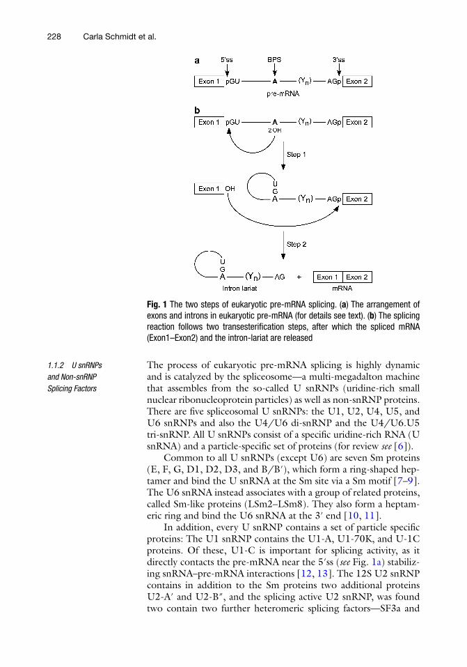
Eukaryotic pre-mRNAs consist of protein-coding sequences (exons) and noncoding sequences (introns). The introns are defi ned by very short, conserved sequences at the 5′ and 3′ splice sites (i.e., the exon/intron and intron/exon junctions) as well as the branch-point site, which contains a conserved adenosine (branch-point adenosine) and in most cases a polypyrimidine tract (Y n ; Fig. 1a ).
During pre-mRNA splicing, the introns are excised and the exons are ligated to yield mature mRNA. This proceeds by two consecutive transesterifi cation reactions (Fig. 1b ): First, the 2′ hydroxyl group of the branch-point adenosine attacks the phos-phodiester bond at the 5′ splice site (5′ss) resulting in a phospho-diester bond between the branch-point adenosine and the fi rst nucleotide of the intron. In the second step of splicing, the free 3′ hydroxyl group of exon1 attacks the phosphodiester bond of the 3′ splice site (3′ss); in this way, exon 1 and exon 2 become ligated and the intron is released in the form of a lariat (Fig. 1b ) [ 1 – 5 ].
1.1 The Spliceosome
1.1.1 Eukaryotic Pre-mRNAs
228
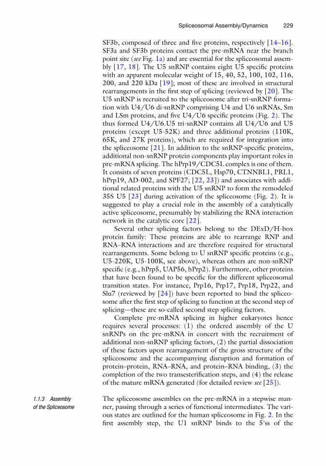
The process of eukaryotic pre-mRNA splicing is highly dynamic and is catalyzed by the spliceosome—a multi-megadalton machine that assembles from the so-called U snRNPs (uridine-rich small nuclear ribonucleoprotein particles) as well as non-snRNP proteins. There are fi ve spliceosomal U snRNPs: the U1, U2, U4, U5, and U6 snRNPs and also the U4/U6 di-snRNP and the U4/U6.U5 tri-snRNP. All U snRNPs consist of a specifi c uridine-rich RNA (U snRNA) and a particle-specifi c set of proteins (for review see [ 6 ]).
Common to all U snRNPs (except U6) are seven Sm proteins (E, F, G, D1, D2, D3, and B/B′), which form a ring-shaped hep-tamer and bind the U snRNA at the Sm site via a Sm motif [ 7 – 9 ]. The U6 snRNA instead associates with a group of related proteins, called Sm-like proteins (LSm2–LSm8). They also form a heptam-eric ring and bind the U6 snRNA at the 3′ end [ 10 , 11 ].
In addition, every U snRNP contains a set of particle specifi c proteins: The U1 snRNP contains the U1-A, U1-70K, and U-1C proteins. Of these, U1-C is important for splicing activity, as it directly contacts the pre-mRNA near the 5′ss ( see Fig. 1a ) stabiliz-ing snRNA–pre-mRNA interactions [ 12 , 13 ]. The 12S U2 snRNP contains in addition to the Sm proteins two additional proteins U2-A′ and U2-B″, and the splicing active U2 snRNP, was found two contain two further heteromeric splicing factors—SF3a and
1.1.2 U snRNPs and Non-snRNP Splicing Factors
Fig. 1 The two steps of eukaryotic pre-mRNA splicing. ( a ) The arrangement of exons and introns in eukaryotic pre-mRNA (for details see text). ( b ) The splicing reaction follows two transesterifi cation steps, after which the spliced mRNA (Exon1–Exon2) and the intron-lariat are released
Carla Schmidt et al.
229
SF3b, composed of three and fi ve proteins, respectively [ 14 – 16 ]. SF3a and SF3b proteins contact the pre-mRNA near the branch point site ( see Fig. 1a ) and are essential for the spliceosomal assem-bly [ 17 , 18 ]. The U5 snRNP contains eight U5 specifi c proteins with an apparent molecular weight of 15, 40, 52, 100, 102, 116, 200, and 220 kDa [ 19 ]; most of these are involved in structural rearrangements in the fi rst step of splicing (reviewed by [ 20 ]. The U5 snRNP is recruited to the spliceosome after tri- snRNP forma-tion with U4/U6 di-snRNP comprising U4 and U6 snRNAs, Sm and LSm proteins, and fi ve U4/U6 specifi c proteins (Fig. 2 ). The thus formed U4/U6.U5 tri-snRNP contains all U4/U6 and U5 proteins (except U5-52K) and three additional proteins (110K, 65K, and 27K proteins), which are required for integration into the spliceosome [ 21 ]. In addition to the snRNP- specifi c proteins, additional non-snRNP protein components play important roles in pre-mRNA splicing. The hPrp19/CDC5L complex is one of them. It consists of seven proteins (CDC5L, Hsp70, CTNNBL1, PRL1, hPrp19, AD-002, and SPF27; [ 22 , 23 ]) and associates with addi-tional related proteins with the U5 snRNP to form the remodeled 35S U5 [ 23 ] during activation of the spliceosome (Fig. 2 ). It is suggested to play a crucial role in the assembly of a catalytically active spliceosome, presumably by stabilizing the RNA interaction network in the catalytic core [ 22 ].
Several other splicing factors belong to the DExD/H-box protein family: These proteins are able to rearrange RNP and RNA–RNA interactions and are therefore required for structural rearrangements. Some belong to U snRNP specifi c proteins (e.g., U5-220K, U5-100K, see above), whereas others are non-snRNP specifi c (e.g., hPrp5, UAP56, hPrp2). Furthermore, other proteins that have been found to be specifi c for the different spliceosomal transition states. For instance, Prp16, Prp17, Prp18, Prp22, and Slu7 (reviewed by [ 24 ]) have been reported to bind the spliceo-some after the fi rst step of splicing to function at the second step of splicing—these are so-called second step splicing factors.
Complete pre-mRNA splicing in higher eukaryotes hence requires several processes: (1) the ordered assembly of the U snRNPs on the pre-mRNA in concert with the recruitment of additional non-snRNP splicing factors, (2) the partial dissociation of these factors upon rearrangement of the gross structure of the spliceosome and the accompanying disruption and formation of protein–protein, RNA–RNA, and protein–RNA binding, (3) the completion of the two transesterifi cation steps, and (4) the release of the mature mRNA generated (for detailed review see [ 25 ]).
The spliceosome assembles on the pre-mRNA in a stepwise man-ner, passing through a series of functional intermediates. The vari-ous states are outlined for the human spliceosome in Fig. 2 . In the fi rst assembly step, the U1 snRNP binds to the 5′ss of the
1.1.3 Assembly of the Spliceosome
Spliceosomal Assembly/Dynamics
230
pre- mRNA, forming the E (“early”) complex [ 12 , 26 ]. The recruit-ment of U2 snRNP leads then to formation of the A complex, which is also called the pre-spliceosome [ 27 , 28 ]. Upon integration of the U4/U6.U5 tri-snRNP and additional splicing factors, called B specifi c proteins, the pre-catalytic spliceosome (B complex) is developed [ 29 ]. Structural RNA and protein rearrangements within the B complex induced by RNA helicases Brr2 (U5-200K) and Snu114 (U5-116K) cause the dissociation of U1 and U4 snRNPs. Dissociation of U1 and U4 together with U4/U6 specifi c proteins and remodeling of U5 initiated by the binding of the hPrp19/CDC5L complex generate the activated spliceosome (B*),
Fig. 2 The stepwise assembly of the spliceosome during pre-mRNA splicing. First, U1 snRNP binds to the 5′ss forming the E complex followed by binding of U2 snRNP (A complex formation). Recruitment of the pre-assembled tri-snRNP (U4/U6.U5) leads to formation of the pre-catalytic B complex. Upon structural rearrangements U1 and U4 snRNPs dissociate and incorporation of the hPrp19/CDC5L complex leads to remodeling of U5 generating the activated B complex. The fi rst step of splicing occurs in this intermediate assembly yielding the C complex, in which the second step of splicing is carried out. The generated mRNA and the post-spliceosomal complex are released and the splicing factors are reconstituted. In addition to the protein complex hPrp19/CDC5L, numerous non-snRNP specifi c proteins, not shown here, join and leave the spliceosome at various points during the cycle
Carla Schmidt et al.
231
in which the fi rst catalytic step of splicing occurs [ 30 , 31 ]. The complex that forms during this process is the catalytically active C complex, which goes on to perform the second step of splicing for which the second step splicing factors (see above) are required [ 32 ]. The fi nal steps are the release of the mature mRNA product, dissociation of the post-spliceosomal intron complex, and recycling of the splicing factors (Fig. 2 ).
So far, only few studies have applied quantitative mass spectrome-try combined with metabolic labeling using stable isotopes to describe dynamic protein changes in ribonucleoprotein complexes. In a fi rst study, the SILAC strategy was applied to analyze the pro-teome of the nucleolus from differentially labeled cells after differ-ent durations of treatment [ 33 ]. The time-dependent composition profi les of protein subunits from RNA polymerase I, snRNPs and ribosomes were recorded [ 33 ]. In a similar manner, the assembly kinetics of the 30S ribosomal subunit of Escherichia coli have been studied by quantitative pulse-chase MS (PC/QMS). 15 N-labeled proteins were incubated with 16S rRNA and, after assembly had taken place for various times, chased with an excess of 14 N-labeled proteins. 30S subunits were then completely assembled and puri-fi ed, and the 15 N/ 14 N ratio in their proteins was used to reveal the binding kinetics [ 34 ]. In this chapter we describe the quantitative analysis of the dynamic protein changes that occur during pre- mRNA splicing by using stable isotope labeling and subsequent mass spectrometry.
The distinct assembly states of the human spliceosome (i.e., A, B, B*, and C complexes, see above) in vitro have been analyzed in previous studies and compared in a semi-quantitative manner to determine differences in their protein compositions [ 27 , 29 , 30 , 32 , 35 ]. However, this approach only monitors the quantitative changes of the protein composition during the transition of one purifi ed state of the spliceosomes to another. Yet no description of the dynamic protein changes that occur during assembly of pro-teins pre-mRNA splicing in a time dependent manner has been applied. We therefore used SILAC quantifi cation to monitor the protein assembly on a pre-mRNA in a time-dependent manner.
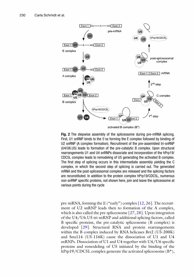
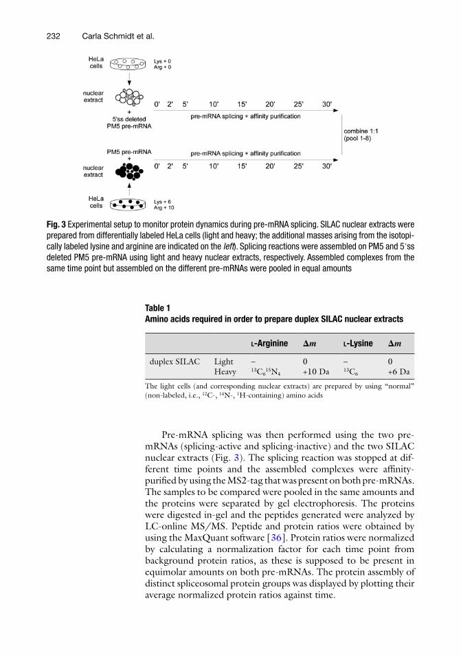
We used MS2-tagged PM5 pre-mRNA, which in previous studies had been successfully applied to purify catalytically active spliceosomes [ 32 ], and a splicing-inactive variant of this pre- mRNA, which was generated by deletion of the 5′ss. A direct com-parison was made between the assembled proteins on the splicing-active and on the splicing-inactive pre-mRNA at different time points during pre-mRNA splicing (Fig. 3 ).
For this purpose, we prepared HeLa nuclear extracts from differentially labeled HeLa cells (light and heavy SILAC cells; Table 1 ).
1.2 Analyzing Dynamic Protein Changes by Quantitative Mass Spectrometry
1.2.1 Protein Assembly and Dynamics of the Human Spliceosome
Spliceosomal Assembly/Dynamics
232
Pre-mRNA splicing was then performed using the two pre- mRNAs (splicing-active and splicing-inactive) and the two SILAC nuclear extracts (Fig. 3 ). The splicing reaction was stopped at dif-ferent time points and the assembled complexes were affi nity- purifi ed by using the MS2-tag that was present on both pre- mRNAs. The samples to be compared were pooled in the same amounts and the proteins were separated by gel electrophoresis. The proteins were digested in-gel and the peptides generated were analyzed by LC-online MS/MS. Peptide and protein ratios were obtained by using the MaxQuant software [ 36 ]. Protein ratios were normalized by calculating a normalization factor for each time point from background protein ratios, as these is supposed to be present in equimolar amounts on both pre-mRNAs. The protein assembly of distinct spliceosomal protein groups was displayed by plotting their average normalized protein ratios against time.
Fig. 3 Experimental setup to monitor protein dynamics during pre-mRNA splicing. SILAC nuclear extracts were prepared from differentially labeled HeLa cells (light and heavy; the additional masses arising from the isotopi-cally labeled lysine and arginine are indicated on the left ). Splicing reactions were assembled on PM5 and 5′ss deleted PM5 pre-mRNA using light and heavy nuclear extracts, respectively. Assembled complexes from the same time point but assembled on the different pre-mRNAs were pooled in equal amounts
Table 1 Amino acids required in order to prepare duplex SILAC nuclear extracts
L -Arginine Δ m L -Lysine Δ m
duplex SILAC Light – 0 – 0 Heavy 13 C 6 15 N 4 +10 Da 13 C 6 +6 Da
The light cells (and corresponding nuclear extracts) are prepared by using “normal” (non-labeled, i.e., 12 C-, 14 N-, 1 H-containing) amino acids
Carla Schmidt et al.
233
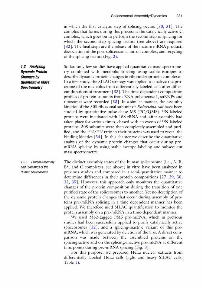
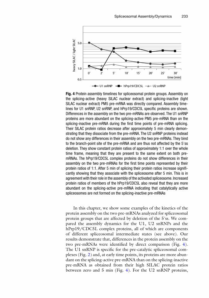
In this chapter, we show some examples of the kinetics of the protein assembly on the two pre-mRNAs analyzed for spliceosomal protein groups that are affected by deletion of the 5′ss. We com-pared the assembly dynamics for the U1, U2 snRNPs and the hPrp19/CDC5L complex proteins, all of which are components of different spliceosomal intermediate states (see above). Our results demonstrate that, differences in the protein assembly on the two pre-mRNAs were identifi ed by direct comparison (Fig. 4 ). The U1 snRNP is specifi c for the pre-catalytic spliceosomal com-plexes (Fig. 2 ) and, at early time points, its proteins are more abun-dant on the splicing-active pre-mRNA than on the splicing- inactive pre-mRNA as obtained from their high SILAC protein ratios between zero and 5 min (Fig. 4 ). For the U2 snRNP proteins,
Fig. 4 Protein assembly timelines for spliceosomal protein groups. Assembly on the splicing-active (heavy SILAC nuclear extract) and splicing-inactive (light SILAC nuclear extract) PM5 pre-mRNA was directly compared. Assembly time-lines for U1 snRNP, U2 snRNP, and hPrp19/CDC5L specifi c proteins are shown. Differences in the assembly on the two pre-mRNAs are observed. The U1 snRNP proteins are more abundant on the splicing-active PM5 pre-mRNA than on the splicing-inactive pre-mRNA during the fi rst time points of pre-mRNA splicing. Their SILAC protein ratios decrease after approximately 5 min clearly demon-strating that they dissociate from the pre-mRNA. The U2 snRNP proteins instead do not show any differences in their assembly on the two pre-mRNAs. They bind to the branch-point site of the pre-mRNA and are thus not affected by the 5′ss deletion. They show constant protein ratios of approximately 1:1 over the whole time frame, meaning that they are present to the same extent on both pre- mRNAs. The hPrp19/CDC5L complex proteins do not show differences in their assembly on the two pre-mRNAs for the fi rst time points represented by their protein ratios of 1:1. After 5 min of splicing their protein ratios increase signifi -cantly showing that they associate with the spliceosome after 5 min. This is in agreement with their role in the assembly of the activated spliceosome. Increased protein ratios of members of the hPrp19/CDC5L also reveal that they are more abundant on the splicing-active pre-mRNA indicating that catalytically active spliceosomes are not formed on the splicing-inactive pre-mRNAs
Spliceosomal Assembly/Dynamics
234
which are present in all intermediate states (Fig. 2 ), no differences were observed between the two pre-mRNAs (Fig. 4 ).
Interestingly, the hPrp19/CDC5L complex, which the spli-ceosome incorporates during its activation (see above and Fig. 2 ), assembles at later time points only on the splicing-active pre-mRNA; the SILAC protein ratios increased signifi cantly after 2 min of incubation for this group of proteins. Thus, our quantitative MS results demonstrate that the different protein groups indeed show different assembly kinetics, and they also show which proteins are affected by the deletion on the 5′ss. The timelines generated for the assembly of whole spliceosomal protein groups during pre-mRNA splicing thus contribute substantially toward gaining an understanding of this dynamic process.
2 Materials
1. HeLa S3 cells (wt). 2. DMEM, high glucose, w/o arginine, w/o lysine. 3. Dialyzed fetal bovine serum (FBS). 4. 100× penicillin–streptomycin. 5. 50 mg/l L -arginine, 50 mg/l 13 C 6 15 N 4 - L -arginine. 6. 50 mg/l L -lysine, 50 mg/l 13 C 6 - L -lysine. 7. 2.0 L spinner fl asks. 8. 2.5 L fermenter (bioreactor). 9. See also Notes 1 – 3 and Table 1 .
1. Phosphate-buffered saline (PBS): 130 mM NaCl, 0.2 mM K-PO 4 , ice-cold.
2. MC buffer: 10 mM HEPES–KOH pH 7.6, 10 mM KOAc, 0.5 mM Mg(OAc) 2 , ice-cold.
3. 0.25 M dithioerythritol (DTE). 4. EDTA-free protease inhibitor cocktail (Roche). 5. Roeder C buffer: 25 % (v/v) glycerol, 20 mM HEPES–KOH
pH 7.6, 420 mM NaCl, 1.5 mM MgCl 2 , 0.2 mM EDTA, ice-cold.
6. 0.1 M phenylmethylsulfonyl fl uoride (PMSF), dissolved in isopropanol ( see Note 4 ).
7. Roeder D buffer: 10 % (v/v) glycerol, 20 mM HEPES–KOH pH 7.6, 100 mM KCl, 1.5 mM MgCl 2 , 0.2 mM EDTA, 0.5 mM DTT (dithiothreitol), 0.5 mM PMSF, ice-cold.
8. Dounce homogenizer. 9. Dialysis tubing (MWCO 6,000–8,000 Da). 10. See also Note 5 .
2.1 SILAC Labeling of HeLa Cells
2.2 Preparation of SILAC HeLa Nuclear Extracts
Carla Schmidt et al.
235
1. Transcription-optimized 5× buffer. 2. 0.1 M ATP, 0.1 M UTP, 0.1 M CTP, 0.01 M GTP. 3. 32 P-αUTP. 4. m 7 GpppG cap (Kedar, Poland). 5. Stock solutions of MgCl 2 (1 M) and DTT (1 M). 6. 10 mg/ml BSA. 7. RNAsin (40 U/μl). 8. SP6 RNA polymerase (2 U/μl). 9. DNA template. 10. RNA extraction buffer: 20 mM Tris–HCl pH 7.5, 150 mM
NaCl, 0.5 % (w/v) SDS, 0.2 mM EDTA pH 8.0. 11. RQ1 DNase. 12. 5 % polyacrylamide gels containing 8 M urea. 13. X-ray fi lm. 14. Phenol–chloroform–isoamyl alcohol (25:24:1). 15. Chloroform. 16. 10 μg/μl glycogen. 17. 100 % (v/v) ethanol, ice-cold. 18. 80 % (v/v) ethanol, ice-cold. 19. 3 M sodium acetate (NaOAc) pH 5.3. 20. RNAse-free water. 21. Vortex mixer. 22. Vacuum centrifuge. 23. See also Notes 6 and 7 .
1. m 7 G(5′)ppp(5′)G-capped and MS2-tagged pre-mRNA ( 32 P-labeled and unlabeled).
2. MS2-MBP fusion protein [ 37 ]. 3. SILAC-labeled HeLa nuclear extracts. 4. Stock solutions of KCl (2 M), MgCl 2 (1 M), ATP (0.1 M),
creatine phosphate (0.5 M). 5. Scintillation counter. 6. See also Notes 8 – 10 .
1. Disposable chromatography columns. 2. Amylose resin. 3. 20 mM HEPES–KOH pH 7.6, 1.5 mM MgCl 2 , 150 mM
NaCl. 4. 50 mM maltose dissolved in 20 mM HEPES–KOH pH 7.6,
1.5 mM MgCl 2 , 150 mM NaCl.
2.3 Transcription of pre-mRNA
2.4 Splice Reaction
2.5 Affi nity Purifi cation of Assembled Spliceosomal Complexes
Spliceosomal Assembly/Dynamics
236
1. 100 % ethanol, ice-cold. 2. 80 % (v/v) ethanol, ice-cold. 3. 0.3 M NaOAc, pH 5.3. 4. NuPAGE 4–12 % Bis-Tris pre-cast gels, 4× sample buffer, 10×
reducing agent, 20× MOPS SDS running buffer, antioxidant (Life Technologies).
5. See also Note 11 .
1. Ultrapure water. 2. Acetonitrile (ACN). 3. 100 mM ammonium bicarbonate (NH 4 CO 3 ), pH 8.0. 4. 10 mM DTT in 100 mM NH 4 CO 3 . 5. 55 mM iodoacetamide (IAA) in 100 mM NH 4 CO 3 . 6. 5 % (v/v) formic acid (FA). 7. Trypsin (sequencing grade, 0.1 μg/μl). 8. Buffer 1: 50 μl H 2 O, 50 μl of 100 mM NH 4 CO 3 , 5 μl of
100 mM CaCl 2 , 15 μl trypsin. 9. Buffer 2: 50 μl H 2 O, 50 μl of 100 mM NH 4 CO 3 , 5 μl of
100 mM CaCl 2 . 10. Gel loader pipette tips. 11. Thermomixer (Eppendorf). 12. Vacuum centrifuge. 13. See also Notes 12 and 13 .
1. 1 % (v/v) FA (loading buffer). 2. See Note 14 .
1. Computer system (Intel Pentium III/800 MHz or higher, 2 GB RAM minimum).
2. MaxQuant software package. 3. GProx software platform. 4. See also Notes 15 and 16 .
3 Methods
1. Prepare custom-made DMEM containing the following ingredients ( see Table 1 for combinations of light and heavy L -arginine and L -lysine to obtain duplex SILAC medium): (a) 500 ml DMEM w/o arginine, w/o lysine. (b) 50 ml dialyzed FBS.
2.6 Quantifi cation and LC-MS Analysis
2.6.1 Mixing “Light” and “Heavy” Assembled Complexes and Gel Electrophoresis
2.6.2 In-Gel Hydrolysis of Proteins and Extraction of Peptides
2.6.3 LC-MS Analysis
2.7 Data Analysis
3.1 SILAC Labeling of HeLa Cells
Carla Schmidt et al.
237
(c) 5 ml of 100× penicillin–streptomycin. (d) 5.55 ml of 50 mg/l l-arginine. (e) 5.55 ml of 50 mg/l l-lysine.
2. Grow HeLa S3 cells in small volumes for at least six passages and then expand to 2.0 L in spinner fl asks (0.5–1.0 × 10 6 cells/ml).
3. Transfer the cells to a 2.5 L fermenter and grow under stan-dard conditions (2.5–5.0 × 10 6 cells/ml).
1. Harvest cells from the fermenter by centrifugation for 5 min at 1,200–1,600 × g and wash cells with ice-cold PBS.
2. Resuspend the cells in 1.25 volumes of MC buffer supple-mented with 1/500 volumes of 0.25 M DTE and 1/100 vol-umes of EDTA-free protease-inhibitor cocktail.
3. Incubate on ice for 5 min. 4. Lyse in a Dounce homogenizer (18 strokes) at 4 °C. 5. Pellet the nuclei by centrifugation for 5 min at 18,000 × g . 6. Dounce (20 strokes) at 4 °C in 1.3 volumes of Roeder C buf-
fer supplemented with 1/500 volumes of 0.25 M DTE and 1/200 volumes of 0.1 M PMSF.
7. Stir for 40 min at 4 °C. 8. Centrifuge for 30 min at approx. 30,000 × g . 9. Dialyze the supernatant three times for 2 h against 50 volumes
of Roeder D buffer. 10. Centrifuge the dialysate for 2 min at 9,000 × g . 11. Prepare aliquots of the supernatant and freeze in liquid nitro-
gen. Store nuclear extracts at −80 °C.
Synthesize pre-mRNA by in vitro transcription using RNA poly-merase and linearized DNA template. To synthesize 32 P-labeled pre-mRNA, add a certain amount of 32 P-αUTP.
1. For in vitro transcription, use 1× transcription buffer, 7.5 mM ATP, 7.5 mM CTP, 7.5 mM UTP, 1.3 mM GTP, 5 mM m 7 GpppG cap, 20 mM MgCl 2 , 10 mM DTT, 0.1 μg/ml BSA, 1 U/μl RNasin, 0.1 μg/μl DNA template, and 2 U/μl SP6 RNA polymerase. Adjust the volume to 50 ( 32 P-labeled pre-mRNA) or 150 μl (non-labeled pre-mRNA) with RNAse-free water.
2. Incubate for approx. 4 h at 40 °C. 3. Digest the DNA template using 1 U of RQ1 DNase/μg tem-
plate and incubate for 20 min at 37 °C. 4. Purify RNA transcripts by gel purifi cation using 5 % polyacryl-
amide gels containing 8 M urea.
3.2 Preparation of SILAC HeLa Nuclear Extracts
3.3 In Vitro Transcription of Pre-mRNA
Spliceosomal Assembly/Dynamics
238
5. Visualize unlabeled RNA by UV-shadowing (254 nm) and 32 P-αUTP-labeled RNA by exposure of an X-ray fi lm.
6. Excise bands from the gel. 7. Extract RNA by incubation with RNA extraction buffer
overnight. 8. Purify extracted RNA further by Phenol–Chloroform–Isoamyl
alcohol PCI extraction and ethanol precipitation (see below). 9. Resuspend the purifi ed RNA in RNase-free water. 10. See also Notes 6 , 7 , 17 , and 18 .
PCI extraction:
1. Mix the sample with 1 volume of PCI and 1 μl of 10 μg/μl glycogen.
2. Vigorously agitate on a vortex (15 min). 3. Separate aqueous and organic phases by centrifugation for
5 min at 13,000 rpm at room temperature. 4. Transfer the aqueous RNA containing phase (upper phase) to
a new tube. 5. Add 1 volume of chloroform. 6. Vigorously agitate on a vortex (15 min). 7. Separate aqueous and organic phases by centrifugation for
5 min at 13,000 rpm at room temperature. 8. Transfer the aqueous phase to a new tube and precipitate RNA
with ethanol.
Ethanol precipitation:
1. Add 3 volumes of ice-cold 100 % ethanol and 1/10 volumes of 3 M NaOAc, pH 5.3.
2. Incubate at −20 °C for at least 2 h. 3. Centrifuge for 30 min at 16,200 × g at 4 °C. 4. Remove the supernatant and wash the pellet with 1 ml ice-
cold 80 % (v/v) ethanol. 5. Spin down for 30 min at 16,200 × g at 4 °C. 6. Remove the supernatant and dry the protein pellet in a vacuum
centrifuge.
To perform in vitro splicing and subsequently purify assembled protein–RNA complexes, use m 7 G(5′)ppp(5′)G-capped and MS2- tagged pre-mRNA. In our laboratory, we use a mixture of 32 P-labeled (radioactive) and non-labeled pre-mRNA, i.e., the non-labeled pre-mRNA is spiked with a small amount of radioac-tive 32 P-labeled pre-mRNA to allow for determination of the con-centration. The amount of pre-mRNA and, thus the molar amounts of assembled protein complexes can then be determined by using
3.4 Spliceosome Assembly
Carla Schmidt et al.
239
a scintillation counter. Use duplex SILAC nuclear extracts to com-pare directly the protein assembly on splicing-active pre-mRNAs with the assembly on splicing-inactive pre-mRNAs ( see Fig. 3 ).
1. Pre-incubate the pre-mRNA with a 20-fold molar excess of MS2-MBP fusion protein for approx. 30 min on ice.
2. Prepare several splicing reactions, each containing 20 pmol of pre-mRNA and 50 % (v/v) HeLa nuclear extract, 65 mM KCl, 3 mM MgCl 2 , 2 mM ATP, and 20 mM creatine phosphate.
3. Incubate for different time intervals at 30 °C. 4. Stop the assembly by placing the reaction vessel on ice.
Affi nity-purify assembled complexes on amylose beads:
1. Use disposable chromatography columns and add the amylose beads.
2. Wash the beads three times with 20 mM HEPES–KOH pH 7.6, 1.5 mM MgCl 2 , 150 mM NaCl.
3. Add the assembled complexes to the beads. 4. Wash again three times. 5. Elute complexes with 50 mM maltose (dissolved in 20 mM
HEPES–KOH pH 7.6, 1.5 mM MgCl 2 , 150 mM NaCl). 6. Perform all steps at 4 °C.
1. Determine the molar amounts of assembled complexes within the samples from different time points by measuring the radio-activity of the pre-mRNA.
2. Pool samples from different time points to be compared in equal molar amounts.
3. Precipitate proteins with ethanol (see above). 4. Redissolve the proteins in SDS-PAGE sample buffer and
perform gel electrophoresis.
In-gel hydrolysis : Carry out all incubation steps at 26 °C in a ther-momixer at 1,050 rpm for 15 min unless otherwise stated. Remove the solutions after incubation steps using gel loader pipette tips.
1. Cut gel slices from entire gel lanes and cut the slices into small pieces.
2. Wash the gel pieces with 150 μl of water. 3. Dehydrate with 150 μl of ACN. 4. Dry the gel pieces in a vacuum centrifuge. 5. Reduce disulfi de bonds of proteins by addition of 100 μl of
10 mM DTT and incubation at 56 °C for 50 min. 6. Dehydrate with 150 μl of ACN.
3.5 Affi nity Purifi cation of Assembled Spliceosomal Complexes
3.6 Quantifi cation and LC-MS Analysis
3.6.1 Mixing “Light” and “Heavy” Assembled Complexes
3.6.2 In-Gel Hydrolysis of Proteins and Extraction of Peptides
Spliceosomal Assembly/Dynamics
240
7. Alkylate reduced cysteine residues by addition of 100 μl of 55 mM IAA and incubation at 26 °C for 20 min.
8. Incubate the gel pieces with 150 μl of 100 mM NH 4 CO 3 for 15 min.
9. Add of 150 μl ACN and incubate for 15 min. 10. Add of 150 μl ACN and incubate for 15 min. 11. Dry the gel pieces in a vacuum centrifuge. 12. Rehydrate gel pieces on ice with buffer 1. 13. Cover the gel pieces with buffer 2 and carry out the tryptic
digestion overnight at 37 °C.
Extraction of peptides : Carry out all incubation steps at 37 °C in a thermomixer at 1,050 rpm for 15 min.
1. Incubate gel pieces with 50 μl of water. 2. Add 50 μl of ACN. 3. Remove the supernatant containing tryptic peptides and col-
lect it in a new microcentrifuge tube. 4. Add 50 μl of 5 % (v/v) FA to the gel pieces. 5. Add 50 μl of ACN. 6. Remove the supernatant and pool it with the fi rst
supernatant. 7. Evaporate supernatants to dryness in a vacuum centrifuge and
store the peptide pellets at −20 °C.
1. Dissolve the samples in loading buffer and analyze them by LC-MS/MS.
2. Analyze samples in technical replicates. 3. See also Note 14 .
1. Analyze the raw data using the MaxQuant software package. 2. Defi ne labeled amino acids and appropriate settings for data-
base search. 3. Use protocols provided [ 38 , 39 ]. 4. See also Notes 15 and 19 .
Normalize the obtained protein ratios by using the ratios of back-ground proteins.
1. Choose a multitude of background (approx. 10–20 proteins). 2. Calculate a normalization factor for every time point in the
assembly. 3. Apply the normalization factor to the protein ratios obtained. 4. See also Note 20 .
3.6.3 LC-MS Analysis
3.7 Data Analysis
3.7.1 Max Quant Data Analysis
3.7.2 Normalization of Protein Ratios
Carla Schmidt et al.
241
Here, we used the protein ratios of ribosomal proteins which have been found to be present in equal amounts within the differ-ent SILAC nuclear extracts.
Clustering of proteins into protein groups can help with the inter-pretation of the results and is thus an important step during data analysis. In this study, we used GProx for clustering of protein groups. Please refer to protocols provided [ 39 ]. See also Note 16 .
4 Notes
1. DMEM, FBS, penicillin–streptomycin, and heavy-labeled amino acids are available from different commercial sources in different purity grades.
2. Dissolve amino acids in DMEM and if necessary adjust the pH of the solution by using fi ltered sodium hydroxide solution.
3. As heavy-labeled amino acids are in most cases high-priced, the concentration of lysine and arginine can be reduced com-pared with normal DMEM. However, the concentration of both amino acids should be adjusted to ensure normal cell growth. The differently labeled cells should in all three cases—i.e., light, medium, and heavy cells—be the same to ensure comparability of the cells (nuclear extracts).
4. PMSF is not very soluble in water and is usually dissolved in isopropanol or ethanol. PMSF should be added freshly, below the liquid surface, to avoid precipitation.
5. DTT, DTE and PMSF solutions should be prepared freshly and added before use.
6. Depending on the promoter, each DNA template requires the use of the appropriate RNA polymerase (e.g., SP6 or T7). This protocol describes the use of SP6 polymerase. When a differ-ent polymerase is used, the protocol may need to be adjusted.
7. When working with RNA, the use of RNase-free water is highly recommended to avoid RNA hydrolysis.
8. For preparation of 32 P-labeled and unlabeled m 7 G(5′)ppp(5′)G-capped and MS2-tagged pre-mRNA, see Subheading 3.3 .
9. For preparation of SILAC-labeled HeLa nuclear extracts, see Subheading 3.2 and Table 1 .
10. For preparation of MS2-MBP fusion protein, see ref. 37 . 11. In our laboratory, the NuPAGE gel system has been found to
be well-suited for subsequent MS analysis. In principle, any other gel system can be applied to separate the purifi ed protein complexes.
3.7.3 Clustering of Protein Groups
Spliceosomal Assembly/Dynamics
242
12. For all buffers and solutions, p.a. grade water and solvents should be used.
13. All buffers for in-gel digestion of proteins and extraction of peptides should be prepared freshly before use.
14. Since every laboratory has its own individual setup for LC-MS/MS, we do not provide a specifi c protocol for this. However, it is worth mentioning that the MaxQuant software [ 36 ], which has been proven to be well suited for the analysis of SILAC experiments, is only compatible with data acquired on high-resolution mass spectrometers (i.e., LTQ-Orbitraps, Exactive and Q-Exactive, and FT-ICR; Thermo Fisher Scientifi c).
15. The MaxQuant software package is freely available ( www. maxquant.org ). Visit the Web site for additional information and support.
16. The software GProx is freely available (http://gprox.source-forge.net/). Visit the Web site for additional information and support.
17. Addition of 32 P-αUTP will generate radioactively labeled pre-mRNA. The incorporation of 32 P-αUTP will be random. The specifi c activity of the labeled pre-mRNA can be calculated from the mixing ratio of UTP to 32 P-αUTP, the number of uridines within the pre-mRNA and the radioactivity of the 32 P-αUTP.
18. For further information, please see standard molecular biology protocols.
19. In theory, any other software can be used to analyze the raw data on the assembly kinetics of protein(-RNA) complexes. However, the MaxQuant software package is well-suited to the analysis of the large SILAC datasets that are generated when protein dynamics are analyzed. In addition, it can be applied fully automated and provides additional tools, e.g., for statistical analysis of the data obtained.
20. It is highly recommended that one performs an initial experi-ment pooling the differentially labeled nuclear extracts in a 1:1 ratio. All proteins should be present in equal amounts and should not be upregulated or downregulated in the different extracts. Background proteins that deviate from the overall 1:1 protein ratios should not be selected for normalization of the data.
Acknowledgements
We thank Klaus Hartmuth for fruitful discussions and Uwe Plessmann and Johanna Lehne for excellent technical assistance.
Carla Schmidt et al.
243
References
1. Burge CB, Tuschl T, Sharp PA (1999) Splicing of precursors to mRNA by the Spliceosome. In: Gesteland RF, Cech TR, Atkins TR (eds) The RNA world, 2nd edn. Cold Spring Harbor Laboratory Press, Cold Spring Harbour, pp 525–560
2. Green MR (1991) Biochemical mechanisms of constitutive and regulated premRNA splicing. Annu Rev Cell Biol 7:559–599
3. Moore MJ, Query CC, Sharp PA (1993) Splicing of precursors to mRNA by the spli-ceosome. In: Gesteland RF, Atkins TR (eds) The RNA world, 1st edn. Cold Spring Harbor Laboratory Press, Cold Spring Harbour, pp 303–357
4. Moore MJ, Sharp PA (1993) Evidence for two active sites in the spliceosome provided by ste-reochemistry of pre-mRNA splicing. Nature 365:364–368
5. Nilsen TW (1998) RNA-RNA interactions in nuclear pre-mRNA splicing. In: Simmons RW, Grunerg-Manaro M (eds) RNA structure and function, 1st edn. Cold Spring Harbor Laboratory Press, Cold Spring Harbour, pp 1793–1809
6. Wahl MC, Will CL, Luhrmann R (2009) The spliceosome: design principles of a dynamic RNP machine. Cell 136:701–718
7. Raker VA, Hartmuth K, Kastner B et al (1999) Spliceosomal U snRNP core assembly: Sm proteins assemble onto an Sm site RNA nona-nucleotide in a specifi c and thermodynamically stable manner. Mol Cell Biol 19:6554–6565
8. Raker VA, Plessel G, Luhrmann R (1996) The snRNP core assembly pathway: identifi cation of stable core protein heteromeric complexes and an snRNP subcore particle in vitro. EMBO J 15:2256–2269
9. Urlaub H, Raker VA, Kostka S et al (2001) Sm protein-Sm site RNA interactions within the inner ring of the spliceosomal snRNP core structure. EMBO J 20:187–196
10. Achsel T, Brahms H, Kastner B et al (1999) A doughnut-shaped heteromer of human Sm-like proteins binds to the 3′-end of U6 snRNA, thereby facilitating U4/U6 duplex formation in vitro. EMBO J 18:5789–5802
11. Vidal VP, Verdone L, Mayes AE et al (1999) Characterization of U6 snRNA-protein inter-actions. RNA 5:1470–1481
12. Heinrichs V, Bach M, Winkelmann G et al (1990) U1-specifi c protein C needed for effi -cient complex formation of U1 snRNP with a 5′ splice site. Science 247:69–72
13. Pomeranz Krummel DA, Oubridge C, Leung AK et al (2009) Crystal structure of human
spliceosomal U1 snRNP at 5.5 A resolution. Nature 458:475–480
14. Behrens SE, Tyc K, Kastner B et al (1993) Small nuclear ribonucleoprotein (RNP) U2 contains numerous additional proteins and has a bipartite RNP structure under splicing con-ditions. Mol Cell Biol 13:307–319
15. Brosi R, Groning K, Behrens SE et al (1993) Interaction of mammalian splicing factor SF3a with U2 snRNP and relation of its 60-kD sub-unit to yeast PRP9. Science 262:102–105
16. Will CL, Urlaub H, Achsel T et al (2002) Characterization of novel SF3b and 17S U2 snRNP proteins, including a human Prp5p homologue and an SF3b DEAD-box protein. EMBO J 21:4978–4988
17. Gozani O, Feld R, Reed R (1996) Evidence that sequence-independent binding of highly conserved U2 snRNP proteins upstream of the branch site is required for assembly of spliceo-somal complex A. Genes Dev 10:233–243
18. Kramer A, Gruter P, Groning K et al (1999) Combined biochemical and electron microscopic analyses reveal the architecture of the mamma-lian U2 snRNP. J Cell Biol 145: 1355–1368
19. Bach M, Winkelmann G, Luhrmann R (1989) 20S small nuclear ribonucleoprotein U5 shows a surprisingly complex protein composition. Proc Natl Acad Sci U S A 86:6038–6042
20. Staley JP, Guthrie C (1998) Mechanical devices of the spliceosome: motors, clocks, springs, and things. Cell 92:315–326
21. Makarova OV, Makarov EM, Luhrmann R (2001) The 65 and 110 kDa SR-related pro-teins of the U4/U6.U5 tri-snRNP are essen-tial for the assembly of mature spliceosomes. EMBO J 20:2553–2563
22. Ajuh P, Kuster B, Panov K et al (2000) Functional analysis of the human CDC5L com-plex and identifi cation of its components by mass spectrometry. EMBO J 19:6569–6581
23. Makarova OV, Makarov EM, Urlaub H et al (2004) A subset of human 35S U5 proteins, including Prp19, function prior to catalytic step 1 of splicing. EMBO J 23:2381–2391
24. Umen JG, Guthrie C (1995) The second cata-lytic step of pre-mRNA splicing. RNA 1: 869–885
25. Will CL, Luhrmann R (2011) Spliceosome structure and function. Cold Spring Harb Perspect Biol 3: pii: a003707
26. Will CL, Rumpler S, Klein GJ et al (1996) In vitro reconstitution of mammalian U1 snRNPs active in splicing: the U1-C protein enhances the formation of early (E) spliceosomal com-plexes. Nucleic Acids Res 24:4614–4623
Spliceosomal Assembly/Dynamics
244
27. Behzadnia N, Golas MM, Hartmuth K et al (2007) Composition and three-dimensional EM structure of double affi nity-purifi ed, human prespliceosomal A complexes. EMBO J 26:1737–1748
28. Hartmuth K, Urlaub H, Vornlocher HP et al (2002) Protein composition of human prespli-ceosomes isolated by a tobramycin affi nity- selection method. Proc Natl Acad Sci U S A 99:16719–16724
29. Deckert J, Hartmuth K, Boehringer D et al (2006) Protein composition and electron microscopy structure of affi nity-purifi ed human spliceosomal B complexes isolated under physiological conditions. Mol Cell Biol 26:5528–5543
30. Bessonov S, Anokhina M, Krasauskas A et al (2010) Characterization of purifi ed human Bact spliceosomal complexes reveals composi-tional and morphological changes during spli-ceosome activation and fi rst step catalysis. RNA 16:2384–2403
31. Makarov EM, Makarova OV, Urlaub H et al (2002) Small nuclear ribonucleoprotein remodeling during catalytic activation of the spliceosome. Science 298:2205–2208
32. Bessonov S, Anokhina M, Will CL et al (2008) Isolation of an active step I spliceosome and com-position of its RNP core. Nature 452:846–850
33. Andersen JS, Lam YW, Leung AKL et al (2005) Nucleolar proteome dynamics. Nature 433:77–83
34. Talkington MW, Siuzdak G, Williamson JR (2005) An assembly landscape for the 30S ribosomal subunit. Nature 438:628–632
35. Agafonov DE, Deckert J, Wolf E et al (2011) Semiquantitative proteomic analysis of the human spliceosome via a novel two- dimensional gel electrophoresis method. Mol Cell Biol 31:2667–2682
36. Cox J, Mann M (2008) MaxQuant enables high peptide identifi cation rates, individualized p.p.b.-range mass accuracies and proteome- wide protein quantifi cation. Nat Biotechnol 26:1367–1372
37. Jurica MS, Licklider LJ, Gygi SR et al (2002) Purifi cation and characterization of native spliceosomes suitable for three-dimensional structural analysis. RNA 8:426–439
38. Cox J, Matic I, Hilger M et al (2009) A practi-cal guide to the MaxQuant computational platform for SILAC-based quantitative pro-teomics. Nat Protoc 4:698–705
39. Rigbolt KT, Vanselow JT, Blagoev B (2011) GProX, a user-friendly platform for bioinfor-matics analysis and visualization of quantitative proteomics data. Mol Cell Proteomics 10: 701–718
Carla Schmidt et al.