Embed Size (px)
Citation preview
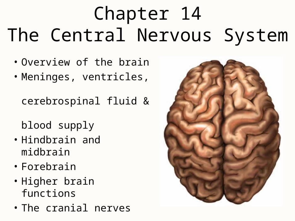
Chapter 14The Central Nervous System
• Overview of the brain
• Meninges, ventricles, cerebrospinal fluid & blood supply
• Hindbrain and midbrain
• Forebrain
• Higher brain functions
• The cranial nerves
Brain – Directional Terms and Landmarks
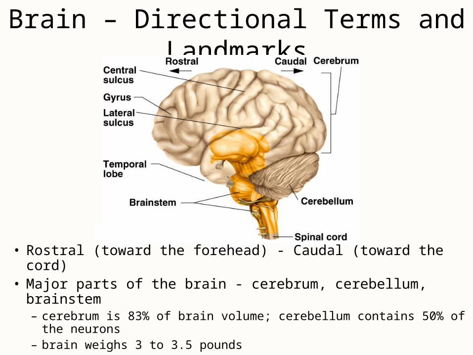
• Rostral (toward the forehead) - Caudal (toward the cord)• Major parts of the brain - cerebrum, cerebellum, brainstem
– cerebrum is 83% of brain volume; cerebellum contains 50% of the neurons
– brain weighs 3 to 3.5 pounds
Brain
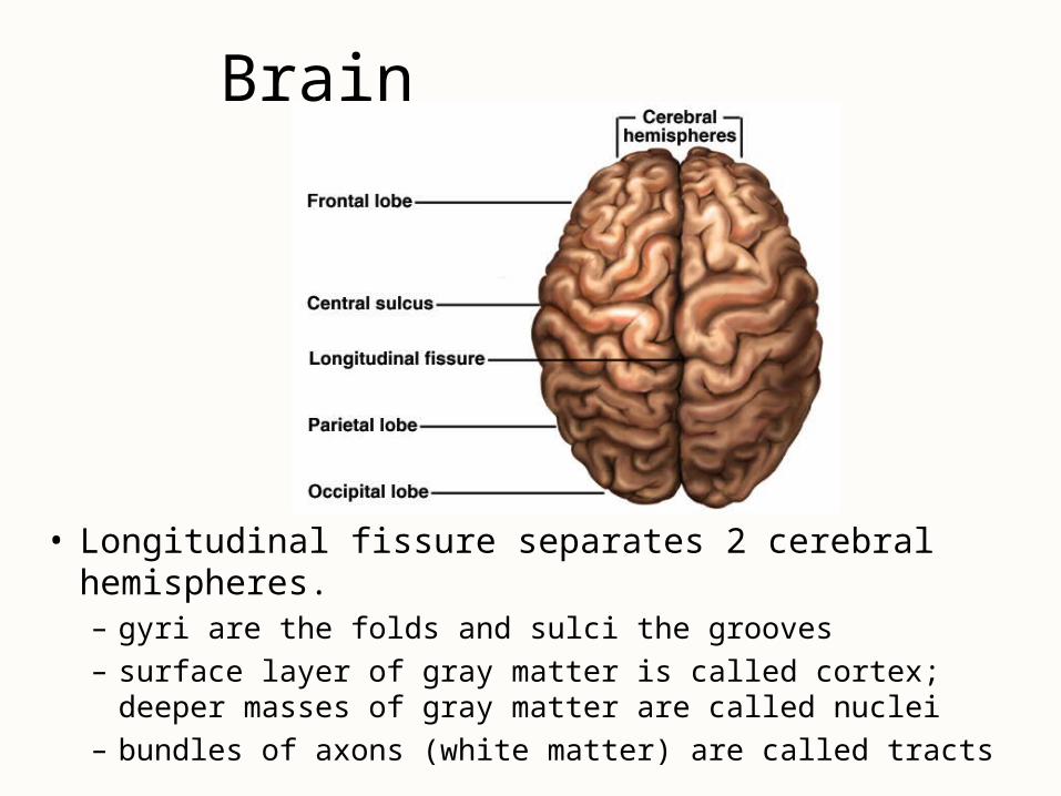
• Longitudinal fissure separates 2 cerebral hemispheres.– gyri are the folds and sulci the grooves
– surface layer of gray matter is called cortex; deeper masses of gray matter are called nuclei
– bundles of axons (white matter) are called tracts
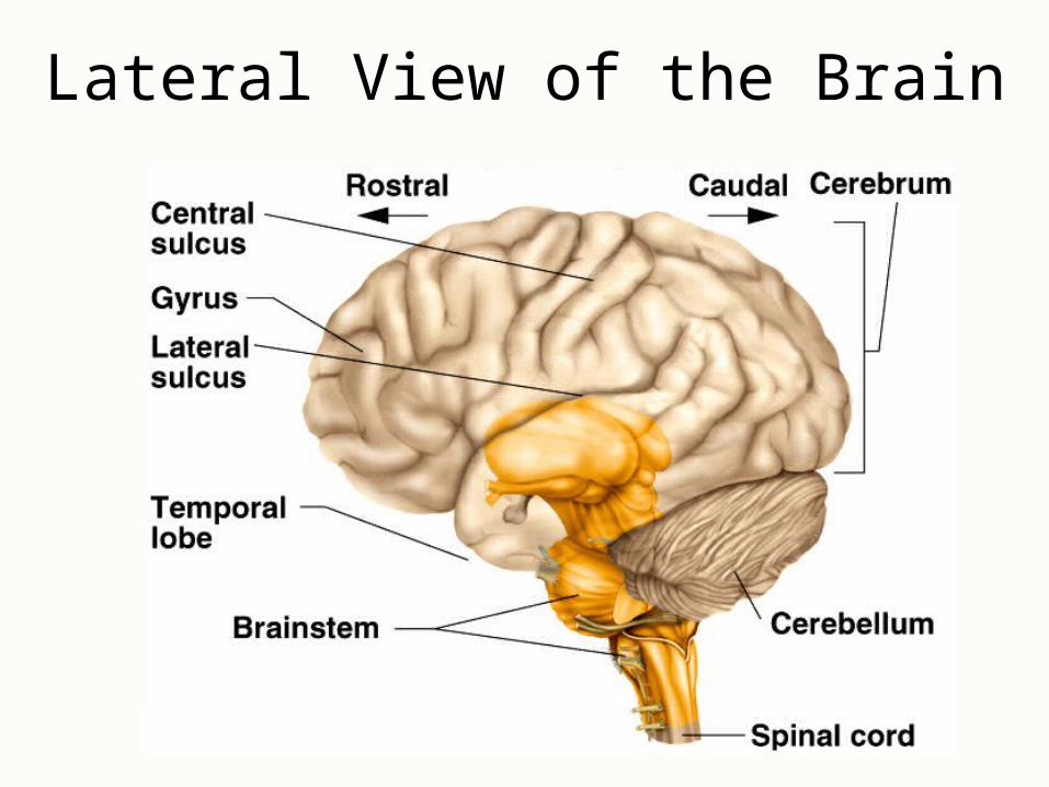
Lateral View of the Brain
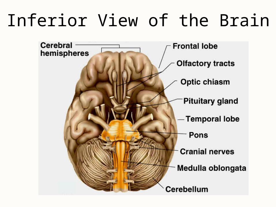
Inferior View of the Brain
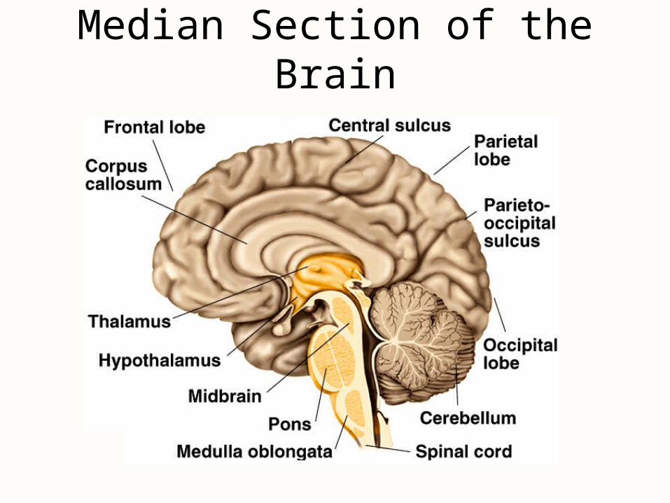
Median Section of the Brain
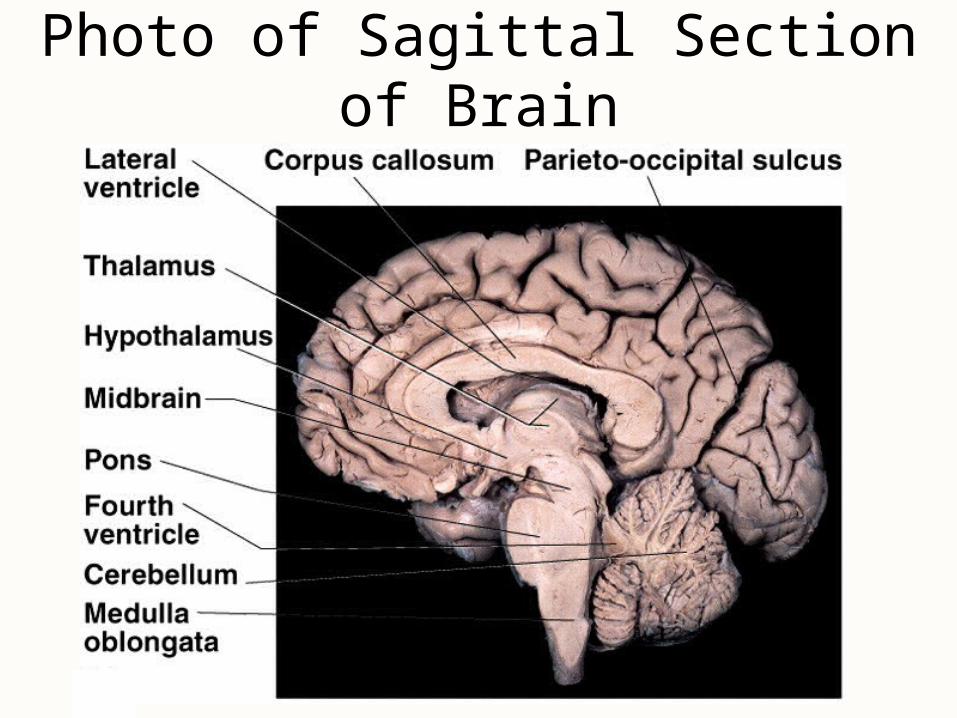
Photo of Sagittal Section of Brain
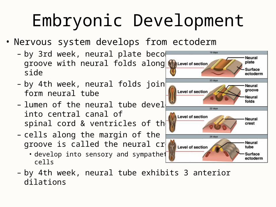
Embryonic Development• Nervous system develops from ectoderm
– by 3rd week, neural plate becomes a groove with neural folds along each side
– by 4th week, neural folds join to form neural tube
– lumen of the neural tube develops into central canal of spinal cord & ventricles of the brain
– cells along the margin of the neural groove is called the neural crest
• develop into sensory and sympathetic neurons & schwann cells
– by 4th week, neural tube exhibits 3 anterior dilations
Brain Development
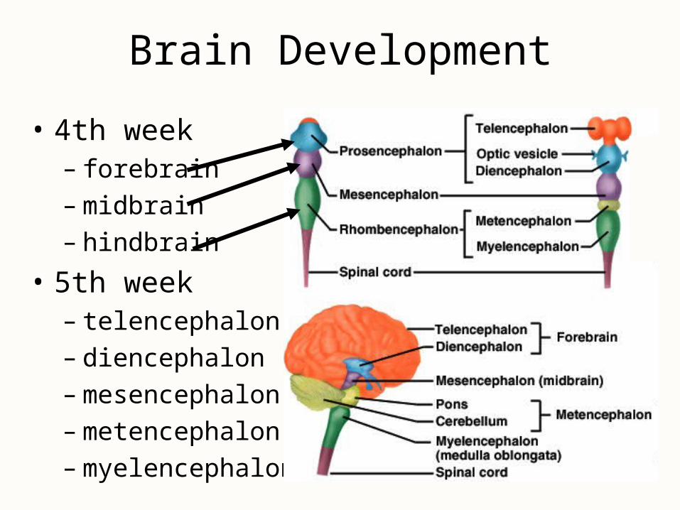
• 4th week– forebrain– midbrain– hindbrain
• 5th week– telencephalon– diencephalon– mesencephalon– metencephalon– myelencephalon
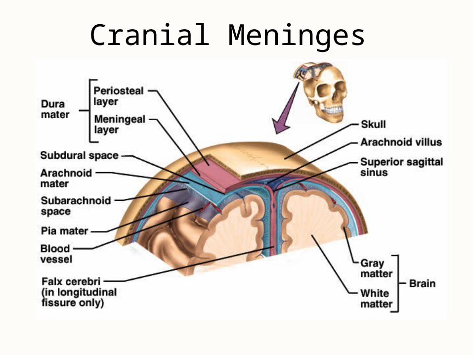
Meninges• Dura mater -- outermost, tough membrane
– outer periosteal layer against bone– where separated from inner meningeal layer forms
dural venous sinuses draining blood from brain– supportive structures formed by dura mater
• falx cerebri, falx cerebelli and tentorium cerebelli
– epidural space filled with fat in lower back region• epidural anaesthesia during childbirth
• Arachnoid mater is spider web filamentous layer
• Pia mater is a thin vascular layer adherent to contours of brain
Cranial Meninges
Meningitis
• Inflammation of the meninges
• Serious disease of infancy and childhood– between 3 months and 2 years of age
• Bacterial and virus invasion of the CNS by way of the nose and throat– pia mater and arachnoid are most likely to be affected
• Signs include high fever, stiff neck, drowsiness and intense headache and may progress to coma
• Diagnose by examining the CSF
Brain Ventricles
Ventricles of the Brain
Ventricles and Cerebrospinal Fluid
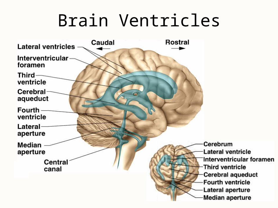
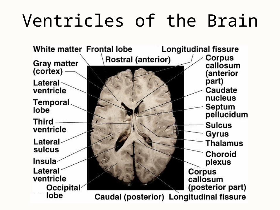
• Internal chambers within the CNS– lateral ventricles found inside cerebral hemispheres– third ventricle is single vertical space under corpus
callosum– cerebral aqueduct runs through midbrain– fourth ventricle is small chamber between pons &
cerebellum– central canal runs down through spinal cord
• Lined with ependymal cells and containing choroid plexus of capillaries that produce CSF
Cerebrospinal Fluid• Clear liquid fills ventricles and canals & bathes its
external surface (in subarachnoid space)
• Brain produces & absorbs about 500 ml/day– filtration of blood through choroid plexus– has more Na+ & Cl- but less K+ & Ca+2 than plasma
• Functions– buoyancy -- floats brain so it neutrally buoyant– protection -- cushions from hitting inside of skull– chemical stability -- rinses away wastes
• Escapes from 4th ventricle to surround the brain
• Absorbed by arachnoid villi into venous sinus
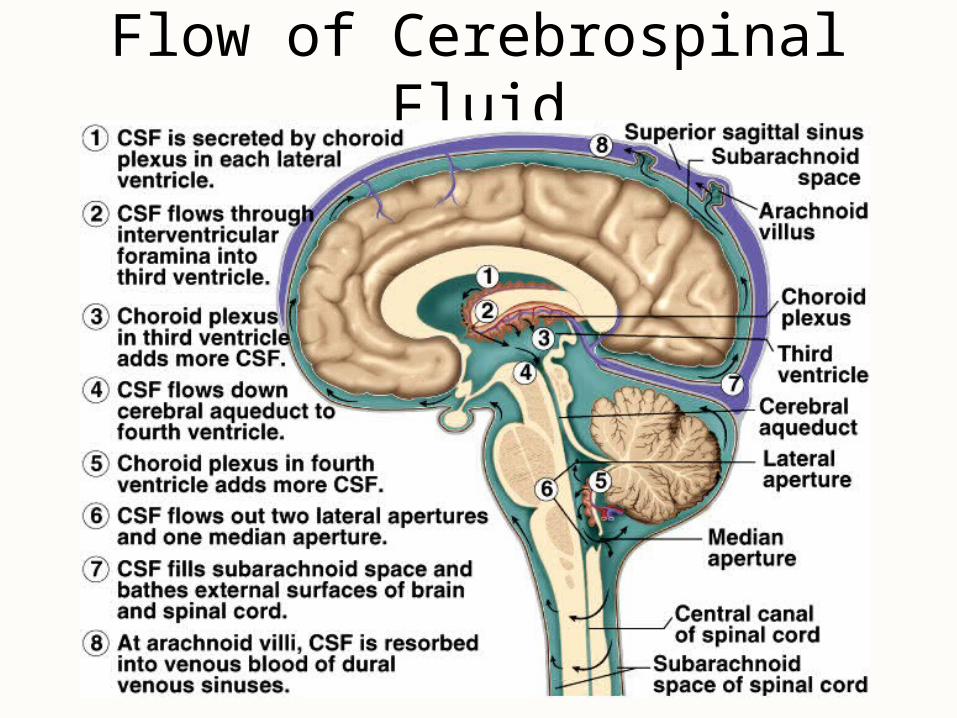
Flow of Cerebrospinal Fluid
Blood-Brain and Blood-CSF Barriers
• Blood-brain barrier is tightly joined endothelium– permeable to lipid-soluble materials (alcohol, O2,
CO2, nicotine and anesthetics)– circumventricular organs in 3rd & 4th ventricles at
breaks in the barrier where blood has direct access • monitoring of glucose, pH, osmolarity & other variations
• allows route for HIV virus to invade the brain
• Blood-CSF barrier at choroid plexus is ependymal cells joined by tight junctions
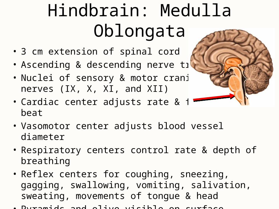
Hindbrain: Medulla Oblongata
• 3 cm extension of spinal cord• Ascending & descending nerve tracts• Nuclei of sensory & motor cranial
nerves (IX, X, XI, and XII)• Cardiac center adjusts rate & force of heart beat• Vasomotor center adjusts blood vessel diameter• Respiratory centers control rate & depth of breathing• Reflex centers for coughing, sneezing, gagging,
swallowing, vomiting, salivation, sweating, movements of tongue & head
• Pyramids and olive visible on surface
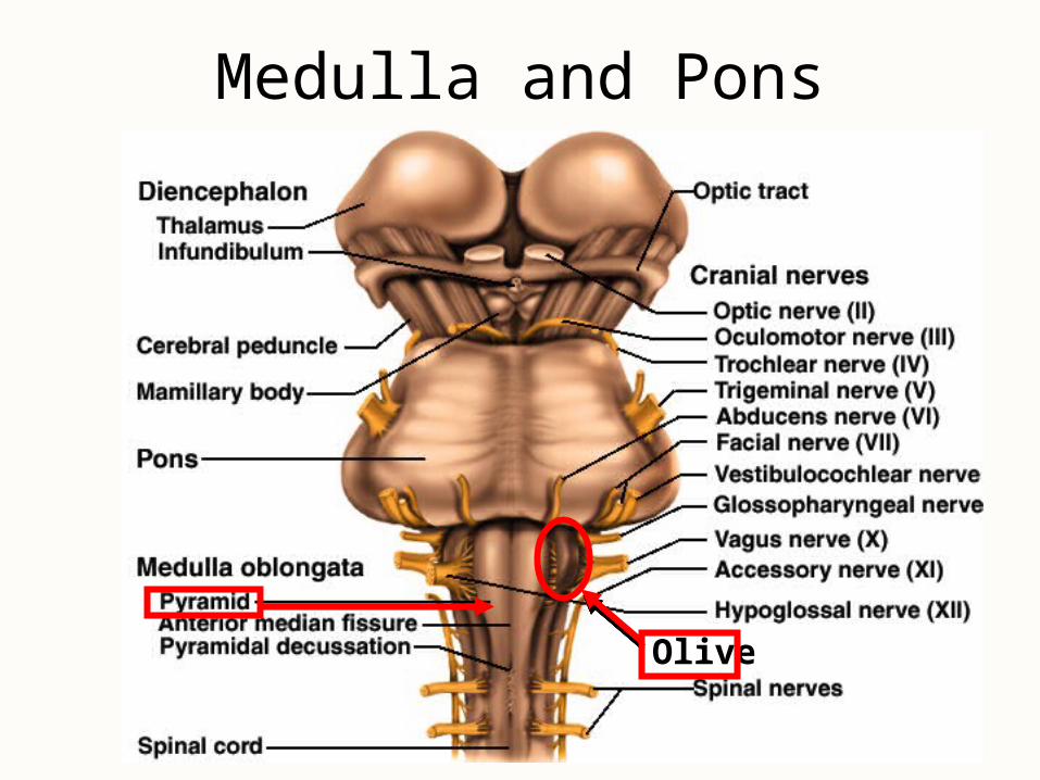
Medulla and Pons
Olive
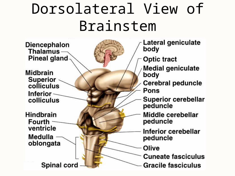
Dorsolateral View of Brainstem
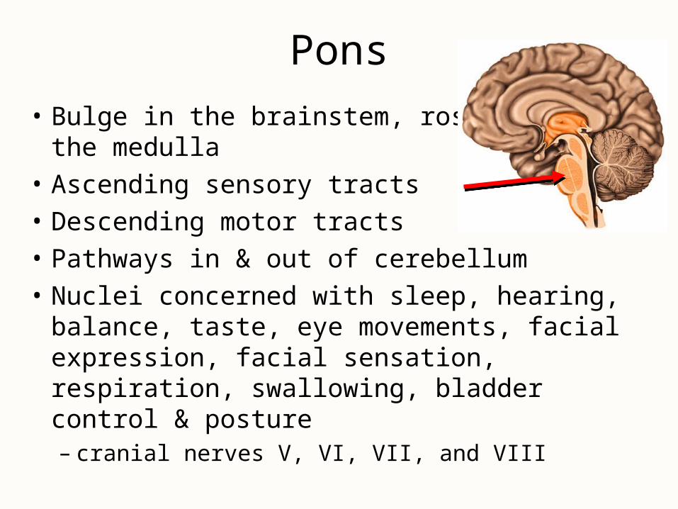
Pons
• Bulge in the brainstem, rostral to the medulla
• Ascending sensory tracts
• Descending motor tracts
• Pathways in & out of cerebellum
• Nuclei concerned with sleep, hearing, balance, taste, eye movements, facial expression, facial sensation, respiration, swallowing, bladder control & posture– cranial nerves V, VI, VII, and VIII
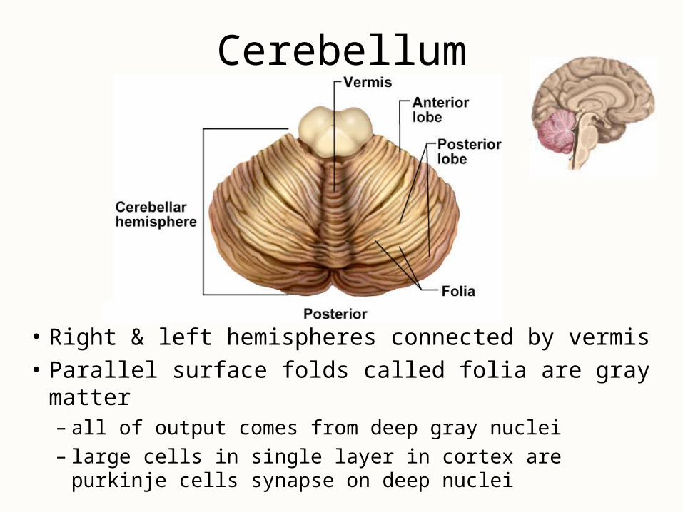
Cerebellum
• Right & left hemispheres connected by vermis
• Parallel surface folds called folia are gray matter– all of output comes from deep gray nuclei– large cells in single layer in cortex are purkinje cells
synapse on deep nuclei
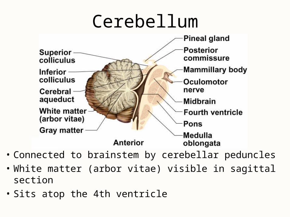
Cerebellum
• Connected to brainstem by cerebellar peduncles
• White matter (arbor vitae) visible in sagittal section
• Sits atop the 4th ventricle
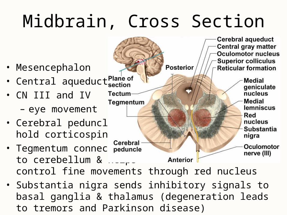
• Mesencephalon
• Central aqueduct
• CN III and IV
– eye movement
• Cerebral peduncles hold corticospinal tract
• Tegmentum connects to cerebellum & helps control fine movements through red nucleus
• Substantia nigra sends inhibitory signals to basal ganglia & thalamus (degeneration leads to tremors and Parkinson disease)
Midbrain, Cross Section
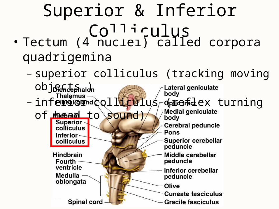
Superior & Inferior Colliculus• Tectum (4 nuclei) called corpora quadrigemina
– superior colliculus (tracking moving objects )– inferior colliculus (reflex turning of head to sound)
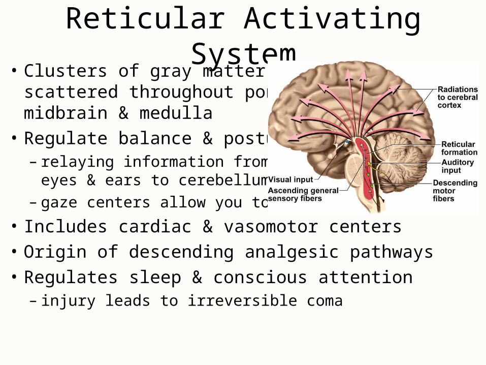
Reticular Activating System• Clusters of gray matter
scattered throughout pons, midbrain & medulla
• Regulate balance & posture – relaying information from
eyes & ears to cerebellum
– gaze centers allow you to track moving object
• Includes cardiac & vasomotor centers• Origin of descending analgesic pathways• Regulates sleep & conscious attention
– injury leads to irreversible coma
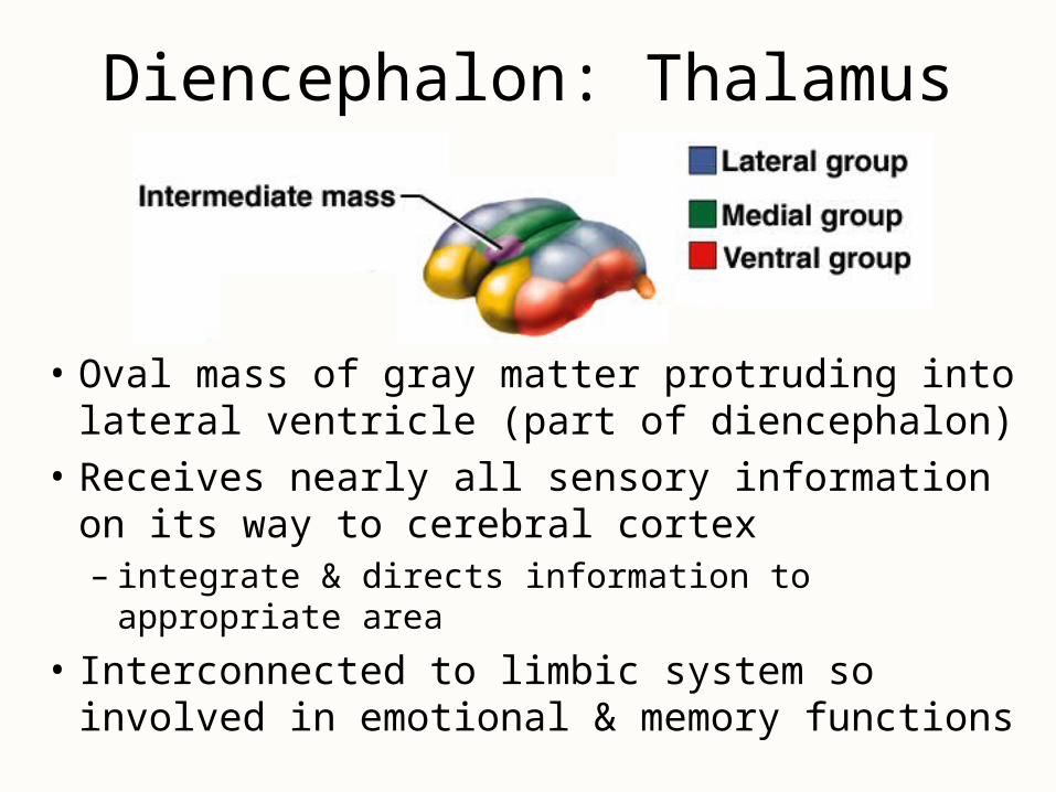
Diencephalon: Thalamus
• Oval mass of gray matter protruding into lateral ventricle (part of diencephalon)
• Receives nearly all sensory information on its way to cerebral cortex– integrate & directs information to appropriate area
• Interconnected to limbic system so involved in emotional & memory functions
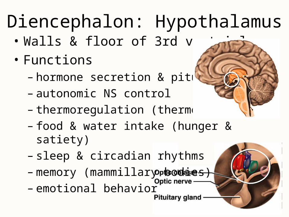
• Walls & floor of 3rd ventricle
• Functions– hormone secretion & pituitary– autonomic NS control– thermoregulation (thermostat)– food & water intake (hunger & satiety)– sleep & circadian rhythms– memory (mammillary bodies)– emotional behavior
Diencephalon: Hypothalamus
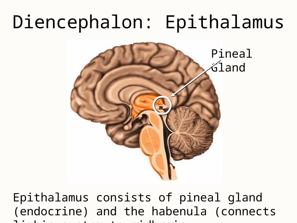
Diencephalon: Epithalamus
Pineal Gland
Epithalamus consists of pineal gland (endocrine) and the habenula (connects limbic system to midbrain.
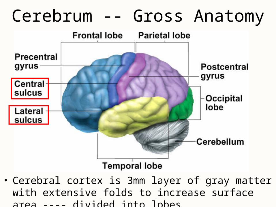
Cerebrum -- Gross Anatomy
• Cerebral cortex is 3mm layer of gray matter with extensive folds to increase surface area ---- divided into lobes
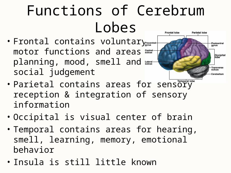
• Frontal contains voluntary motor functions and areas for planning, mood, smell and social judgement
• Parietal contains areas for sensory reception & integration of sensory information
• Occipital is visual center of brain
• Temporal contains areas for hearing, smell, learning, memory, emotional behavior
• Insula is still little known
Functions of Cerebrum Lobes
Tracts of Cerebral White Matter
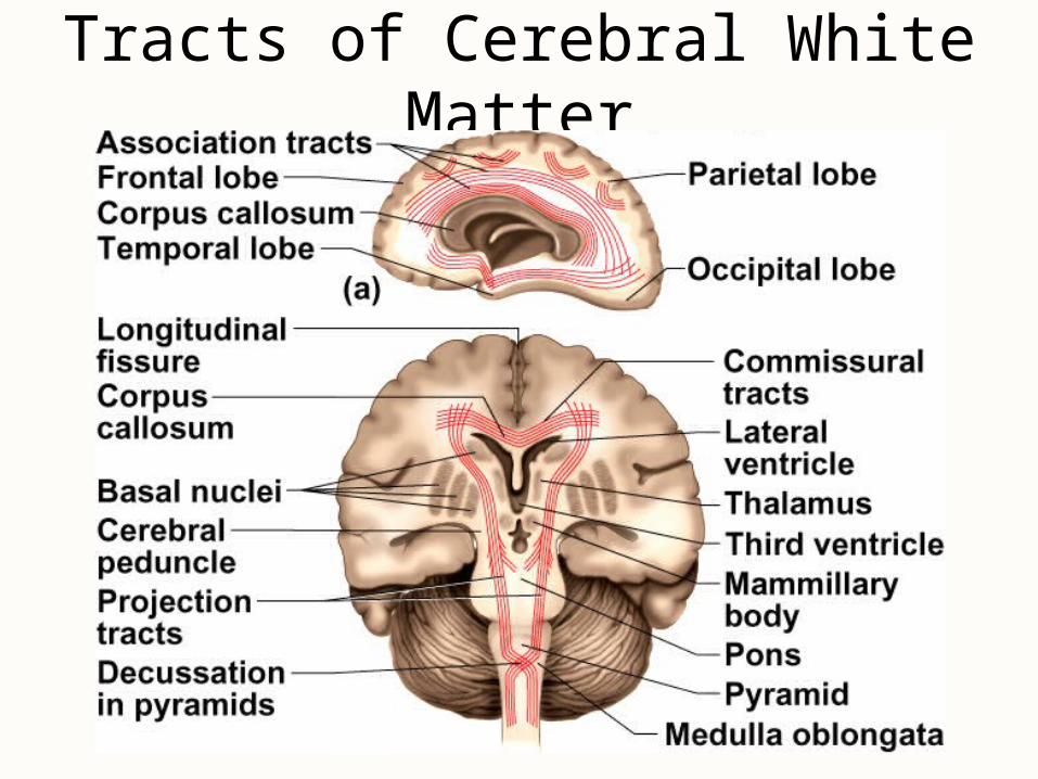
Tracts of Cerebral White Matter
• Most of volume of cerebrum is white matter
• Types of tracts– projection tracts
• extend vertically from brain to spinal cord forming internal capsule
– commissural tracts• cross from one hemisphere to the other
– corpus callosum is wide band of white fiber tracts
– anterior & posterior commissures are pencil-lead sized
– association tracts• connect lobes & gyri of each hemisphere to each other
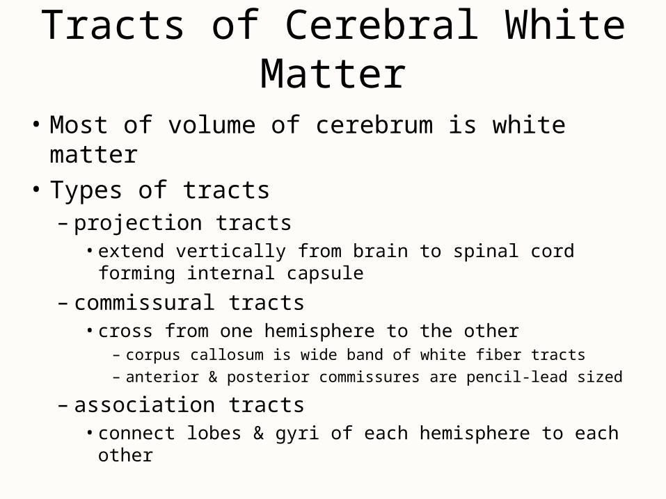
Cerebral Cortex• Surface layer of gray matter -- 3 mm thick• Neocortex (six-layered tissue)
– newest part of the cortex (paleocortex & archicortex)– layers vary in thickness in different regions of brain
• 2 types of cells– stellate cells
• have dendrites projectingin all directions
– pyramidal cells • have an axon that passes
out of the area
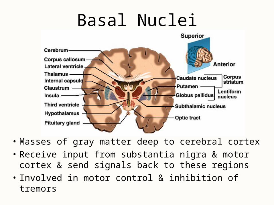
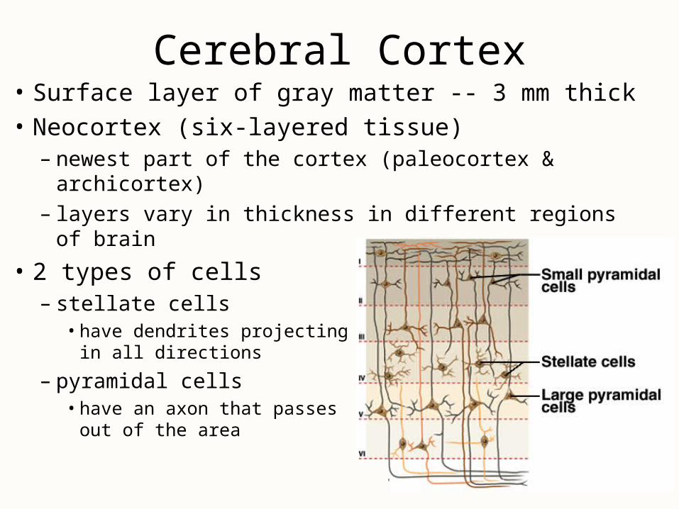
Basal Nuclei
• Masses of gray matter deep to cerebral cortex
• Receive input from substantia nigra & motor cortex & send signals back to these regions
• Involved in motor control & inhibition of tremors
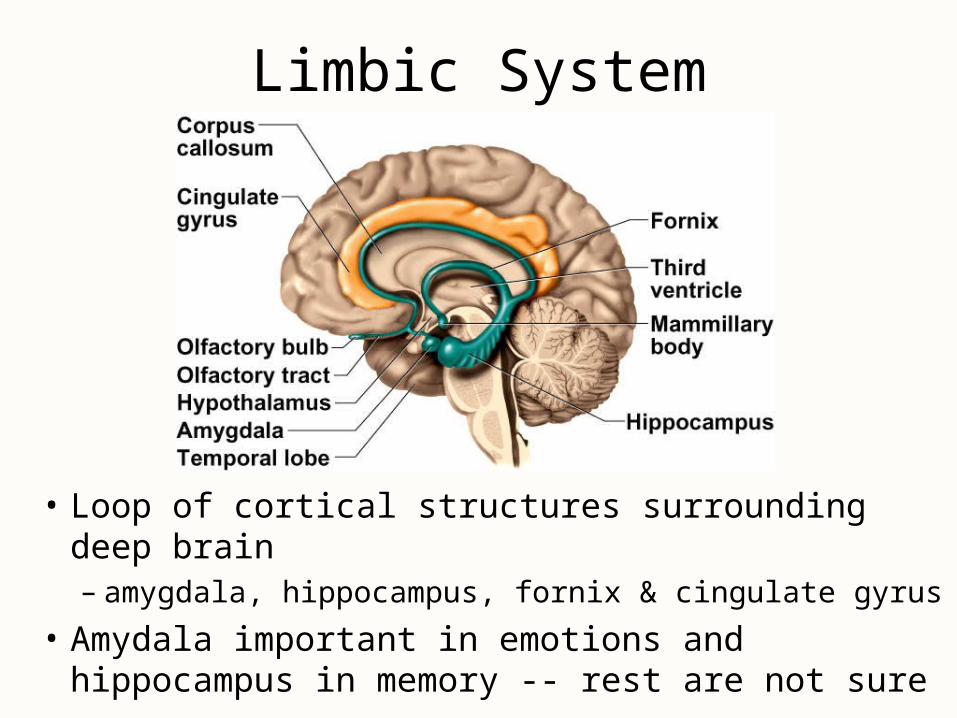
Limbic System
• Loop of cortical structures surrounding deep brain– amygdala, hippocampus, fornix & cingulate gyrus
• Amydala important in emotions and hippocampus in memory -- rest are not sure
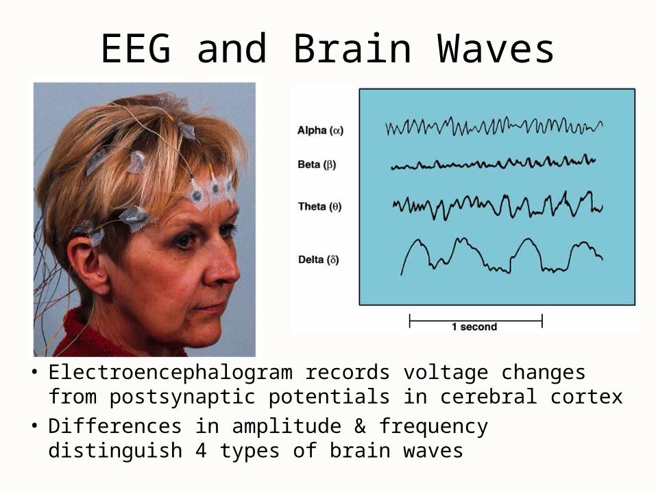
EEG and Brain Waves
• Electroencephalogram records voltage changes from postsynaptic potentials in cerebral cortex
• Differences in amplitude & frequency distinguish 4 types of brain waves
Brain Waves & Sleep• States of consciousness can be correlated with EEG• 4 types of brain waves
– alpha occur when awake & resting with eyes closed– beta occur with eyes open performing mental tasks– theta occur during sleep or emotional stress– delta occur during deep sleep
• Sleep is temporary state of unconsciousness– coma is state of unconsciousness with no possible arousal– reticular formation seems to regulate state of alertness– suprachiasmatic nucleus acts as biological clock to set our
circadian rhythm of sleep and waking
Stages of Sleep• Non-REM sleep occurs in stages
– 4 stages occurring in first 30 to 45 minutes of sleep• stage 1 is drifting sensation (would claim was not sleeping)
• stage 2 still easily aroused
• stage 3 vital signs change -- BP, pulse & breathing rates drop– reached in 20 minutes
• stage 4 is deep sleep -- difficult to arouse
– seems to have a restorative effect
• REM sleep occurs about 5 times a night– rapid eye movements under the eyelids, vital signs increase,
EEG resembles awake person, dreams and penile erections occur– may help sort & strengthen information from memory
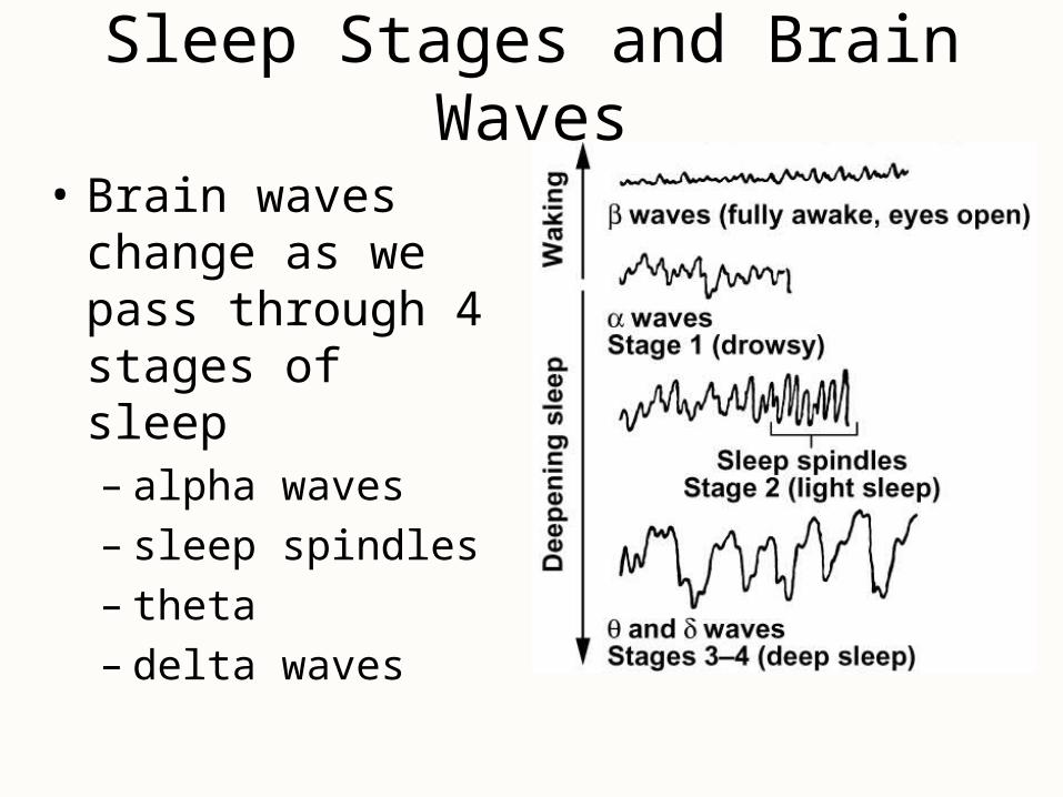
Sleep Stages and Brain Waves
• Brain waves change as we pass through 4 stages of sleep– alpha waves– sleep spindles– theta – delta waves
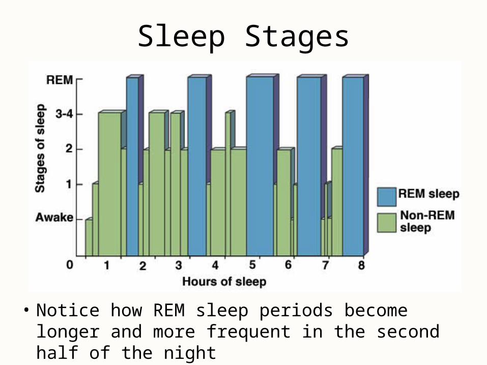
Sleep Stages
• Notice how REM sleep periods become longer and more frequent in the second half of the night
Cognition
• Cognition is mental processes such as awareness, perception, thinking, knowledge & memory– 75% of brain is association areas where integration of
sensory & motor information occurs
• Examples of effects of brain lesions– parietal lobe -- contralateral neglect syndrome– temporal lobe -- agnosia (inability to recognize objects)
or prosopagnosia (inability to recognize faces)– frontal lobe -- problems with personality (inability to
plan & execute appropriate behavior)
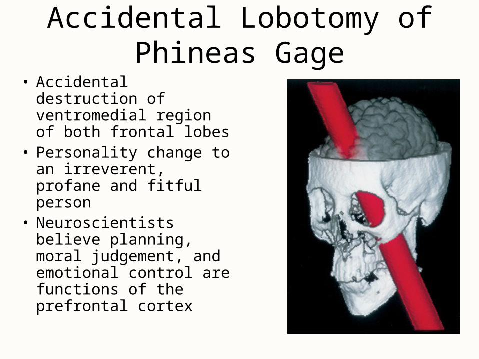
Accidental Lobotomy of Phineas Gage
• Accidental destruction of ventromedial region of both frontal lobes
• Personality change to an irreverent, profane and fitful person
• Neuroscientists believe planning, moral judgement, and emotional control are functions of the prefrontal cortex
Memory• Information management requires learning,
memory & forgetting (eliminating the trivia)– pathological inability to forget have trouble with
reading comprehension– anterograde amnesia -- can not store new data– retrograde amnesia -- can not remember old data
• Hippocampus is important in organizing sensory & cognitive information into a memory– lesion to it causes inability to form new memories
• Cerebellum helps learn motor skills
• Amygdala important in emotional memory
Emotion
• Prefrontal cortex controls how emotions are expressed (seat of judgement)
• Emotions form in hypothalamus & amygdala– artificial stimulation produces fear, anger, pleasure,
love, parental affection, etc.– electrode in median forebrain bundle in rat or human
and a foot pedal• press all day to the exclusion of food (report a quiet, relaxed
feeling)
• Much of our behavior is learned by rewards and punishments or responses of others to them
Somesthetic Sensation
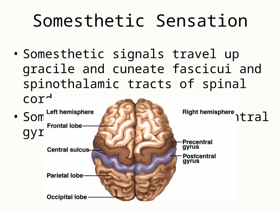
• Somesthetic signals travel up gracile and cuneate fascicui and spinothalamic tracts of spinal cord
• Somatosensory area is postcentral gyrus
Sensory Homunculus
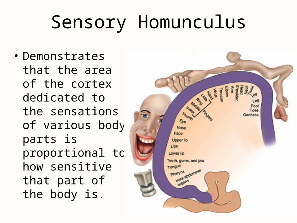
• Demonstrates that the area of the cortex dedicated to the sensations of various body parts is proportional to how sensitive that part of the body is.
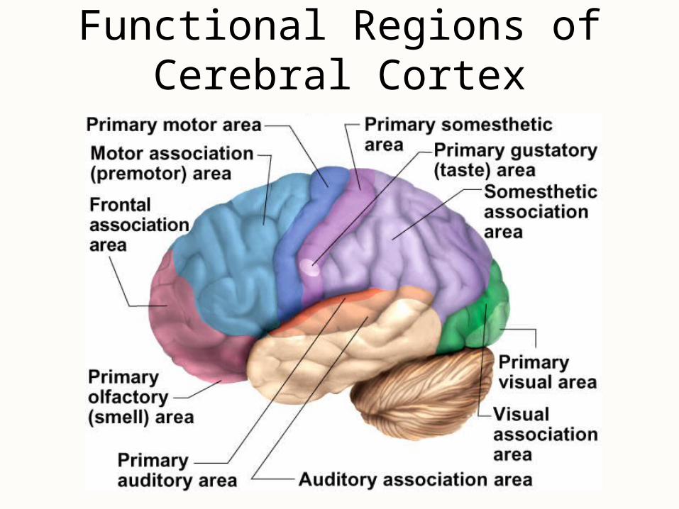
Functional Regions of Cerebral Cortex

Special Senses
• Organs of smell, vision, hearing & equilibrium project to specialized regions of the brain
• Locations– taste is lower end of postcentral gyrus– smell is medial temporal lobe & inferior frontal lobe– vision is occipital lobe– hearing is superior temporal lobe– equilibrium is mainly the cerebellum, but to unknown
areas of cerebral cortex via the thalamus
Sensory Association Areas
• Association areas interpret sensory information
• Somesthetic association area (parietal lobe)– position of limbs, location of touch or pain, and
shape, weight & texture of an object
• Visual association area (occipital lobe)– identify the things we see– faces are recognized in temporal lobe
• Auditory association area (temporal lobe)– remember the name of a piece of music or identify a
person by his voice
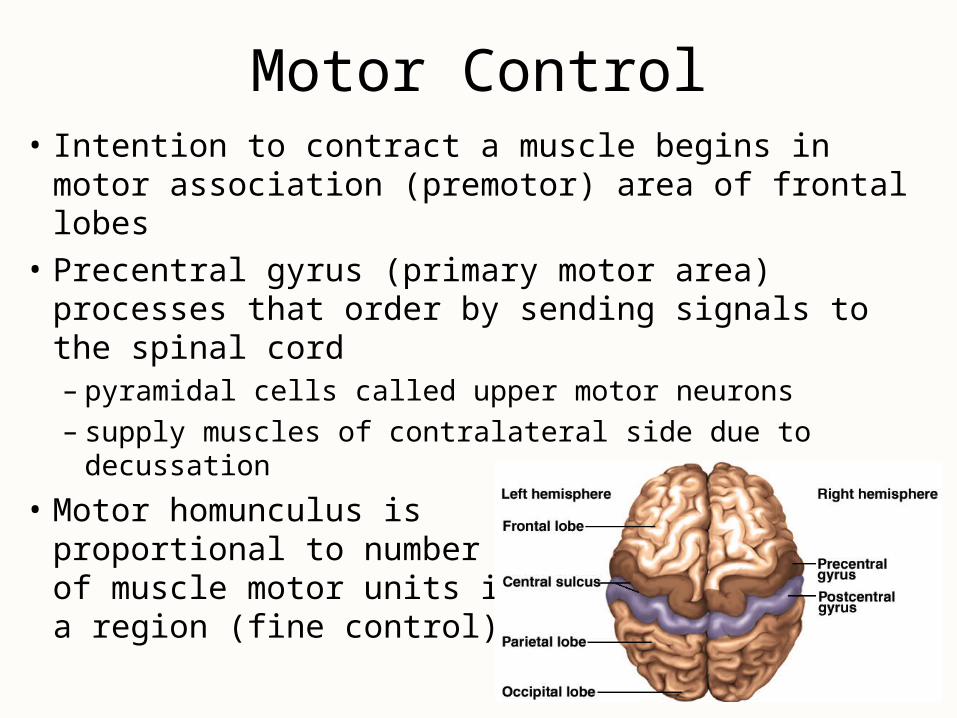
Motor Control• Intention to contract a muscle begins in motor
association (premotor) area of frontal lobes
• Precentral gyrus (primary motor area) processes that order by sending signals to the spinal cord– pyramidal cells called upper motor neurons– supply muscles of contralateral side due to decussation
• Motor homunculus is proportional to number of muscle motor units in a region (fine control)
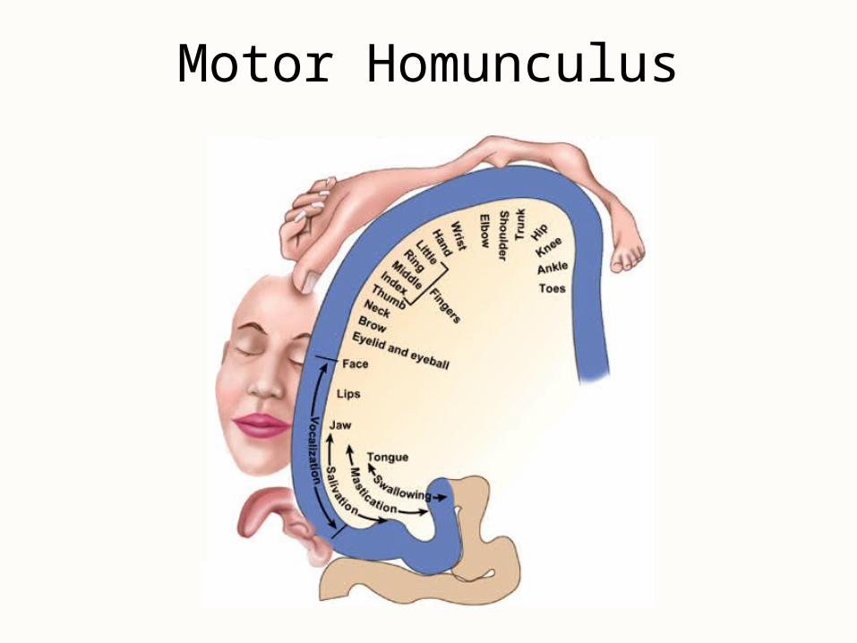
Motor Homunculus
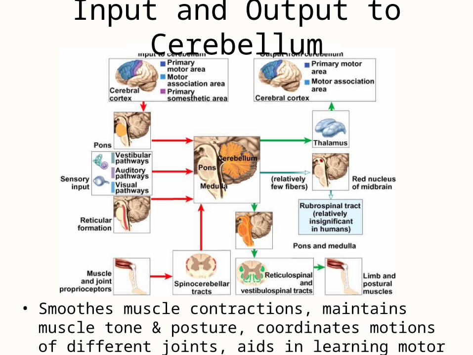
Input and Output to Cerebellum
• Smoothes muscle contractions, maintains muscle tone & posture, coordinates motions of different joints, aids in learning motor skills & coordinates eye movements
Language
• Includes reading, writing, speaking & understanding words
• Wernicke’s area permits recognition of spoken & written language & creates plan of speech– angular gyrus processes text into a form we can speak
• Broca’s area generates motor program for larynx, tongue, cheeks & lips – transmits that to primary motor cortex for action
• Affective language area lesions produce aprosodia– area area as Broca’s on opposite hemisphere
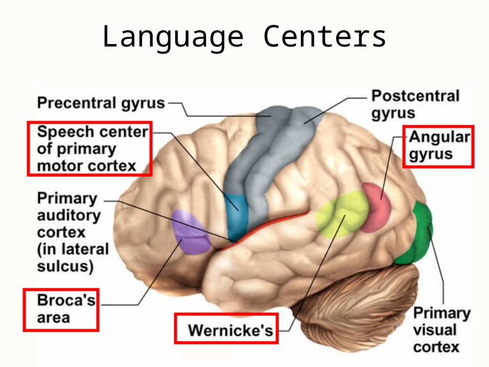
Language Centers
Aphasia• Any language deficit resulting from lesions in
same hemisphere as Wernicke’s & Broca’s areas
• Lesion to Broca’s = nonfluent aphasia– slow speech, difficulty in choosing words– entire vocabulary may be 2 to 3 words
• Lesion to Wernicke’s = fluent aphasia– speech normal & excessive, but makes little sense
• Anomic aphasia = speech & understanding are normal but text & pictures make no sense
• Others = understanding only 1st half of words or writing only consonants
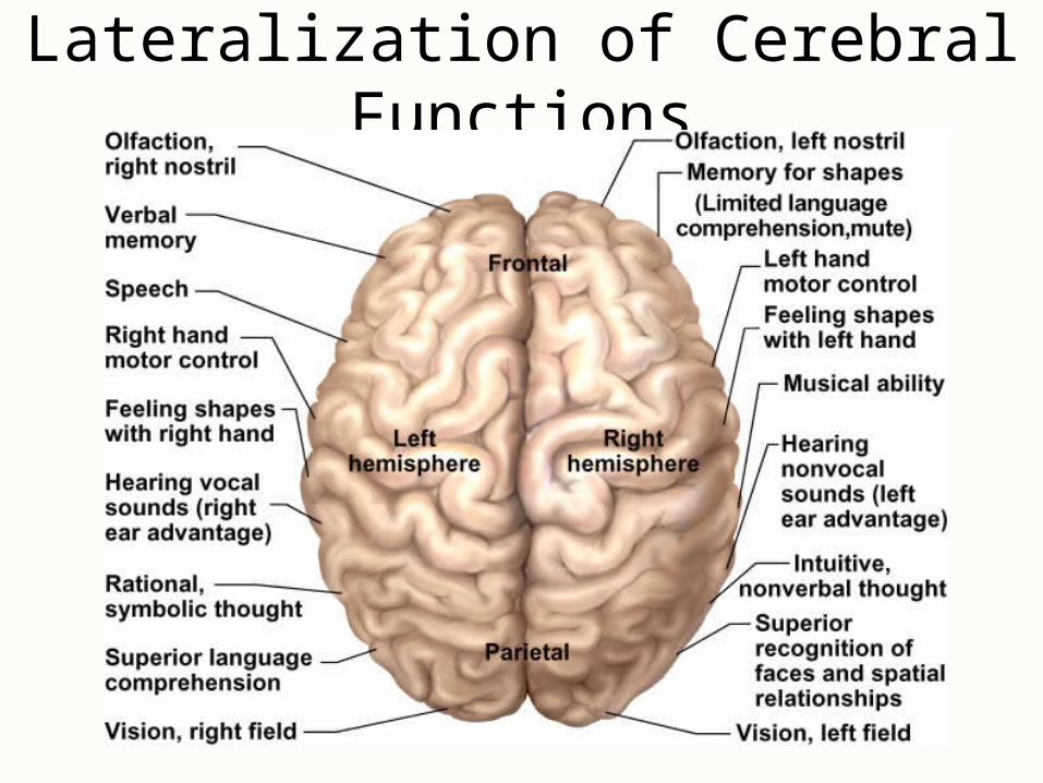
Lateralization of Cerebral Functions
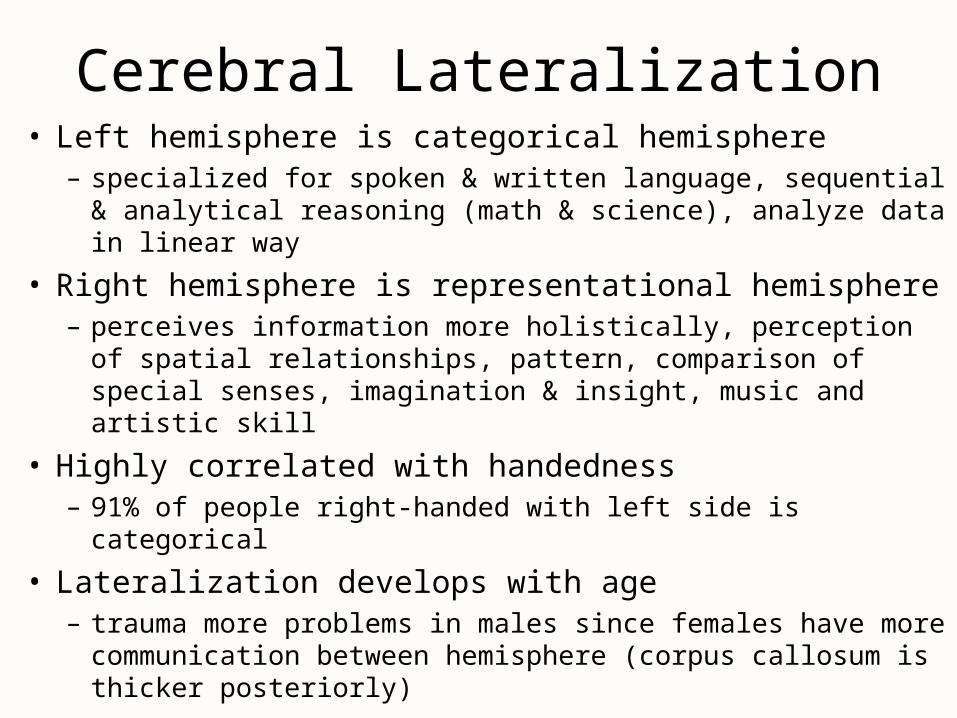
Cerebral Lateralization• Left hemisphere is categorical hemisphere
– specialized for spoken & written language, sequential & analytical reasoning (math & science), analyze data in linear way
• Right hemisphere is representational hemisphere– perceives information more holistically, perception of spatial
relationships, pattern, comparison of special senses, imagination & insight, music and artistic skill
• Highly correlated with handedness – 91% of people right-handed with left side is categorical
• Lateralization develops with age– trauma more problems in males since females have more
communication between hemisphere (corpus callosum is thicker posteriorly)
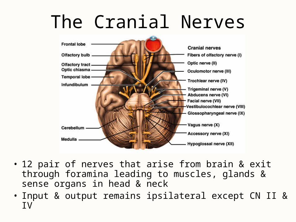
The Cranial Nerves
• 12 pair of nerves that arise from brain & exit through foramina leading to muscles, glands & sense organs in head & neck
• Input & output remains ipsilateral except CN II & IV
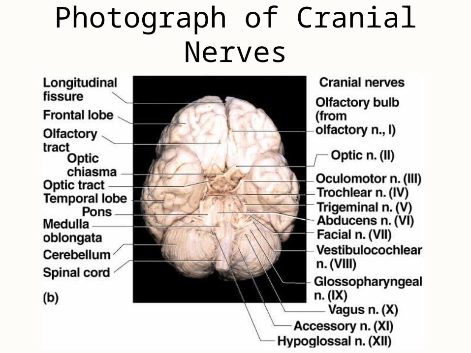
Photograph of Cranial Nerves
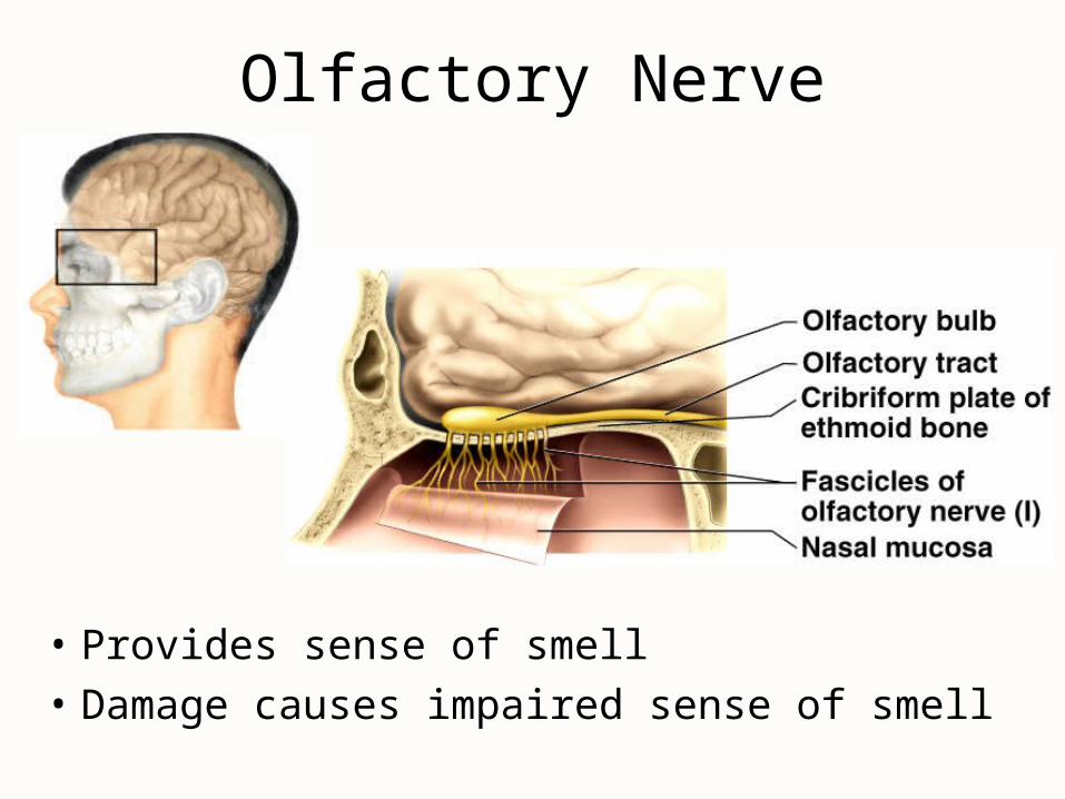
Olfactory Nerve
• Provides sense of smell
• Damage causes impaired sense of smell
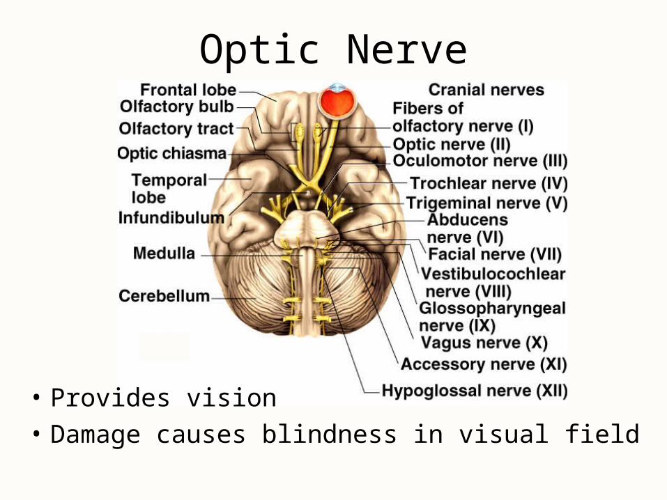
Optic Nerve
• Provides vision
• Damage causes blindness in visual field
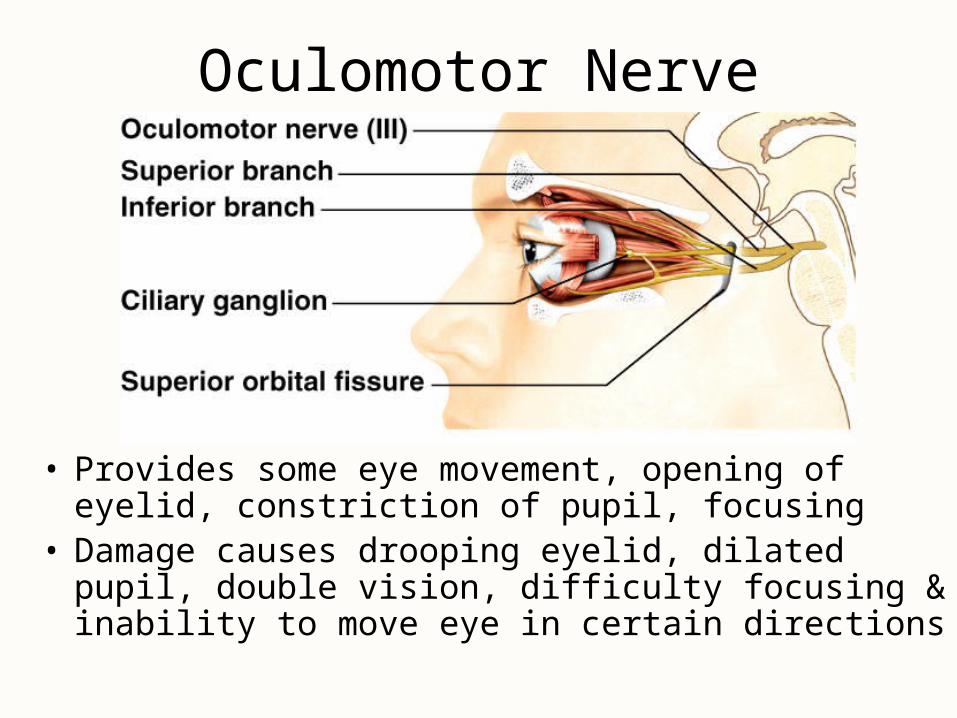
Oculomotor Nerve
• Provides some eye movement, opening of eyelid, constriction of pupil, focusing
• Damage causes drooping eyelid, dilated pupil, double vision, difficulty focusing & inability to move eye in certain directions
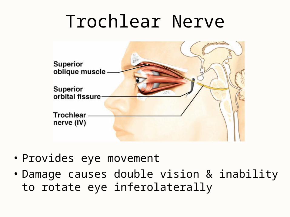
Trochlear Nerve
• Provides eye movement
• Damage causes double vision & inability to rotate eye inferolaterally
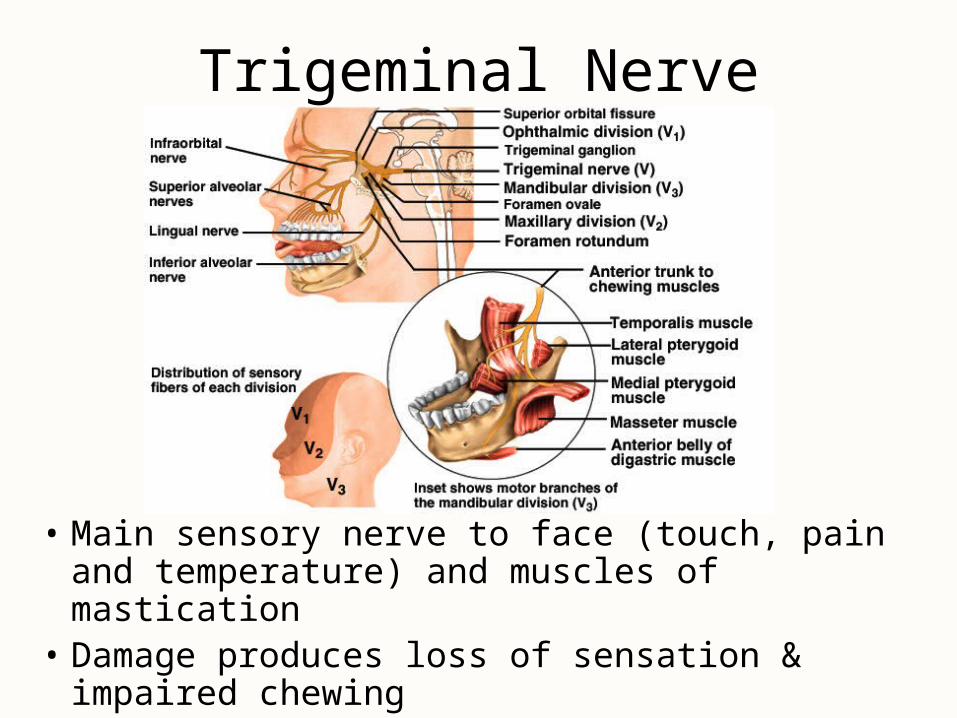
Trigeminal Nerve
• Main sensory nerve to face (touch, pain and temperature) and muscles of mastication
• Damage produces loss of sensation & impaired chewing
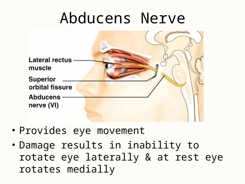
Abducens Nerve
• Provides eye movement
• Damage results in inability to rotate eye laterally & at rest eye rotates medially
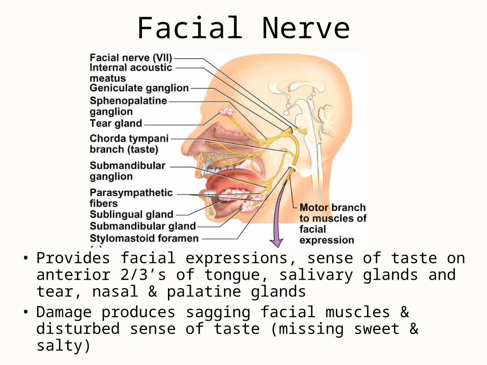
Facial Nerve
• Provides facial expressions, sense of taste on anterior 2/3’s of tongue, salivary glands and tear, nasal & palatine glands
• Damage produces sagging facial muscles & disturbed sense of taste (missing sweet & salty)
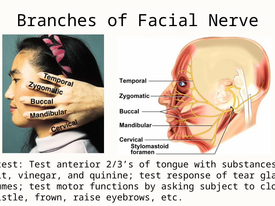
Branches of Facial Nerve
Clinical test: Test anterior 2/3’s of tongue with substances such as sugar, salt, vinegar, and quinine; test response of tear glands to ammonia fumes; test motor functions by asking subject to close eyes,smile, whistle, frown, raise eyebrows, etc.
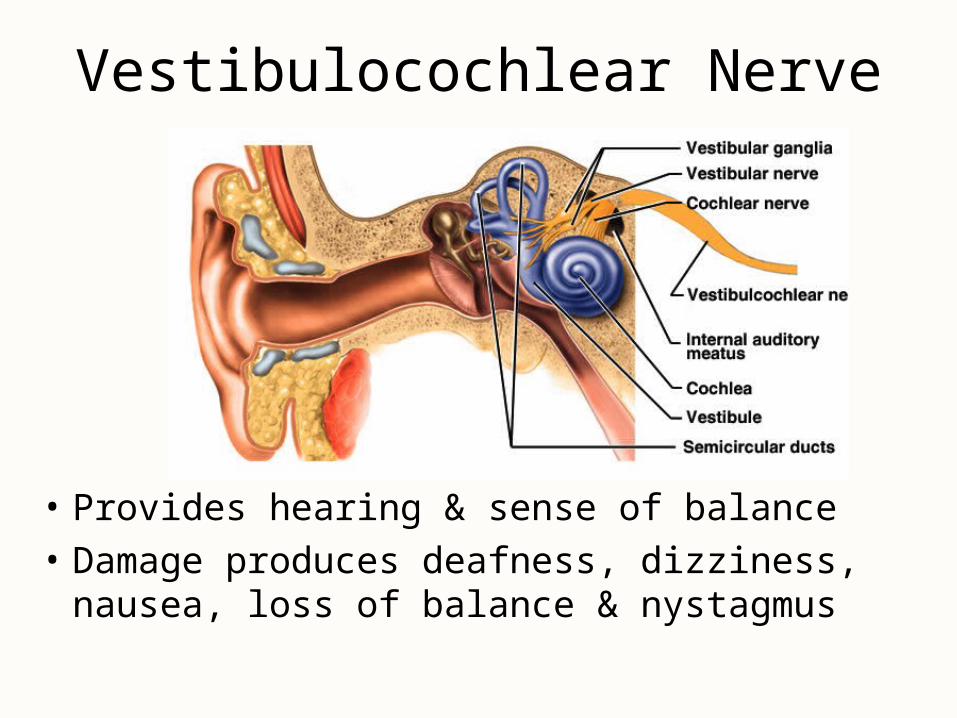
Vestibulocochlear Nerve
• Provides hearing & sense of balance
• Damage produces deafness, dizziness, nausea, loss of balance & nystagmus
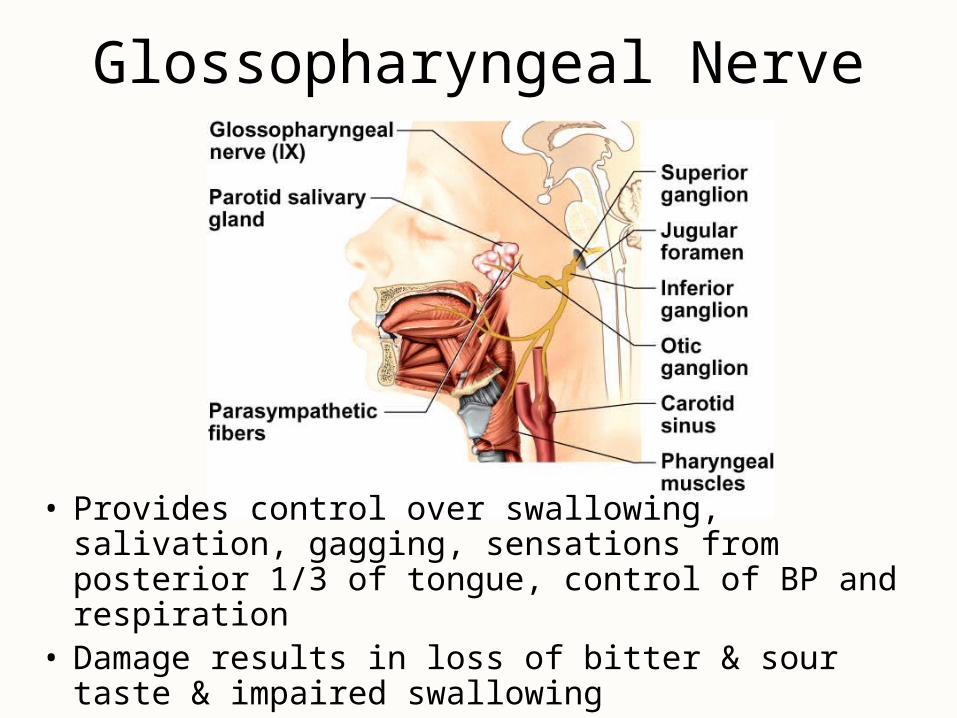
Glossopharyngeal Nerve
• Provides control over swallowing, salivation, gagging, sensations from posterior 1/3 of tongue, control of BP and respiration
• Damage results in loss of bitter & sour taste & impaired swallowing
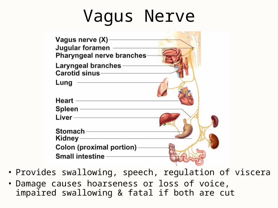
Vagus Nerve
• Provides swallowing, speech, regulation of viscera• Damage causes hoarseness or loss of voice, impaired
swallowing & fatal if both are cut
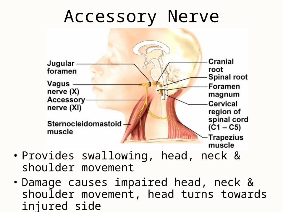
Accessory Nerve
• Provides swallowing, head, neck & shoulder movement
• Damage causes impaired head, neck & shoulder movement, head turns towards injured side
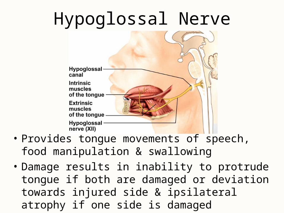
Hypoglossal Nerve
• Provides tongue movements of speech, food manipulation & swallowing
• Damage results in inability to protrude tongue if both are damaged or deviation towards injured side & ipsilateral atrophy if one side is damaged
Cranial Nerve Disorders
• Trigeminal neuralgia (tic douloureux)– recurring episodes of intense stabbing pain in
trigeminal nerve area (near mouth or nose)– pain triggered by touch, drinking, washing face– treatment is cutting of nerve
• Bell palsy– degenerative disorder of facial nerve– paralysis of facial muscles on one side– may appear abruptly & disappear within 3-5 weeks
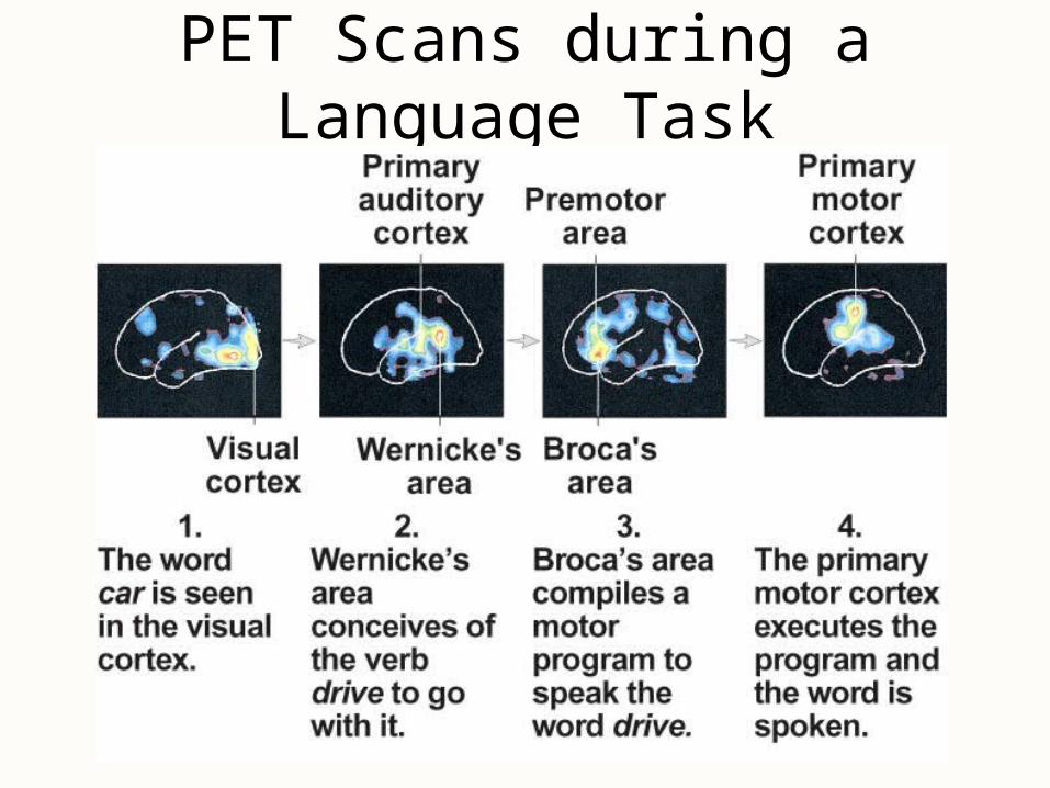
PET Scans during a Language Task