Embed Size (px)
Citation preview
159
Indications for Facial and Neck Trauma Surgery
Chapter 14
INDICATIONS FOR FACIAL AND NECK TRAUMA SURGERY
BRENT FELDT, MD,* and MANUEL LOPEZ, MD†
INTRODUCTION
HISTORY OF MAXILLOFACIAL TRAUMA SURGICAL REPAIRAncient Through Mediaeval PeriodsThe 18th and 19th CenturiesThe 20th Century
INDICATIONS FOR SURGERYMechanism of InjuryCasualty Evacuation From TheaterTiming of Facial Fracture Repair Timing of Neck Exploration
SUMMARY
*Major, Medical Corps, US Air Force; Otolaryngologist, David Grant US Air Force Medical Center, 101 Bordin Circle, Travis Air Force Base, California 94535
†Facial Plastic Surgeon, Lopez Plastic Surgery, 1314 East Sonterra Boulevard, Suite 5104, San Antonio, Texas 78258
160
Otolaryngology/Head and Neck Combat Casualty Care
Thus, the person intending to practice this kind of surgery must serve in the army, and accompany it on expeditions abroad; for in this way he would become experienced in this practice.1
— Hippocrates
INTRODUCTION
As long as humans have battled against nature and each other, there have been facial and neck in-juries. Because the face and neck are often exposed, these areas of the body require specialized treatment when injured. This chapter will review the history of
the treatment of facial and neck injuries, including improvements made in patient medical evacuation from theater, and describe some of the indications and limitations on in-theater repair of facial and neck injuries.
HISTORY OF MAXILLOFACIAL TRAUMA SURGICAL REPAIR
Ancient Through Mediaeval Periods
The treatment of facial trauma injuries can be dated to around 5,000 bce, when the Sumerian physician Hammurabi described payment for physicians who set broken bones.2 Techniques of local pedicle flap repair of cheek defects and forehead wounds were performed in ancient India around 1,500 bce, as described by Graham.3 In the Edwin Smith papyrus, circa 1,600 bce, Egyptian surgeons described replacing a dislocated mandible by “putting thumbs upon the rami of the mandible and fingers under the chin and cause them to fall back so that they rest in their places.”4 The pa-
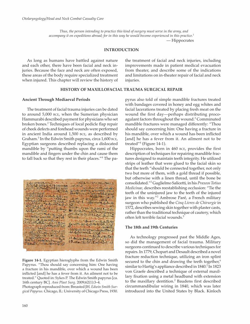
Figure 14-1. Egyptian hieroglyphs from the Edwin Smith Papyrus. “Thou should say concerning him: One having a fracture in his mandible, over which a wound has been inflicted [and] he has a fever from it. An ailment not to be treated.” Quoted in: Sykes P. The Edwin Smith papyrus [ca. 16th century BC]. Ann Plast Surg. 2009;62(1):3–4.Photograph reproduced from: Breasted JH. Edwin Smith Sur-gical Papyrus. Chicago, IL: University of Chicago Press, 1930.
pyrus also told of simple mandible fractures treated with bandages covered in honey and egg whites and facial lacerations treated by placing fresh meat on the wound the first day—perhaps distributing proco-agulant factors throughout the wound.2 Comminuted mandible fractures were managed differently: “Thou should say concerning him: One having a fracture in his mandible, over which a wound has been inflicted [and] he has a fever from it. An ailment not to be treated”4 (Figure 14-1).
Hippocrates, born in 460 bce, provides the first description of techniques for repairing mandible frac-tures designed to maintain teeth integrity. He utilized strips of leather that were glued to the facial skin so that the teeth “should be connected together, not only two but more of them, with a gold thread if possible, but otherwise with a linen thread, until the bone be consolidated.”2 Guglielmo Salicetti, in his Praxeos Totius Medicinae, describes reestablishing occlusion: “Tie the teeth of the uninjured jaw to the teeth of the injured jaw in this way.”2 Ambrose Paré, a French military surgeon who published the Cinq Livres de Chirurgie in 1572, describes sewing skin together with plaster casts, rather than the traditional technique of cautery, which often left terrible facial wounds.5
The 18th and 19th Centuries
As technology progressed past the Middle Ages, so did the management of facial trauma. Military surgeons continued to describe various techniques for repairs. In 1779, Chopart and Desault described a novel fracture reduction technique, utilizing an iron splint secured to the chin and drawing the teeth together,6 similar to Hartig’s appliance described in 1840.2 In 1823 von Graefe described a technique of external maxil-lary fixation using a metal headband with extension to the maxillary dentition.2 Baudens first described circummandibular wiring in 1840, which was later introduced into the United States by Black. Kinloch
161
Indications for Facial and Neck Trauma Surgery
first used transosseous wiring of the mandible bone ends in 1858 using silver wire.2 Guerin first noted in 1866 that fractures of the orbit often involved the pterygoid plates.7
The definitive treatment of facial wounds in the 19th century was sometimes one of benign neglect. The Medical and Surgical History of the British Army in the Crimea, Volume II (1858), describes the reasoning for this strategy: “Wounds of the face . . . are not generally of so serious a nature as their first appearance might lead the uninitiated to expect. The reason of this . . . seems obviously to be the very free supply of blood which this part receives. This leads us to not remove bony fragments unless the comminution be great, or the fragment completely separated from the soft parts. Even partially detached teeth will often be found not to have lost their vitality and, if carefully readjusted, will become useful.”8 These comments would likely be readily accepted by many facial trauma surgeons today. Sir Harold Gillies advocated for every patient with maxillofacial trauma to be placed prone, to pre-vent the soft tissue from falling into the airway.2
Thomas Gunning deserves special recognition. He developed a mandible splint using vulcanized rubber that enclosed the mandible teeth and obtained maxil-lary teeth impressions. The splint was then secured using thread or wires, with a cut-away portion in the front to allow passage of food. Gunning became well-known in America for his treatment of facial fractures, particularly jaw fractures. He was consulted when William H Seward, the secretary of state to Abraham Lincoln, sustained a mandible fracture from a carriage accident. Although Seward sustained a wound infec-tion secondary to contemporary treatment techniques, Gunning was able to help him regain normal occlu-sion. Gunning himself sustained a mandible fracture after falling off his horse. He reduced the fracture himself, then used interdental suture and his splint to restore occlusion, and saw patients in his clinic the next day.2
The 20th Century
In the early 20th century the understanding of facial trauma surged forward. In 1901 the work of Rene Le Fort was published in France (translated into English in 1941), describing for the first time the most com-mon midface fracture patterns.9 These descriptions are still used today by clinicians to describe the types of fracture sustained by facial trauma patients in the middle third of the facial skeleton. The development of radiographs and improved techniques for skin grafting and flap reconstruction enabled surgeons to increase the accuracy of their treatment. The treatment of facial
trauma was also advanced when Dr Ivan Magill devel-oped endotracheal anesthesia in the early part of the century, allowing surgeons to work without waiting for ether to be provided.
The first military facial trauma units were devel-oped before World War I. During the battle of Somme, Gillies created the first military unit dedicated to maxillofacial injuries, which treated over 2,000 injuries beginning on the first day of the battle.10 During the war, the work of Gillies, Fry, and Fraser in England and Blair, Ivy, and Smith from the United States formed the basis of many of the techniques used to repair facial trauma injuries.2
However, facial and neck injuries were still treated with benign neglect; surgeons would allow the wounds to heal with secondary intention, followed by scar revi-sion and reconstruction weeks to months later.10 This often caused significant deformity from contraction and scarring. The few advancements made during World War I were often lost as surgeons returned to their home countries and resumed private practice. There were no significant conferences or collaboration efforts to continue improving upon the lessons learned in the war. Although the American Association of Oral Surgeons, the American Academy of Ophthalmology and Otolaryngology, and the American Society of Plastic and Reconstructive Surgery were founded in this period,10 these organizations initially did not fo-cus on development of trauma management schemes nor push for cooperation and research in this area.
Significant advances in facial and neck trauma sur-gery occurred during World War II. The development of aircraft evacuation, use of antibiotics, improvements in anesthesia and transfusions, and advances in exter-nal fixation devices allowed improved outcomes in facial trauma patients. For example the biphasic exter-nal fixation device, also known as the Joe Hall Morris device, was developed during this period to aid in external fixation of complicated mandible fractures.11
A major change occurred when British plastic sur-geon Patrick Clarkson began treating facial trauma patients differently. The prevailing paradigm Clarkson deviated from was treatment with secondary intention healing with delayed reconstruction. Many surgeons believed healing with a deformity was preferable to possibly causing facial skin loss. During the battle of Cassino in Italy in 1944, Clarkson and his team of dentists, ophthalmologists, neurosurgeons, and plastic surgeons began to treat patients with early wound closure. This resulted in evacuation of only 20% of wounded soldiers back to Britain.10
As Clarkson and his colleagues continued this new treatment strategy, they noted improvements in patient outcomes. Patients treated with early closure
162
Otolaryngology/Head and Neck Combat Casualty Care
had not only faster healing but also faster union of underlying fractures. Since internal fixation had not yet been developed, this allowed for earlier bone grafting.10 Most fractures were definitively repaired with wiring and cast metal splints. The infection rate was 3%, and 95% of patients with isolated soft tissue injury returned to duty within 11 days. The unit’s se-questration rate dropped from 70% to 10% compared to prior treatments in North Africa earlier in the war.10 Unfortunately, many of the advancements made dur-ing World War II were lost following the war for the same reason as after World War I.
The next advancement in treating head and neck facial trauma came in the 1950s with the develop-ment of advanced internal fixation techniques. In 1950 Robert Danis in Belgium described internal fixation in Théorie et Pratique de l’Osteosynthèse.12 His technique allowed bones to heal without callous for-mation, which he termed “per primam,” or primary healing. The first successful results of open reduc-tion and internal fixation (ORIF) were described by Maurice Muller in Switzerland in 1951. Muller and colleagues developed the Arbeitsgemeinschaft für Osteosynthesefragen (AO) in 1958 and began collect-ing data on outcomes and improving internal fixation techniques.12 The first AO cranio-maxillofacial trauma course was conducted by Joachim Prein in Basel, Swit-
zerland, in 1974.12 ORIF became the primary surgical management technique for complex facial fractures over the next few decades.
The Vietnam conflict provided another opportunity for developments in the treatment of wartime facial and neck injuries. Since most of the lessons learned in World War II were lost, much of the treatment of facial fractures was based on the individual surgeon’s experi-ence, and patients were evacuated to the United States for definitive treatment. Again, wartime experience was not adequately reported, and data describing the techniques and timing of facial and neck trauma surgi-cal repairs from Vietnam is lacking in the literature.13
During the remainder of the 20th century, research and development of novel techniques for ORIF and soft tissue repair continued, led by the developments of Muller and others. Many companies began developing titanium plates, absorbable plates, specialized sutures, and splints for use both in traumatic injuries and for repairing surgical defects. Surgeons around the world described unique techniques for repairing bony and soft tissue injuries. The principles of AO continued to guide many of the surgical techniques for facial fracture treat-ment. By the beginning of the 21st century, the strategy for treating facial trauma relied on addressing imme-diate life-threatening injuries first, then reducing and plating fractures with adequate soft-tissue coverage.14
INDICATIONS FOR SURGERY
The indications and timing of surgical repair of facial and neck trauma has undergone many changes over the past few decades. This section describes gen-eral topics related to timing of surgical repair in theater. For more detailed indications for surgical airways and specific injuries, see chapters 12, Airway Management; 17, Acute Soft-Tissue Injuries and Repair; 27, Complex Head and Neck Reconstruction in Theater; and 28, Penetrating and Blunt Neck Trauma.
Mechanism of Injury
In the Vietnam conflict, mechanism of injury was most commonly fragmentation wounds (62%), followed by bullets (23%), and blasts (3%).14 In the conflicts in Iraq and Afghanistan injuries were more commonly caused by blasts (58%–74%).14,15 Many more casualties have suffered polytrauma in these conflicts as well: only 5% of Vietnam wounded had more than two areas of the body injured compared to 69% of those transported by a critical care air transport team (CCATT).14 The difference in injury patterns may be explained by other factors such as urban versus jungle tactics and the use of improvised explosive devices versus fragmentation grenades.16,17
Casualty Evacuation From Theater
A unique aspect of the recent conflicts in Iraq and Afghanistan is the rapid mobility afforded to wounded American and allied soldiers. Moving from Role 1 (combat medic and buddy care) to Role 4 (definitive care out of theater) has improved dramatically (Fig-ure 14-2). Prior to the advances made during these conflicts, many injured service members would wait weeks or months to return to an advanced level of care in Germany or the United States. The average time to return to the United States for the injured in Vietnam was 45 days, but in the Iraq and Afghanistan conflicts that time has been reduced to an average of 4 days.18 This rapid evacuation has reduced the need for definitive treatment of complex traumatic wounds while in theater.
CCATTs and acute lung rescue teams (ALRTs) were created to transport severely injured patients to higher roles of care.14,15,19,20 CCATT missions to evacu-ate wounded personnel from Afghanistan began in October 2001 and from Iraq in 2003. Composed of a critical care-trained physician, critical care nurse, and a respiratory therapist, the teams can travel on aircraft of opportunity, usually a C-141 Starlifter or
163
Indications for Facial and Neck Trauma Surgery
C-17 Globemaster III.14 Unlike in many previous long-range medical evacuations using the now-retired C-9, these teams do not need specialized equipment integrated into the aircraft itself, but bring all neces-sary equipment and supplies with them. Data on CCATT missions is available in the US Transportation Command Regulating and Command and Control Evacuation System (TRAC2ES). From 2001 to 2006,
Role 1Soldier and combat medic
Tactical Combat Casualty CareScene safety
Hemorrhage control
Role 2Battalion aid stationsForward Surgical Team
Advanced Trauma Life SupportResuscitation
Damage control surgery
Role 3Combat hospital
Advanced Trauma Life SupportResuscitation
Damage control/definitive surgerySpecialty consultants
CASEVAC
CASEVAC TACTICAL AEROVAC/MEDEVAC
Role 4Strategic evacuation
[en route] medical centerDamage control/definitive surgery
Specialty consultantsShort-term recovery
Rehabilitation centerPhysical medicine/rehabilitation
Physical therapyOccupational therapy
Speech therapy
Definitive care medical centerDamage control/definitive surgery
Specialty consultantsLong-term recovery
Rehabilitation
STRATEGIC AEROVAC
STRATEGIC AEROVAC
AFTER EVACUATION FROM THEATERTHEATER
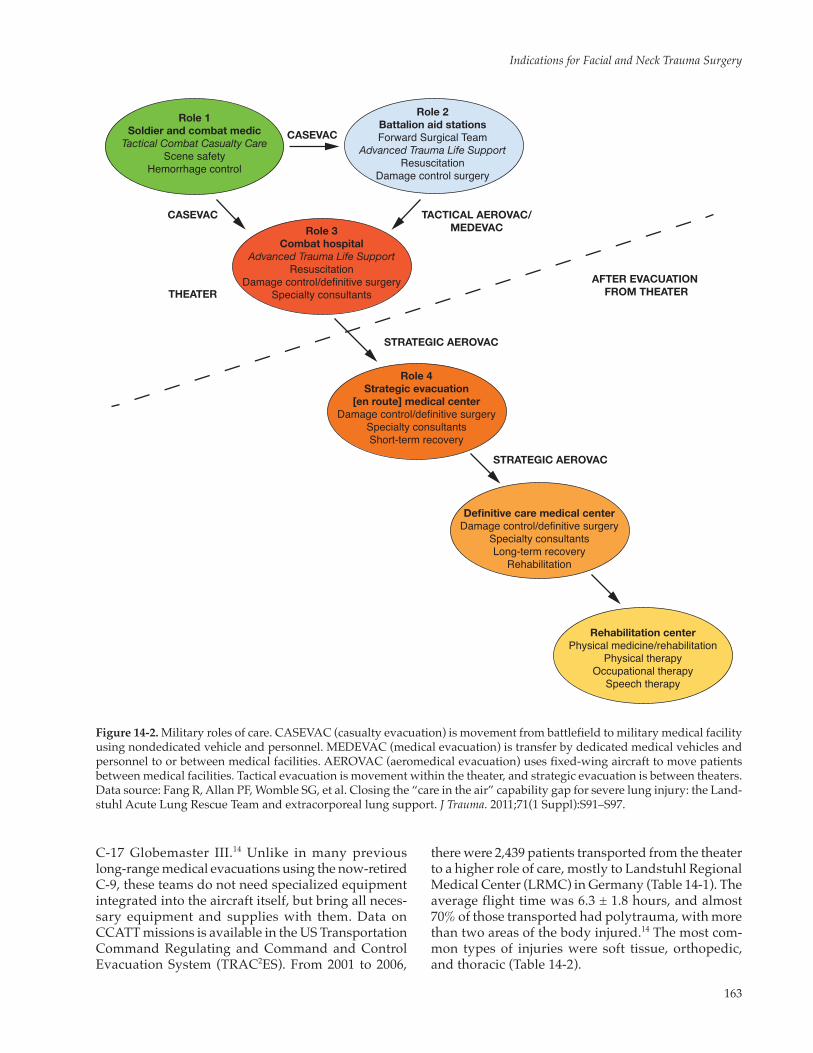
Figure 14-2. Military roles of care. CASEVAC (casualty evacuation) is movement from battlefield to military medical facility using nondedicated vehicle and personnel. MEDEVAC (medical evacuation) is transfer by dedicated medical vehicles and personnel to or between medical facilities. AEROVAC (aeromedical evacuation) uses fixed-wing aircraft to move patients between medical facilities. Tactical evacuation is movement within the theater, and strategic evacuation is between theaters.Data source: Fang R, Allan PF, Womble SG, et al. Closing the “care in the air” capability gap for severe lung injury: the Land-stuhl Acute Lung Rescue Team and extracorporeal lung support. J Trauma. 2011;71(1 Suppl):S91–S97.
there were 2,439 patients transported from the theater to a higher role of care, mostly to Landstuhl Regional Medical Center (LRMC) in Germany (Table 14-1). The average flight time was 6.3 ± 1.8 hours, and almost 70% of those transported had polytrauma, with more than two areas of the body injured.14 The most com-mon types of injuries were soft tissue, orthopedic, and thoracic (Table 14-2).
164
Otolaryngology/Head and Neck Combat Casualty Care
TABLE 14-1
CASUALTIES EVACUATED FROM THEATER TO LRMC BY CCATT, OCTOBER 2001 TO MAY 2006*
Category N (%)
Military 1,749 (88%) Army 1,263 (63%) Marines 348 (18%) Air Force 48 (2%) Navy 46 (2%) NATO 27 (2%) Other Nation Military 16 (1%)US Civilian 183 (9.4%)Foreign National 43 (2%)
*N = 1,995.CCATT: critical care air transport teamLRMC: Landstuhl Regional Medical CenterNATO: North Atlantic Treaty OrganizationData source: Bridges E, Evers K. Wartime critical care air transport. Mil Med. 2009;174(4):370–375.
Most of those transported by CCATT had battle in-juries (64%), and most required mechanical ventilation during transport (63%).14 During Vietnam, by contrast, almost no patients were transported on ventilators. CCATTs transport patients who have been “stabi-lized,” that is, one who has a secured airway, acces-sible hemorrhage controlled, and extremity fractures immobilized.15 Physiologic stability is not implied, thus these patients may require continued hemodynamic and cardiopulmonary resuscitation en-route.
Like the CCATT, the ALRT has provided a unique capability to transport critically injured patients from the theater to higher roles of care. Unlike the “sta-bilized” patient transported by CCATT, the ALRT moves patients with unstable cardio-pulmonary sta-tus.20 These patients have deteriorating pulmonary status, and conventional ventilation techniques are inadequate to treat them.15 The ALRT consists of two critical care-trained physicians and nurses and two respiratory technicians. These personnel are able to provide advanced cardiopulmonary support, includ-ing extracorporeal membranous oxygenation (ECMO) during transport from the theater to LRMC.15 The ef-forts of the CCATT and ALRT personnel have made inter-theater transport of patients much easier, which has reduced the burden on staff and supplies within the theater and has saved many lives.
Timing of Facial Fracture Repair
Due to the rapid evacuation of allied soldiers from the theater for treatment, early in the conflict only local or host-nation personnel, who could not be evacuated,
TABLE 14-2
DISTRIBUTION OF CASUALTIES EVACUATED BY CCATT, OCTOBER 2001 TO MAY 2006*
Injury Type Total (%)
Soft tissue trauma 948 (64%)Orthopedic trauma 636 (43%) Upper extremity fracture 170 (11%) Lower extremity/pelvic fracture 323 (22%) Fracture upper/lower extremity 131 (9%)Pulmonary/thoracic 523 (35%)Skull fracture 396 (27%)Neurologic 475 (32%)Vascular 361 (24%)Gastrointestinal/abdominal 328 (22%)Burns 254 (17%)Ocular 208 (14%)Amputation 202 (14%)Vertebral fracture 134 (9%)Genitourinary/renal 114 (8%)Cardiac 19 (1%)
*N = 1,491CCATT: critical care air transport teamData source: Bridges E, Evers K. Wartime critical care air transport. Mil Med. 2009;174(4):370–375.
were being treated definitively in theater. Prior to May 2005, most allied military personnel with facial fractures were transported to LRMC for definitive treatment.21 Concerns about Acinetobacter baumannii infection and delayed evacuation for polytrauma inju-ries precluded definitive in-theater treatment of facial fractures. The rate of Acinetobacter-related bloodstream infections was high at the beginning of the conflict, and maintaining sterility in combat hospitals was difficult, causing reluctance to place ORIF hardware in allied patients.21–23 However, it was noted that the local and insurgent patients who underwent ORIF of facial fractures did not have an increased rate of wound infections or need for revisions, so in May 2005 allied personnel meeting strict guidelines also began under-going ORIF of facial fractures (Table 14-3).21
Following these guidelines, definitive in-theater treatment of facial fractures has become the standard approach for injured allied soldiers. Coordination with CCATT missions allowed surgeons to know when patients would be evacuated to LRMC, so they could determine the timing of facial fracture ORIF and know if it would delay CCATT evacuation. The initial study on in-theater facial fracture ORIF by Lopez and Arnholt evaluated the 16 American patients who were available for follow-up after their ORIF. None of the patients developed an Acinetobacter infection, and only 1 of the 16 required later plate removal and revision.21
165
Indications for Facial and Neck Trauma Surgery
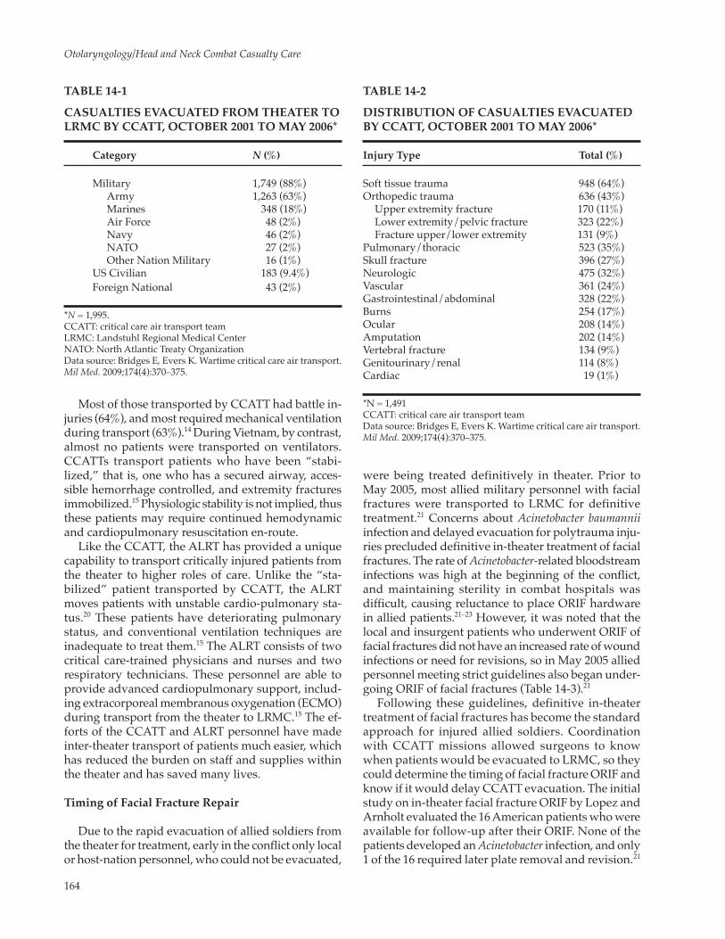
Figure 14-3. A US soldier who sustained a right mandible ramus fracture, facial soft tissue avulsion, and right ear avulsion (a). At 8 months postoperative repair in theater (b), the patient has scarring, but had required no further maxillofacial treatment.
TABLE 14-3
GUIDELINES FOR IN-THEATER TREATMENT OF ALLIED PERSONNEL WITH FACIAL FRAC-TURES
Criteria Description
1 The fracture site was exposed either through a soft tissue wound or a surgical approach (eg, a frontal sinus fracture exposed by a bicoronal flap during a craniotomy).
2 Definitive treatment of the fracture would not delay evacuation of the patient from the theater.
3 Treatment of the facial fracture would allow the patient to remain in theater.
Data source: Lopez MA, Arnholt JL. Safety of definitive in-theater repair of facial fractures. Arch Facial Plast Surg. 2007;9(6):400–405.
a b
In-theater management of facial fractures has several advantages. It has been shown that delaying fracture fixation can lead to increased technical dif-ficulties and infectious complications.24,25 Soft tissue contracture around an untreated fracture and bony fibrosis can make it more difficult to completely reduce
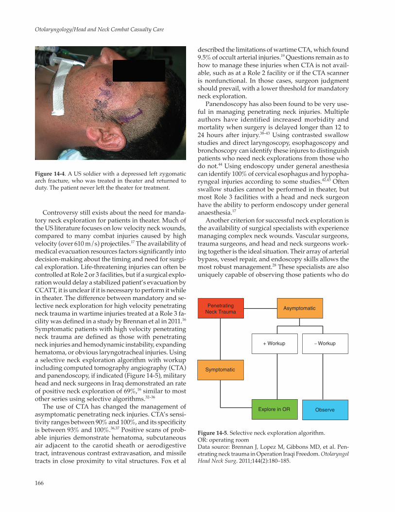
the fracture. Nerve injuries and malocclusion can occur with mandible fractures that have delayed treatment.24 Primary treatment and closure of soft tissue injuries over an ORIF also reduces the need for further facial surgery in patients who are evacuated to higher roles of care (Figure 14-3).21 Also, some patients treated in theater are able to return to their units, reducing critical personnel shortages (Figure 14-4).
Timing of Neck Exploration
Penetrating neck trauma in World War I resulted in a mortality rate of about 16%, possibly because nonsurgical management prevailed. The mortality rate was reduced to 7% during World War II, due to mandatory neck explorations.17,26,27 In Vietnam, surgi-cal management of penetrating neck trauma reduced the mortality rate to between 4% and 7%. Brennan et al reported a 3.7% mortality rate during a 4-year period of the Iraq and Afghanistan conflicts.16 This is comparable to a perioperative mortality of about 3% for civilian patients undergoing neck exploration for low velocity penetrating neck trauma.28 High velocity neck wounds are common in wartime injuries,17,29–31 as are injures to vascular, nervous, and laryngotracheal structures.16,30
166
Otolaryngology/Head and Neck Combat Casualty Care
Figure 14-4. A US soldier with a depressed left zygomatic arch fracture, who was treated in theater and returned to duty. The patient never left the theater for treatment.
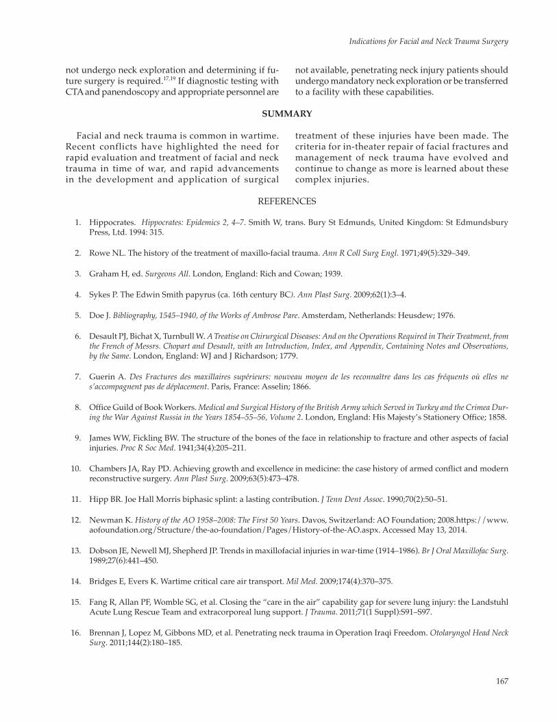
Figure 14-5. Selective neck exploration algorithm.OR: operating roomData source: Brennan J, Lopez M, Gibbons MD, et al. Pen-etrating neck trauma in Operation Iraqi Freedom. Otolaryngol Head Neck Surg. 2011;144(2):180–185.
Controversy still exists about the need for manda-tory neck exploration for patients in theater. Much of the US literature focuses on low velocity neck wounds, compared to many combat injuries caused by high velocity (over 610 m/s) projectiles.17 The availability of medical evacuation resources factors significantly into decision-making about the timing and need for surgi-cal exploration. Life-threatening injuries can often be controlled at Role 2 or 3 facilities, but if a surgical explo-ration would delay a stabilized patient’s evacuation by CCATT, it is unclear if it is necessary to perform it while in theater. The difference between mandatory and se-lective neck exploration for high velocity penetrating neck trauma in wartime injuries treated at a Role 3 fa-cility was defined in a study by Brennan et al in 2011.16 Symptomatic patients with high velocity penetrating neck trauma are defined as those with penetrating neck injuries and hemodynamic instability, expanding hematoma, or obvious laryngotracheal injuries. Using a selective neck exploration algorithm with workup including computed tomography angiography (CTA) and panendoscopy, if indicated (Figure 14-5), military head and neck surgeons in Iraq demonstrated an rate of positive neck exploration of 69%,16 similar to most other series using selective algorithms.32–36
The use of CTA has changed the management of asymptomatic penetrating neck injuries. CTA’s sensi-tivity ranges between 90% and 100%, and its specificity is between 93% and 100%.36,37 Positive scans of prob-able injuries demonstrate hematoma, subcutaneous air adjacent to the carotid sheath or aerodigestive tract, intravenous contrast extravasation, and missile tracts in close proximity to vital structures. Fox et al
described the limitations of wartime CTA, which found 9.5% of occult arterial injuries.19 Questions remain as to how to manage these injuries when CTA is not avail-able, such as at a Role 2 facility or if the CTA scanner is nonfunctional. In those cases, surgeon judgment should prevail, with a lower threshold for mandatory neck exploration.
Panendoscopy has also been found to be very use-ful in managing penetrating neck injuries. Multiple authors have identified increased morbidity and mortality when surgery is delayed longer than 12 to 24 hours after injury.38–43 Using contrasted swallow studies and direct laryngoscopy, esophagoscopy and bronchoscopy can identify these injures to distinguish patients who need neck explorations from those who do not.44 Using endoscopy under general anesthesia can identify 100% of cervical esophagus and hypopha-ryngeal injuries according to some studies.42,43 Often swallow studies cannot be performed in theater, but most Role 3 facilities with a head and neck surgeon have the ability to perform endoscopy under general anaesthesia.17
Another criterion for successful neck exploration is the availability of surgical specialists with experience managing complex neck wounds. Vascular surgeons, trauma surgeons, and head and neck surgeons work-ing together is the ideal situation. Their array of arterial bypass, vessel repair, and endoscopy skills allows the most robust management.28 These specialists are also uniquely capable of observing those patients who do
PenetratingNeck Trauma
Asymptomatic
Explore in OR
+ Workup
Symptomatic
– Workup
Observe
167
Indications for Facial and Neck Trauma Surgery
not undergo neck exploration and determining if fu-ture surgery is required.17,19 If diagnostic testing with CTA and panendoscopy and appropriate personnel are
not available, penetrating neck injury patients should undergo mandatory neck exploration or be transferred to a facility with these capabilities.
SUMMARY
Facial and neck trauma is common in wartime. Recent conflicts have highlighted the need for rapid evaluation and treatment of facial and neck trauma in time of war, and rapid advancements in the development and application of surgical
treatment of these injuries have been made. The criteria for in-theater repair of facial fractures and management of neck trauma have evolved and continue to change as more is learned about these complex injuries.
REFERENCES
1. Hippocrates. Hippocrates: Epidemics 2, 4–7. Smith W, trans. Bury St Edmunds, United Kingdom: St Edmundsbury Press, Ltd. 1994: 315.
2. Rowe NL. The history of the treatment of maxillo-facial trauma. Ann R Coll Surg Engl. 1971;49(5):329–349.
3. Graham H, ed. Surgeons All. London, England: Rich and Cowan; 1939.
4. Sykes P. The Edwin Smith papyrus (ca. 16th century BC). Ann Plast Surg. 2009;62(1):3–4.
5. Doe J. Bibliography, 1545–1940, of the Works of Ambrose Pare. Amsterdam, Netherlands: Heusdew; 1976.
6. Desault PJ, Bichat X, Turnbull W. A Treatise on Chirurgical Diseases: And on the Operations Required in Their Treatment, from the French of Messrs. Chopart and Desault, with an Introduction, Index, and Appendix, Containing Notes and Observations, by the Same. London, England: WJ and J Richardson; 1779.
7. Guerin A. Des Fractures des maxillaires supérieurs: nouveau moyen de les reconnaître dans les cas fréquents où elles ne s’accompagnent pas de déplacement. Paris, France: Asselin; 1866.
8. Office Guild of Book Workers. Medical and Surgical History of the British Army which Served in Turkey and the Crimea Dur-ing the War Against Russia in the Years 1854–55–56, Volume 2. London, England: His Majesty’s Stationery Office; 1858.
9. James WW, Fickling BW. The structure of the bones of the face in relationship to fracture and other aspects of facial injuries. Proc R Soc Med. 1941;34(4):205–211.
10. Chambers JA, Ray PD. Achieving growth and excellence in medicine: the case history of armed conflict and modern reconstructive surgery. Ann Plast Surg. 2009;63(5):473–478.
11. Hipp BR. Joe Hall Morris biphasic splint: a lasting contribution. J Tenn Dent Assoc. 1990;70(2):50–51.
12. Newman K. History of the AO 1958–2008: The First 50 Years. Davos, Switzerland: AO Foundation; 2008.https://www.aofoundation.org/Structure/the-ao-foundation/Pages/History-of-the-AO.aspx. Accessed May 13, 2014.
13. Dobson JE, Newell MJ, Shepherd JP. Trends in maxillofacial injuries in war-time (1914–1986). Br J Oral Maxillofac Surg. 1989;27(6):441–450.
14. Bridges E, Evers K. Wartime critical care air transport. Mil Med. 2009;174(4):370–375.
15. Fang R, Allan PF, Womble SG, et al. Closing the “care in the air” capability gap for severe lung injury: the Landstuhl Acute Lung Rescue Team and extracorporeal lung support. J Trauma. 2011;71(1 Suppl):S91–S97.
16. Brennan J, Lopez M, Gibbons MD, et al. Penetrating neck trauma in Operation Iraqi Freedom. Otolaryngol Head Neck Surg. 2011;144(2):180–185.
168
Otolaryngology/Head and Neck Combat Casualty Care
17. Brennan JA, Meyers AD, Jafek BW. Penetrating neck trauma: a 5-year review of the literature, 1983 to 1988. Am J Oto-laryngol. 1990;11(3):191–197.
18. Champion HR, Bellamy RF, Roberts CP, Leppaniemi A. A profile of combat injury. J Trauma. 2003;54(5 Suppl):S13–S19.
19. Fox CJ, Gillespie DL, Weber MA, et al. Delayed evaluation of combat-related penetrating neck trauma. J Vasc Surg. 2006;44(1):86–93.
20. Dorlac GR, Fang R, Pruitt VM, et al. Air transport of patients with severe lung injury: development and utilization of the Acute Lung Rescue Team. J Trauma. 2009;66(4 Suppl):S164–S171.
21. Lopez MA, Arnholt JL. Safety of definitive in-theater repair of facial fractures. Arch Facial Plast Surg. 2007;9(6):400–405.
22. Davis KA, Moran KA, McAllister CK, Gray PJ. Multidrug-resistant Acinetobacter extremity infections in soldiers. Emerg Infect Dis. 2005;11(8):1218–1224.
23. Centers for Disease Control and Prevention. Acinetobacter baumannii infections among patients at military medical facilities treating injured US service members, 2002–2004. MMWR Morb Mortal Wkly Rep. 2004;53(45):1063–1066.
24. Biller JA, Pletcher SD, Goldberg AN, Murr AH. Complications and the time to repair of mandible fractures. Laryngo-scope. 2005;115(5):769–772.
25. Maloney PL, Lincoln RE, Coyne CP. A protocol for the management of compound mandibular fractures based on the time from injury to treatment. J Oral Maxillofac Surg. 2001;59(8):879–884; discussion 885–886.
26. Obeid FN, Haddad GS, Horst HM, Bivins BA. A critical reappraisal of a mandatory exploration policy for penetrating wounds of the neck. Surg Gynecol Obstet. 1985;160(6):517–522.
27. Narrod JA, Moore EE. Selective management of penetrating neck injuries. A prospective study. Arch Surg. 1984;119(5):574–578.
28. Biffl WL, Moore EE, Rehse DH, Offner PJ, Franciose RJ, Burch JM. Selective management of penetrating neck trauma based on cervical level of injury. Am J Surg. 1997;174(6):678–682.
29. Feldt BA, Salinas NL, Rasmussen TE, Brennan J. The joint facial and invasive neck trauma (J-FAINT) project, Iraq and Afghanistan 2003-2011. Otolaryngol Head Neck Surg. 2013;148(3):403–408.
30. Owens BD, Kragh JF Jr, Wenke JC, Macaitis J, Wade CE, Holcomb JB. Combat wounds in operation Iraqi Freedom and operation Enduring Freedom. J Trauma. 2008;64(2):295–299.
31. Clouse WD, Rasmussen TE, Peck MA, et al. In-theater management of vascular injury: 2 years of the Balad Vascular Registry. J Am Coll Surg. 2007;204(4):625-632.
32. Carducci B, Lowe RA, Dalsey W. Penetrating neck trauma: consensus and controversies. Ann Emerg Med. 1986;15(2):208–215.
33. Jurkovich GJ, Zingarelli W, Wallace J, Curreri PW. Penetrating neck trauma: diagnostic studies in the asymptomatic patient. J Trauma. 1985;25(9):819–822.
34. Sclafani SJ, Panetta T, Goldstein AS, et al. The management of arterial injuries caused by penetration of zone III of the neck. J Trauma. 1985;25(9):871–881.
35. Velmahos GC, Souter I, Degiannis E, Mokoena T, Saadia R. Selective surgical management in penetrating neck injuries. Can J Surg. 1994;37(6):487–491.
36. Osborn TM, Bell RB, Qaisi W, Long WB. Computed tomographic angiography as an aid to clinical decision making in the selective management of penetrating injuries to the neck: a reduction in the need for operative exploration. J Trauma. 2008;64(6):1466–1471.
169
Indications for Facial and Neck Trauma Surgery
37. Munera F, Danton G, Rivas LA, Henry RP, Ferrari MG. Multidetector row computed tomography in the management of penetrating neck injuries. Semin Ultrasound CT MR. 2009;30(3):195–204.
38. Demetriades D, Velmahos GG, Asensio JA. Cervical pharyngoesophageal and laryngotracheal injuries. World J Surg. 2001;25(8):1044–1048.
39. Asensio JA, Berne J, Demetriades D, et al. Penetrating esophageal injuries: time interval of safety for preoperative evaluation—how long is safe? J Trauma. 1997;43(2):319–324.
40. Stanley RB Jr, Armstrong WB, Fetterman BL, Shindo ML. Management of external penetrating injuries into the hypopharyngeal-cervical esophageal funnel. J Trauma. 1997;42(4):675–679.
41. Bryant AS, Cerfolio RJ. Esophageal trauma. Thorac Surg Clin. 2007;17(1):63–72.
42. Ahmed N , Massier C, Tassie J, Whalen J, Chung R. Diagnosis of penetrating injuries of the pharynx and esophagus in the severely injured patient. J Trauma. 2009;67(1):152–154.
43. Armstrong WB, Detar TR, Stanley RB. Diagnosis and management of external penetrating cervical esophageal injuries. Ann Otol Rhinol Laryngol. 1994;103(11):863–871.
44. Tisherman SA, Bokhari F, Collier B, et al. Clinical practice guideline: penetrating zone II neck trauma. J Trauma. 2008;64(5):1392–1405.

170
Otolaryngology/Head and Neck Combat Casualty Care


![Cervical Lymphadenopathy Diagnosis and Management1].pdf · –PET CT + MRI neck –No evidence of Primary ... (SPIRO, STRONG 1973) ... INDICATIONS FOR PROPHYLACTIC NECK DISSECTION](https://img.pdfslide.us/doc/110x75/5b3272737f8b9a2c328d70ce/cervical-lymphadenopathy-diagnosis-and-1pdf-pet-ct-mri-neck-no-evidence.jpg)