Embed Size (px)
Citation preview
CHAPTER 11
Polymer-Based Prodrugs forCancer Chemotherapy
QIHANG SUNb, JINQIANG WANGa, MACIEJ RADOSZb
AND YOUQING SHEN*a
a Center for Bionanoengineering and State Key Laboratory of Chemical
Engineering, Department of Chemical and Biological Engineering, Zhejiang
University, Hangzhou 310027, P. R. China; b Department of Chemical and
Petroleum Engineering, Soft Materials Laboratory, University of Wyoming,
Laramie, WY 82071, USA
*E-mail: [email protected]
11.1 Introduction
Chemotherapy has achieved great success in cancer treatment during recent
past decades, but it is still challenged by poor solubility, low tumor selectivity,
and associated toxicity of most anticancer drugs.1 The prodrug strategy is one
of the most commonly used chemical/biochemical strategies towards improv-
ing the therapeutic index of anticancer drugs.2
A prodrug is defined as a chemically modified drug derivative that is inactive
or less active but metabolized in vivo to release the parent active component in
the pharmacological environment3 to improve the drug’s desirability, including
water solubility, patient acceptability (e.g. decreasing pain on injection), and
pharmacokinetics (absorption, biodistribution, metabolism, and elimination;
ADME). A typical prodrug normally consists of three parts (Figure 11.1): (1)
the parent drug exerting therapeutic effects; (2) the chemical linker bridging the
parent drug and the modifier; and (3) the modifier endowing the prodrug with
RSC Polymer Chemistry Series No. 3
Functional Polymers for Nanomedicine
Edited by Youqing Shen
# The Royal Society of Chemistry 2013
Published by the Royal Society of Chemistry, www.rsc.org
245
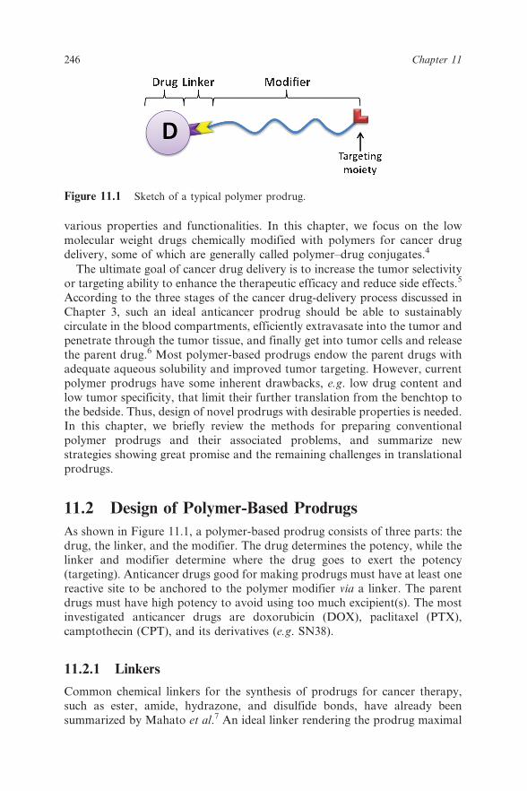
various properties and functionalities. In this chapter, we focus on the low
molecular weight drugs chemically modified with polymers for cancer drug
delivery, some of which are generally called polymer–drug conjugates.4
The ultimate goal of cancer drug delivery is to increase the tumor selectivity
or targeting ability to enhance the therapeutic efficacy and reduce side effects.5
According to the three stages of the cancer drug-delivery process discussed in
Chapter 3, such an ideal anticancer prodrug should be able to sustainably
circulate in the blood compartments, efficiently extravasate into the tumor and
penetrate through the tumor tissue, and finally get into tumor cells and release
the parent drug.6 Most polymer-based prodrugs endow the parent drugs withadequate aqueous solubility and improved tumor targeting. However, current
polymer prodrugs have some inherent drawbacks, e.g. low drug content and
low tumor specificity, that limit their further translation from the benchtop to
the bedside. Thus, design of novel prodrugs with desirable properties is needed.
In this chapter, we briefly review the methods for preparing conventional
polymer prodrugs and their associated problems, and summarize new
strategies showing great promise and the remaining challenges in translational
prodrugs.
11.2 Design of Polymer-Based Prodrugs
As shown in Figure 11.1, a polymer-based prodrug consists of three parts: the
drug, the linker, and the modifier. The drug determines the potency, while the
linker and modifier determine where the drug goes to exert the potency
(targeting). Anticancer drugs good for making prodrugs must have at least one
reactive site to be anchored to the polymer modifier via a linker. The parent
drugs must have high potency to avoid using too much excipient(s). The mostinvestigated anticancer drugs are doxorubicin (DOX), paclitaxel (PTX),
camptothecin (CPT), and its derivatives (e.g. SN38).
11.2.1 Linkers
Common chemical linkers for the synthesis of prodrugs for cancer therapy,
such as ester, amide, hydrazone, and disulfide bonds, have already been
summarized by Mahato et al.7 An ideal linker rendering the prodrug maximal
Figure 11.1 Sketch of a typical polymer prodrug.
246 Chapter 11
therapeutic efficacy and minimal toxicity would be the one that is stable in the
blood compartments but labile in cancer cells.
The linker should first be stable in blood circulation to ensure low toxicity.8
For example, the PK1 conjugate in which DOX was covalently bound to
poly[N-(2-hydroxypropyl)methacrylamide] (PHPMA) via a blood-stable but
lysosome-labile peptidyl linker had very low dose-limiting toxicity.9 In phase I
clinical and pharmacokinetic studies, PK1 had a maximum tolerated dose
(MTD) of 320 mg m22, and no congestive cardiac failure despite individual
cumulative doses up to 1,680 mg m22.9 However, both the PHPMA-
PTX prodrug named PNU16694510 and the poly(methacryloylglycinamide)
(PMAG)-CPT prodrug11 experienced dose-limiting toxicity in phase I clinical
study because their easily hydrolysable ester linkers released the drugs while in
circulation.12
Upon reaching the intracellular target, the prodrug must efficiently release
the parent drug to exert its pharmaceutical action because only the liberated
drug becomes active.13 Stable prodrugs, e.g. drugs bonded to poly(lactic-co-
glycolic acid) (PLGA)14 or poly(L-aspartic acid) [P(Asp)],15 showed low or
even no anticancer activity. It is preferable that the linker be cleavable in the
tumor microenvironment. This is achieved by using labile linkers responsive to
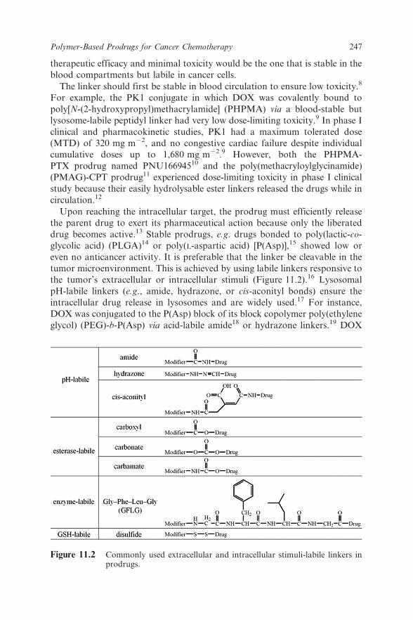
the tumor’s extracellular or intracellular stimuli (Figure 11.2).16 Lysosomal
pH-labile linkers (e.g., amide, hydrazone, or cis-aconityl bonds) ensure the
intracellular drug release in lysosomes and are widely used.17 For instance,
DOX was conjugated to the P(Asp) block of its block copolymer poly(ethylene
glycol) (PEG)-b-P(Asp) via acid-labile amide18 or hydrazone linkers.19 DOX
Figure 11.2 Commonly used extracellular and intracellular stimuli-labile linkers inprodrugs.
Polymer-Based Prodrugs for Cancer Chemotherapy 247
conjugated to PHPMA and biodegradable star polymers via such linkers
demonstrated a fast DOX release at pH 4.20 An interesting example is the
dual pH-responsive prodrug PPC-Hyd-DOX-DA developed by Du and co-
workers.21 At tumor extracellular pH (y6.8), the prodrug reversed its surface
charge from negative to positive via acid-labile amide bonds for fast
internalization; at endosomal or lysosomal pH (y5.0), the drug was fast
released due to the breakable hydrazone bonds. Esterases are ubiquitously
distributed in the body and can readily hydrolyze ester bonds in prodrugs;
thereby esterase-labile linkers, such as carboxyl, carbonate, and carbamate
esters, are also employed.22 For instance, in IT-101, CPT was conjugated to the
linear b-cyclodextrin (b-CD) and PEG copolymer via the ester bond.23 The
conformation of released CPT was not changed compared to the parent CPT
and the release rate of the conjugated CPT can be tuned.24 Lysosomal
degradable peptides [e.g., glycylphenylalanylleucylglycine (GFLG)], which are
cleavable by lysosomal enzymes, are also useful linkers in prodrugs.25
Recently, the disulfide bond,26 which can be cleaved by intracellular
glutathione (GSH), has attracted increasing attention as linkers for intracellu-
larly triggered drug release. The underlying rationale is the elevated
intracellular GSH concentration, particularly in cancer cells, but low in the
blood.27
11.2.2 Modifiers
The modifier may consist of a polymer chain and targeting moieties and others
such as a tracer moiety. The main roles of the polymer chain are to endow the
prodrug with water solubility and a long blood-circulation time for altered
ADME and tumor targeting capability. The targeting moiety facilitates the
prodrug’s active targeting, as discussed in Chapter 2.
Most anticancer drugs are water insoluble, giving them poor bioavail-
ability.28 Anchoring drugs to water-soluble polymer chains makes them water
soluble. For instance, PTX is an extremely water-insoluble anticancer drug
(,0.01 mg mL21).29 Conjugating PTX to a PEG30 or poly(L-glutamic acid)
(PGA)31 resulted in highly water-soluble prodrugs with antitumor effects
superior to PTX itself.31,32
It is generally assumed that by prolonging the blood circulation time, a
polymer prodrug has more opportunity to pass through the hyperpermeable
tumor blood vessels and extravasate into tumor tissue via the EPR effect.1b
Prodrugs with a stealth property may evade the reticuloendothelial system
(RES) screening33 and thus circulate for a long time in the blood
compartments, resulting in greatly increased tumor drug concentrations (10-
fold or higher) and MTD relative to administration of the free drug.34 The
stealth character of a polymer prodrug is mainly determined by the polymer’s
properties. The polymer must be water soluble and, very importantly, not
immunogenic. A very interesting example is the natural biopolymer dextran. It
is usually assumed to be non-immunogenic, but drugs conjugated to dextran
248 Chapter 11
and carboxymethyldextran can be captured by the RES, causing dose-limiting
toxicity.35 PHPMA36 and PEG37 are the most studied bio-inert polymers able
to render prodrugs stealthy. For example, NKTR-102, a PEG prodrug of
irinotecan (a chemotherapy drug that is metabolized to its active metabolite
SN38 in the body), increased the half-life of SN38 90-fold (from 4 h to 15 days)
and 10-fold in colorectal and lung cancer treatment, respectively.38 Other
PEG-based prodrugs of docetaxel (NKTR-105), SN38 (EZN-2208), and CPT
(Pegamotecan) also showed longer half-life times and increased accumulation
at tumor sites and thereby tumor growth suppression.39
Incorporating targeting ligands in the polymer modifier, such as antibodies,
peptides, aptamers, and folic acid, may promote tumor targeting (building on
the EPR effect) by receptor-mediated delivery,40 as discussed in Chapter 2. For
example, PK1 conjugated with galactosamine, named PK2,41 delivered 3.3 ¡
5.6% of the dose to the tumor and 16.9 ¡ 3.9% to the liver, whereas
PK1 showed no obvious targeting.42 Zhu et al.43 and Borgman et al.44
also proved that incorporating targeting ligands in polymer modifiers in-
creased the prodrug’s tumor accumulation. More examples have been reviewed
elsewhere.45
11.2.3 Drawbacks of Current Polymer-Based Prodrugs
An inherent dilemma in polymer prodrugs is their drug-loading content versus
the water solubility and thereby the stealth capability. A high drug-loading
content in a nanocarrier reduces use of excipients and minimizes the related
side effects, and should be considered as a demanding criterion to judge a
prodrug’s quality.46 As most parent drugs are highly hydrophobic, the
modifier or the water-soluble polymer chain must be sufficiently long to make
the prodrug water soluble. Therefore, in most polymer prodrugs the drug
content is less than 10 wt%.47 Typical examples are PEG-DOX prodrugs using
PEG with different molecular weights from 5,000 to 20,000 g mol21, which
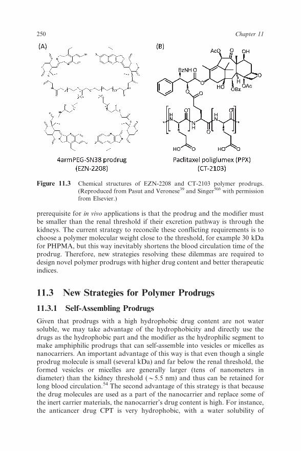
contain 2.7–8.0 wt% DOX.48 A four-armed PEG-SN38 prodrug named EZN-
2208, prepared by coupling SN38 to multiarm PEGs (Figure 11.3A), had an
increased drug content, but only of 3.7 wt%, compared to the CPT-PEG-diol
prodrug Pegamotecan.39 As for PHPMA–drug conjugates, for instance PK1,
the drug contents are also as low as 8 wt%.49 Some prodrugs have high drug-
loading contents. For instance, the prodrug CT-2103, PTX conjugated to PGA
through its 29-hydroxyl group (Figure 11.3B),50 has a drug content of
approximately 37 wt% and a PGA-20(S)-CPT prodrug has a 30–35 wt% drug
content.51 However, increasing the drug content not only lowers the prodrug
water solubility but may also cause opsonization,37 resulting in rapid blood
clearance.
Another complication is also related to the polymer chain’s molecular
weight. To have a long blood-circulation time for passive tumor targeting, the
molecular weight of a prodrug must be higher than the polymer’s renal
threshold, e.g. 40 kDa for PEG52 and 45 kDa for PHPMA,53 but the safety
Polymer-Based Prodrugs for Cancer Chemotherapy 249
prerequisite for in vivo applications is that the prodrug and the modifier must
be smaller than the renal threshold if their excretion pathway is through the
kidneys. The current strategy to reconcile these conflicting requirements is to
choose a polymer molecular weight close to the threshold, for example 30 kDa
for PHPMA, but this way inevitably shortens the blood circulation time of theprodrug. Therefore, new strategies resolving these dilemmas are required to
design novel polymer prodrugs with higher drug content and better therapeutic
indices.
11.3 New Strategies for Polymer Prodrugs
11.3.1 Self-Assembling Prodrugs
Given that prodrugs with a high hydrophobic drug content are not water
soluble, we may take advantage of the hydrophobicity and directly use the
drugs as the hydrophobic part and the modifier as the hydrophilic segment to
make amphiphilic prodrugs that can self-assemble into vesicles or micelles asnanocarriers. An important advantage of this way is that even though a single
prodrug molecule is small (several kDa) and far below the renal threshold, the
formed vesicles or micelles are generally larger (tens of nanometers in
diameter) than the kidney threshold (y5.5 nm) and thus can be retained for
long blood circulation.54 The second advantage of this strategy is that because
the drug molecules are used as a part of the nanocarrier and replace some of
the inert carrier materials, the nanocarrier’s drug content is high. For instance,
the anticancer drug CPT is very hydrophobic, with a water solubility of
Figure 11.3 Chemical structures of EZN-2208 and CT-2103 polymer prodrugs.
(Reproduced from Pasut and Veronese39 and Singer50b with permission
from Elsevier.)
250 Chapter 11
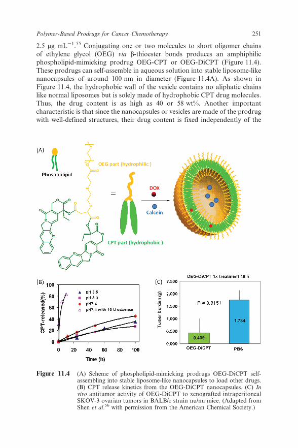
2.5 mg mL21.55 Conjugating one or two molecules to short oligomer chains
of ethylene glycol (OEG) via b-thioester bonds produces an amphiphilic
phospholipid-mimicking prodrug OEG-CPT or OEG-DiCPT (Figure 11.4).
These prodrugs can self-assemble in aqueous solution into stable liposome-like
nanocapsules of around 100 nm in diameter (Figure 11.4A). As shown in
Figure 11.4, the hydrophobic wall of the vesicle contains no aliphatic chains
like normal liposomes but is solely made of hydrophobic CPT drug molecules.
Thus, the drug content is as high as 40 or 58 wt%. Another important
characteristic is that since the nanocapsules or vesicles are made of the prodrug
with well-defined structures, their drug content is fixed independently of the
Figure 11.4 (A) Scheme of phospholipid-mimicking prodrugs OEG-DiCPT self-assembling into stable liposome-like nanocapsules to load other drugs.(B) CPT release kinetics from the OEG-DiCPT nanocapsules. (C) Invivo antitumor activity of OEG-DiCPT to xenografted intraperitonealSKOV-3 ovarian tumors in BALB/c strain nu/nu mice. (Adapted fromShen et al.56 with permission from the American Chemical Society.)
Polymer-Based Prodrugs for Cancer Chemotherapy 251
nanocarrier size and preparation methods. This character makes it easy to
scale-up for translation, as discussed in Chapter 3. The nanocapsules have no
burst release but can release CPT quickly once inside the cells (Figure 11.4B).
In vivo tests showed that the nanocapsules had strong antitumor activity
(Figure 11.4C).56
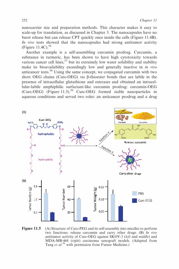
Another example is a self-assembling curcumin prodrug. Curcumin, a
substance in turmeric, has been shown to have high cytotoxicity towards
various cancer cell lines,57 but its extremely low water solubility and stability
make its bioavailability exceedingly low and generally inactive in in vivo
anticancer tests.58 Using the same concept, we conjugated curcumin with two
short OEG chains (Curc-OEG) via b-thioester bonds that are labile in the
presence of intracellular glutathione and esterases and obtained an intracel-
lular-labile amphiphilic surfactant-like curcumin prodrug: curcumin-OEG
(Curc-OEG) (Figure 11.5).59 Curc-OEG formed stable nanoparticles in
aqueous conditions and served two roles: an anticancer prodrug and a drug
Figure 11.5 (A) Structure of Curc-PEG and its self-assembly into micelles to performtwo functions: release curcumin and carry other drugs. (B) In vivoantitumor activity of Curc-OEG against SKOV-3 (left and middle) andMDA-MB-468 (right) carcinoma xenograft models. (Adapted fromTang et al.59 with permission from Future Medicine.)
252 Chapter 11
carrier (Figure 11.5A). As an anticancer prodrug, the formed nanoparticles
had a high and fixed curcumin-loading content of 25.3 wt%, and released
active curcumin in the intracellular environment. Curc-OEG had high
inhibition ability for several cancer cell lines due to apoptosis. Intravenously
injected Curc-OEG had a bioavailability (the area under curve value) 250 times
of that of the parent curcumin, and thus a high overall tumor concentration.
As a result, it significantly reduced tumor weights and tumor numbers in the
athymic mice xenografted with intraperitoneal SKOV-3 tumors and subcuta-
neous (mammary fat pad) MDA-MB-468 tumors (Figure 11.5B). Preliminary
systemic toxicity studies found that Curc-OEG did not cause acute or
subchronic toxicities in mouse visceral organs at high doses. As a drug carrier,
Curc-OEG nanoparticles could carry other anticancer drugs, such as DOX,
and ship them into drug-resistant cells, greatly enhancing the cytotoxicity of
the loaded drug. Thus, Curc-OEG is a promising prototype that merits further
study for cancer therapy.
11.3.2 Prodrug Micelles
Besides the phospholipid-mimicking amphiphilic prodrugs, another type of
prodrug capable of self-assembly is the diblock copolymer in which one block
is a water-soluble chain such as PEG and the other is conjugated with
hydrophobic drug molecules as the water-insoluble block. Such block
copolymers form micelles in aqueous solution with drug molecules anchored
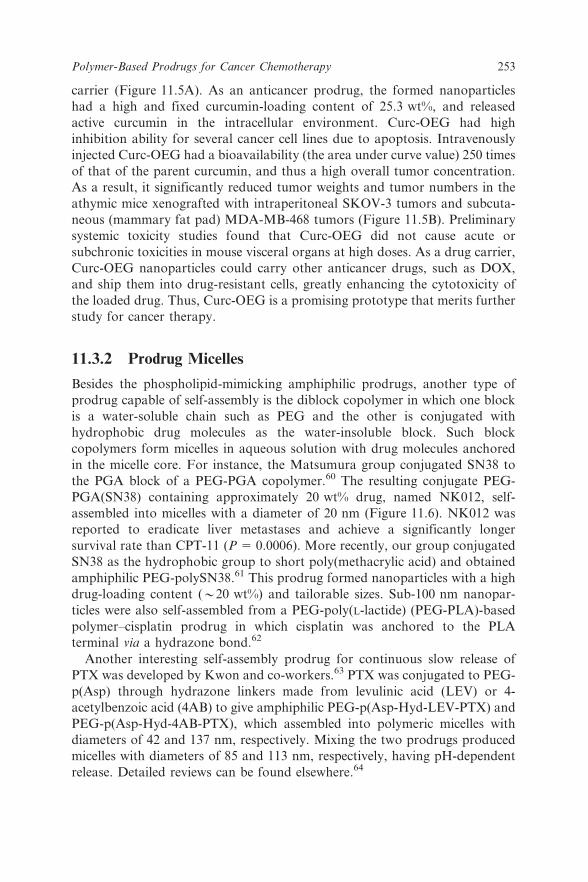
in the micelle core. For instance, the Matsumura group conjugated SN38 to
the PGA block of a PEG-PGA copolymer.60 The resulting conjugate PEG-
PGA(SN38) containing approximately 20 wt% drug, named NK012, self-
assembled into micelles with a diameter of 20 nm (Figure 11.6). NK012 was
reported to eradicate liver metastases and achieve a significantly longer
survival rate than CPT-11 (P 5 0.0006). More recently, our group conjugated
SN38 as the hydrophobic group to short poly(methacrylic acid) and obtained
amphiphilic PEG-polySN38.61 This prodrug formed nanoparticles with a high
drug-loading content (y20 wt%) and tailorable sizes. Sub-100 nm nanopar-
ticles were also self-assembled from a PEG-poly(L-lactide) (PEG-PLA)-based
polymer–cisplatin prodrug in which cisplatin was anchored to the PLA
terminal via a hydrazone bond.62
Another interesting self-assembly prodrug for continuous slow release of
PTX was developed by Kwon and co-workers.63 PTX was conjugated to PEG-
p(Asp) through hydrazone linkers made from levulinic acid (LEV) or 4-
acetylbenzoic acid (4AB) to give amphiphilic PEG-p(Asp-Hyd-LEV-PTX) and
PEG-p(Asp-Hyd-4AB-PTX), which assembled into polymeric micelles with
diameters of 42 and 137 nm, respectively. Mixing the two prodrugs produced
micelles with diameters of 85 and 113 nm, respectively, having pH-dependent
release. Detailed reviews can be found elsewhere.64
Polymer-Based Prodrugs for Cancer Chemotherapy 253
11.3.3 Drug Polymers
An alternative to using the drug molecules themselves as a part of the
nanocarrier is to directly use biofunctional drug molecules as monomers to
prepare backbone-type polymer–drug conjugates, or drug polymers. One suchexample is polycurcumin (PCurc; Figure 11.7).65 Inspired by the bihydroxyl
functionality of the curcumin molecule, curcumin was used as a co-monomer
to make the curcumin-containing PCurc by polycondensation polymerization.
A series of water-soluble PCurcs were prepared with different covalent bonds
(e.g., ester, disulfide, acetal) with high and fixed curcumin-loading content and
efficiency in comparison with previously developed curcumin-loading nano-
medicines.66 For instance, curcumin was condensed with divinyl ether to
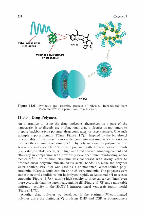
produce linear polycurcumin linked via acetal bonds. To make the polymerwater soluble, PEG-diol was used as a co-monomer. Water-soluble poly-
curcumin, PCurc 8, could contain up to 25 wt% curcumin. The polymers were
stable at neutral conditions, but hydrolyzed rapidly at lysosomal pH to release
curcumin (Figure 11.7A), causing high toxicity to three cancer cell lines (even
more cytotoxic than the parent curcumin itself) (Figure 11.7B) and remarkable
antitumor activity in the SKOV-3 intraperitoneal xenograft tumor model
(Figure 11.7C).
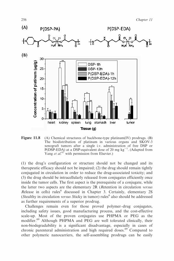
Another drug polymer we developed is the platinum(IV)-coordinated
polymer using the platinum(IV) prodrugs DHP and DSP as co-monomers
Figure 11.6 Synthesis and assembly process of NK012. (Reproduced fromMatsumura60a with permission from Elsevier.)
254 Chapter 11
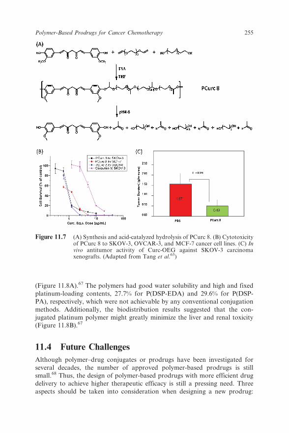
(Figure 11.8A).67 The polymers had good water solubility and high and fixed
platinum-loading contents, 27.7% for P(DSP-EDA) and 29.6% for P(DSP-
PA), respectively, which were not achievable by any conventional conjugation
methods. Additionally, the biodistribution results suggested that the con-
jugated platinum polymer might greatly minimize the liver and renal toxicity
(Figure 11.8B).67
11.4 Future Challenges
Although polymer–drug conjugates or prodrugs have been investigated for
several decades, the number of approved polymer-based prodrugs is still
small.68 Thus, the design of polymer-based prodrugs with more efficient drug
delivery to achieve higher therapeutic efficacy is still a pressing need. Three
aspects should be taken into consideration when designing a new prodrug:
Figure 11.7 (A) Synthesis and acid-catalyzed hydrolysis of PCurc 8. (B) Cytotoxicityof PCurc 8 to SKOV-3, OVCAR-3, and MCF-7 cancer cell lines. (C) Invivo antitumor activity of Curc-OEG against SKOV-3 carcinomaxenografts. (Adapted from Tang et al.65)
Polymer-Based Prodrugs for Cancer Chemotherapy 255
(1) the drug’s configuration or structure should not be changed and its
therapeutic efficacy should not be impaired; (2) the drug should remain tightly
conjugated in circulation in order to reduce the drug-associated toxicity; and
(3) the drug should be intracellularly released from conjugates efficiently once
inside the tumor cells. The first aspect is the prerequisite of a conjugate, while
the latter two aspects are the elementary 2R (Retention in circulation versus
Release in cells) rules6 discussed in Chapter 3. Certainly, elementary 2S
(Stealthy in circulation versus Sticky in tumor) rules6 also should be addressed
as further requirements of a superior prodrug.
Challenges remain even for those proved polymer–drug conjugates,
including safety issues, good manufacturing process, and the cost-effective
scale-up. Most of the proven conjugates use PHPMA or PEG as the
modifier.69 Although PHPMA and PEG are well tolerated clinically, their
non-biodegradability is a significant disadvantage, especially in cases of
chronic parenteral administration and high required doses.69 Compared to
other polymeric nanocarriers, the self-assembling prodrugs can be easily
Figure 11.8 (A) Chemical structures of backbone-type platinum(IV) prodrugs. (B)The biodistribution of platinum in various organs and SKOV-3xenograft tumors after a single i.v. administration of free DSP orP(DSP-EDA) at a DSP-equivalent dose of 20 mg kg21. (Adapted fromYang et al.67 with permission from Elsevier.)
256 Chapter 11
formulated with a fixed composition and a relatively good manufacturing
process, providing a novel design concept for translation of polymer-based
prodrugs from benchtop to the bedside.
References
1. (a) R. Tong, D. A. Christian, L. Tang, H. Cabral, J. R. Baker, Jr.,
K. Kataoka, D. E. Discher and J. Cheng, MRS Bull., 2009, 34, 422–431;
(b) F. Danhier, O. Feron and V. Preat, J. Controlled Release, 2010, 148,135–146.
2. J. S. Sohn, J. I. Jin, M. Hess and B. W. Jo, Polym. Chem., 2010, 1, 778–
792.
3. J. Rautio, H. Kumpulainen, T. Heimbach, R. Oliyai, D. Oh, T. Jarvinen
and J. Savolainen, Nat. Rev. Drug Discovery, 2008, 7, 255–270.
4. F. Greco and M. J. Vicent, Adv. Drug Delivery Rev., 2009, 61, 1203–1213.
5. I. Brigger, C. Dubernet and P. Couvreur, Adv. Drug Delivery Rev., 2002,
54, 631–651.
6. Q. Sun, M. Radosz and Y. Shen, J. Controlled Release, 2012, 164, 156–169.
7. R. Mahato, W. Tai and K. Cheng, Adv. Drug Delivery Rev., 2011, 63, 659–
670.
8. (a) J. W. Singer, S. Shaffer, B. Baker, A. Bernareggi, S. Stromatt,
D. Nienstedt and M. Besman, Anti-Cancer Drugs, 2005, 16, 243–254; (b)
S. Jaracz, J. Chen, L. V. Kuznetsova and L. Ojima, Bioorg. Med. Chem.,
2005, 13, 5043–5054.
9. P. A. Vasey, S. B. Kaye, R. Morrison, C. Twelves, P. Wilson, R. Duncan,
A. H. Thomson, L. S. Murray, T. E. Hilditch, T. Murray, S. Burtles,
D. Fraier, E. Frigerio and J. Cassidy, Clin. Cancer Res., 1999, 5, 83–94.
10. J. M. M. Terwogt, W. W. T. Huinink, J. H. M. Schellens, M. Schot,
I. A. M. Mandjes, M. G. Zurlo, M. Rocchetti, H. Rosing, F. J. Koopman
and J. H. Beijnen, Anti-Cancer Drugs, 2001, 12, 315–323.
11. N. E. Schoemaker, C. van Kesteren, H. Rosing, S. Jansen, M. Swart,
J. Lieverst, D. Fraier, M. Breda, C. Pellizzoni, R. Spinelli, M. G. Porro,
J. H. Beijnen, J. H. M. Schellens and W. W. T. Huinink, Br. J. Cancer.,
2002, 87, 608–614.
12. R. Duncan, Nat. Rev. Cancer, 2006, 6, 688–701.
13. (a) J. Kopecek, P. Kopeckova, T. Minko, Z. R. Lu and C. M. Peterson,
J. Controlled Release, 2001, 74, 147–158; (b) A. Malugin, P. Kopeckova
and J. Kopecek, J. Controlled Release, 2007, 124, 6–10.
14. H. S. Yoo, K. H. Lee, J. E. Oh and T. G. Park, J. Controlled Release, 2000,
68, 419–431.
15. M. Yokoyama, S. Fukushima, R. Uehara, K. Okamoto, K. Kataoka,
Y. Sakurai and T. Okano, J. Controlled Release, 1998, 50, 79–92.
16. (a) C. Wei, J. Guo and C. Wang, Macromol. Rapid Commun., 2011, 32,
451–455; (b) L. Wong, M. Kavallaris and V. Bulmus, Polym. Chem., 2011,2, 385–393.
Polymer-Based Prodrugs for Cancer Chemotherapy 257
17. (a) B. Rihova, T. Etrych, M. Sirova, L. Kovar, O. Hovorka, M. Kovar,
A. Benda and K. Ulbrich, Mol. Pharmaceutics, 2010, 7, 1027–1040; (b)
T. Etrych, L. Kovar, J. Strohalm, P. Chytil, B. Rihova and K. Ulbrich,
J. Controlled Release, 2011, 154, 241–248.
18. M. Yokoyama, G. S. Kwon, T. Okano, Y. Sakurai, T. Seto and
K. Kataoka, Bioconjugate Chem., 1992, 3, 295–301.
19. (a) M. Harada, I. Bobe, H. Saito, N. Shibata, R. Tanaka, T. Hayashi and
Y. Kato, Cancer Sci., 2011, 102, 192–199; (b) A. Ponta and Y. Bae, Pharm.
Res., 2010, 27, 2330–2342; (c) Y. Bae, A. W. G. Alani, N. C. Rockich,
T. S. Z. C. Lai and G. S. Kwon, Pharm. Res., 2010, 27, 2421–2432.
20. (a) K. Ulbrich, T. Etrych, P. Chytil, M. Pechar, M. Jelinkova and
B. Rihova, Int. J. Pharm., 2004, 277, 63–72; (b) K. Ulbrich, T. Etrych,
P. Chytil, M. Jelinkova and B. Rihova, J. Controlled Release, 2003, 87, 33–
47.
21. J.-Z. Du, X.-J. Du, C.-Q. Mao and J. Wang, J. Am. Chem. Soc., 2011, 133,
17560–17563.
22. (a) B. M. Liederer and R. T. Borchardt, J. Pharm. Sci., 2006, 95, 1177–
1195; (b) R. B. Greenwald, Y. H. Choe, J. McGuire and C. D. Conover,
Adv. Drug Delivery Rev., 2003, 55, 217–250.
23. M. E. Davis, Adv. Drug Delivery Rev., 2009, 61, 1189–1192.
24. J. J. Cheng, K. T. Khin, G. S. Jensen, A. J. Liu and M. E. Davis,
Bioconjugate Chem., 2003, 14, 1007–1017.
25. (a) Y. Shiose, H. Kuga, H. Ohki, M. Ikeda, F. Yamashita and
M. Hashida, Bioconjugate Chem., 2009, 20, 60–70; (b) V. Subr,
J. Strohalm, K. Ulbrich, R. Duncan and I. C. Hume, J. Controlled
Release, 1992, 18, 123–132.
26. (a) G. Saito, J. A. Swanson and K. D. Lee, Adv. Drug Delivery Rev., 2003,
55, 199–215; (b) Y. E. Kurtoglu, R. S. Navath, B. Wang, S. Kannan,
R. Romero and R. M. Kannan, Biomaterials, 2009, 30, 2112–2121.
27. D. P. Jones, J. L. Carlson, P. S. Samiec, P. Sternberg, V. C. Mody,
R. L. Reed and L. A. S. Brown, Clin. Chim. Acta, 1998, 275, 175–184.
28. R. Duncan, Nat. Rev. Drug Discovery, 2003, 2, 347–360.
29. D. M. Vyas, Pharmacochem. Libr., 1995, 22, 103–130.
30. C. Li, D. F. Yu, T. Inoue, D. J. Yang, L. Milas, N. R. Hunter, E. E. Kim
and S. Wallace, Anti-Cancer Drugs, 1996, 7, 642–648.
31. C. Li, D. F. Yu, R. A. Newman, F. Cabral, L. C. Stephens, N. Hunter,
L. Milas and S. Wallace, Cancer Res., 1998, 58, 2404–2409.
32. Y. Y. Zou, H. Fu, S. Ghosh, D. Farquhar and J. Klostergaard, Clin.
Cancer Res., 2004, 10, 7382–7391.
33. S. D. Li and L. Huang, Mol. Pharmaceutics, 2008, 5, 496–504.
34. D. W. Northfelt, F. J. Martin, P. Working, P. A. Volberding, J. Russell,
M. Newman, M. A. Amantea and L. D. Kaplan, J. Clin. Pharm., 1996, 36,
55–63.
258 Chapter 11
35. S. A. Veltkamp, E. O. Witteveen, A. Capriati, A. Crea, F. Animati,
M. Voogel-Fuchs, I. J. G. M. van den Heuvel, J. H. Beijnen, E. E. Voest
and J. H. M. Schellens, Clin. Cancer Res., 2008, 14, 7535–7544.
36. K. Ulbrich and V. Subr, Adv. Drug Delivery Rev., 2010, 62, 150–166.
37. K. Knop, R. Hoogenboom, D. Fischer and U. S. Schubert, Angew. Chem.
Int. Ed., 2010, 49, 6288–6308.
38. M. A. Eldon, C.-M. Staschen, T. X. Viegas and M. D. Bentley, Mol.
Cancer Ther., 2007, 6, 3577S–3578S.
39. G. Pasut and F. M. Veronese, Adv. Drug Delivery Rev., 2009, 61, 1177–
1188.
40. (a) T. M. Allen, Nat. Rev. Cancer, 2002, 2, 750–763; (b) R. A. Petros and
J. M. DeSimone, Nat. Rev. Drug Discovery, 2010, 9, 615–627.
41. P. J. Julyan, L. W. Seymour, D. R. Ferry, S. Daryani, C. M. Boivin,
J. Doran, M. David, D. Anderson, C. Christodoulou, A. M. Young,
S. Hesslewood and D. J. Kerr, J. Controlled Release, 1999, 57, 281–290.
42. L. W. Seymour, D. R. Ferry, D. Anderson, S. Hesslewood, P. J. Julyan,
R. Poyner, J. Doran, A. M. Young, S. Burtles and D. J. Kerr, J. Clin.
Oncol., 2002, 20, 1668–1676.
43. S. Zhu, L. Qian, M. Hong, L. Zhang, Y. Pei and Y. Jiang, Adv. Mater.,
2011, 23, H84–H89.
44. M. P. Borgman, O. Aras, S. Geyser-Stoops, E. A. Sausville and
H. Ghandehari, Mol. Pharmaceutics, 2009, 6, 1836–1847.
45. (a) G. Trapani, N. Denora, A. Trapani and V. Laquintana, J. Drug
Targeting, 2012, 20, 1–22; (b) M. Wang and M. Thanou, Pharmacol. Res.,
2010, 62, 90–99.
46. J. Szebeni, Toxicology, 2005, 216, 106–121.
47. H. S. Yoo and T. G. Park, J. Controlled Release, 2004, 100, 247–256.
48. F. M. Veronese, O. Schiavon, G. Pasut, R. Mendichi, L. Andersson,
A. Tsirk, J. Ford, G. F. Wu, S. Kneller, J. Davies and R. Duncan,
Bioconjugate Chem., 2005, 16, 775–784.
49. R. Duncan, J. K. Coatsworth and S. Burtles, Hum. Exp. Toxicol., 1998,
17, 93–104.
50. (a) J. W. Singer, B. Baker, P. De Vries, A. Kumar, S. Shaffer, E. Vawter,
M. Bolton and P. Garzone, in Polymer Drugs in the Clinical Stage:
Advantages and Prospects (Advances in Experimental Medicine and Biology,
vol. 519), ed. H. Maeda, A. Kabanov, K. Kataoka and T. Okano, Kluwer/
Plenum, New York, 2003, pp. 81–99; (b) J. W. Singer, J. Controlled Release,
2005, 109, 120–126.
51. R. L. Bhatt, P. de Vries, J. Tulinsky, G. Bellamy, B. Baker, J. W. Singer
and P. Klein, J. Med. Chem., 2003, 46, 190–193.
52. K. D. Jensen, A. Nori, M. Tijerina, P. Kopeckova and J. Kopecek,
J. Controlled Release, 2003, 87, 89–105.
53. L. W. Seymour, R. Duncan, J. Strohalm and J. Kopecek, J. Biomed.
Mater. Res., 1987, 21, 1341–1358.
Polymer-Based Prodrugs for Cancer Chemotherapy 259
54. (a) V. P. Torchilin, Eur. J. Pharm. Sci., 2000, 11, S81–S91; (b) H. Maeda,
J. Wu, T. Sawa, Y. Matsumura and K. Hori, J. Controlled Release, 2000,
65, 271–284.
55. W. D. Kingsbury, J. C. Boehm, D. R. Jakas, K. G. Holden, S. M. Hecht,
G. Gallagher, M. J. Caranfa, F. L. McCabe, L. F. Faucette, R. K. Johnson
and R. P. Hertzberg, J. Med. Chem., 1991, 34, 98–107.
56. Y. Shen, E. Jin, B. Zhang, C. J. Murphy, M. Sui, J. Zhao, J. Wang,J. Tang, M. Fan, E. Van Kirk and W. J. Murdoch, J. Am. Chem. Soc.,
2010, 132, 4259–4265.
57. G. Bar-Sela, R. Epelbaum and M. Schaffer, Curr. Med. Chem., 2010, 17,
190–197.
58. P. Anand, A. B. Kunnumakkara, R. A. Newman and B. B. Aggarwal,
Mol. Pharmaceutics, 2007, 4, 807–818.
59. H. D. Tang, C. J. Murphy, B. Zhang, Y. Q. Shen, M. H. Sui, E. A. Van
Kirk, X. W. Feng and W. J. Murdoch, Nanomedicine (London, U. K.),2010, 5, 855–865.
60. (a) Y. Matsumura, Adv. Drug Delivery Rev., 2011, 63, 184–192; (b)
A. Takahashi, N. Ohkohchi, M. Yasunaga, J.-I. Kuroda, Y. Koga,
H. Kenmotsu, T. Kinoshita and Y. Matsumura, Clin. Cancer Res., 2010,
16, 4822–4831.
61. Y. Shen, Polymer preprints American Chemical Society, Division of
Polymer Chemistry, 2012, 53 (1), 439.
62. S. Aryal, C.-M. J. Hu and L. Zhang, ACS Nano, 2010, 4, 251–258.63. A. W. G. Alani, Y. Bae, D. A. Rao and G. S. Kwon, Biomaterials, 2010,
31, 1765–1772.
64. (a) M. J. Vicent and R. Duncan, Trends Biotechnol., 2006, 24, 39–47; (b)
R. Satchi-Fainaro, R. Duncan and C. M. Barnes, Adv. Polym. Sci., 2006,
193, 1–65; (c) F. Greco and M. J. Vicent, Front. Biosci., 2008, 13, 2744–
2756; (d) R. Haag and F. Kratz, Angew. Chem. Int. Ed., 2006, 45, 1198–
1215.
65. H. D. Tang, C. J. Murphy, B. Zhang, Y. Q. Shen, E. A. Van Kirk,W. J. Murdoch and M. Radosz, Biomaterials, 2010, 31, 7139–7149.
66. (a) M. S. Cartiera, E. C. Ferreira, C. Caputo, M. E. Egan, M. J. Caplan
and W. M. Saltzman, Mol. Pharmaceutics, 2010, 7, 86–93; (b) W. Shi,
S. Dolai, S. Rizk, A. Hussain, H. Tariq, S. Averick, W. L’Amoreaux, A. El
Ldrissi, P. Banerjee and K. Raja, Org. Lett., 2007, 9, 5461–5464.
67. J. Yang, W. Liu, M. Sui, J. Tang and Y. Shen, Biomaterials, 2011, 32,
9136–9143.
68. C. Li and S. Wallace, Adv. Drug Delivery Rev., 2008, 60, 886–898.69. R. Duncan, Curr. Opin. Biotechnol., 2011, 22, 492–501.
260 Chapter 11