Embed Size (px)
Citation preview
Chapter 11 523
Breast imaging
XXX 524
XXX 526
XXX 528
CHAPTER 11 Breast imaging 524
Mammography Mammography refers to the breast imaging using x-rays. It is used for
detecting assymptomatic breast cancer (i.e. screening), and for the diag-
nosis of clinical and mammographic-detected abnormalities.
How are images acquired?
Technology
- Film-screen mammography: uses low energy x-rays to expose radi-ographic film
- Digital mammography: the x-rays „expose‟ a digital receptor plate which allows for later electronic image adjustment and analysis e.g. magnification. Benefits shown in denser breasts and younger wom-en.
- Tomosynthesis: Also called 3D mammography, tomosynthesis in-volves taking a series of low dose exposures across an arc allowing post-processing reconstruction that resembles a CT scan – giving „slices‟ through the breast. This limits the problem of tissue overlap called superimposition. Current role in screening and diagnostic mammography is under evaluation.
Views
Breast is held in firm compression during x-ray exposure by a plastic
plate. Some women find this uncomfortable and it is important that they
are reassured about the importance of this compression to: - Minimize movement - Spread tissues to improve visualization of abnormalities - Decrease breast thickness to reduce radiation exposure .
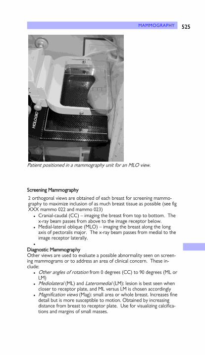
MAMMOGRAPHY1 525
Patient positioned in a mammography unit for an MLO view.
Screening Mammography
2 orthogonal views are obtained of each breast for screening mammo-graphy to maximize inclusion of as much breast tissue as possible (see fig XXX mammo 022 and mammo 023)
- Cranial-caudal (CC) – imaging the breast from top to bottom. The x-ray beam passes from above to the image receptor below.
- Medial-lateral oblique (MLO) – imaging the breast along the long axis of pectoralis major. The x-ray beam passes from medial to the image receptor laterally.
-
Diagnostic Mammography Other views are used to evaluate a possible abnormality seen on screen-ing mammograms or to address an area of clinical concern. These in-clude:
- Other angles of rotation from 0 degrees (CC) to 90 degrees (ML or LM)
- Mediolateral (ML) and Lateromedial (LM): lesion is best seen when closer to receptor plate, and ML versus LM is chosen accordingly
- Magnification views (Mag): small area or whole breast. Increases fine detail but is more susceptible to motion. Obtained by increasing distance from breast to receptor plate. Use for visualizing calcifica-tions and margins of small masses.
CHAPTER 11 Breast imaging 526
- Focal compression: small paddle used to compress overlying tissue away from area of interest
- Rolled views: the top of the breast is rolled relative to the bottom to spread out the tissues and provide localization for images only seen in one plane.
- Extended CC view (XCC) to see axillary or far lateral tissue - Tangential views: put the skin or an abnormality in tangential to the
xray beam to aid localization – e.g. of skin calcifications - Cleavage views: used for assessing medial abnormalities
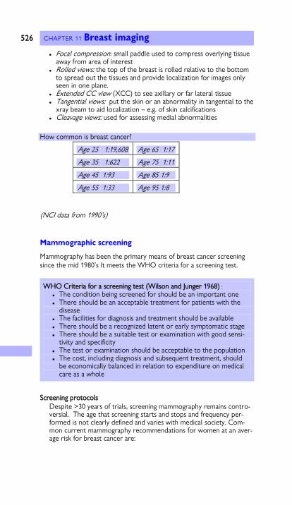
How common is breast cancer?
Age 25 1:19,608 Age 65 1:17
Age 35 1:622 Age 75 1:11
Age 45 1:93 Age 85 1:9
Age 55 1:33 Age 95 1:8
(NCI data from 1990‟s)
Mammographic screening
Mammography has been the primary means of breast cancer screening
since the mid 1980‟s It meets the WHO criteria for a screening test.
WHO Criteria for a screening test (Wilson and Junger 1968) :
- The condition being screened for should be an important one - There should be an acceptable treatment for patients with the
disease - The facilities for diagnosis and treatment should be available - There should be a recognized latent or early symptomatic stage - There should be a suitable test or examination with good sensi-
tivity and specificity - The test or examination should be acceptable to the population - The cost, including diagnosis and subsequent treatment, should
be economically balanced in relation to expenditure on medical care as a whole
Screening protocols
Despite >30 years of trials, screening mammography remains contro-versial. The age that screening starts and stops and frequency per-formed is not clearly defined and varies with medical society. Com-mon current mammography recommendations for women at an aver-age risk for breast cancer are:
MAMMOGRAPHIC SCREENING1 527
40-49 50-74 >75
ACR Annual Annual Annual*
ACS 2003 Annual Annual
AMA 2002 Annual Annual Annual*
WHO 2009 -- 1-2 years** --
USPSTF -- biennial --
ACR – American College of Radiology
ACS – American Cancer Society
WHO – World Health Organization
USPSTF – U.S. Preventative Services Task Force
** ages 50 -69
*When should screening stop?
There is no upper limit defined by ACR/ACS/AMA but, screening is
unlikely to be of benefit if life expectancy is <5-7 years on basis of age or
co-morbidities.
Evidence for screening
Based on the USPSTF 2009 meta-analyses: there is a known reduction in mortality due to screening mammography:
Ages Mortality Reduction Number of women screened to save one life
40-49 14% 1904
50-59 17% 1334
60-69 32% 377
70-79 10% Not defined from data
Randomized clinical trials (RCTs) have demonstrated a 25-30% reduction in breast cancer mortality in women 40-69 years who are screened an-nually or biennially with mammograms.
Limitations and controversies of screening mammography
Due to inherent difficulties of randomized screening studies, to eva-luate efficacy , morbidity and mortality, recommendations are conti-nually changing - Increased breast density is associated with both a lower sensitivity
for screening mammography and a higher risk of breast cancer.
Density Sensitivity Specificity Fatty 88% 97% Scattered 82% 93% Heterogeneous 69% 91% Very dense 62% 90%
Carney Ann Int Med 2003;138:168
CHAPTER 11 Breast imaging 528
- Longer screening intervals decrease the chance of catching faster
growing cancers earlier - May provide earlier diagnosis without change in outcome - Potential identification of low grade cancers (and in-situ cancers)
that may not be clinically significant but leads to over treatment („pseudo-disease)
- Reader/expertise dependent o Sensitivity : 60% to >90% o Specificity from 90% to 95%.
- False positives lead to increased anxiety and benign biopsies (cost and complication implications
What is a „call back/recall‟ Screening mammograms are usually read after patients have left the mammographic facility. Patients whose studies are felt to be abnor-mal (BIRADS 0 (see PXXX) are „called back‟ for diagnostic mammo-graphy (additional views and/or ultrasound)
Diagnostic mammography
DIAGNOSTIC mammography should be performed when:
Abnormality found on screening mammogram
Short interval follow up of probably benign but abnormal mammogram
Patient/physician identified breast lump
Focal breast pain/tenderness
Suspected abscess
Spontaneous nipple discharge
New nipple changes (e.g. inversion)
Normal mammogram
By convention, labeling of the projection used is placed on the lateral or cephalad aspect of the patient on the film.
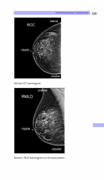
MAMMOGRAPHIC SCREENING1 529
Normal CC mammogram
Normal MLO mammogram on the same patient.
CHAPTER 11 Breast imaging 530
How do I read a mammogram?
Mammographic interpretation is almost all pattern recognition due to the limited anatomy present.
Technical Adequacy - No motion - Adequate tissue within field of view on both views. (CC view
should not differ from MLO more than 10 mm) - Inferior mammary fold (fold under breast) included - Pectoralis muscle included on MLO to nipple level.
Search Pattern
The entire mammogram needs to be searched in a systematic manner,
e.g: - Each breast from cranial to caudal - Each breast from nipple to chest wall left to right - Compare one breast to other zone by zone Compare each view with comparison older mammograms
Breast Density is assessed in all patients according to set criteria. All
breasts contain some combination of glandular/fibrotic tissue and fat.
1. Fatty: <25% glandular
2. Scattered: 25-50%
3. Heterogeneously dense: 51-75%
4. Extremely dense: >75% glandular
Comparison films are key in mammography due to the lack of normal anatomy and inter-patient variability. Comparison with films 2+ years ago decreases call back rates and may help detect smaller cancers.
What are we looking for?
- Breast density (see above) - Symmetry – most breast have a relatively symmetrical distribution
of glandular and fatty tissue - Signs of cancer that may include:
o Asymmetrical areas o Masses o Architectural distortion o Calcifications
Patterns of disease
When an abnormality is detected, individual characteristics are evaluated
to assess the level of concern for malignancy.
The most worrisome feature determines management with any abnor-
mality
MAMMOGRAPHIC SCREENING1 531
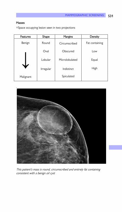
Masses
=Space occupying lesion seen in two projections
Features Shape Margins Density
Benign Round Circumscribed Fat containing
↓ Oval Obscured Low
Lobular Microlobulated Equal
Irregular Indistinct High
Malignant Spiculated
.
This patient‟s mass is round, circumscribed and entirely fat containing consistent with a benign oil cyst
CHAPTER 11 Breast imaging 532
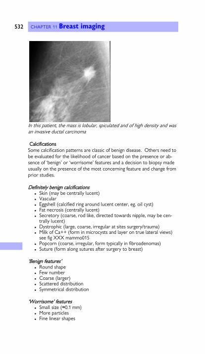
In this patient, the mass is lobular, spiculated and of high density and was an invasive ductal carcinoma Calcifications
Some calcification patterns are classic of benign disease. Others need to
be evaluated for the likelihood of cancer based on the presence or ab-
sence of „benign‟ or „worrisome‟ features and a decision to biopsy made
usually on the presence of the most concerning feature and change from
prior studies.
Definitely benign calcifications - Skin (may be centrally lucent) - Vascular - Eggshell (calcified ring around lucent center, eg. oil cyst) - Fat necrosis (centrally lucent) - Secretory (coarse, rod like, directed towards nipple, may be cen-
trally lucent) - Dystrophic (large, coarse, irregular at sites surgery/trauma) - Milk of Ca++ (form in microcysts and layer on true lateral views)
see fig XXX mammo015 - Popcorn (coarse, irregular, form typically in fibroadenomas) - Suture (form along sutures after surgery to breast)
„Benign features‟ - Round shape - Few number - Coarse (larger) - Scattered distribution - Symmetrical distribution
„Worrisome‟ features - Small size (≈0.1 mm) - More particles - Fine linear shapes
MAMMOGRAPHIC SCREENING1 533
- Branching shapes - Pleomorphic (varying in size and shape) - Clustered distribution (> 5 in a cm
3)
- Segmental, ductal branching or linear distribution
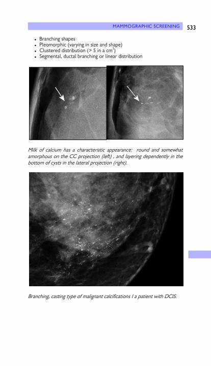
Milk of calcium has a characteristic appearance: round and somewhat amorphous on the CC projection (left) , and layering dependently in the bottom of cysts in the lateral projection (right).
Branching, casting type of malignant calcifications I a patient with DCIS.
CHAPTER 11 Breast imaging 534
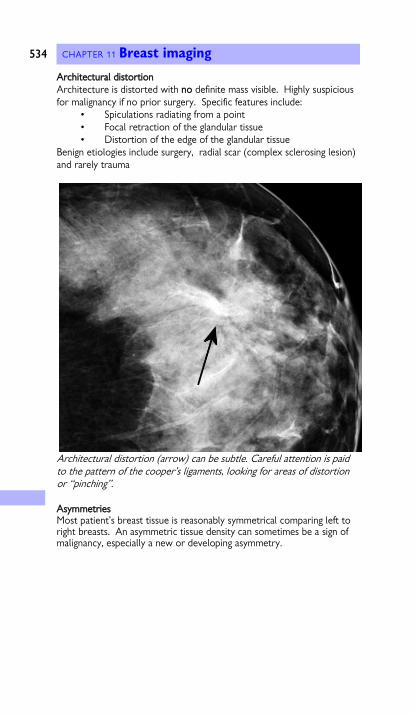
Architectural distortion
Architecture is distorted with no definite mass visible. Highly suspicious
for malignancy if no prior surgery. Specific features include:
• Spiculations radiating from a point
• Focal retraction of the glandular tissue
• Distortion of the edge of the glandular tissue
Benign etiologies include surgery, radial scar (complex sclerosing lesion)
and rarely trauma
Architectural distortion (arrow) can be subtle. Careful attention is paid to the pattern of the cooper‟s ligaments, looking for areas of distortion or “pinching”.
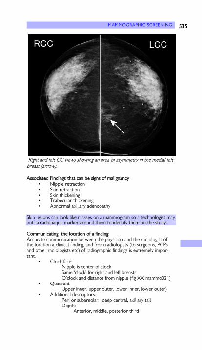
Asymmetries Most patient‟s breast tissue is reasonably symmetrical comparing left to right breasts. An asymmetric tissue density can sometimes be a sign of malignancy, especially a new or developing asymmetry.
MAMMOGRAPHIC SCREENING1 535
Right and left CC views showing an area of asymmetry in the medial left breast (arrow).
Associated Findings that can be signs of malignancy • Nipple retraction • Skin retraction • Skin thickening • Trabecular thickening • Abnormal axillary adenopathy
Skin lesions can look like masses on a mammogram so a technologist may puts a radiopaque marker around them to identify them on the study. Communicating the location of a finding: Accurate communication between the physician and the radiologist of the location a clinical finding, and from radiologists (to surgeons, PCPs and other radiologists etc) of radiographic findings is extremely impor-tant.
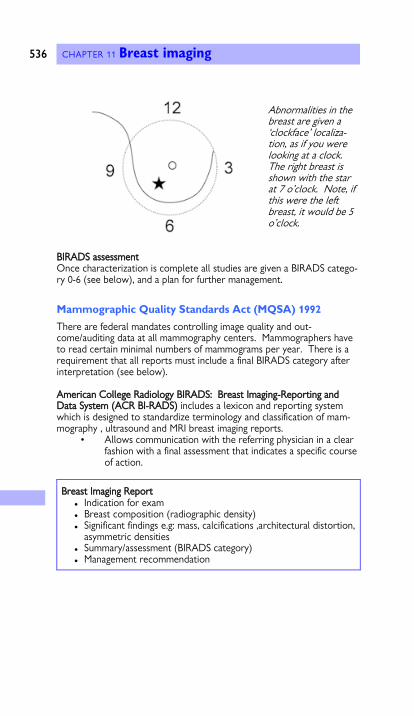
• Clock face Nipple is center of clock Same „clock‟ for right and left breasts O‟clock and distance from nipple (fig XX mammo021)
• Quadrant Upper inner, upper outer, lower inner, lower outer)
• Additional descriptors: Peri or subareolar, deep central, axillary tail Depth:
Anterior, middle, posterior third
CHAPTER 11 Breast imaging 536
Abnormalities in the breast are given a „clockface‟ localiza-tion, as if you were looking at a clock. The right breast is shown with the star at 7 o‟clock. Note, if this were the left breast, it would be 5 o‟clock.
BIRADS assessment Once characterization is complete all studies are given a BIRADS catego-ry 0-6 (see below), and a plan for further management.
Mammographic Quality Standards Act (MQSA) 1992
There are federal mandates controlling image quality and out-come/auditing data at all mammography centers. Mammographers have to read certain minimal numbers of mammograms per year. There is a requirement that all reports must include a final BIRADS category after interpretation (see below). American College Radiology BIRADS: Breast Imaging-Reporting and Data System (ACR BI-RADS) includes a lexicon and reporting system which is designed to standardize terminology and classification of mam-mography , ultrasound and MRI breast imaging reports.
• Allows communication with the referring physician in a clear fashion with a final assessment that indicates a specific course of action.
Breast Imaging Report - Indication for exam - Breast composition (radiographic density) - Significant findings e.g: mass, calcifications ,architectural distortion,
asymmetric densities - Summary/assessment (BIRADS category) - Management recommendation
MAMMOGRAPHIC SCREENING1 537
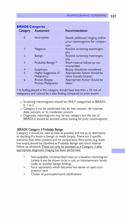
BIRADS Categories
Category Assessment Recommendation
0 Incomplete Needs additional imaging and/or
prior mammograms for compar-
ison 1 Negative Routine screening mammogra-
phy 2 Benign Routine screening mammogra-
phy 3 Probably Benign * Short-interval follow-up rec-
ommended 4 Suspicious Biopsy should be considered 5 Highly Suggestive of
Malignancy Appropriate Action should be taken (usually biopsy)
6 Known Biopsy-Proven Malignancy
Appropriate Action should be taken
* A finding placed in this category should have less than a 2% risk of malignancy and cannot be a new finding compared to prior exams
- Screening mammograms should be ONLY categorized as BIRADS
0, 1 or 2 - Category 4 can be subdivided into 4a: low concern, 4b: interme-
diate concern, or 4c: moderate concern - Diagnostic mammograms may be any category but the use of
BIRADS 0 should be avoided unless waiting for prior mammograms.
BIRADS Category 3 Probably Benign Category 3 should be used as little as possible and not as an alternative to deciding if a lesion is benign or needs biopsy. There are 3 specific scenarios that when present and no comparisons films exists (e.g. base-line exam),should be classified as Probably Benign and short interval follow-up obtained. These can only be assessed as a Category 3 after appropriate diagnostic imaging has been performed.
• Non-palpable, circumscribed mass on a baseline mammogram (unless it can be shown to be a cyst, an intramammary lymph node, or another benign finding),
• Focal asymmetry which becomes less dense on spot com-pression view
• Cluster of punctuate/round calcifications
CHAPTER 11 Breast imaging 538
Ultrasound Ultrasound evaluates different tissue properties than mammography.
When do you do breast ultrasound?
- Focal clinical abnormalities Lump, focal pain - Primary study in evaluating a palpable mass in young (<35), lactating
or pregnant patient - Evaluation of a mammographic abnormality
Mass - solid versus cystic, if solid likelihood of malignancy Focal or regional asymmetric tissue
- Evaluation of an MRI abnormality - Evaluation for abscess or seroma - Guidance for interventional procedures
Whole breast screening ultrasound is still controversial and under as-sessment. ACRIN 6666 trial evaluated screening US only in patients at higher than average risk and with dense breasts. In this study:
o Supplemental cancer detection over mammogra-phy consistently 3-4 cases per1000.
o PPV of biopsy prompted by US approx. 9%
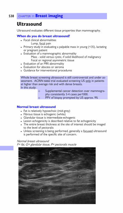
Normal breast ultrasound
- Fat is relatively hypoechoic (mid-grey) - Fibrous tissue is echogenic (white) - Glandular tissue is intermediate-echogenic - Lesion echogenicity is described relative to fat echogenicity - The entire breast thickness at the site of interest should be imaged
to the level of pectoralis - Unless screening is being performed, generally a focused ultrasound
is performed of the specific site of concern.
Normal breast ultrasound F= fat, G= glandular tissue, P= pectoralis muscle
ULTRASOUND1 539
As with mammography, finding are classified based on their imaging
characteristics and a BI-RADS assessment made.
Limitations of ultrasound and pitfalls - High false positive rate for screening - User/equipment variability - Ultrasound parameters incorrectly - Limited penetration esp. large breasts with fatty composition - Pseudolesions:
Fat lobules Cooper‟s ligaments
- Chest wall – eg: ribs misinterpreted as lesions - Nipple shadowing – difficult to assess behind the nipple (special - techniques required)
Patterns of disease breast ultrasound
The reference for echogenicity in the breast is the echogenity of breast
fat (i.e. all other substances are compared to the echogenity of fat, which
should be set at a mid-gray level).
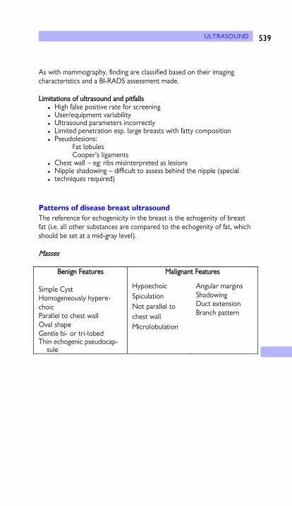
Masses
Benign Features Malignant Features
Simple Cyst
Homogeneously hypere-
choic
Parallel to chest wall
Oval shape
Gentle bi- or tri-lobed Thin echogenic pseudocap-
sule
Hypoechoic
Spiculation
Not parallel to
chest wall
Microlobulation
Angular margins
Shadowing
Duct extension
Branch pattern
CHAPTER 11 Breast imaging 540
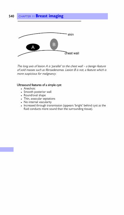
The long axis of lesion A is „parallel‟ to the chest wall – a benign feature of sold masses such as fibroadenomas. Lesion B is not, a feature which is more suspicious for malignancy.
Ultrasound features of a simple cyst - Anechoic - Smooth posterior wall - Round/oval shape - Thin, avascular septations - No internal vascularity - Increased through transmission (appears „bright‟ behind cyst as the
fluid conducts more sound than the surrounding tissue).
MRI1 541
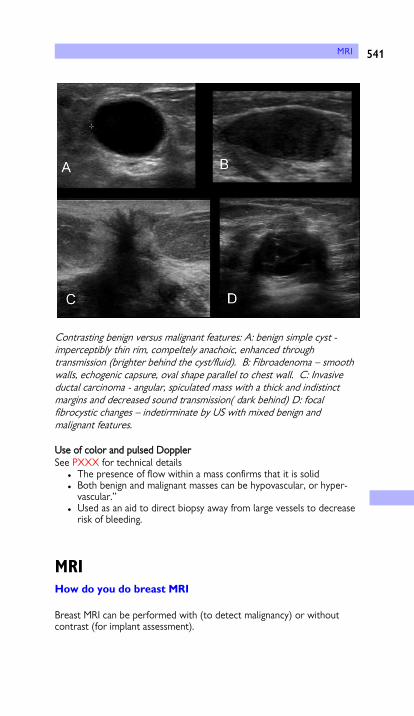
Contrasting benign versus malignant features: A: benign simple cyst - imperceptibly thin rim, compeltely anachoic, enhanced through transmission (brighter behind the cyst/fluid). B: Fibroadenoma – smooth walls, echogenic capsure, oval shape parallel to chest wall. C: Invasive ductal carcinoma - angular, spiculated mass with a thick and indistinct margins and decreased sound transmission( dark behind) D: focal fibrocystic changes – indetirminate by US with mixed benign and malignant features.
Use of color and pulsed Doppler
See PXXX for technical details - The presence of flow within a mass confirms that it is solid - Both benign and malignant masses can be hypovascular, or hyper-
vascular.” - Used as an aid to direct biopsy away from large vessels to decrease
risk of bleeding.
MRI How do you do breast MRI
Breast MRI can be performed with (to detect malignancy) or without contrast (for implant assessment).
CHAPTER 11 Breast imaging 542
Contrast enhanced breast MRI adds physiologic information. The breasts are scanned serially (usually 4-5 times) before and after the injection of i.v. gadolinium to assess for areas of contrast enhancement (increased signal on T1 following contrast).. Malignant tumors tend to enhance early and vigorously with fast washout of the contrast.
- Patients are scanned face-down with their breasts dependent in specialized breast coils that lie close to the breasts.
- Specialized software for the rapid scanning is needed as well as for kinetic analysis of the contrast enhancement (see PXX).
- Breast MRI should only be performed in centers that are able to perform MRI guided interventional procedures.
Protocols and contrast
Typical standard sequences: - T2 weighted - T1 weighted with and without fat saturation - TI with fat saturation scanned rapidly and repeatedly after gadoli-
nium injection <90seconds per series o Plus subtraction images (enhanced minus unen-
hanced images) o Shows only the areas of enhancement
- OR: T1 with no fat saturation plus subtraction images (enhanced minus unenhanced images)
o Shows only the areas of enhancement - Either axial (preferably) or sagittal images may be the primary imag-
ing plane for the dynamic sequences.
When do you do breast MRI?
These guidelines are developing and vary between facilities and medical
societies. The following are based on the recommendations from the American College Radiology and the American Cancer Society 2010.
Breast Cancer Staging Many centers use it routinely to stage all new breast cancer diagnoses.
- Ipsilateral disease: Additional unsuspected malignancies detected in 10-15% patients
- Contralateral disease : Additional unsuspected malignancies de-tected in 3-5.%
o Surgical Management changed in 19% of patients - Some centers use it as part of staging in the setting of isolated DCIS
o Can show extent of non-calcified portions Unknown Primary
- Looking for occult breast cancer as a cause of metastatic axillary adenocarcinoma
- MRI detects the primary cancer in up to 70% of these patients
High risk screening
MRI1 543
Screening breast MRI remains controversial due to its high cost and low
specificity (high rate of false positives). No current trials have assessed
effect on mortality and have only assessed high risk individuals.
ACS Recommend annual MRI screening (based on evidence) - BRCA mutation - First-degree relative of BRCA carrier, but untested - Lifetime risk ~20–25% or greater (from calculated models)
Recommend annual MRI screening (based on expert consensus opinion) - Radiation to chest between age 10 and 30 years - Li-Fraumeni syndrome and first-degree relatives - Cowden and Bannayan-Riley-Ruvalcaba syndromes and first-degree
relatives
Insufficient Evidence to Recommend for or Against MRI Screening
(judged on case by base basis) - Lifetime risk 15–20%, as defined by BRCAPRO or other models that
are largely dependent on family history - Lobular carcinoma in situ (LCIS) - Atypical lobular hyperplasia (ALH) - Heterogeneously or extremely dense breast on mammography - Women with a personal history of breast cancer, including ductal
carcinoma in situ (DCIS)
Recommend Against MRI - Women at <15% lifetime risk
Breast MRI detects approximately twice as many cancers as mammogra-
phy in high risk individuals.
Evaluation by a genetics unit should be considered before commitment to routine screening by MRI due to the cost involved and the significant risk of false positives (from 20-40% on a first screening exam, dropping to less than 10% on subsequent exams)
Diagnostic The role of MRI in diagnostic breast imaging is not yet clearly defined. Patients who are BI-RADS 0 on the basis of screening exam should un-dergo standard mammographic/ultrasound evaluation before MRI Consider in:
- Worrisome nipple discharge (esp bloody) or nipple retraction in a patient with a negative diagnostic mammogram and ultrasound.
- Evaluation for recurrence at lumpectomy site in a patient with nega-tive mammographic work up but clinical suspicion, or an equivocal mammogram.
- Evaluation of implant integrity
A negative MRI should never exclude biopsy of a mammographically or ultrasound suspicious lesion
CHAPTER 11 Breast imaging 544
Implants - Silicone implant integrity can be evaluated using specific pulse se-
quences that suppress water and fat signal - No contrast is used (so does not also evaluate for cancer) - Saline implants do not require MRI imaging as collapse if ruptured
Advantages of BMR
- No ionizing radiation - Multiplanar imaging, images entire breast volume and chest wall - 3D lesion mapping - >90% sensitivity for invasive carcinoma - Detects occult, multifocal or residual malignancy - May estimate tumor size better - Ability to image regional lymph nodes
Disadvantages of BMR
- High equipment and examination (≈ 8-10x mammography) costs - Limited scanner and biopsy availability - Injection of gadolinium required (NSF) - Learning curve for interpretation - False positives (limited specificity 30-50%) - Variable enhancement of in situ - 5% false negatives due to non- enhancing invasive ca
ACR BI-RADs for BMR - Uniform terminology for interpretation - Similar to mammography
o 0-6 BIRADS categories
MRI1 545
Patterns of disease breast MRI
- As with other modalities, when reading a breast MRI one evaluates o Characteristics of structural abnormalities
(masses, distortion , margins, etc) - Unique to BMRI is evaluation of enhancement patterns (kinetics)
o Extent, degree and type of enhancement
Fat T1 bright before fat supression
Normal tissue T1 dark pre-contrast, progres-
sively signal with contrast
Normal lymph nodes T2 bright, hypervascular with
washout kinetics
Cysts T2 bright
Complicated cysts and duct ecta-
sia (proteinaceous debris or
blood)
Variable signal on T1 and T2
Carcinoma T2 iso-intense to dark, usually
rapidly enhancing and washout.
DCIS T2 isointense. Usually clumped
enhancement with widely variable
kinetics. Low grade may not en-
hance
Computer assisted analysis (detection)
Many centers use post-processing software evaluate the kinetics of con-
trast processed by a voxel of tissue. It color-codes each voxel on the
image according to the time-activity curve of the contrast. One common
display is shown on color fig xx. Mammo_025.
- Benign curve: slowly accumulates contrast ( as seen in normal tissue
and fibrocystic tissue) - Malignant curve: rapid peak followed by rapid washout - Indeterminate or “Plateau” :in between the prior two
Unfortunately there is considerable overlap between these kinetic curves
– „benign‟ curves may be seen in DCIS, invasive lobular and low grade
invasive ductal cancers and „malignant‟ curves in normal lymph nodes and
vascular tumors such as papillomas and hemangiomas.
Common conditions
Diffuse Hormonal Enhancement:
The enhancement pattern of the breast is hormonally sensitive and can
obscure small foci of malignancy.
CHAPTER 11 Breast imaging 546
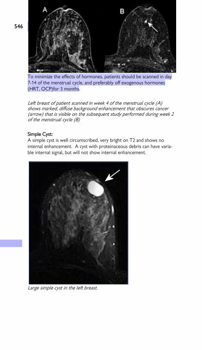
To minimize the effects of hormones, patients should be scanned in day
7-14 of the menstrual cycle, and preferably off exogenous hormones
(HRT, OCP)for 3 months.
Left breast of patient scanned in week 4 of the menstrual cycle (A) shows marked, diffuse background enhancement that obscures cancer (arrow) that is visible on the subsequent study performed during week 2 of the menstrual cycle (B)
Simple Cyst:
A simple cyst is well circumscribed, very bright on T2 and shows no
internal enhancement. A cyst with proteinaceous debris can have varia-
ble internal signal, but will not show internal enhancement.
Large simple cyst in the left breast.
MRI1 547
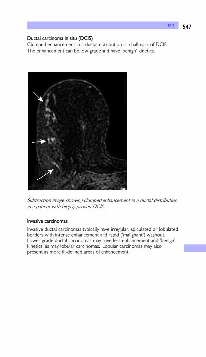
Ductal carcinoma in situ (DCIS)
Clumped enhancement in a ductal distribution is a hallmark of DCIS.
The enhancement can be low grade and have „benign‟ kinetics.
Subtraction image showing clumped enhancement in a ductal distribution in a patient with biopsy proven DCIS.
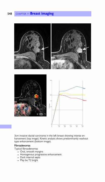
Invasive carcinomas
Invasive ductal carcinomas typically have irregular, spiculated or lobulated borders with intense enhancement and rapid („malignant‟) washout. Lower grade ductal carcinomas may have less enhancement and „benign‟ kinetics, as may lobular carcinomas. Lobular carcinomas may also present as more ill-defined areas of enhancement.
CHAPTER 11 Breast imaging 548
3cm invasive ductal carcinoma in the left breast showing intense en-hancement (top image). Kinetic analysis shows predominantly washout type enhancement (bottom image).
Fibroadenomas
Typical fibroadenomas - Oval, smooth margins - Homogenous progressive enhancement - Dark internal septa - May be T2 bright
PROCEDURES1 549
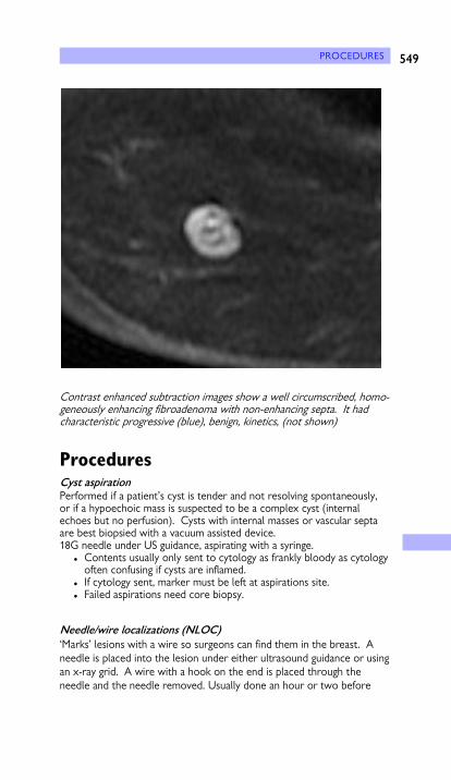
Contrast enhanced subtraction images show a well circumscribed, homo-geneously enhancing fibroadenoma with non-enhancing septa. It had characteristic progressive (blue), benign, kinetics, (not shown)
Procedures Cyst aspiration Performed if a patient‟s cyst is tender and not resolving spontaneously, or if a hypoechoic mass is suspected to be a complex cyst (internal echoes but no perfusion). Cysts with internal masses or vascular septa are best biopsied with a vacuum assisted device. 18G needle under US guidance, aspirating with a syringe.
- Contents usually only sent to cytology as frankly bloody as cytology often confusing if cysts are inflamed.
- If cytology sent, marker must be left at aspirations site. - Failed aspirations need core biopsy.
Needle/wire localizations (NLOC)
„Marks‟ lesions with a wire so surgeons can find them in the breast. A
needle is placed into the lesion under either ultrasound guidance or using
an x-ray grid. A wire with a hook on the end is placed through the
needle and the needle removed. Usually done an hour or two before
CHAPTER 11 Breast imaging 550
surgery. At surgery a piece of tissue around the hook of the wire is re-
moved. - Lesions not amenable to percutaneous biopsy due to:
Location in the breast (eg. calcifications close to the chest wall) Lesions very close to the nipple. Medical conditions preclude laying prone for 20 minutes re-quired for a stereotactic biopsy
- Localizing lesions that need surgical excision (e.g. known cancer or atypia)
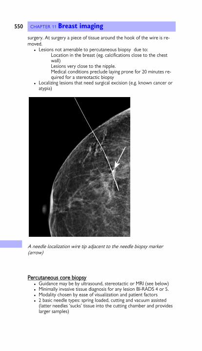
A needle localization wire tip adjacent to the needle biopsy marker (arrow)
Percutaneous core biopsy - Guidance may be by ultrasound, stereotactic or MRI (see below) - Minimally invasive tissue diagnosis for any lesion BI-RADS 4 or 5. - Modality chosen by ease of visualization and patient factors - 2 basic needle types: spring loaded, cutting and vacuum assisted
(latter needles „sucks‟ tissue into the cutting chamber and provides larger samples)

PROCEDURES1 551
- All biopsies: o Sterile prep o Lidocaine +/- epinephrine for local anesthesia o 2-5mm skin incision with a scalpel
- A tiny metallic clip is left at the biopsy site to confirm sampling site and allow NLOC if further excision needed
Patients usually need to discontinue anticoagulant therapy for core needle biopsies. This may involve them transferring to injectable anti-coagulants temporarily e.g. lovenox (enoxaparin). Some centers stop aspirin and plavix (clopidogrel) for a week before biopsy.
Ultrasound guided biopsy
Ultrasound biopsy is the method of choice if lesions can be seen by
ultrasound - cheapest, fastest and the most comfortable for patients. - Fine needle aspiration.
o US can guide FNA of suspicious lesions. o 18-23G needles o Generally core biopsy is preferred due to its
much higher sensitivity (98+% compared to 80%) - Core cutting needles
o Well defined lesions - Vacuum assisted needles
o Ill defined or largely cystic lesions o Very small lesions o Lesions against chest wall
Stereotactic biopsy
Stereotactic biopsy uses x-ray guidance to guide core needle biopsy placement. Generally this is performed for calcifications, masses or suspicious densities with no ultrasound correlate.
- Patient lies prone on stereo table with breast through hole in table in light compression
- 2 images taken 15 degrees from center with digital mammography - Computer calculates x, y and z coordinates of lesion - Local anesthesia applied - Vacuum assisted needle takes 9 or 10g samples. - Sample is x-rayed to confirm sampling - Image pairs taken after each adjustment to plan next action
CHAPTER 11 Breast imaging 552
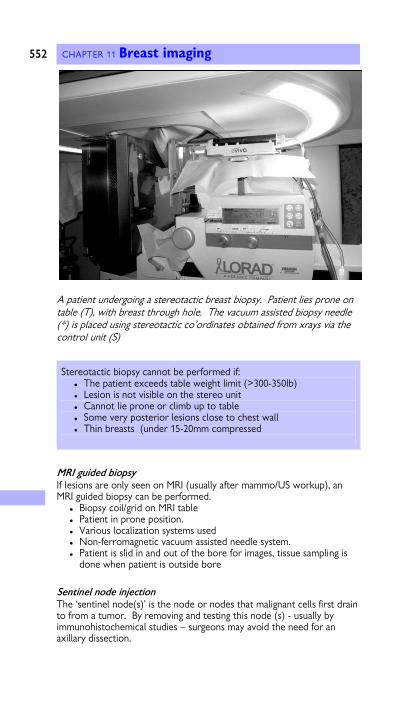
A patient undergoing a stereotactic breast biopsy. Patient lies prone on table (T), with breast through hole. The vacuum assisted biopsy needle (*) is placed using stereotactic co'ordinates obtained from xrays via the control unit (S)
Stereotactic biopsy cannot be performed if: - The patient exceeds table weight limit (>300-350lb) - Lesion is not visible on the stereo unit - Cannot lie prone or climb up to table - Some very posterior lesions close to chest wall - Thin breasts (under 15-20mm compressed
MRI guided biopsy If lesions are only seen on MRI (usually after mammo/US workup), an MRI guided biopsy can be performed.
- Biopsy coil/grid on MRI table - Patient in prone position. - Various localization systems used - Non-ferromagnetic vacuum assisted needle system. - Patient is slid in and out of the bore for images, tissue sampling is
done when patient is outside bore
Sentinel node injection The „sentinel node(s)‟ is the node or nodes that malignant cells first drain to from a tumor. By removing and testing this node (s) - usually by immunohistochemical studies – surgeons may avoid the need for an axillary dissection.
MANAGEMENT PROBLEMS1 553
The sentinel nodes (usually 1-3) can be identified by using one more of these tests:
- Injecting blue dye around the tumor at surgery and following the lymphatics visually.
- Injecting 99m
Tc sulfur colloid around the tumor - Injecting
99mTc sulfur colloid intradermally above the tumor
- Injecting 99m
Tc sulfur colloid under the areolar (the breast drains to the areolar then outwards to the axilla +/- the internal mammary nodes).
The 99m
Tc sulfur colloid is injected either by palpation, by ultrasound or by x-ray guidance about 2 hours before surgery. The sentinel nodes are then localized by a gamma probe at the time of surgery. Sometimes imaging is first performed with a gamma camera in nuclear medicine if isolated internal mammary node drainage is suspected with medial le-sions.
Management problems Palpable masses
- Refer for DIAGNOSTIC mammography and ultrasound - Imaging can rarely be negative in cancer in presence of palpable
mass Patients <35 may have ultrasound as initial investigation.
Discrete lumps if new/growing should be referred to a clinical pro-vider such as a breast surgeon for consideration of biopsy even if imaging negative
Breast pain - Rarely a tumor can be painful, more commonly pain is due to fibro-
cystic changes, cysts or hormonal. - Diffuse, bilateral or regional breast pain should have routine screen-
ing mammography only - Focal pain („can you point to it with a finger?‟) should have mammo-
graphy plus ultrasound.
It is vital that physicians accurately convey the site of concern (mass, pain) to the radiologist, recording breast side, size, position (clock face) and distance from nipple.
Nipple discharge - Bloody nipple discharge is the most concerning, but cancer-
associated nipple discharge can also be clear or serous - Unilateral spontaneous more concerning than bilateral or expressi-
ble only
CHAPTER 11 Breast imaging 554
- Mammography +/- magnified views and/or peri-areolar ultrasound - Consider MRI for bloody nipple discharge
References Breast Cancer Screening. A summary of the evidence for the US Pre-ventative ServicesTask Force. Ann.Intern.Med 2002;137:345-360.
Update: www.ahrq.gov/clinic/uspstf09/breastcancer/brcanrs.htm
American Cancer Society Guidelines for Breast Screening with MRI as an Adjunct to Mammography. CA Cancer J Clin 2007; 57:75-89
Lehman, et al: Indications for Breast MRI in Patient With Newly Diag-nosed Breast Cancer: Preoperative Staging of the Affected and Contrala-teral Breast in Patients With a Known Cancer. www.medscape.com