Embed Size (px)
Citation preview
Pediatric Clinical Practice Guidelines for Nurses in Primary Care 2011
CHAPTER 11 – CARDIOVASCULAR SYSTEMFirst Nations and Inuit Health Branch (FNIHB) Pediatric Clinical Practice Guidelines for Nurses in Primary Care.
The section on Rheumatic Fever (Carditis) has been updated as of December 2017. The remaining content of this chapter was reviewed in September 2011.
Table of Contents
INTRODUCTION ....................................................................................................11–1
ASSESSMENT OF THE CARDIOVASCULAR SYSTEM .......................................11–1
In Infants ..........................................................................................................11–1
In Children ........................................................................................................11–2
Medical History (Specific to Cardiovascular System) ......................................11–2
Physical Findings .............................................................................................11–2
COMMON PROBLEMS OF THE CARDIOVASCULAR SYSTEM ..........................11–3
Heart Murmurs .................................................................................................11–3
Innocent Heart Murmur ....................................................................................11–4
EMERGENCY PROBLEMS OF THE CARDIOVASCULAR SYSTEM ...................11–5
Cardiac Failure .................................................................................................11–5
Cyanosis in the Newborn (Birth to 6 Weeks) ...................................................11–6
Rheumatic Fever (Carditis) ..............................................................................11–8
Viral Myocarditis ...............................................................................................11–9
SOURCES ............................................................................................................11–12

Cardiovascular System 11–1
Pediatric Clinical Practice Guidelines for Nurses in Primary Care 2011
INTRODUCTION
Cardiovascular disease is uncommon in childhood. The major problems seen include congenital heart disease (usually abnormalities of the great vessels, hypoplastic heart, pulmonary or aortic atresia and tetralogy of Fallot), cardiac failure, rheumatic fever carditis and myocarditis.
Functional or innocent heart murmurs are common.
Congestive heart failure at birth is rare and usually suggests severe valvular deformities.
Symptoms of ventricular septal defect, including heart failure, usually occur at approximately 6 weeks of age.
For more information on the history and physical examination of the cardiovascular system in older children and adolescents, see the chapter “Cardiovascular System” in the adult clinical practice guidelines.
ASSESSMENT OF THE CARDIOVASCULAR SYSTEM
Symptoms of cardiovascular disease vary with the age of the child.
Ask about:
– Rapid or noisy breathing – Cough – Cyanosis – Sleeping patterns – Exercise tolerance: indicated in a young child by
ability to feed and in an older child by ability to keep up with peers during play
IN INFANTS
CYANOSIS
– An abnormality of oxygen transport related to heart, lungs or blood or inadequate oxygenation of blood due to mixing of venous and arterial blood. Transport problems include impairment of the oxygen-carrying capacity of hemoglobin, as for example, in carbon monoxide poisoning, and hypoxemia secondary to ventilation/perfusion mismatches as for example in pneumonia
– Causes bluish discolouration of mucous membranes, nail beds and skin, is a significant clinical finding and is related to inadequate oxygenation of arterial blood
– May be transient (related to increased oxygen demand by tissues, for example, during feeding in infants or during play in toddlers) or permanent from birth
EXERCISE INTOLERANCE
– Eats slowly or poorly – Tires easily during feeding and with poor
weight gain – Cyanosis appears with feeding (exertional) – Often described by parents or caregiver as a
“good baby”: always quiet, sleeps a lot, parents may find baby less energetic compared to siblings at same age
DIFFICULTY BREATHING
– Tachypnea – Chest retractions – Nasal flaring – Anxious appearance – Grunting
EXCESSIVE PERSPIRATION
– Infant’s head described as “always wet” – Infant perspires freely and easily, especially
with excretion and feeding
SLOW GROWTH
– Child usually exhibits slow weight gain, relative to height gain; difficulty in feeding may contribute to this problem
– Metabolic demands increased
RESPIRATORY INFECTIONS
– More common with congestive heart failure – More severe with increased pulmonary flow
Cardiovascular System11–2
Pediatric Clinical Practice Guidelines for Nurses in Primary Care2011
IN CHILDREN
– Slow growth – Respiratory infections – Chest pain – Palpitations – Dizzy spells or blackouts – Exercise intolerance – Squatting with cyanotic episodes
(“tetralogy spells”)
MEDICAL HISTORY (SPECIFIC TO CARDIOVASCULAR SYSTEM)
– Prematurity (associated with a higher prevalence of congenital cardiac malformation)
– History of illnesses related to heart disease (for example, strep throat)
– “Flu-like” illness – Joint pains or swelling – Down syndrome (associated with a higher
prevalence of congenital heart disease)
PHYSICAL FINDINGS
An examination of the cardiovascular system involves more than just examining the heart. The examination generally covers two systems: the central cardiovascular system (head, neck and precordium [anterior chest]) and the peripheral vascular system (extremities). Examination of the cardiovascular system must also include a full assessment of the lungs and neuromental status (for signs of confusion, irritability or stupor).
VITAL SIGNS
– Heart rate – Respiratory rate – Blood pressure (in both an upper and a lower
limb, if possible) – Temperature (may be elevated with myocarditis
or acute rheumatic fever) – Cardiovascular problems may present as failure
to thrive (weight and height below specified percentiles for age) or as a sharp decline in the growth curve across a major percentile line, therefore always document height and weight for all well baby and child examinations
INSPECTION
– Respiratory distress – Cyanosis: central and peripheral – Hands and feet: cyanosis, clubbing – Precordium: visible pulsations – Edema (hands, feet, sacrum)
PALPATION
– Apical beat is located at fourth intercostal space, lateral to the mid-clavicular line in infants, and at fifth intercostal space, lateral to the mid-clavicular line in older children
– Brief, localized apical tap is normal – Apical beat may be laterally displaced, which
indicates cardiomegaly – Thrills or heaves may be palpable through chest
wall; check supraclavicular area for thrills (in children with a thin chest wall, normal heart movements can be easily palpated and should not be confused with true thrills and heaves)
– Hepatomegaly – Check for presence, rate, rhythm, amplitude
and equivalence of peripheral pulses, especially femoral pulses (which are bounding in patent ductus arteriosus, absent in coarctation of aorta)
– Check for synchrony of radial and femoral pulses – Capillary refill (normal < 3 seconds) – Edema: pitting (rated 0 to 4) and level (how far up
the feet and legs the edema extends); sacral edema – Skin: temperature, turgor
AUSCULTATION
– S1 and S2 heart sounds – Physiologic splitting of S2 heart sound – Added heart sounds (S3 and S4): determine their
location and relation to respiration – Murmurs: determine location (where murmurs are
best heard), radiation, their timing in cardiac cycle, intensity, grade (see Table 1, “Characteristics of Heart Murmurs of Various Grades”) and quality
– Bruits: may occur in carotid arteries, abdominal aorta, renal arteries, iliac arteries, femoral arteries
– Crackles in lungs: may indicate heart failure (in infants and children, this usually occurs as a late sign)
Cardiovascular System 11–3
Pediatric Clinical Practice Guidelines for Nurses in Primary Care 2011
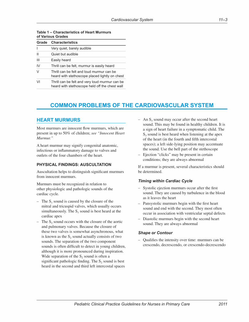
Table 1 – Characteristics of Heart Murmurs of Various Grades
Grade Characteristics
I Very quiet, barely audible
II Quiet but audible
III Easily heard
IV Thrill can be felt, murmur is easily heard
V Thrill can be felt and loud murmur can be heard with stethoscope placed lightly on chest
VI Thrill can be felt and very loud murmur can be heard with stethoscope held off the chest wall
COMMON PROBLEMS OF THE CARDIOVASCULAR SYSTEM
HEART MURMURS
Most murmurs are innocent flow murmurs, which are present in up to 50% of children; see “Innocent Heart Murmur.”
A heart murmur may signify congenital anatomic, infectious or inflammatory damage to valves and outlets of the four chambers of the heart.
PHYSICAL FINDINGS: AUSCULTATION
Auscultation helps to distinguish significant murmurs from innocent murmurs.
Murmurs must be recognized in relation to other physiologic and pathologic sounds of the cardiac cycle.
– The S1 sound is caused by the closure of the mitral and tricuspid valves, which usually occurs simultaneously. The S1 sound is best heard at the cardiac apex
– The S2 sound occurs with the closure of the aortic and pulmonary valves. Because the closure of these two valves is somewhat asynchronous, what is known as the S2 sound actually consists of two sounds. The separation of the two component sounds is often difficult to detect in young children, although it is more pronounced during inspiration. Wide separation of the S2 sound is often a significant pathologic finding. The S2 sound is best heard in the second and third left intercostal spaces
– An S3 sound may occur after the second heart sound. This may be found in healthy children. It is a sign of heart failure in a symptomatic child. The S3 sound is best heard when listening at the apex of the heart (in the fourth and fifth intercostal spaces); a left side-lying position may accentuate the sound. Use the bell part of the stethoscope
– Ejection “clicks” may be present in certain conditions; they are always abnormal
If a murmur is present, several characteristics should be determined.
Timing within Cardiac Cycle
– Systolic ejection murmurs occur after the first sound. They are caused by turbulence in the blood as it leaves the heart
– Pansystolic murmurs begin with the first heart sound and end with the second. They most often occur in association with ventricular septal defects
– Diastolic murmurs begin with the second heart sound. They are always abnormal
Shape or Contour
– Qualifies the intensity over time: murmurs can be crescendo, decrescendo, or crescendo-decrescendo
Cardiovascular System11–4
Pediatric Clinical Practice Guidelines for Nurses in Primary Care2011
Location on the Thorax
There are four general auscultatory areas:
– Aortic: left ventricular outflow murmur (usually ejection)
– Pulmonary: right ventricular outflow murmur, patent ductus arteriosus
– Tricuspid: tricuspid murmurs increase on inspiration; ventricular septal defects are heard best in this area
– Mitral: murmur at the cardiac apex
Radiation
Radiation of the murmur to the back, sides and neck should be carefully auscultated. Radiation of the murmur may give important diagnostic clues (for example, aortic stenosis radiates to the neck).
Intensity of Murmur
– Intensity expressed as a fraction of VI (for example, I/VI, II/VI), where a very loud murmur = V/VI or VI/VI, a loud murmur = III/VI or IV/VI, and a soft murmur = I/VI or II/VI (see Table 1, “Characteristics of Heart Murmurs of Various Grades”)
– Intensity (loudness) does not necessarily correlate with the severity of the condition. Soft murmurs may be dangerous, whereas loud murmurs are not necessarily so. A murmur associated with a thrill has an intensity of at least IV/VI
– Intensity may also increase with increased blood flow, as with exercise
Pitch
– Can be low, medium or high and is determined by whether it can be auscultated best with the bell or the diaphragm of a stethoscope
Quality
– Blowing – Harsh – Musical – Rumbling – Clanging
INNOCENT HEART MURMUR
Heart murmur that occurs in the absence of anatomic or physiologic abnormalities of the heart and therefore has no clinical significance. Such murmurs occur in 50–80% of children.
TYPES OF INNOCENT HEART MURMURS
Still’s murmur – vibratory, systolic ejection murmur (SEM), lower left sternal border (LLSB) or apex; ages 3 to 6 years.
Venous hum – infraclavicular hum, continuous, heard on right side more than left side; ages 3 to 6 years.
Peripheral pulmonic stenosis – pulmonic area, systolic, low pitched, radiates to axilla and back, seen in neonates; disappears usually by 3 to 6 months of age.
Pulmonary ejection – soft, blowing, upper left sternal border, systolic ejection murmur (SEM).
PATHOPHYSIOLOGY
Most innocent heart murmurs are produced by the forward flow of blood, which creates turbulence in the chambers of the heart or the great vessels. These murmurs are often more pronounced in high-output states, such as during a fever. Because the intensity of the murmur parallels the ejection velocity of blood from the ventricles, innocent murmurs usually occur during early to mid-systole, are short in duration, have a crescendo-decrescendo contour (especially an ejection murmur), are less than 3/6 in intensity and are never diastolic.
CLINICAL FEATURES
Innocent heart murmurs are asymptomatic and are usually found on routine physical examination.
DIAGNOSTIC TESTS
– Electrocardiogram (ECG) – Echocardiography (only as ordered by a physician)
MANAGEMENT
Reassure the parents or caregiver that no immediate treatment is necessary.
Referral
Refer the asymptomatic child electively to a physician for assessment when a murmur is found.
Cardiovascular System 11–5
Pediatric Clinical Practice Guidelines for Nurses in Primary Care 2011
EMERGENCY PROBLEMS OF THE CARDIOVASCULAR SYSTEM
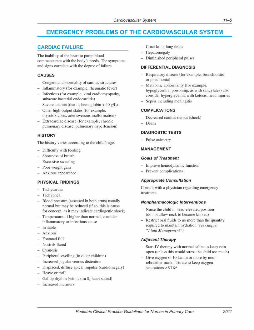
CARDIAC FAILURE
The inability of the heart to pump blood commensurate with the body’s needs. The symptoms and signs correlate with the degree of failure.
CAUSES
– Congenital abnormality of cardiac structures – Inflammatory (for example, rheumatic fever) – Infectious (for example, viral cardiomyopathy,
subacute bacterial endocarditis) – Severe anemia (that is, hemoglobin < 40 g/L) – Other high-output states (for example,
thyrotoxicosis, arteriovenous malformation) – Extracardiac disease (for example, chronic
pulmonary disease, pulmonary hypertension)
HISTORY
The history varies according to the child’s age.
– Difficulty with feeding – Shortness of breath – Excessive sweating – Poor weight gain – Anxious appearance
PHYSICAL FINDINGS
– Tachycardia – Tachypnea – Blood pressure (assessed in both arms) usually
normal but may be reduced (if so, this is cause for concern, as it may indicate cardiogenic shock)
– Temperature: if higher than normal, consider inflammatory or infectious cause
– Irritable – Anxious – Fontanel full – Nostrils flared – Cyanosis – Peripheral swelling (in older children) – Increased jugular venous distention – Displaced, diffuse apical impulse (cardiomegaly) – Heave or thrill – Gallop rhythm (with extra S3 heart sound) – Increased murmurs
– Crackles in lung fields – Hepatomegaly – Diminished peripheral pulses
DIFFERENTIAL DIAGNOSIS
– Respiratory disease (for example, bronchiolitis or pneumonia)
– Metabolic abnormality (for example, hypoglycemia; poisoning, as with salicylates) also consider hyperglycemia with ketosis, head injuries
– Sepsis including meningitis
COMPLICATIONS
– Decreased cardiac output (shock) – Death
DIAGNOSTIC TESTS
– Pulse oximetry
MANAGEMENT
Goals of Treatment
– Improve hemodynamic function – Prevent complications
Appropriate Consultation
Consult with a physician regarding emergency treatment.
Nonpharmacologic Interventions
– Nurse the child in head-elevated position (do not allow neck to become kinked)
– Restrict oral fluids to no more than the quantity required to maintain hydration (see chapter “Fluid Management”)
Adjuvant Therapy
– Start IV therapy with normal saline to keep vein open (unless this would stress the child too much)
– Give oxygen 6–10 L/min or more by non-rebreather mask.1 Titrate to keep oxygen saturations > 97%2
Cardiovascular System11–6
Pediatric Clinical Practice Guidelines for Nurses in Primary Care2011
Pharmacologic Interventions
Drugs used to treat heart failure in children are to be ordered by a physician.
– Diuretics to decrease volume:
furosemide (Lasix), 1 mg/kg IV stat (may be given
PO if IV access not available)
– ACE inhibitors may be prescribed by a physician for afterload reduction
– Digoxin may be used in some cases to increase contractility
Monitoring and Follow-Up
Acute Phase
Monitor ABCs (airway, breathing and circulation), vital signs, pulse oximetry (if available), heart and lung sounds, intake and output until child is transferred to hospital.
Over the Long Term
Children with cardiac illness should be monitored regularly within the community to ensure normal growth and development and to watch for complications. Frequency of follow-up depends on the severity of the condition.
Referral
Medevac immediately.
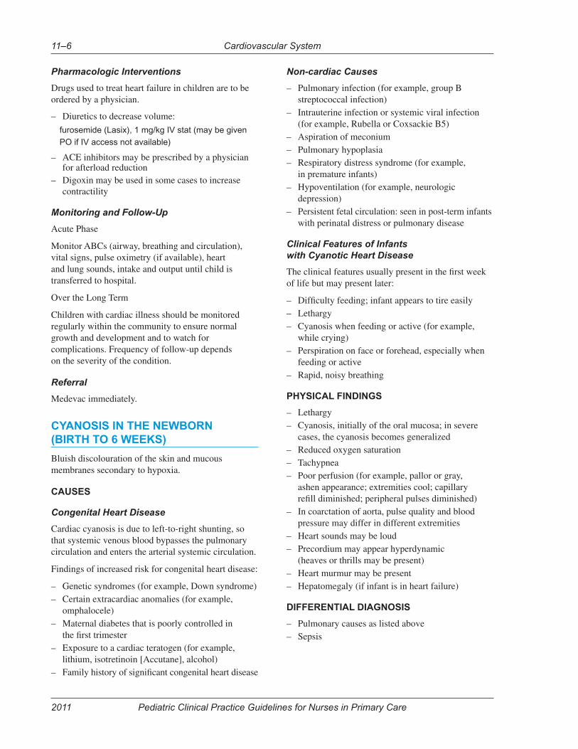
CYANOSIS IN THE NEWBORN (BIRTH TO 6 WEEKS)
Bluish discolouration of the skin and mucous membranes secondary to hypoxia.
CAUSES
Congenital Heart Disease
Cardiac cyanosis is due to left-to-right shunting, so that systemic venous blood bypasses the pulmonary circulation and enters the arterial systemic circulation.
Findings of increased risk for congenital heart disease:
– Genetic syndromes (for example, Down syndrome) – Certain extracardiac anomalies (for example,
omphalocele) – Maternal diabetes that is poorly controlled in
the first trimester – Exposure to a cardiac teratogen (for example,
lithium, isotretinoin [Accutane], alcohol) – Family history of significant congenital heart disease
Non-cardiac Causes
– Pulmonary infection (for example, group B streptococcal infection)
– Intrauterine infection or systemic viral infection (for example, Rubella or Coxsackie B5)
– Aspiration of meconium – Pulmonary hypoplasia – Respiratory distress syndrome (for example,
in premature infants) – Hypoventilation (for example, neurologic
depression) – Persistent fetal circulation: seen in post-term infants
with perinatal distress or pulmonary disease
Clinical Features of Infants with Cyanotic Heart Disease
The clinical features usually present in the first week of life but may present later:
– Difficulty feeding; infant appears to tire easily – Lethargy – Cyanosis when feeding or active (for example,
while crying) – Perspiration on face or forehead, especially when
feeding or active – Rapid, noisy breathing
PHYSICAL FINDINGS
– Lethargy – Cyanosis, initially of the oral mucosa; in severe
cases, the cyanosis becomes generalized – Reduced oxygen saturation – Tachypnea – Poor perfusion (for example, pallor or gray,
ashen appearance; extremities cool; capillary refill diminished; peripheral pulses diminished)
– In coarctation of aorta, pulse quality and blood pressure may differ in different extremities
– Heart sounds may be loud – Precordium may appear hyperdynamic
(heaves or thrills may be present) – Heart murmur may be present – Hepatomegaly (if infant is in heart failure)
DIFFERENTIAL DIAGNOSIS
– Pulmonary causes as listed above – Sepsis
Cardiovascular System 11–7
Pediatric Clinical Practice Guidelines for Nurses in Primary Care 2011
COMPLICATIONS – Cardiac failure (see “Cardiac Failure”) – Failure to thrive (see “Failure to Thrive” in the
pediatric chapter “Hematology, Endocrinology, Metabolism and Immunology”)
– Death
DIAGNOSTIC TESTS – Pulse oximetry
MANAGEMENT
Appropriate ConsultationConsult a physician immediately and prepare to medevac.
Adjuvant Therapy – Give oxygen 6–10 L/min (or more, if necessary)
by non-rebreather mask. Titrate to keep oxygen saturations > 97%2
– Consider intravenous (IV) therapy with normal saline if infant is feeding poorly or is in significant clinical distress
Nonpharmacologic Interventions – Nurse in an upright position – Feed small amounts frequently
Monitoring and Follow-Up – Monitor level of consciousness, vital signs, heart
and lung sounds, perfusion, pulse oximetry – Hydration status (intake and output) (see “Clinical
Features of Dehydration” in the chapter “Fluid Management”)
– Watch for signs of cardiac failure (see “Cardiac Failure”)
Referral – Medevac as soon as possible
Pediatric Clinical Practice Guidelines for Nurses in Primary Care2017
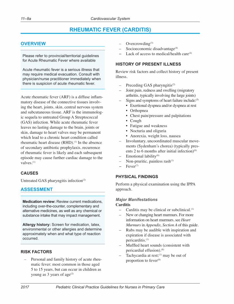
OVERVIEW
Please refer to provincial/territorial guidelines for Acute Rheumatic Fever where available Acute rheumatic fever is a serious illness that may require medical evacuation. Consult with physician/nurse practitioner immediately when there is suspicion of acute rheumatic fever.
-matory disease of the connective tissues involv-ing the heart, joints, skin, central nervous system and subcutaneous tissue. ARF is the immunolog-ic sequela to untreated Group A Streptococcal (GAS) infection. While acute rheumatic fever leaves no lasting damage to the brain, joints or skin, damage to heart valves may be permanent which lead to a chronic heart condition called rheumatic heart disease (RHD).(1) In the absence of secondary antibiotic prophylaxis, recurrence of rheumatic fever is likely and each subsequent episode may cause further cardiac damage to the valves.(1)
CAUSES
Untreated GAS pharyngitis infection(2)
ASSESSMENT
Medication review: Review current medications, including over-the-counter, complementary and alternative medicines, as well as any chemical or substance intake that may impact management. Allergy history: Screen for medication, latex, environmental or other allergies and determine approximately when and what type of reaction occurred.
RISK FACTORS
– Personal and family history of acute rheu-matic fever; most common in those aged 5 to 15 years, but can occur in children as young as 3 years of age(2)
– Overcrowding(3)
– Socioeconomic disadvantage(4)
− Lack of access to medical/health care(4)
HISTORY OF PRESENT ILLNESS
Review risk factors and collect history of present illness.
– Preceding GAS pharyngitis(2)
– Joint pain, redness and swelling (migratory arthritis, typically involving the large joints)
– Signs and symptoms of heart failure include:(5)
• Exertional dyspnea and/or dyspnea at rest• Orthopnea• Chest pain/pressure and palpitations• Cough• Fatigue and weakness• Nocturia and oliguria• Anorexia, weight loss, nausea
– Involuntary, uncoordinated muscular move-ments (Sydenham’s chorea) (typically pres-ents 2 to 6 months after initial infection)(6)
– Emotional lability(6) – Non-pruritic, painless rash(1)
− Fever(7)
PHYSICAL FINDINGS
Perform a physical examination using the IPPA approach.
Major ManifestationsCarditis
– Carditis may be clinical or subclinical.(1)
– New or changing heart murmurs. For more information on heart murmurs, see Heart Murmurs in Appendix, Section A of this guide.
– Rubs may be audible with inspiration and expiration if disease is associated with pericarditis.(1)
– pericardial effusion).(8)
– Tachycardia at rest;(1) may be out of proportion to fever(8)
RHEUMATIC FEVER (CARDITIS)
Cardiovascular System11–8a
Pediatric Clinical Practice Guidelines for Nurses in Primary Care 2017
– Tachypnea, orthopnea, jugular venous disten-tion, crackles, hepatomegaly, a gallop rhythm, edema and swelling of the peripheral extrem-ities if the client is in heart failure.(9)
− Cardiomegaly, pulmonary congestion and
may be observed on chest x-ray.(10)
Arthritis – Large joints are usually affected, especially
the knees and ankles.(1)
–presence of 2 or more of the following:(1)
• Limitation of movement• Hot joint• Pain in the joint and/or tenderness
Sydenham’s Chorea – Jerky, uncoordinated movements of the
extremities that disappear during sleep,(1) dysphonia and possible emotional lability(6)
– Female predominance(7)
− Can be a standalone criterion for the diagnosis of acute rheumatic fever without additional manifestations(1)
Subcutaneous Nodules – Usually located over a bony prominence
or near tendons(11)
− -sionally painful protuberances found on ex-
the knees, elbows, and wrists; also seen in the occiput and along the spinous processes of the thoracic and lumbar vertebrae(7)
Erythema Marginatum –
dark-skinned people)(1)
– An evanescent, pink rash with a pale center and rounded or serpiginous margins(7)
– The rash is usually present on the trunk and proximal extremities; it is almost never on the face(7)
– Blanches with pressure(7)
– medication(1)
− Rarely seen as the sole major criterion for acute rheumatic fever and should be accompanied by additional major criteria in order to make the diagnosis(1)
Minor Manifestations – Fever(7)
– Arthralgia(7)
– Raised C-reactive protein (CRP) and eryth-rocyte sedimentation rate (ESR)(7)
− Prolonged PR interval on electrocardio-gram (ECG)(7)
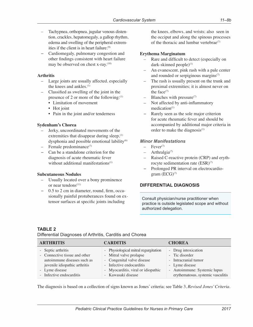
DIFFERENTIAL DIAGNOSIS
Consult physician/nurse practitioner when practice is outside legislated scope and without authorized delegation.
TABLE 2 Differential Diagnoses of Arthritis, Carditis and Chorea
ARTHRITIS CARDITIS CHOREA- Septic arthritis- Connective tissue and other
autoimmune diseases such as juvenile idiopathic arthritis
- Lyme disease- Infective endocarditis
- Physiological mitral regurgitation- Mitral valve prolapse- Congenital valve disease- Infective endocarditis- Myocarditis, viral or idiopathic- Kawasaki disease
- Drug intoxication- Tic disorder- Intracranial tumor- Lyme disease- Autoimmune: Systemic lupus
erythematosus, systemic vasculitis
The diagnosis is based on a collection of signs known as Jones’ criteria; see Table 3, Revised Jones’ Criteria.
Cardiovascular System 11–8b
Pediatric Clinical Practice Guidelines for Nurses in Primary Care2017
COMPLICATIONS
– Rheumatic heart disease due to permanent damage to the heart valves, which leads to valvular disease, cardiac myopathy and sequelae such as:(1) • Heart failure• • Systemic embolism• Stroke• Endocarditis• Need for cardiac surgery
– Recurrent attack, which worsens the damage to the heart(12)
− Death(2)
DIAGNOSTIC TESTS
Consult physician/nurse practitioner when practice is outside legislated scope and without authorized delegation.
Diagnostic test selection is based on client history,
test availability. Testing should be carried out as per provincial/territorial policies and procedures. The tests below are for consideration. Current-
the diagnosis of ARF.(1) ARF remains a clinical diagnosis and relies on health professionals being aware of its diagnostic features.(1)
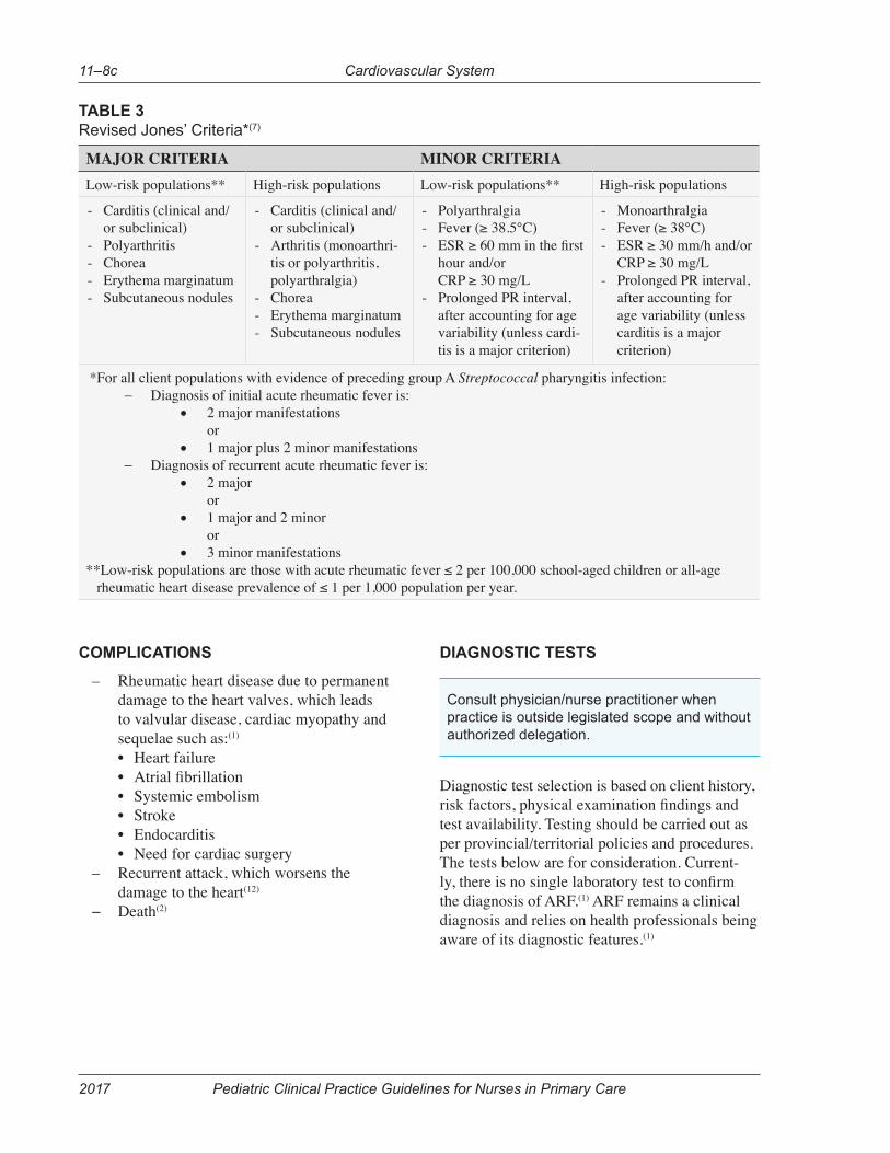
TABLE 3 Revised Jones’ Criteria*(7)
MAJOR CRITERIA MINOR CRITERIALow-risk populations** High-risk populations Low-risk populations** High-risk populations
- Carditis (clinical and/or subclinical)
- Polyarthritis- Chorea- Erythema marginatum- Subcutaneous nodules
- Carditis (clinical and/or subclinical)
- Arthritis (monoarthri-tis or polyarthritis, polyarthralgia)
- Chorea- Erythema marginatum- Subcutaneous nodules
- Polyarthralgia- Fever (≥ 38.5°C)-
hour and/or CRP ≥ 30 mg/L
- Prolonged PR interval, after accounting for age variability (unless cardi-tis is a major criterion)
- Monoarthralgia- Fever (≥ 38°C)- ESR ≥ 30 mm/h and/or
CRP ≥ 30 mg/L- Prolonged PR interval,
after accounting for age variability (unless carditis is a major criterion)
*For all client populations with evidence of preceding group A Streptococcal pharyngitis infection:− Diagnosis of initial acute rheumatic fever is:
• 2 major manifestations or
• 1 major plus 2 minor manifestations− Diagnosis of recurrent acute rheumatic fever is:
• 2 major or
• 1 major and 2 minor or
• 3 minor manifestations**Low-risk populations are those with acute rheumatic fever ≤ 2 per 100,000 school-aged children or all-age rheumatic heart disease prevalence of ≤ 1 per 1,000 population per year.
Cardiovascular System11–8c
Pediatric Clinical Practice Guidelines for Nurses in Primary Care 2017
Laboratory – Rapid Antigen Detection Test (RADT)
(if available) – Throat culture and sensitivity (C+S)
for group A Streptococcus(10)
– WBC, ESR(13)
− Blood culture and sensitivity (C+S) if febrile(13)
Other Diagnostic Tests – Chest x-ray(13)
− ECG to assess for prolonged PR interval(1)
MANAGEMENT
Consult physician/nurse practitioner when practice is outside legislated scope and without authorized delegation.
Note: The diagnosis and treatment of rheumatic fever requires medical evacuation. Emergency treatment of heart failure may be necessary; see FNIHB Pediatric and Adolescent Care Clinical Practice Guidelines – Chapter 11 – Cardiovascular System – Cardiac Failure.
GOALS OF TREATMENT
– Provide symptomatic relief − Prevent complications
NON-PHARMACOLOGIC INTERVENTIONS
Interventions − Bed rest while awaiting medical evacuation
PHARMACOLOGICAL INTERVENTIONS
In addition to consulting a physician/nurse practitioner, review the drug monograph, the FNIHB Nursing Station Formulary or provincial/territorial formulary before initiating treatment. Note: With the exception of acetaminophen or medications to treat heart failure (if required), medications should not be started until the diagnosis has been clearly established. After consultation with receiving facility, antibiotics may be started after throat swab(s) have been collected.(1)
Antipyretic/AnalgesicAcetaminophen(14)
– Acetaminophen 10 to 15 mg/kg/dose PO q4-6h PRN
– [caution-hepatic, INR, renal]− Maximum from all sources: acetaminophen 75
mg/kg/day or 4,000 mg/day, whichever is less
Antibiotic Therapy – Antibiotics should be initiated to eradicate
residual GAS infection in all cases while the diagnosis of ARF is being established.(1)
– A full course of antibiotics should be given.(1) – Oral therapy such as penicillin, amoxicillin,
cephalexin or clindamycin may be considered. FNIHB
Pediatric and Adolescent Care Clinical Practice Guidelines – Chapter 9 – Ears, Nose, Throat and Mouth – Bacterial Pharyngotonsillitis – Antibiot-ic Therapy.
MONITORING AND FOLLOW-UP
Consult physician/nurse practitioner when practice is outside legislated scope and without authorized delegation.
MONITORING
– Monitor vital signs as indicated by client’s condition, including oxygen saturation.
– Monitor intake and output. – Monitor for signs of cardiac failure. If cli-
ent is in cardiac failure, see FNIHB Pediat-ric and Adolescent Care Clinical Practice Guidelines – Chapter 11 – Cardiovascular System – Cardiac Failure.
− If administering an agent with risk of anaphy-laxis, monitor the client closely for 30 minutes.
FOLLOW-UP
Post-acute Phase – All clients should receive regular fol-
low-up. The frequency and duration of review is dependent on individual clinical needs and should become more frequent in the event of symptom onset, symptomatic
(1)
Cardiovascular System 11–8d
Pediatric Clinical Practice Guidelines for Nurses in Primary Care2017
– Discuss the importance of ongoing second-ary prophylaxis during every health profes-sional interaction with the client.(1)
– Because of the risk of recurrence, contin-ual antibiotic prophylaxis with benzathine penicillin G must be maintained.
– The risk of recurrence is greatest in the 5 years after the initial episode.
– Prophylaxis is initiated immediately after completion of a full therapeutic course of antibiotics, as initiated by a physician/nurse practitioner.
– The physician/nurse practitioner should also determine any discontinuation of prophylaxis.
– The duration of antibiotic prophylaxis can range from 5 years to 30 years, as it depends on a number of factors, including age, clinical pattern, environment and time elapsed since the last episode of ARF.(1)
− Clients may need to be on other cardiac and anticoagulation therapies, depending on the severity of the heart damage due to ARF.
Referral− Arrange for medical evacuation.
APPENDIX SECTION A: SUPPLEMENTAL CLINICAL MANAGEMENT INFORMATION
Heart Murmurs – Those most commonly heard during acute
rheumatic fever are:(8) • Apical pansystolic murmur is a high-
pitched, blowing-quality murmur of mitral regurgitation that radiates to the left axilla. The murmur is unaffected by respiration or position.
• Apical diastolic murmur (known as Carey-Coombs murmur) is heard with active carditis and accompanies severe
• Basal diastolic murmur is an early dia-stolic murmur of aortic regurgitation and is a high-pitched, blowing, decrescendo heard best along the right upper and mid-left sternal border after deep expiration while the client is leaning forward.
Approaches to Disease PreventionEffective control programs for acute rheumatic fever integrate features of primordial, primary and secondary prevention.
Primordial Prevention− Primordial prevention refers to broad
social, economic and environmental ini-tiatives undertaken to prevent or limit the impact of GAS infection in a population.
Primary Prevention− Primary prevention refers to medical in-
tervention using antibiotics to reduce GAS transmission, acquisition, colonization and carriage, or treating GAS infection effec-tively to prevent the development of acute rheumatic fever.(13)
Secondary Prevention− Secondary prevention refers to medical
intervention using long-term prophylactic antibiotics to reduce repeated acquisition of GAS that might induce recurrent episodes of acute rheumatic fever in order to prevent the development of rheumatic heart dis-ease, or for those clients who have estab-lished rheumatic heart disease in order to prevent disease progression.
BIBLIOGRAPHYThe following references and other sources have informed the updating of this Clinical Practice Guideline.
REFERENCES
1. Heart Foundation of New Zealand. New Zealand guidelines for rheumatic fever: Diag-nosis, management and secondary prevention of acute rheumatic fever and rheumatic heart disease: 2014 update. [Internet] Auckland (NZ): The National Heart Foundation of New Zealand; 2014 [cited 2015 May 20]. Available from: http://assets.heartfoundation.org.nz/shop/marketing/non-stock-resources/diagnosis-management-rheu-matic-fever-guideline.pdf
Cardiovascular System11–8e
Pediatric Clinical Practice Guidelines for Nurses in Primary Care 2017
2. Gerber MA, Baltimore RS, Eaton CB, Gewitz M, Rowley AH, Shulman ST, et al. Prevention of Rheumatic Fever and Diagnosis and Treatment
Statement From the American Heart Association Rheumatic Fever, Endocarditis, and Kawasaki Disease Committee of the Council on Cardiovas-cular Disease. Circulation. [Internet] Hagerstown, MD : Lippincott Williams & Wilkins; 2009. 119(11), 1541–1551. doi:10.1161/CIRCULATIO-NAHA.109.191959. Available from: http://www.circ.ahajournals.org/con-tent/119/11/1541
3. Gordon J, Kirlew M, Schreiber Y, Saginur R, Bocking N, Blakelock B, et al. Acute rheumatic fever in First Nations communities in northwest-ern Ontario. Canadian Family Physician. Don Mills, Ont.: College of Family Physicians of Canada; 2015. 61(10), 881–886. Available from: http://www.cfp.ca/content/61/10/881
4. Victoria State Government. Rheumatic heart disease. Department of Health and Human Services. [Internet] Melbourne, Victoria (AU): Department of Health and Human Services, State Government of Victoria (AU); 2014 [cited 2015 May 6]. Available from: http://www.betterhealth.vic.gov.au/health/con-ditionsandtreatments/rheumatic-heart-disease.
5. Dumitru I, Baker MM. Heart Failure: Practice Essentials, Background, Pathophysiology. [Internet] New York, NY: Medscape; 2015 [cited 2015 December 14]. Available from: http://emedicine.medscape.com with authorized username and password.
6. Madden S, Kelly L. Update on acute rheumatic fever: It still exists in remote communities. Canadi-an Family Physician. [Internet] Don Mills, ON: Col-lege of Family Physicians of Canada; 2009. 55(5), 475–478. Available from: http://www.ncbi.nlm.nih.gov/pmc/articles/PMC2682299/ with payment.
7. Gewitz MH, Baltimore RS, Tani LY, Sable CA, Shulman ST, Kaplan EL. Revision of the Jones Criteria for the diagnosis of acute rheumat-ic fever in the era of doppler echocardiography:
Association. Circulation. [Internet] Hagerstown, MD : Lippincott Williams & Wilkins; 2015 May 19. doi:10.1161/CIR.0000000000000205. Available from: http://circ.ahajournals.org/con-tent/early/2015/04/23/CIR.0000000000000205
8. Chin TK, Chin EM. Pediatric Rheumatic Heart Disease: Background, Pathophysiology, Epide-miology. [Internet] New York, NY: Medscape; 2014 [cited 2015 December 14]. Available from http://emedicine.medscape.com with authorized username and password.
9. Colucci WS. Evaluation of the patient with suspected heart failure. [Internet] Waltham, MA (US): UptoDate; 2016. [cited 2017 January 12]. Available from: http://www.uptodate.com with authorized username and password.
10. Chin TK, Li D. Pediatric rheumatic fever: Background, pathophysiology, epidemiology. [Internet] New York, NY: Medscape; 2014 [cited 2015 December 7]. Available from: http://reference.medscape.com with authorized username and password.
11. Steer A, Gibofsky A. Acute rheumatic fever: Clinical manifestations and diagnosis. [Internet] Waltham, MA (US): UptoDate; 2015. Available from: https://www.uptodate.com with authorized username and password.
12. World Heart Federation. Rheumatic heart disease. [Internet] Geneva, Switzerland: World Heart Federation; 2012 [cited 2016 May 17]. Available from: https://www.world-heart-federa-tion.org/programmes/rheumatic-heart-disease/
Cardiovascular System 11–8f
Pediatric Clinical Practice Guidelines for Nurses in Primary Care2017
13. RHD Australia (ARF/RHD writing group). The Australian guideline for prevention, diagno-sis and management of acute rheumatic fever and rheumatic heart disease, 2nd ed. Quick reference guides. National Heart Foundation of Australia and the Cardiac Society of Australia and New Zealand. [Internet] Casuarina NT (AU): Men-zies School of Health Research; c2014 [cited 2015 December 16]. Available from: http://www.
quick_reference_guides_0.pdf
14. Acetaminophen. [Internet] Hudson, OH: Lexi-Comp. Inc.; 2015 [cited 2015 April 23]. Available from: http://online.lexi.com with authorized username and password.
OTHER SOURCES
Health Canada. First Nations and Inuit Health Branch (FNIHB) Nursing Station Formulary and
Canadian Pharmacists Association. Acetamino-phen (Tylenol). [Internet] Ottawa, ON: e-Thera-peutics; 2012 [cited 2015 December 13]. Avail-able from: http://www.e-therapeutics.ca with authorized username and password.
Canadian Pharmacists Association. Compendium of Therapeutic Choices: Canada’s Trusted Refer-ence for Primary Care Therapeutics (CTC 7). Ottawa: Canadian Pharmacists Association; c2014.
Acetaminophen. [Internet] Hudson, OH: Lexi-Comp. Inc.; 2015 [cited 2015 December 13]. Available from: http://online.lexi.com with autho-rized username and password.
Canadian Pharmacists Association. Penicillin V. [Internet] Ottawa, ON: e-Therapeutics; 2009 [cited 2015 December 6]. Available from: http://www.e-therapeutics.ca with authorized username and password.
Penicillin V. [Internet] Hudson, OH: Lexi-Comp. Inc.; 2015 [cited 2015 December 6]. Available from: http://online.lexi.com with authorized user-name and password.
Canadian Pharmacists Association. Amoxicillin (Amoxicillin). [Internet] Ottawa, ON: e-Thera-peutics; 2015 [cited 2015 December 6]. Available from: http://www.e-therapeutics.ca with autho-rized username and password.
Amoxicillin. [Internet] Hudson, OH: Lexi-Comp. Inc.; 2015 [cited 2015 December 6]. Available from: http://online.lexi.com with authorized username and password.
Canadian Pharmacists Association. Cephalexin. [Internet] Ottawa, ON: e-Therapeutics; 2009 [cited 2015 December 6]. Available from: http://www.e-therapeutics.ca with authorized username and password.
Cephalexin. [Internet] Hudson, OH: Lexi-Comp. Inc.; 2015 [cited 2015 December 6]. Available from: http://online.lexi.com with authorized user-name and password.
Canadian Pharmacists Association. Clindamycin (Dalacin C). [Internet] Ottawa, ON: e-Therapeu-tics; 2014 [cited 2015 December 6]. Available from: http://www.e-therapeutics.ca with autho-rized username and password.
Clindamycin. [Internet] Hudson, OH: Lexi-Comp. Inc.; 2015 [cited 2015 December 6]. Available from: http://online.lexi.com with authorized username and password.
Canadian Pharmacists Association. Penicillin G. [Internet] Ottawa, ON: e-Therapeutics; 2009 [cited 2015 December 6]. Available from: http://www.e-therapeutics.ca with authorized username and password.
Penicillin G. [Internet] Hudson, OH: Lexi-Comp. Inc.; 2015 [cited 2015 December 6]. Available from: http://online.lexi.com with authorized username and password.
Cardiovascular System11–8g
Cardiovascular System 11–9
Pediatric Clinical Practice Guidelines for Nurses in Primary Care 2011
VIRAL MYOCARDITISMyocarditis is an inflammatory disorder of the myocardium with necrosis of the myocytes and associated inflammatory infiltrate.
PATHOPHYSIOLOGYMyocarditis generally results in a decrease in myocardial function, with concomitant enlargement of the heart and an increase in the end-diastolic volume caused by increased preload. Progressive increase in left ventricular end-diastolic volume increases left atrial, pulmonary venous and arterial pressures, resulting in increasing hydrostatic forces. These increased forces lead to both pulmonary edema and congestive heart failure.
CAUSESIt is usually caused by a viral infection. Parvovirus B19 and human herpesvirus-6 are the most frequent pathogens in patients with acute myocarditis. Infecting organisms may include the following:
– Parvovirus B19 – Herpesvirus – Coxsackievirus types A and B (especially type B) – Adenovirus (most commonly types 2 and 5) – Cytomegalovirus – Echovirus – Epstein-Barr virus – Hepatitis C virus – Human immunodeficiency virus – Influenza and parainfluenza – Measles – Mumps, associated with endocardial
fibroelastosis (EFE) – Poliomyelitis virus – Rubella – Varicella
Risk FactorsYounger patients, especially newborns and infants and immunocompromised individuals may have increased susceptibility to myocarditis.
HISTORYClinical presentation varies widely. In mild forms, few or no symptoms are noted. In severe cases, patients may present with acute cardiac decompensation and progress to death.
Cardiovascular System11–10
Pediatric Clinical Practice Guidelines for Nurses in Primary Care2011
In newborns and infants, symptoms may sometimes appear suddenly and may include:
– Irritability – Failure to thrive – Feeding difficulties – Fever and other symptoms of infection – Lethargy – Low urine output (a sign of decreasing kidney
function) – Pale hands and feet (a sign of poor circulation) – Rapid breathing – Rapid heart rate
Symptoms in children over age 2 may also include:
– Belly area pain and nausea – Cough – Fatigue – Swelling (edema) in the legs, feet and face – Recent, nonspecific, flu-like illness – Older children present with similar symptoms
as above and may experience lack of energy and general malaise
– Chest pain: Although rare in young children, this may be the initial presentation for older children and adolescents and should be considered a serious symptom accordingly
PHYSICAL FINDINGS
Neonates/Infants
– Hypothermia or hyperthermia – Tachypnea – Tachycardia – Cyanosis – Cool extremities – Decreased capillary refill – Pale or mottled skin may be present – Wheezing, and diaphoresis with feeding – Irritability – Somnolence – Hypotonia – Seizures – Oliguria – End-organ damage (for example, renal failure
may develop because of direct viral infestation or because of low cardiac output
Older Children
– Low grade fever – Tachycardia, weak pulse – Jugular venous distention and edema of the lower
extremities may be present – Heart sounds may be muffled, especially in the
presence of pericarditis – An S3 may be present – Heart murmur caused by atrioventricular valve
regurgitation may be heard – Crackles may be heard in older children – Hepatomegaly may be present in younger children – Cool extremities – Decreased capillary refill – Pale or mottled skin may be present
Adolescents
Presentation in adolescents is similar to that of children between 6 and 12 years old. However, the following symptoms may be more prominent:
– Decreased exercise tolerance – Lack of energy, malaise – Chest pain – Low-grade fever – Arrhythmia – Cough – Low cardiac output
DIFFERENTIAL DIAGNOSIS
– Myocarditis, nonviral – Pericarditis, viral – Aortic stenosis, valvular – Enteroviral infections – Cardiomyopathy, dilated – Glycogen-storage disease type I or type II – Coarctation of the aorta – Coronary artery anomalies
DIAGNOSTIC TESTS
Chest x-ray may show cardiomegaly and cardiac failure.
Electrocardiography (ECG)
Cardiovascular System 11–11
Pediatric Clinical Practice Guidelines for Nurses in Primary Care 2011
In some patients with mild cardiac involvement, ECG changes may be the only abnormal findings suggestive of myocarditis.
– Low-voltage QRS (< 5 mm throughout the limb leads) is the classic pattern
– Pseudoinfarction patterns with pathologic Q waves and poor progression of R waves in the precordial leads may also be present
– T-wave flattening or inversion is a common finding associated with small or absent Q waves in V5 and V6
– Left ventricular hypertrophy with strain may be present
– Other nonspecific findings include a prolonged PR segment and prolonged QT interval
– Sinus tachycardia is the most common finding – Premature ventricular contractions and atrial
tachycardia have been reported – Junctional tachycardia is common and may
worsen congestive heart failure – Occasional second-degree and third-degree
atrioventricular block may be present – Ventricular tachycardia is commonly associated
and may be the initial presentation
COMPLICATIONS
– Arrhythmia – Cardiac failure (see “Cardiac Failure”) – Thromboembolism – Decrease in ventricular function – Dilated cardiomyopathy
MANAGEMENT
Goals of Treatment
– Stabilize cardiovascular function – Prevent complications
Appropriate Consultation
Consult a physician urgently if you suspect this condition.
Adjuvant Therapy
– Give supplemental oxygen as necessary via non-rebreather mask. Titrate to keep oxygen saturations > 97%
– Start an intravenous line with normal saline. Run at a rate sufficient to maintain hydration depending on oral intake of child. Do not overhydrate. Keep line open until consultation with an emergency physician. Always weigh infant before starting any intravenous fluids as a measure of hydration
Nonpharmacologic Interventions
– Bed rest is necessary during the acute phase of the illness
– Nurse in an upright position
Pharmacologic Interventions
Consult a physician for medication orders. Medications may include the following, when indicated: see “Cardiac Failure”.
– Diuretics to decrease volume:
furosemide (Lasix), 1 mg/kg IV stat (may be given PO if IV access not available)
– ACE inhibitors may be prescribed by a physician for afterload reduction
– Digoxin may be used in some cases to increase contractility
– Antiarrhythmics – Anticoagulants
Monitoring and Follow-Up
Acute Phase
Monitor ABCs (airway, breathing and circulation), vital signs, pulse oximetry, heart and lung sounds, neuromental status, intake and output and medication response and adverse effects closely until child is transferred to hospital.
Over the Long Term
Children with cardiac illness should be monitored regularly within the community to ensure normal growth and development and to watch for complications. Frequency of follow-up depends on the severity of the condition.
Referral
Medevac to a facility with intensive and cardiology care.
Cardiovascular System11–12
Pediatric Clinical Practice Guidelines for Nurses in Primary Care2011
SOURCES
Internet addresses are valid as of February 2012.
BOOKS AND MONOGRAPHS
Bickley LS. Bates’ guide to physical examination and history taking. 7th ed. Baltimore, MD: Lippincott Williams & Wilkins; 1999.
Cheng A, et al. The Hospital for Sick Children handbook of pediatrics. 10th ed. Elsevier; 2003.
Colman R, Somogyi R (Editors-in-chief). Toronto notes – MCCQE 2008 review notes. 24th ed. Toronto, ON: University of Toronto, Faculty of Medicine; 2008.
Gray J (Editor-in-chief). Therapeutic choices. 5th ed. Ottawa, ON: Canadian Pharmacists Association; 2007.
Health Canada. Canadian immunization guide. 7th ed. Ottawa, ON: Health Canada; 2006.
Jensen B, Regier L (Editors). The Rx files. 7th ed. Saskatoon, SK; 2008
Rudolph CD, et al. Rudolph’s pediatrics. 21st ed. McGraw-Hill; 2003.
Uphold CR, Graham MV. Clinical guidelines in child health. 3rd ed. Gainesville, FL: Barmarrae Books; 2003.
Repchinsky C (Editor-in-chief). CPS Compendium of pharmaceuticals and specialties. Ottawa, ON: Canadian Pharmacists Association; 2007.
JOURNAL ARTICLES
Arnold JMO, Liu P, Demers C, et al. Canadian Cardiovascular Society consensus conference recommendations on heart failure 2006: Diagnosis and management. Can J Cardiol 2006;22(1):23-45.
Birdi N, Hosking M, Clulow MK, Duffy CM, et al. Acute rheumatic fever and poststreptococcal reactive arthritis: Diagnostic and treatment practices of pediatric subspecialists in Canada. J Rheumatol 2001;28:1681-88.
INTERNET GUIDELINES / DOCUMENTS / OTHER PUBLICATIONS
Cilliers A, Manyemba J, Saloojee HH. (2003). Anti-inflammatory treatment for carditis in acute rheumatic fever. Available at: http://summaries.cochrane.org/CD003176/corticosterids-andimmunotherapy-to-prevent-heart-damage-as-a-resultof-rheumatic-fever
Chin TK, Chin EM, Siddiqui T, Sundell, A-K. (2008, October). Rheumatic Heart Disease: Treatment & Medication. Available at: http://emedicine.medscape.com/article/891897-overview
Hong W, Tang W. (2008, September). Myocarditis. Available at: http://emedicine.medscape.com/article/156330-overview
Poothirikovil V. (2008, November). Cardiomyopathy, Dilated. Available at: http://emedicine.medscape.com/article/895187-overview
Rodriguez-Cruz E, Ross RD. (2008, October). Myocarditis, Viral. Available at: http://emedicine.medscape.com/article/890740-overview
Satou GM, Herzberg G, Erickson LC. (2006, June; updated 2009, March). Heart Failure, Congestive.Available at: http://emedicine.medscape.com/article/901307-overview
END NOTES
1 Emergency Nurses Association (ENA) (2007) Trauma Nursing Core Competencies Provider Manual. 6th Edition. p. 70.
2 Debbie M. Popovich, Nancy Richiuso, Gale Danek. Pediatric health care providers’ knowledge of pulse oximetry Pediatric Nursing, Jan-Feb, 2004; p. 2. Available at: http://findarticles.com/p/articles/mi_m0FSZ/is_1_30/ai_n17206797/pg_2/?tag=content;col1
3 UpToDate Online 18.1. 2010. Treatment and prevention of rheumatic fever. And UpToDate Online 18.1. 2010. Antibiotic therapy of GAS pharyngitis, which is based on Gerber, MA, Baltimore, RS, Eaton, CB, et al. Prevention of rheumatic fever and diagnosis and treatment of acute streptococcal pharyngitis: A scientific statement from the American Heart Association Rheumatic Fever, Endocarditis, and Kawasaki Disease Committee of the Council on Cardiovascular Disease in the Young, the Interdisciplinary Council on Functional Genomics and Translational Biology, and the Interdisciplinary Council on Quality of Care and Outcomes Research. Circulation 2009;119(11):1541-51. Available by subscription: www.uptodate.com
4 Secondary prevention of rheumatic fever (prevention of recurrent attacks). UpToDate Online 18.1. Available by subscription: www.uptodate.com