Embed Size (px)
Citation preview

CHAPTER 1
ITRODUCTIO

Introduction Chapter I
1
ITRODUCTIO
Malaysia is known for its richness in plants that have been around for millions of
years. Strategically located on the equator where there exists a balance of hot and wet
climate, therefore the environment has a wide range of plants to form a majestic plant
kingdom. From ancient times, preparations of plants have been used as remedies
against disease. Today, with the advent of modern technology scientists are able to
identify the active principles of medicinal plants such as morphine in opium poppy
which acts as a pain killer. By the end of nineteenth century organic chemists had
begun the investigation on alkaloids and their usage in traditional or modern
medicine. Therefore, the studies on various compounds such as alkaloids have been
carried out widely due to the demand of the pharmaceutical industries.
Natural products have played an important role in drugs discovery. Natural products
are those chemical compounds derived from living organisms such as plants, animals,
insects, and the study of natural products is the investigation of their structures,
formations, use, and purpose in the organism. Drug derived from natural products are
usually secondary metabolites and their derivatives. Today those must be pure and
highly characterized compounds. Until the late 1800’s, organic chemistry was almost
exclusively the study and use of natural products. The natural products that were
studied and used tend to be the compounds that occurred in the largest amounts,
mostly from plants, and were most easily isolated in a pure, or sometimes not very
pure, from the technique such as simple distillation, steam distillation or extraction
with acid or base. We now employ different solvents, for example hexane and CO2
nowadays are used to extract the non-polar constituents, methanol and ethanol to
extract the polar constituents. Modern isolation techniques include all types of

Introduction Chapter I
2
chromatography (HPLC, TLC, CC, GC), often guided by bioassays, to isolate the
active compounds.
This thesis deals mainly with the isolation and structural elucidation of the alkaloids
contained in bark and leaves of Ochrosia oppositifolia which belongs to the family of
Apocynaceae.
The objectives of this research are:
i. to isolate the phytochemicals from the Ochrosia oppositifolia.
ii. to identify the chemical structures of the isolated compounds.
iii. to evaluate the antiplasmodial activity of the crudes and the isolated compounds.
1.1 Apocynaceae: Distribution and Habitat
The Apocynaceae or dogbane family is a family of flowering plants, including trees,
shrubs, herbs, or lianas. Many species are tall trees, found in the tropical rainforest,
and most are from the tropic and subtropics, but some grow in tropical environments.
There are also some perennial herbs from temperate zones. Many of these plants have
milky sap; ans some species are poisonous if ingested. Some genera of Apocynaceae,
such as Adenium how ever, have both clear and milky latex sap, and others, such as
Pachypodium, possess clear sap.1
The Apocynaceae family is one of the largest plant families; comprises of 400 genera
and 1500 species. In Peninsular Malaysia, there are only 32 genera and 120 species.
The alkaloids in the Apocynaceae are important in native medicine, such as bark of
Ochrosia oppositifolia which had used as a medicine to treat the cancer in ancient
Hawaii, while more recent tests of the crude alkaloid extract showed antiplasmodial
properties 2. These plants are distributed mainly in the tropical and sub tropical region.

Introduction Chapter I
3
1.2 General Appearance and Morphology 3, 4
The leaves are simple, usually opposite and decussate, or whorled; stipules are usually
absent. The flowers are bisexual and actinomorphic or sometimes weakly
zygomorphic. The calyx is synsepalous and usually 5-lobed. The corolla is
sympetalous and usually 5-lobed. The stamens are distinct, as many as corolla lobes
and alternate with them, and adnate to the corolla tube (or erigynous zone). The
anthers are introrse and commonly adherent to the surface of the stigma. The
ynoecium consists of a single compound pistil of 2 carpels that may be distinct at the
level of the superior or rarely partly inferior ovary but which are united by a single
style. When distinct, each ovary typically has few to numerous ovules on marginal
placentae; when connate, the placentation is axile or intruded parietal.
A nectary consisting of 5 glands or an annular ring is usually found at the base of the
ovary. The fruit is commonly a follicle, capsule, or berry. The seeds usually are flat
and winged or have a tuft of hairs at one end.
This is a large Family with about 1500 species found mainly in tropical regions. It
includes many of the most well-known tropical ornamental plants (Oleander,
Frangipani,Allamanda, Mandevilla). Many are large trees with buttress roots found in
rainforests some are smaller, evergreen or deciduous trees, shrubs or climbers from
other warm areas of the world, and one or two are found in temperate regions (Vinca).
The sap of most plants is milky latex,which is often of economic importance for
medicinal use, or further production of rubber. This sap is often toxic.
Fruit type is highly diversified in the family, and it is diagnostic of many genera.
Genera 1-produce 1, 2-celled berries from a flower; genus 5 produces 2, 1-celled
berries from a flower; 6 and 7 produce mostly fleshy follicles containing deeply
indented seeds with ruminate endosperm; 8 has follicles and winged seeds; 9 produces

Introduction Chapter I
4
follicles and seeds with 2 comas; 10-12 have follicles with globose seeds; 13-18 have
drupes mostly with fleshy mesocarp; 19 has samaroid fruit; 20 has spiny capsules with
seeds winged all around; and 21-44 have free or fused follicles and comose seeds.
Double flowers are known only from cultivated forms of erium oleander,
Tabernaemontana divaricata, and Wrightia religiosa.
Plants of the Apocynaceae are often poisonous and are rich in alkaloids or glycosides,
especially in the seeds and latex. Some species are valuable sources of medicine,
secticides, fibers, and rubber.
1.3 Classification of Apocynaceae
Apocynaceae can be divided into Plumerioideae subfamily, which is further divided
into a few tribes (Scheme 1.1). Each tribe comprises of several genera. The genus
Ochrosia is classified as member of the tribe Rauvolfieae. 5

Introduction Chapter I
5

Introduction Chapter I
6
1.3.1 Genus Ochrosia
Ochrosia is a genus of sea-shores, which was observed in Singapore a century ago.
There are 32 ochrosia in Malaysia which mostly occurs in the Pangkor island.Trees
with latex. Branches stout. leaves in whorls 3-5, rarely opposite, lateralveins
numerous, sub parallel, almost at a right angle to midvein. Cymes sub terminal,
peduncalate. Calyx deeply divided, usually without glands. Corolla salver form;
tube slightly dilated above middle, to 1cm throat without scales; lobes overlapping
to right. Stamens inserted in widening of corolla tube; anthers free from pistil head,
narrowly oblong, rounded at base; disc absent. Ovaries 2, free or basally connate;
ovules 2-6, biseriate on each side of a prominent placenta. Style filiform; pistil head
shortly 2-cleff atapex. Drupes 1 or 2, smooth, endocapp thick, hard, seeds 2-4 per
locale, flat, not compose; endosperm none; cotyledons large, flat.6
The nearest
related genus is probably Cerbera.

Introduction Chapter I
7
1.3.2 Ochrosia oppositifolia
Ochrosia oppositifolia is an evergreen tree that can vary greatly in size, growing to a
maximum height of 15 m (50ft) or more. This species produces five-petalled white
flowers with yellow centers. The flowers usually drop to the ground like confetti. The
fallen flowers provide a clue to finding the tree. The fruit comes in pairs, is elliptical
in shape, and is about 5-8 cm long and turns yellow when it is ripe. The seed is about
10-20 cm, apparently growing quite slowly.
Ochrosia oppositifolia occurs in Indonesia and along the southern coastal region of
Sri Lanka. This plant is commonly called Muda Kaduru in Sri Lanka. The timber is
moderately hard and even grained. Its roots are reputed to nullify the effect of eating
poisonous fish and crabs. It is also used as a bitter, stomachic and carminative. The
seeds of Ochrosia oppositifolia are edible and also reputed in indigenous medicine. 7
Ochrosia oppositifolia belonging to Apocynaceae family is an alkaloid-rich plant.
Plants of this genus find wide use in the traditional system of medicine.7 The presence
of alkaloids was first observed by J.Polsson. A literature search on the alkaloids of
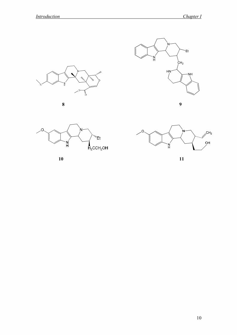
Ochrosia oppositifolia revealed the presence of (isoreserpiline 1, reserpiline,
ochroposinine 7, epirauvanine, bleekerine, 10-hydroxyapparicine, 10-
methoxyapparacine, 10-methoxydihydrocorynantheol 10, ochrolofuanine, reserpinine
3, isoreserpinine 8 and 9-methoxyellipticine 4).8
Table 1.2 shows the alkaloids isolated from Ochrosia.9, 10

Introduction Chapter I
8
Table 1.2 Alkaloids Isolated from Ochrosia
Compound Mol.Wt. Mol. Formula
Isoreserpiline 1 412 C23H28N2O5
Neisosposinine 2 428 C23H28N2O6
Reserpinine 3 382 C22H26N2O4
9-methoxyellipticine 4 276 C18H16N2O
10-Hydroxyapparacine 5 280 C18H20N2O
10-methoxyapparacine 6 294 C19H22N2O
Ochropposinine 7 411 C23H27N2O+
5
Isoreserpinine 8 382 C22H26N2O4
Ochrolifuanine 9 438 C29H34N4
10-methoxydihydrocorynantheol 10 382 C22H26N2O4
10- methoxycorynantheol 11 326 C20H26N202

Introduction Chapter I
9
1 2
3 4
5 R= OH 7
6 R= CH3
Introduction Chapter I
10
NH
N
Et
CH2
HNNH
8 9
10 11

Introduction Chapter I
11
Figure 1.1: Bark and Leaves of Ochrosia oppositifolia

CHAPTER 2
GEERAL CHEMICAL
ASPECTS

General Chemical Aspects Chapter II
12
GEERAL CHEMICAL ASPECTS
2.1 Alkaloids
Plants produced two types of metabolites, i.e. primary metabolite and secondary
metabolite. The former includes polysaccharide, protein fatty acid and nucleic
acid, and the latter includes chemicals with no apparent function in the primary
metabolism of the organism and these substances tend to be of restricted
taxonomic distribution and such metabolites have a history of use as bioactive
agents like antiplasmodial activities.
2.2 Secondary Metabolites
Secondary metabolites are often unique to a particular species or group of
organism and some act many act as antifeedants, sex attractants, repellents,
antimalarial, antibacterial, antifungal, antiviral, and antibiotic agents. 11
While
others may not have any apparent biological role.
Alkaloids are the most potent of all the secondary metabolites since the last
century. Their potency extends to almost universal toxicity and it would be correct
to start that the action of my alkaloid on the body is inherently stressful. 12
The
following paragraph shall discuss briefly the alkaloids and their chemicals aspect.

General Chemical Aspects Chapter II
13
2.3 Definition of Alkaloids 13-15
The term alkaloids or alkali-like was first proposed by W. Meisner in
1819.Alkaloids are defined as nitrogen containing basic substances, having a
complex structure, naturally origin and limited distribution on earth. Alkaloids
always contain their nitrogen atom as part of the heterocyclic system, and they
usually possess some pharmacological activity. Particular alkaloid is usually
restricted to certain genera and families of plant kingdom, rarely being present in
large groups of plants. They are biosynthetically exist as salt and related to acid
amino such as ornithine and lysine. A precise definition of the term alkaloid
(alkali-like) is somewhat difficult because there is no clear-cut boundary between
alkaloids and naturally occurring complex amines16
. Many of alkaloids are derived
from plant sources. They are basic, contain one or more nitrogen atoms (usually in
a heterocyclic ring) and usually have a marked physiological action on man or
other animals (Table 2.1).
Alkaloids can be separately categorized into three groups based on their
biogenesis; true alkaloids, protoalkaloids and pseudoalkaloids.
2.3.1 True Alkaloid
The true alkaloids normally contain nitrogen in heterocyclic rings, they derived
from amino acid. They normally occur in the plants as the salt of an organic acid.
Examples of this group are hygrine 12 and cryptostyline 13.

General Chemical Aspects Chapter II
14
2.3.2 Proto Alkaloid
The protoalkaloids are relative simple amine in which the amino acid nitrogen is
not in a heterocyclic ring. Like the true alkaloids, they are derived from amino
acid and are basic. Examples of this groups are mescalin 14, ephedrine 15, , -
dimethyltripthamine 16, serotonin 17 and cathinone 18.
2.3.3 Pseudo Alkaloid
The pseudoalkaloids are not derived from amino acid precursor and usually basic.
They are nitrogen containing in the molecule but they have a carbon skeletons
derived from terpenoids (mono-, sesqui-, di-, and triterpenoids), steroidal,
hemiterpenoids and other acetate derivatives. For example of this group are
actinidine 19, deoxynupharidine 20 and alchorneine 21.
Alkaloids represent a fascinating group of natural product for a number of reasons.
Many reveal important biological and pharmacological activities and for several
decades have been therapeutically applied in the treatments of various diseases.
Some of the alkaloids with impressive activities which have prompted the
development of broadly applied drugs on the pharmaceutical market, include the
well known cytotoxic bisindole alkaloids vincaleucoblastine 22 and vinblastine 23
from Catharanthus, the diterpenoid alkaloid taxol from Taxus, the highly
important analgesic morphine 24, the spasmolytics tubocurarine 25 and
papaverine 26, the vasodilating agents vincamine 27 and ajmalicine 28, emetine
29 with its emetic activity, and the antiarrhythmic alkaloids quinidine 30 and
ajmaline 31. 17

General Chemical Aspects Chapter II
15
The diversity of alkaloid structures forced scientists to concentrate during recent
decades on the elucidation of biosynthetic pathways at the enzymatic level. Now
the first example exist for the detection of series of enzymes catalyzing multistep
biosynthetic sequences, e.g. in the
field of isoquinoline alkaloids18
, indole bases19
and pyrrolizadine alkaloids20
.
Successful study of the molecular genetics of alkaloid formation has been
undertaken21
including the first example of heterologous expression of appropriate
enzymes catalyzing alkaloid
metabolism.22-24

General Chemical Aspects Chapter II
16
Table 2.1 Physiological activities of some alkaloids 25, 45, 67
Alkaloids Physiological activities
Nicotine 41
Atropine 42
Cytochalasin D 43
Haplophyllidine 44
Perforine 45
Bucharaine 46
Mescaline 47
Ephedrine 48
Gliotoxin 49
Papaverine 50
Laudanosine 51
(+)-Cepharanthine 52
(-)-mecambrine 53
Thebaine 54
Heroine 55
Erysotrine 56
Coccuvinine 57
Strychnine 33
Tylophorine 58
Canthin-6-one 59
Tecomine 60
Toxiferine 70
Toxic, stimulate respiratory processes, inhibition
in all sympathetic and parasympathetic
Antagonism to muscarinic receptors
(parasympathetic inhibition)
Cytotoxic and antitumor activity
Ataractic and sedative
Hypoglycemic activity
Suppress aggressive tendencies
Inhibit the pressor effect epinephrine and
produce hyperthemia and uterine contraction
Therapeutic agent
Bacteriostatic agent
Decrease the tonus of the smooth muscle.
Reduce intraocular pressure
Effective against human tuberculosis and
leprosy
Increases motility of isolated rat or rabbit
Duodenum
Increased intestinal muscle tone in rabbit
Exhibited significant cytotoxicity
Act as a muscle contractor
Neuromuscular blocking agents
Antifungal and leishmanicidal activities
Toxic to many bacteria and other unicellular
organism
Causes muscle relaxation

General Chemical Aspects Chapter II
17
12 13
14 15
NH
N(CH3)2
NH
NH2HO
CH3
NH2
H
O
16 17 18

General Chemical Aspects Chapter II
18
N
H3CCH3
N
CH3
H
CH3
O
N
N NH3C
H3C
CH3
OCH3
19 20 21
22 23
N+
O
OHO
NO
O
OH
24 25

General Chemical Aspects Chapter II
19
N
H3CO
OCH3
H3CO
HO
N
N
HO
O
O
26 27
O
N
O
O
HN
28 29
30 31

General Chemical Aspects Chapter II
20
Usually all the alkaloids occur in multicomponent mixture and separation of
alkaloids from other groups of natural product is the first requirement for detailed
qualitative, quantitative and structural analysis of single alkaloids.
The first crude drug to be investigated chemically was opium, the dried latex of
the poppy Papaver somniferum. In 1803, Derosne isolated a semipure alkaloid
from opium and named it as narcotine 32. Further examination of opium by
Serturner in 1805 led to the isolation of morphine structure. In the years 1817-
1820 in the laboratory of Pelletier and Caventou at the Faculty of Pharmacy in
Paris, the researchers obtained many active alkaloids. Among the alkaloids
obtained were strychnine 33, emetine 29, brucine 34, piperine 35, caffeine 36,
quinine 37, cinchonine 38 and colchicine 39. They also obtained coniine 40 in
1826.26
Certain families have a marked tendency to elaborate alkaloids: this is true for the
monocotyledons (Annonaceae, Apocynaceae, Fumariceae, Lauraceae,
Loganiaceae, Magnoliaceae, Menispermaceae, Papaveriaceae, Ranunculaceae,
Rubiaceae, Rutaceae, and Solanaceae). Within these families, some genera
produce alkaloids and others do not. The biological activity of the alkaloids,
together with their impressive structural diversity, largely accounts for the
enormous research effort that has been devoted to their characterization,
pharmacologic evaluation and synthesis.

General Chemical Aspects Chapter II
21
The probable function of alkaloids in plants seems to be to deter animals and
herbivorous insects. A part from their toxicity, the action of the alkaloids is not
easily summarized because each has its own individual character. Instead a list
will give some idea of their range and categories. As the individual examples
provided are the best known, and thus often the most notorious or dramatically
active, a more toxic picture of the alkaloidal range may be conveyed than is
actually justified.27
O
ONCH3
OH
O
OCH3
OCH3
HH3CO
N
N
OOH
H H
H
32 33
N
N
N
O
O
H3CO
HNOCH3
OCH3
H3CO
H3CO
H
H
H
H
H
H H
29 34

General Chemical Aspects Chapter II
22
O
O
N
O
N
NN
N
O
H3C
CH3
CH3O
35 36
N
N
H3CO
HO
N
N
HO
37 38
O
NH
COCH3H3CO
H3CO
OCH3
OCH3
NH
39 40
General Chemical Aspects Chapter II
23
H3CN
OCOCHC6H5
CH2OH
41 42 43
N OOCH3
HO N OOCH3
HO
HONH
O
OH
OH
O
44 45 46
NH2
OCH3
H3CO
H3CO
H H
C6H5 CH3
OH NHCH3
N
NH
OH
OH
H
O
O
47 48 49
General Chemical Aspects Chapter II
24
N
H3CO
HO
H3CO
NCH3
H3CO
H3CO
H3CO
H3CO
H
NCH3
H3CO
H3CO
H
H3CN O
O
H
OCH3
O
O
50 51 52
53 54 55
N
H3CO
H3CO
H3CO
NH3CO
H3CO
N
OCH3
H3CO
H3CO
OCH3
HH
56 57 58

General Chemical Aspects Chapter II
25
N
O
H3C
CH3
CH3N
N
O
59 60
The role of alkaloids in the plants are still unknown, but the researchers suggested
that the alkaloids are as the end product of metabolism or waste product, and it
functions as a protection against predator attack, storage reservoir of nitrogen for
protein synthesis, plants stimulants or regulator in many activities, such as growth,
metabolism and reproduction and a model for the chemical synthesis of analogue
with excellent properties. 28

General Chemical Aspects Chapter II
26
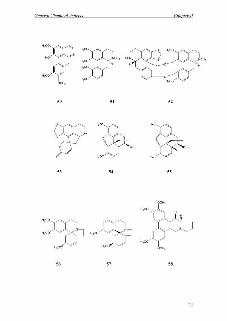
2.4 Classification of Alkaloids
Alkaloids are usually classified according to their common structural motif, based
on the metabolic pathway used to construct the molecule. When not much was
known about the biosynthesis of alkaloids, they were grouped under the names of
known compounds, even some non-nitrogenous ones, for example the
phenanthrenes, since this moiety appeared in the finished product or by the plant
or animals from which they were isolated. When more is learned about a certain
alkaloid, the grouping is changed to reflect the new knowledge, usually talking the
name of a biologically-important amine that stands out in the synthesis process.
Alkaloids may be classified by various methods which are based on the nature of
the classification such as: 29
• Biogenesis.
• Structural relationship based on chromophore of fundamental skeleton.
• Nitrogen atom and its immediate environment.
• Botanical origin.
• Spectroscopic criteria.
Several examples of common alkaloids ring skeleton are illustrated in Scheme
2.1.30

General Chemical Aspects Chapter II
27
Scheme 2.1: Example of alkaloid ring skeleton

General Chemical Aspects Chapter II
28
2.4.1 Indole Alkaloids
Indole is an electron-rich aromatic compound with characteristic properties and is
widely distributed in natural product and in proteins as the important constituent
of essential amino acid tryptophan 71. It is known to form a hydrophobic
environment in proteins and to be involved in enzymatic reactions, in addition to
the redox activities and various weak interactions.31
It shows versatile metal
binding abilities through the nitrogen and carbon atoms. They include the 'animal
alkaloids'; adrenaline, noradrenaline, serotonin (5-hydroxytryptamine or 5-HT),
the tranquillizing alkaloids of passion flower, the ophthalmic alkaloids related to
physostigmine from the calabar bean, the uterine stimulants ergotamine and
ergometrine from the fungus ergot of rye, and lysergic acid diethylamide (LSD).
Also included are the alkaloids of the Indian snakeroot (Rauwolfia serpentia),
including reserpine, having powerful hypotensive effects. In addition, there are
some examples of central nervous stimulants: strychnine, johimbine and
psilocybin. All these alkaloids have their effect on the neuromuscular system by
interacting with adrenergic receptors. Finally, the infamous two antileukaemic
medications vincristine and vinblastine isolated from the Madagascar periwinkle
(Catharanthus rosea).32
Indole alkaloids exhibit numerous biological activities (anti-tumor, anti-microbial,
anti-hypertensive and central nervous system stimulant 1). They can be found in
plants of the Apocynaceae, Rubiaceae, and Loganiaceae families.

General Chemical Aspects Chapter II
29
Among the Apocynaceae, the genus Ochrosia is rich in indole alkaloids. The
previous chemical studies of genus Ochrosia have reported many occurrences of
indole alkaloids especially of the corynanthean or C-type. 33
They are useful
chemical markers of the genus, and also have a great value for the classification of
the individual species within the genus which is very difficult if the classification
was to be based only upon the morphological character of the plants.
The 1H NMR and
13C NMR proton and carbon values of simple indole, C8H7N, are
given below:
Figure 2.1: 1H NMR and
13C NMR of C8H7N
The majority of indole alkaloids from the plant of Apocynaceae can be classified
into nine main types according to the structural characteristics of their skeleton
(Table 2.2). 34-36
Eight main types have been defined. There are vincosan, vallesiachotaman,
corynanthean, strychnan, aspidospermatan (all belong to the class Ι skeleton with
an intact secologanin), plumeran, eburnan (belong to the class ΙΙ skeleton,
rearranged secologanin) and ibogan (class ΙΙΙ skeleton, rearranged monoterpene).
The ninth type, tacaman (class ΙΙΙ skeleton) has been added by Verpoorte and
Beek to account for the isolation of a few tacamines (Table 2.2). 37, 38

General Chemical Aspects Chapter II
30
Table 2.2 : Types of Indole Skeleton, Classes and the Examples
Type of indole alkaloid Skeleton Example
Corynanthean or C-type Class Ι corynanthine
Vincosan or D-type Class Ι vincoside
Vallesiachtamon or V-type Class Ι vallesiachotamine

General Chemical Aspects Chapter II
31
Types of Indole Skeleton, Classes and the Examples (continued)
Type of indole alkaloid Skeleton Examples
Strychnan or S-type Class Ι strychnine
Aspidospermatan or A-type Class Ι condylocarpine
Eburanan or E-type Class ΙΙ vincamine

General Chemical Aspects Chapter II
32
Types of Indole Skeleton, Classes and the Examples (continued)
Type of alkaloid Skeleton Examples
N
N
CO2Me
OH
OH
Plumeran or P-type Class ΙΙ kopsine
Ibogan or J-type Class ΙΙΙ catharantine
Tacaman type Class ΙΙΙ tacamine

General Chemical Aspects Chapter II
33
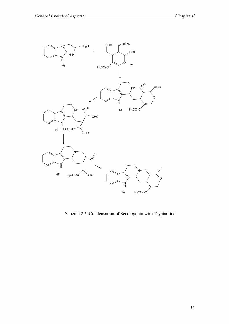
2.4.2 Biosynthesis of Indole Alkaloids: 39, 40, 41
Biosynthesis refers to the manner in which organic substances are synthesized,
altered or degraded by organism (plant or animal).
The complexity of indole alkaloid structure are formally derived from a Mannich
condensation of tryptamine 61 as the indole nucleus and a C9 or C10 monoterpene
moiety, derived from secologanin 62. Secologanine is made up of two molecules
of mevalonic acid.
Condensation of secologanine with tryptamine leads to strictosidine 63, a vincosan
skeleton alkaloid. Hydrolysis of the sugar residue and the opening of the cyclic
acetal function give the dialdehyde 64. Ring closure of 64 yields the tetracyclic
system 65. Minor rearrangements generate ajmalicine 66, a corynanthian type of
alkaloid (type C) which is the main alkaloid type in Ochrosia species. These
transformations are described in (Scheme 2.2).
Strictosidine 6342
is also the precursor of many other type of indole alkaloids;
(type A), (type C), (type V), (type E) and Tacaman type. (Scheme 2.3)
General Chemical Aspects Chapter II
34
NH
H2N
CO2HCHO
O
OGlu
H3CO2C
CH2
NH
NH
O
OGlu
H3CO2C
NH
NH
CHO
H3COOC
CHO
NH
N
H3COOC CHO
NH
N
O
H3COOC
62
63
64
65
66
61
Scheme 2.2: Condensation of Secologanin with Tryptamine

General Chemical Aspects Chapter II
35
Scheme 2.3: Steps in the Biosynthesis of Indole Alkaloids

General Chemical Aspects Chapter II
36
2.5 Pharmacological Activity
Before their recognition as useful therapeutic agents, alkaloids were renowned for
their poisonous properties. Of the many biologically active indole alkaloids, only a
few were used today as therapeutic agents of value in human medicine. For
example vincristine 67 was used to treat acute leukemia.43
The dimeric alkaloids
from Catharanthus roseus form an important class of antitumour agents, widely
used in combination chemotherapy regimens for treating leukamias and many
solid tumours. Vinflunine 68 which is a novel vinca alkaloid synthesized from
vinorelbine 69 using superacidic chemistry and characterised by superior in vivo
activity to vinorelbine in preclinical tumour models. 44, 45

General Chemical Aspects Chapter II
37
67 68
69 70
71

CHAPTER 3
RESULTS AD DISCUSSIO

Result And Discussion Chapter III
38
RESULTS AD DISCUSSIO
One Malaysian species from the family of Apocynaceae, Ochrosia oppositifolia,
has been studied for its components content. The plant materials were collected from
Pangkor islands in 2007. The sample was identified by L.E.Teo , University Malaya
and deposited at the herbarium unit (specimen no; KL 5349).
The isolation process was carried out using the conventional methods and the
structural elucidation was carried out using spectroscopic techniques, notably NMR,
IR, MS and UV and also by comparison with the literature values.
3.1 Isolation and Structural Elucidation of Compounds from Ochrosia
oppositifolia
Six compounds were isolated. The investigation on the crude extract from the bark of
Ochrosia oppositifolia have resulted in the isolation of three compounds, one alkaloid
namely, isoreserpiline 1, and two ferulic acid ester derivatives; 2-propenoic acid, 3-(4-
hydroxy-3,5-dimethoxyphenyl)-,methylester 73 and 17-methoxy-carbonyl-14-
heptadecaenyl- 4-hydroxy-3-methoxy cinnamate 74.
Isolation and structural elucidation of the crude alkaloids from the leaves of Ochrosia
oppositifolia yielded three alkaloids; neisosposinine 2, reserpinine 3 and alkaloid D
72.
The following sub chapters will discuss briefly the structural elucidation of all
compounds.

Result And Discussion Chapter III
39
3.2 Alkaloid from the Bark of Ochrosia oppositifolia
3.2.1 Alkaloid A: Isoreserpiline. 146
NH
N
O
H3CO
H3CO
1
2
4
56
78
9
10
1112
13 14
1617
1819
2021
22
H
15H
3
H
H3CO2C
CH3
H
1
Isoreserpiline 1 was isolated as a brownish amorphous solid. The mass spectrum
showed
pseudo-molecular ion peak at m/z 413.2, [M+H]+, which was consistent with the
molecular formula of C23H28N2O5. Another significant fragmentation was observed at
m/z 353 (M-COOCH3)+ as depicted in Scheme 3.2.
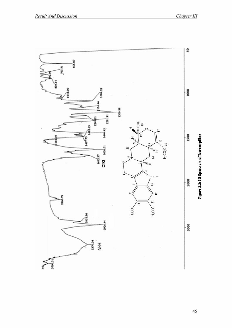
The UV spectrum revealed maxima at 226, 299 nm which were characteristic for an
indole system 47
. In addition, the IR spectrum showed a peak at 1692 cm-1
which
indicate the presence of the carbonyl group, 46
and a band at 3370 cm-1
presence of the
NH/OH groups. (Figure 3.3)
The 1H NMR spectrum (Figure 3.1) indicated the presence of two aromatic protons at
δ 6.88 and 6.79 attached to C-9 and C-12 respectively, and a deshielded singlet at δ
7.54 which belongs to H-17, indicative of a corynanthean skeleton. 47
Another three singlets appeared at δ 3.90, 3.87 and 3.46 can be attributed to the
protons of three methoxyls attached to C-10, C-11 and C- 22 respectively.

Result And Discussion Chapter III
40
The presence of the N-H protons was confirmed by the existence of a broad downfield
signal at δ 7.73 in the 1H NMR spectrum.
There were a total of twelve aliphatic proton signals observed in the 1H NMR
spectrum. Eight methylene proton signals appeared between δ 1.47- 3.05 which
belong to the protons attached to C-5, C-6, C-14 and C-21. The signal for another four
aliphatic protons δ 3.30, 2.8, 4.4 and 1.5 were assigned to H-3, H-15, H-19 and H-20
respectively (Figure 3.2). Finally a doublet in the high field region at δ 1.4 (d, 6.8 Hz)
may be attributed to the methyl group attached to C-19.
The 1H,
13C data (Table 3.2.1) and DEPT indicated the presence of 23 carbons; 8
quaternary carbons; δ 168.2 (C-22), 146.5 (C-11), 144.9 (C-10), 133.3 (C-2), 130.2
(C-13), 120.0 (C-8), 109.6 (C-16) and 107.9 ( C-7), 7 methines; δ 155.8 (C-17), 100.4
(C-9), 94.9 (C-12), 72.6 (C-19), 60.0 (C-3), 38.5 (C-20) and 31.4 (C-15), 4
methylenes; δ 56.5 (C-21) , 53.7 (C-5), 34.4 (C-14) and 21.9 (C-6), 1 methyl; δ 18.6
(C-18), and 3 methoxyls at δ 56.4 (C10-OMe), 56.5 (C11-OMe) and 50.8 (C22- OMe)
respectively.
The carbonyl (C-22) appeared at δ 168.1. Two aromatic methine signals were
observed at 100.4 and 94.9 could be assigned to C-9 and C-12 respectively. C-17
appeared at δ 155.8 and the carbonyl is proven by the correlation between carbonyl
and H-17 in HMBC (Figure 3.5). The HMBC spectrum also showed correlation
between C-20 and the methyl protons of C-18, while H-14 (δ 1.5) revealed correlation
with C-3. Other correlations were shown in Figure 3.5.
The complete assignments of carbons and protons were confirmed with DEPT,
HMQC and HMBC spectra. Analysis of all spectral data obtained and comparison
with literature

Result And Discussion Chapter III
41
confirmed the identify of alkaloid A as isoreserpiline 1 which was previously isolated
from Rauvolfia grandiflora Mart. 46
Scheme 3.2: Possible Mass Fragmentation of Isoreserpiline A 1
NH
N
O
MeO
MeO
H
Me
MeO2C
m/z 412
NH
N
O
MeO
MeO
H
Me
+
m/z 353
+
- COOCH3 .
.

Result And Discussion Chapter III
42
Table 3.2.1: 1H NMR [ 400 MHz, 1H (J,Hz)] and
13C NMR [ 100 MHz, бC] of 1 in
CDCl3
Position 1H (J,Hz)
13C
13C
45
2 133.3 133.14
3 3.30 (br d, 11.48 Hz ) 60.0 59.91
4
5 2.5 (m) 53.7 53.67
2.9 (m)
6 2.7 (m) 21.9 21.82
2.81 (m)
7 107.90 107.82
8 120.0 119.94
9 6.88 (s) 100.4 100.35
10 144.9 144.78
11 146.50 146.41
12 6.79 (s) 94.9 94.82
13 130.2 130.13
14 2.6 (m) 34.4 34.26
1.5 (q , 9.76 Hz)
15 2.8 (m) 31.4 31.29
16 109.6 109.49
17 7.54 (s) 155.8 155.75
18 1.4 (d , 6.8 Hz) 18.6 18.49
19 4.4 (m) 72.6 72.47
20 1.5 (m) 38.5 38.41
21 3.05 (m) 56.5 56.25
2.7 (dd , 1.6, 12.2 Hz)
22 168.2 168.03
OMe 3.90 (s) 56.4 56.25
OMe 3.87 (s) 56.5 56.41
OMe 3.46 (s) 50.8 51.10
NH 7.73 (s)

Result And Discussion Chapter III
43

Result And Discussion Chapter III
44
Result And Discussion Chapter III
45

Result And Discussion Chapter III
46

Result And Discussion Chapter III
47

Result And Discussion Chapter III
48
Scheme 3.6: The HMBC Correlations of Isoreserpiline 1
H 13
C

Result And Discussion Chapter III
49
3.3 Alkaloids from the Leaves of Ochrosia oppositifolia
3.3.1 Alkaloid B: eisosposinine. 2 48
2
Neisosposinine 2 was isolated as a brownish amorphous solid. The mass spectrum
revealed a pseudo-molecular ion peak at m/z 429.1 [M+H]+ corresponding to the
molecular formula of C23H28N2O6. The other prominent fragmentation peaks were
observed at m/z 413, 397 and 369 due to the loss of the methyl group [M-CH3]+,
methoxyl group [M-OCH3]+ and carbomethoxyl group [M-CO2CH3]
+ respectively.
The fragmentation patterns are shown in Scheme 3.3.
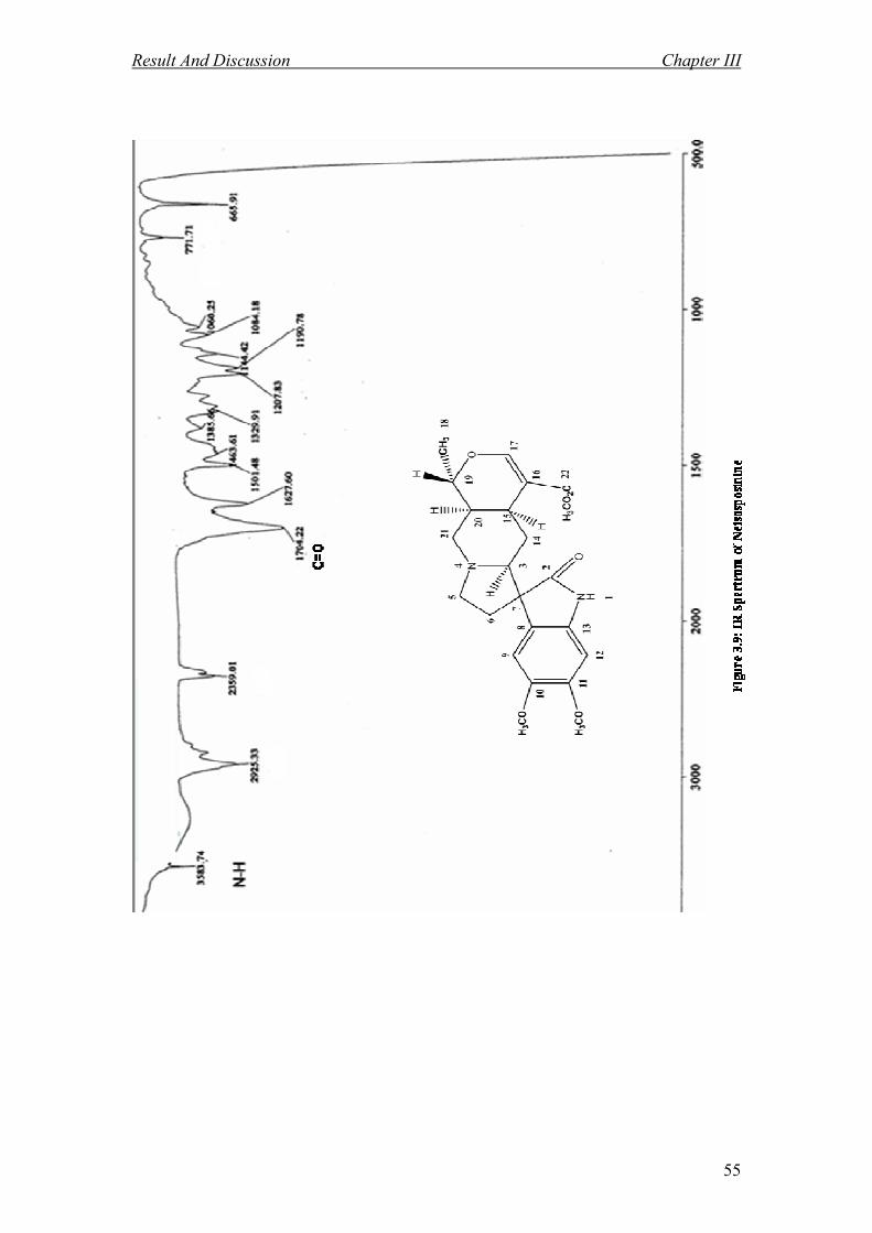
The UV spectrum revealed maxima at 215, 246 and 302 nm which were
characteristic for an oxindole system.49
The IR spectrum (Figure 3.9) displayed a band
at 3583 cm-1
presence of the NH/OH groups. In addition, a peak was observed at 1704
cm-1
(C=O) and 1190 cm-1
(C-O band). 48
The 1H NMR spectrum (Figure 3.7) showed three singlets at δ 7.40, 6.81 and 6.49
which were assigned to the isolated an olefinic proton H-17 and two aromatic protons

Result And Discussion Chapter III
50
H-9 and H-12 respectively. The spectrum also showed a broad singlet in down-field
region at δ 7.79 due to the presence of NH suggested the possibility of an oxindole
skeleton.47
This was further confirmed by the existence of a carbon signal at δ 182.2
(C=O).
In high field region, three singlet signals were appeared at δ 3.87, 3.85 and 3.60
attributed to three methoxyl groups attached to C-10, C-11 and C- 22 respectively.
The typical signal at δ 1.25 confirmed the existence of one methyl group C-18
attached to C-19. This signal appeared as a doublet with J value 5.96 Hz. The
hypothesis was also supported further by the 1H-1H COSY experiment that showed
the following fragments; C-3-C-15, C-5-C-6 and C-18-C-21 which were similar to the
previous alkaloid (Isoreserpiline 1).
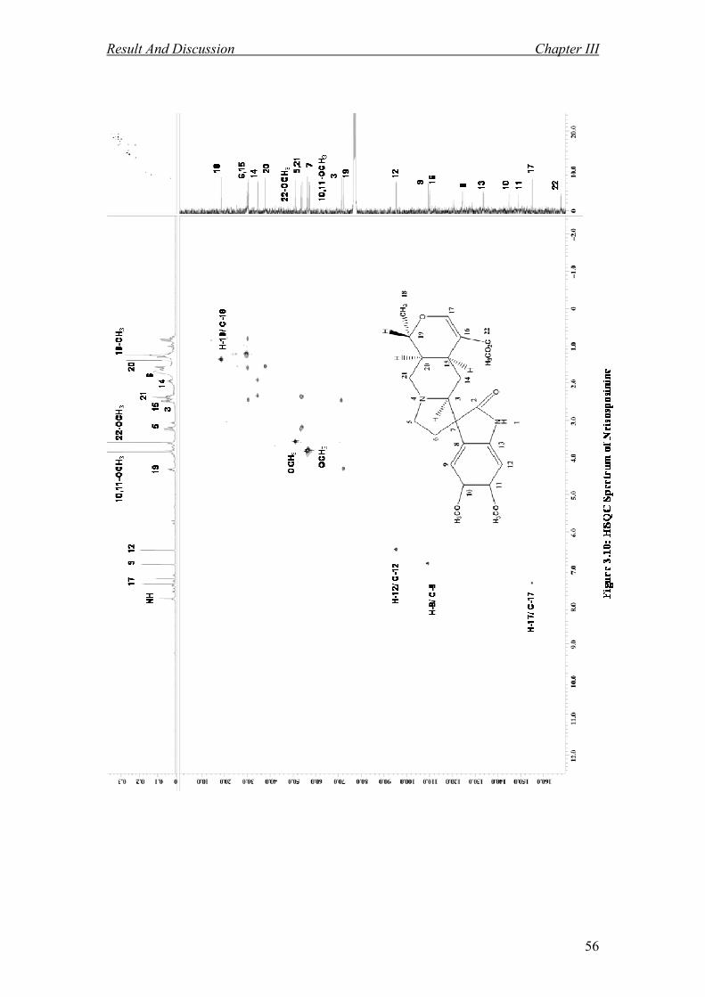
The 13
C NMR (Figure 3.8), DEPT and HMQC showed the presence of 23 carbon
atoms; 8 quaternary carbons; δ 181.2 (C-2), 168.0 (C-22), 146.2 (C-11), 144.9 (C-10),
133.8 (C-13), 124.2 (C-8), 109.9 (C-16) and 56.3 (C-7), 7 methines; δ 155.0 (C-17),
109.3 (C-9), 95.8 (C-12), 72.0 (C-19), 71.3 (C-3), 37.9 (C-20) and 30.4(C-15), and 4
methylene; δ 54.08 (C-21), 54.06 (C-5), 34.5 (C-14) and 30.1 (C-6), 1 methyl; δ 18.5
(C-18), and 3 methoxyls at δ 56. 7 (C10-OMe), 56.6 (C11-OMe) and 51.1 (C22-OMe)
respectively.
Detailed analysis of the spectral data and also data comparison with the literature
value (Table 3.3.1) confirmed that Alkaloid B 2 is assigned as neisosposinine which
was isolated previously from eisosperma oppositifolia. 48

Result And Discussion Chapter III
51
NH
N O
H3CO2CO
H3CO
H3CO
CH3
m/z 428
NH
N O
O
H3CO
H3CO
CH3
+
m/z 369
NH
N O
H3CO2CO
H3CO
H3CO
m/z 413
H
HH
+
NH
N O
H3CO2COH3CO
CH3
H
+
m/z 397
-CO2CH3
-OCH3
-CH3
.
.
.
Scheme 3.3: Possible Mass Fragmentation of Neisosposinine B 2

Result And Discussion Chapter III
52
Table 3.3.1: 1H NMR [ 400 MHz, 1H (J,Hz)] and
13C NMR [ 100 MHz, бC] of 2 in CDCl3
Position 1H (J,Hz)
13C
13C
48
2 181.2 181.3
3 2.48 (m ) 71.3 72.2
4
5 3.20 (t , 6.84 Hz) 54.06 53.67 3.09 (d , 11.88 Hz )
6 1.59 (m) 30.1 30.1
0.86 (q , 11.8 Hz )
7 56.3 57. 2
8 124.2 124.6
9 6.81 (s) 109.3 109. 5
10 144.9 144.2
11 146.2 145. 8
12 6.49 (s) 95.8 95.9
13 133.8 133.6
14 1.96 (m) 34.5 34.4
2.48 (m)
15 2.40 (m) 30.4 30.4
16 109.9 110.0
17 7.40 (s) 155.0 155.1
18 1.25 (d , 5.96 Hz) 18.5 18.4
19 4.32(m) 72.0 72.4
20 1.48 (m) 37.9 38.0
21 2.38 (m) 54.08 56.3
2.40 (m)
22 168.0 167.7
OMe 3.87 (s) 56.7 56.8
OMe 3.85 (s) 56.6 56.3
OMe 3.60 (s) 51.1 51.0
NH 7.79 (s)

Result And Discussion Chapter III
53

Result And Discussion Chapter III
54
Result And Discussion Chapter III
55
Result And Discussion Chapter III
56

Result And Discussion Chapter III
57

Result And Discussion Chapter III
58
Scheme 3.12: The HMBC Correlations of Neisosposinine 2
H 13
C

Result And Discussion Chapter III
59
3.3.2 Alkaloid C: Reserpinine. 3 50
3
Reserpinine 3 was isolated as a brownish amorphous solid. The mass spectrum
showed a [M+H]+ ion at m/z 383.2 corresponding to the molecular formula of
C22H26N2O4. The UV spectrum revealed maxima at 225 and 300 nm which were
attributed typical of an indole system.51
The IR spectrum (Figure 3.15) displayed a
stretching of NH/OH at 3402 cm-1
. In addition, a peak was observed at 1706 cm-1
which indicated the presence of the carbonyl group. 50
The 1H NMR spectrum (Figure 3.13) of alkaloid C 3 is reminiscent of alkaloid A 1
(isoreserpiline). The different between isoreserpiline 1 and resrpinine 3 is existing of
one methoxyl group which attached to the C-11 in the isoreserpiline. The spectrum
indicated the presence of three aromatic protons at δ 7.28; 6.76 and 6.71 attached to
C-9, C-12 and C-10 respectively. The deshielded signal of H-17 appeared at δ 7.53 for
confirming the corynanthean skeleton type of reserpinine 3. In the aliphatic region,
two singlets observed at δ 3.80 and 3.70 may be attributed to two methoxyl groups
attached to C-11 and C- 22 respectively. The signal for another four aliphatic protons

Result And Discussion Chapter III
60
δ 3.27, 2.8, 4.6 and 1.7 were assigned to H-3, H-15, H-19 and H-20 respectively.
Finally a doublet in the high field region at 1.4 (d, 6.8 Hz) may be attributed to the
methyl group attached to C-19.
The 13
C NMR (Figure 3.14) and DEPT showed the presence of 22 carbon atoms; 7
quaternary carbons; δ 168.2 (C-22), 156.0 (C-11), 136.8 (C-2), 133.4 (C-13), 121.7
(C-8), 109.6 (C-16) and 107.8 (C-7), 8 methines; δ 155.9 (C-17), 118.6 (C-9), 109.5
(C-10), 95.07 (C-12), 72.3 (C-19), 59.9 (C-3), 38.5 (C-20) and 31.4 (C-15), 4
methylens; δ 56.3 (C-21), 53.6 (C-5), 34.2 (C-14) and 21.8 (C-6), 1 methyl; δ 18.6 (C-
18), and 2 methoxyls at δ 55.8 (C11-OMe) and 51.2 (C22-OMe) respectively.
Detailed Analysis of the spectral data obtained and assessment with literature value
confirmed that alkaloid C 3 is reserpinine which was previously isolated from
Rauvolfia bahiensis.50
Complete assignments of compound C 3 were listed in table
3.3.2

Result And Discussion Chapter III
61
Table 3.3.2: 1H NMR [ 400 MHz, 1H (J,Hz)] and
13C NMR [ 100 MHz, бC] of 3 in
CDCl3
Position 1H (J,Hz)
13C
13C
49
2 136.8 133.14
3 3.27 (br d, 11.28 Hz ) 59.9 60.0
4
5 2.5 (m) 53.6 53.67 2.9 (m)
6 2.8 (m) 21.8 21.82
2.7 (m)
7 107.8 107.7
8 121.7 121.9
9 7.28 (d, 8.56 Hz ) 118.6 118.2
10 6.71 (dd, 2.4, 8.7 Hz) 109.5 108.7
11 156.0 155.8
12 6.76 (d, 2.2 Hz ) 95.07 95.0
13 133.4 130.13
14 2.5 (m) 34.2 34.2
1.6 (q , 9.86 Hz)
15 2.8 (m) 31.4 31.29
16 109.6 109.49
17 7.53 (s) 155.9 155.7
18 1.4 (d , 6.8 Hz) 18.6 18.49
19 4.6 (m) 72.3 72.47
20 1.7 (m) 38.5 38.41
21 3.2 (m) 56.3 56.25
2.7 (dd , 1.6, 12.2 Hz)
22 168.2 168.03
OMe 3.8 (s) 55.8 56.25
OMe 3.7 (s) 51.2 51.10
NH 7.8 (s)

Result And Discussion Chapter III
62

Result And Discussion Chapter III
63

Result And Discussion Chapter III
64

Result And Discussion Chapter III
65

Result And Discussion Chapter III
66

Result And Discussion Chapter III
67
Scheme 3.18: The HMBC Correlations of Reserpinine 3
H 13
C

Result And Discussion Chapter III
68
3.3.3 Alkaloid D: 72
72
Alkaloid D 72 was isolated as a brownish amorphous solid. The mass spectrum
revealed a pseudo-molecular ion peak at m/z 325.1 [M+H]+ corresponding to the
molecular formula of C21H28N2O. The UV spectrum revealed maximum at 250, 285
nm. The IR spectrum (Figure 3.21) showed a band of NH/OH at 3378 cm-1
.
The 1H NMR spectrum (Figure 3.19) indicated the presence of three aromatic protons
at δ 7.1, 6.8 and 6.7 attached to C-12, C-9 and C-11, respectively.
In up field region, one singlet peak was appeared at δ 3.8 attributed to one methoxyl
group.
There were a total of twelve aliphatic proton signals observed in the 1H NMR
spectrum. Fourteen methylene proton signals appeared between δ 1.2- 3.3 which may
be attributed to the protons attached to C-5, C-6, C-14, C-16, C-17, C-18 and C-22.
The signal for another four aliphatic protons δ 3.2, 1.5, 1.3 and 3.6 were assigned to
H-3, H-15, H-20 and H-21 respectively (Figure 3.19). Finally a doublet in the high
field region at δ 0.9 may be attributed to the methyl group attached to C-20.

Result And Discussion Chapter III
69
The 13
C NMR (Figure 3.20) and DEPT showed the presence of 21 carbon atoms; 5
quaternary carbons; δ 154.0 (C-10), 135.8 (C-2), 131.2 (C-13), 127.4 (C-8) and 107.9
(C-7), 7 methines; δ 111.5 (C-12), 111.05 (C-11), 100.4 (C-9), 60.03(C-3), 55.8 (C-
21), 41.7 (C-15) and 37.1 (C-20), 7 methylens; δ 60.5 (C-22), 53.2 (C-5), 35.5 (C-14),
35.3 (C-18), 30.0 (C-16), 23.5 (C-17) and 21.7 (C-6), 1 methyl; δ 11.1 (C-19) and 1
methoxyl; δ 55.9 ( C10-OMe ).
The assignments of the position of each carbon were established with DEPT, HMQC
and HMBC spectra. Complete assignments of compound D 72 were listed in table
3.3.3 that led to the proposed structure as depicted above (alkaloid D 72).
Experiments such as NOESY and HRMS need to be done to confirm the structure of
alkaloid D 72, however, these are not achievable at the moment due to lack of sample.

Result And Discussion Chapter III
70
Table 3.3.3: 1H NMR [ 400 MHz, 1H (J,Hz)] and
13C NMR [ 100 MHz, бC]
of 72 in CDCl3
Position 1H (J,Hz)
13C
2 135.8
3 3.2 (d, 11.0 Hz ) 60.03
4
5 2.9 (m) 53.2 2.4 (m)
6 2.8 (m) 21.7
2.3 (m)
7 107.9
8 127.4
9 6.8 (s) 100.4
10 154.0
11 6.7 (d, 6.4 Hz) 111.05
12 7.1 (d, 10.2 Hz) 111.5
13 131.2
14 1.2 (m) 35.5
1.4 (m)
15 1.5(m) 41.7
16 1.1 (m) 30.0
1.3 (m)
17 1.0 (m) 23.5
1.7(m)
18 1.9 (m) 35.3
2.1(m)
19 0.9 (d, 15.1) 11.1
20 1.3 (m) 37.1
21 3.6 (m) 55.8
22 1.9 (m) 60.5
3.5 (m)
OMe 3.8 (s) 55.9
NH 8.0 (s)

Result And Discussion Chapter III
71

Result And Discussion Chapter III
72

Result And Discussion Chapter III
73

Result And Discussion Chapter III
74

Result And Discussion Chapter III
75

Result And Discussion Chapter III
76
Scheme 3.24: The HMBC Correlations of Alkaloid D 72
H 13
C

Result And Discussion Chapter III
77
3.4 Ferulic Acid Esters from the Bark of Ochrosia oppositifolia
3.4.1 Ferulic Acid Ester E: 2-propenoic acid, 3-(4-hydroxy-3,5-
dimethoxyphenyl)-, methyl ester. 73
73
Ferulic acid ester E 73 was isolated as a whitish amorphous solid. The mass spectrum
revealed a pseudo-molecular ion peak at m/z 238.09 [M+H]+ corresponding to the
molecular formula of C12H14O5. The UV spectrum revealed maximum at 196, 273 and
328 nm. The IR spectrum (Figure 3.27) showed a band of OH at 3401 cm-1
. In
addition, a peak was observed at 1704 cm-1
which indicated the presence of the
conjugated carbonyl of an ester.
The 1H NMR spectrum (Figure 3.25) showed two overlapping aromatic protons at δ
6.77 attributed to H-6` and H-2` respectively. In the up field region, three singlets
appeared at δ 3.92 and 3.91 and 3.79 attributed to three methoxyls attached to C- 3`,
C- 5` and C- 1 respectively.

Result And Discussion Chapter III
78
The hypothesis was also supported further by the cross peaks of COSY experiment
that showed existence of two sets of doublet (J = 16 Hz) in the 1H NMR at δ 6.29 and
7.59 which corresponded to the resonances of H-2 and H-3.
The 13
C NMR (Figure 3.26) and DEPT showed the presence of 12 carbon atoms; four
quaternary δ 147.2 (C-3` and C-5`), 137.1 (C-4`), and 125.9 (C-1`); four methines δ
145.2 (C-3), 115.6 (C-2), and 105.09 (C-2` and C-6`); three methoxyl δ 56.4 (OMe 3`
and OMe 5`) and 51.7 (OMe 1). One peak appeared in δ 167.6 which is carbonyl of
(C-1). The cross peak between H-3 and C-1 as observed from the HMBC spectrum of
compound E 73 (Figure 3.29) indicated that C-1 is vicinal to the olefinic carbon, C-3.
The comprising of data; DEPT, HMQC and HMBC spectra, established the identify
of compound E 73.52
Complete assignments of compound E 73 were listed in table
3.4.1.

Result And Discussion Chapter III
79
Table 3.4.1: 1H NMR [400 MHz, 1H (J,Hz)] and
13C NMR [ 100 MHz, бC]
of 73 in CDCl3
Position 1H (J,Hz)
13C
1 167.6
2 6.29 (d, 16 Hz) 115.6
3 7.59 (d, 16 Hz) 145.2
1` 125.9
2` 6.77(s) 105.1
3` 147.2
4` 137.1
5` 147.2
6` 6.77 (s) 105.1
OMe 3` 3.92 (s) 56.4
OMe 5` 3.91 (s) 56.4
OMe 1 3.79 (s) 51.7
OH 5.80 (s)

Result And Discussion Chapter III
80

Result And Discussion Chapter III
81

Result And Discussion Chapter III
82

Result And Discussion Chapter III
83

Result And Discussion Chapter III
84

Result And Discussion Chapter III
85
Scheme 3.30: The HMBC Correlations of: 2-propenoic acid, 3-(4-hydroxy-3,5-
dimethoxyphenyl)-, methyl ester 73
H 13
C

Result And Discussion Chapter III
86
3.4.2 Ferulic Acid Ester F: 17-methoxy-carbonyl-14- heptadecaenyl- 4-hydroxy-
3-methoxy cinnamate. 74
1816
15
142-139`8`7`1`
2`
3`4`
5`
6` HC CH C O
O
(CH2)12 CH2
CH
CH CH2
O
OCH3
H3CO
HO
17
CH2
1
74
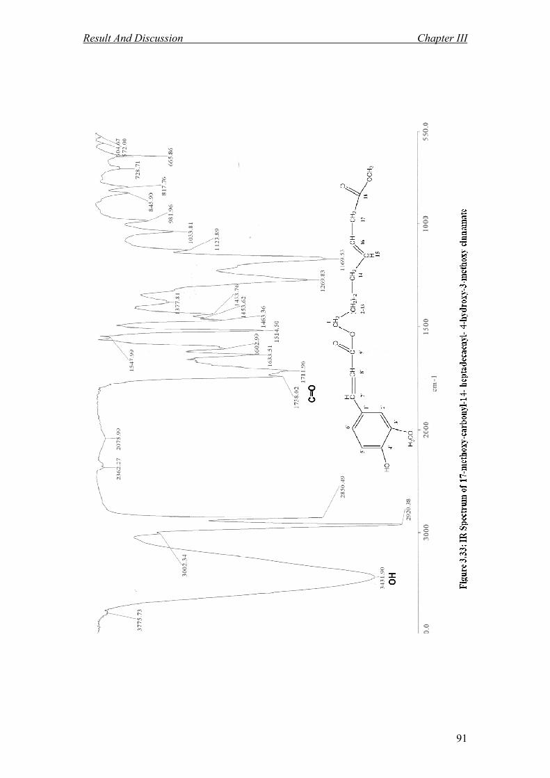
Ferulic acid ester F 74 was isolated as a whitish amorphous solid. The mass spectrum
revealed a pseudo-molecular ion peak at m/z 488.3 [M+H]+ corresponding to the
molecular formula of C29H44O6. The UV spectrum revealed maximum at 196, 273 and
328 nm. The IR spectrum (Figure 3.33) showed a band of OH at 3431 cm-1
. In
addition, two peaks were observed at 1711 and 1738 cm-1
which implied the presence
of two carbonyl group.
The 1H NMR spectrum (Figure 3.31) indicated the presence of three aromatic protons
at δ 7.10, 7.01 and 6.89 attached to C-6`, C-2` and C-5`, respectively.
In up field region, two singlets appeared at δ 3.91 and 3.64 attributed to two methoxyl
groups attached to C-3` and C-18 respectively.
The cross peak of COSY showed one coupling set (H-7`- H-8`). This was confirmed
by the existence of two sets of doublet (J = 16 Hz) in the 1H NMR at δ 7.50 and 6.27
which corresponded to the resonances of H-7` and H 8`, respectively.
Result And Discussion Chapter III
87
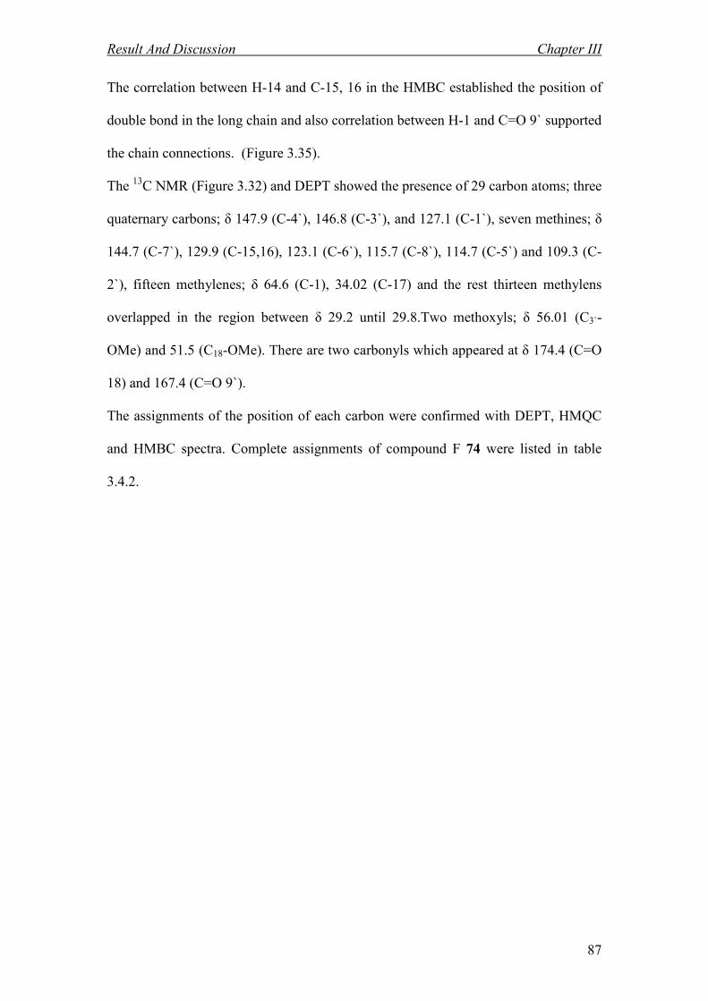
The correlation between H-14 and C-15, 16 in the HMBC established the position of
double bond in the long chain and also correlation between H-1 and C=O 9` supported
the chain connections. (Figure 3.35).
The 13
C NMR (Figure 3.32) and DEPT showed the presence of 29 carbon atoms; three
quaternary carbons; δ 147.9 (C-4`), 146.8 (C-3`), and 127.1 (C-1`), seven methines; δ
144.7 (C-7`), 129.9 (C-15,16), 123.1 (C-6`), 115.7 (C-8`), 114.7 (C-5`) and 109.3 (C-
2`), fifteen methylenes; δ 64.6 (C-1), 34.02 (C-17) and the rest thirteen methylens
overlapped in the region between δ 29.2 until 29.8.Two methoxyls; δ 56.01 (C3`-
OMe) and 51.5 (C18-OMe). There are two carbonyls which appeared at δ 174.4 (C=O
18) and 167.4 (C=O 9`).
The assignments of the position of each carbon were confirmed with DEPT, HMQC
and HMBC spectra. Complete assignments of compound F 74 were listed in table
3.4.2.

Result And Discussion Chapter III
88
Table 3.4.2: 1H NMR [400 MHz, 1H (J,Hz)] and
13C NMR [ 100 MHz, бC]
of 74 in CDCl3
Position 1H (J,Hz)
13C
1 4.1 (t, 6.8, Hz) 64.6
2-13 1.28-1.58 29.2-29.7
14 2.1 ( d, 5.4 Hz) 29.8
15 5.3 (m) 129.9
16 5.3( m) 129.9
17 2.3 (t, 7.7 Hz ) 34.02
18 174.4
1` 127.1
2` 7.01(s) 109.3
3` 146.8
4` 147.9
5` 6.90 (d, 8.2 Hz) 114.7
6` 7.10 (d, 8.2 Hz) 123.1
7` 7.50 (d, 16 Hz) 144.7
8` 6.27(d, 16 Hz) 115.7
9` 167.4
OMe 18 3.64 (s) 51.5
OMe 3` 3.91 (s) 56.01
OH 5.9 (s)

Result And Discussion Chapter III
89

Result And Discussion Chapter III
90
Result And Discussion Chapter III
91

Result And Discussion Chapter III
92

Result And Discussion Chapter III
93

Result And Discussion Chapter III
94
1816
15
142-139`8`7`1`
2`
3`4`
5`
6` HC CH C O
O
(CH2)12 CH2
CH
CH CH2
O
OCH3
H3CO
HO
17
CH2
1
Scheme 3.36: The HMBC Correlations of: 17-methoxy-carbonyl-
14- heptadecaenyl- 4-hydroxy-3-methoxy cinnamate 74
H 13
C

CHAPTER 4
ATIPLASMODIAL
ACTIVITY

Antiplasmodial Activity Chapter iv
95
ATIPLASMODIAL ACTIVITY
4.1 Introduction
In this studies the antiplasmodial activity of the crude extracts and pure
compounds of Ochrosia oppositifolia were investigated.
Among the natural products, indole alkaloids represent an interesting class of
compounds. Screening carried out to date has revealed several substances active in
vitro under the micro molar range and with a good selectivity index. Nevertheless,
in vivo activity has been confirmed only in a small number of cases, and there is a
need to undertake research focused on the mode of action of these compounds.
Antiplasmodial indole alkaloids can be separated into three main categories.
The first category contains the alkaloids with a molecular weight higher than 400
and an important steric crowding: (1) indole analogues of emetine
(usambarensine, ochrolifuanine, strychnopentamine); (2) and other bisindole
alkaloids such as voacamine and ergoline derivatives, matopensines and
isosungucines. Several of these alkaloids have been shown to be much more active
against chloroquine-resistant strains. This phenomenon deserves to be elucidated.
Considering the complexity of this group of compounds, very few attempts have
been made to modify them chemically, as has been the case (in cancer therapy) for
vinca alkaloids. This approach could nevertheless be very interesting.
The second category of antiplasmodial indole alkaloids contains unsaturated
monomeric heterocycle. These are chemically much more attainable. There are
two main models that have been developed: (1) derivatives of cryptolepine (the
Antiplasmodial Activity Chapter iv
96
most interesting compound being 2, 7- dibromocryptolepine); (2) derivatives of
tryptanthrin. Considering this last group, there is still a lot of investigation needed
concering their in vivo potentiality.
Finally, the last group of interesting indolic compounds includes monoindole
alkaloids able to reverse the resistance to chloroquine. Among these, the most
interesting compound seems to be malagashanine, found in strychnos myrtoides
from Madagascar. This compound is active in vivo, but further clinical assays will
be necessary to confirm the interest of this unconventional approach53
.
Malaysia is known with its green tropical vegetation and forest. Its diverse nature
and uses are claimed to possess medicinal value. The Malaysians also practice
traditional and herbal remedies as an alternative choice of treatment of malaria.
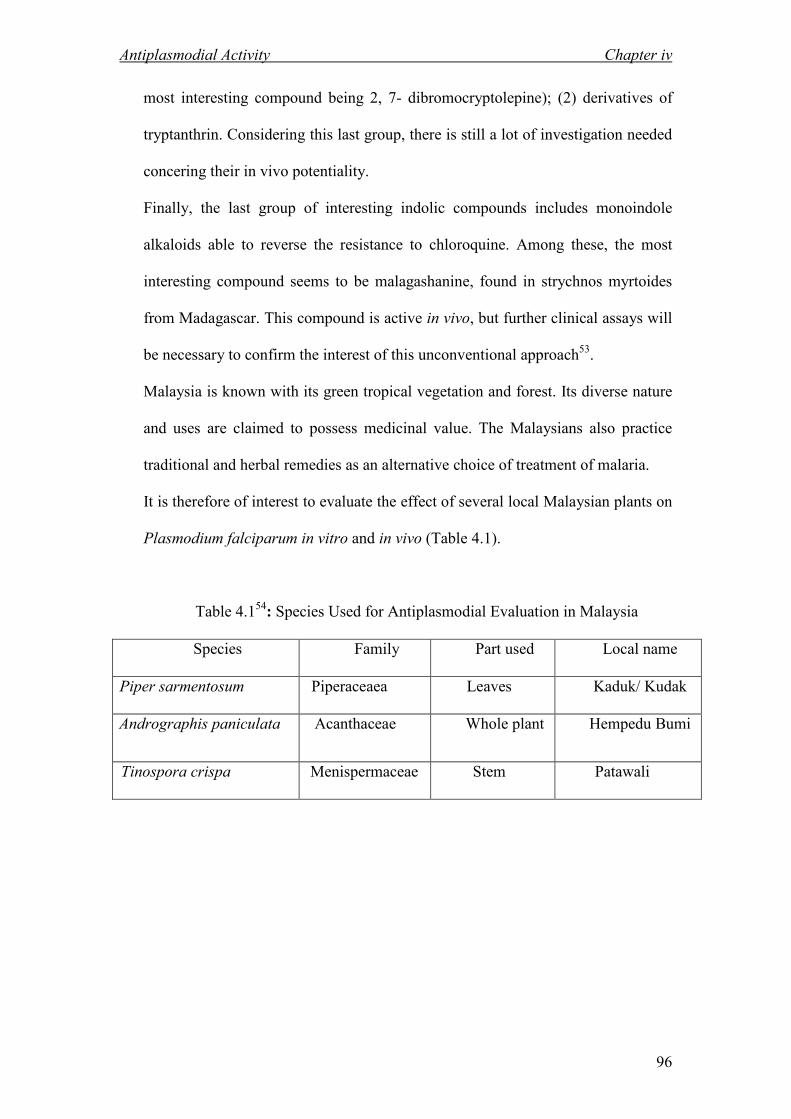
It is therefore of interest to evaluate the effect of several local Malaysian plants on
Plasmodium falciparum in vitro and in vivo (Table 4.1).
Table 4.154
: Species Used for Antiplasmodial Evaluation in Malaysia
Species Family Part used Local name
Piper sarmentosum Piperaceaea Leaves Kaduk/ Kudak
Andrographis paniculata Acanthaceae Whole plant Hempedu Bumi
Tinospora crispa Menispermaceae Stem Patawali

Antiplasmodial Activity Chapter iv
97
4.2 Result and Discussion
Seven samples were tested including three crude extracts and four isolated
compounds for in-vitro inhibitory activity against P. falciparum. Among the crude
extracts, hexane crude extract of bark showed the most potent inhibitory activity,
with the IC 50 value of 0.05051 µg/mL (Table 4.2), the other crude extracts and
compounds especially hexane crude of leaves showed very weak or no inhibitory
activity against P. falsiparum.
Four indole alkaloids showed inhibitory activity; inhibitory concentration ranged
from 0.0123 to 1.1251 µmol L-1
(Tables 4.3). The result for the antiplasmodial
activity of the pure alkaloids indicated the alkaloid D possess the most potent
activity with the IC 50 value of 0.0123 µmol L-1
.
Table 4.2: Inhibition Growth Percentage of Plasmodium falciparum and Probit
Analysis with SPSS 11.5 (crude extracts)
% Inhibition at Concentration (µg/mL) Crude extracts
100 10 1 0.1 0.01
IC50 (µg/mL)
DCM crude of
Leaves
100 68.24 39.54 30.12 16.20 1.61338
DCM crude of
Bark
100 71.26 54.45 39.07 34.61 0.46951
Hexane crude
of Bark
100 91.78 69.73 63.65 22.04 0.05051

Antiplasmodial Activity Chapter iv
98
Table 4.3: Inhibition Growth Percentage of Plasmodium falciparum and Probit
Analysis with SPSS 11.5 (alkaloids)
% Inhibition at Concentration (µg/mL) Alkaloids
10 1 0.1 0.01 0.001
IC50
(µmol L-1
)
Reserpinine 3 58.84 53.83 49.15 38.14 22.92 1.1251
Neisosposinine 2 55.13 54.43 51.55 41.88 31.37 0.7473
Isoreserpiline 1 76.31 64.4 40.59 33.33 29.25 0.2911
Alkaloid D 72 100 99.64 61.3 56.1 46.59 0.0123

CHAPTER 5
COCLUSIO

Conclusion Chapter v
99
COCLUSIO
The first part of the study was dedicated to the investigation of the alkaloid
components of Ochrosia oppositifolia (KL 5349) from Apocynaceae family,
collected from Pangkor Island (Perak). This plant produced monoterpene indole
alkaloids and have been the subject of several studies.7,8
Four indole alkaloids were
isolated from the bark and leaves of Ochrosia oppositifolia namely; isoreserpiline
1, neisosposinine 2, reserpinine 3 and alkaloid D 72 which showed high
antiplasmodial activity. In addition two ferulic acid esters; ferulic acid esters E 73
and F 74, were isolated from the methanol crude extract of the bark.
Isoreserpiline and reserpinine belong to the corynanthian type of alkaloid while
neisosposinine is an oxindole alkaloid.
The second part discussed the antiplasmodial activity of the crudes and isolated
compounds. In antiplasmodial assay, hexane crude of bark showed most potent
inhibitory activity, with IC50 0.05051 µg/mL against P.falsiparum while hexane
crude of leaves displayed very weak inhibitory activity against P.falsiparum.
Four pure indole alklaloids namely reserpinine 3, neisosposinine 2, isoreserpiline 1
and alkaloid D 72 showed inhibitory activity ranging from 0.0123 to 1.1251
µmol L-1
(Table 4.3).among the pure alkaloids alkaloid D showed the most potent
activity with an IC 50 value of 0.0123 µmol L-1
. Further studies of this plant can be
done such as mechanism studies which can serve to provide lead compounds for the
treatment of malaria.

CHAPTER 6
EXPERIMETAL

Experimental Chapter vi
100
EXPERIMETAL
6.1 General Methods
Spectra were recorded on the following instruments as follows:
Ultra Violet Spectra (UV)
Ultra Violet spectra were obtained in methanol on a Shimadzu UV-250 uv-visible
spectrophotometer and the wavelength which the spectrum was recorded is 190-
500nm.
Infrared Spectra (IR)
The infrared spectra were recorded on a Perkin Elmer FTIR (model 1600)
spectrophotometer. Solvent used for dilution the sample is CHCl3.
Optical Rotation (OR)
The optical rotation was obtained on Jasco DIP-1000 Digital polarimeter with
tungsten lamp at 25oC.
Mass Spectra (MS)
The mass spectra were measured on Waters Micromass ZQ. The Automass
Thermofinnigan was used for HR ESI+ and ESI
- analysis.

Experimental Chapter vi
101
MR Spectra
NMR spectra were recorded in deuterated chloroform (CDCl3) and deuterated
methanol (CD3OH) on a JEOL JNM-FX 400MHz, JEOL ECA 400MHz, Bruker
Top Spin 400 MHz and Varian 600MHz. Chemical shifts (δ) were expressed in ppm
and the coupling constants are given in Hz.
Column Chromatography (CC) and Thin Layer Chromatography (TLC)
Column chromatography were prepared by using Silica Gel 60F, 70-230 mesh
ASTM (Merck 7734); Silica Gel 60F, 230-400 mesh ASTM (Merck 9385); Silica
gel 40-63µm (Silicycle R12030B); Silica gel 60-200µm (Silicycle R10040B); Silica
Gel 60GF254, (Merck 1.07730.1000). Analytical Thin Layer Chromatography (TLC)
was performed on commercially Aluminium supported silica gel 60F254 TLC sheets
(Merck 1.05554.0001); Glass supported silica gel 60F254 TLC plates (Merck
1.05715.0001); Glass backed TLC Amino plates (Silicycle TLG-R52030B-203).
6.2 Reagents
Mayer’s Reagent (Potassium mercuric iodide) was used for screening the alkaloid
compounds. A positive result indicated when white precipitate was formed under
acidic condition. The Mayer’s Reagent was prepared as follows:
1.4 g mercuric iodide was dissolved in 60 ml distilled water were mixed with a
solution of 5.0 g of potassium iodide in 10 ml of distilled water.

Experimental Chapter vi
102
Dragendorff’s Reagent (Potassium bismuth iodide) was also used to identify the
presence of alkaloids spotting on TLC. A positive result is indicated by the
formation of orange spots on the developed TLC. The Dragendorff’s Reagent was
made as follows:
Solution A: Bismuth (III) nitrate (1.7 g) was dissolved in a mixture of 20 ml glacial
acetic acid and 80 ml of distilled water.
Solution B: Potassium iodide (16 g) was dissolved in 40 ml of distilled water.
Solution A and B were mixed together to give stock solution. Finally, the spray
reagent was prepared by diluting stock solution (40 ml) with 40 ml glacial acetic
acid in 120 ml distilled water.
6.3 Plant Material
The plant materials were collected from Pangkor Islands in 2007. The sample was
identified by L.E.Teo , University Malaya and deposited at the herbarium unit
(specimen no. KL 5349).
The location of the plant species was shown in Table 6.1.
Table 6.1: Plant Species and Location
Species Specimen umber Location
Ochrosia oppositifolia KL 5349 Pangkor Island, Perak
Experimental Chapter vi
103
6.4 Extraction
6.4.1 Extraction of Ochrosia oppositifolia (Bark)
The extraction of the bark (1kg) was carried out by extracted exhaustively with
hexane after wetting with 10% ammonia solution for 48 hours to removed non-polar
organic compound, waxes and fats. Then the extract was dried on the rotary
evaporator. The plant material was dried and again wetted with 10 % ammonia
solution and left for overnight. They were then re-extracted successively with
dichloromethane (CH2Cl2) and methanol (MeOH). After removal of the solvents,
the hexane crude extract (1.2 g), dichloromethane (15 g) and methanol (15 g) were
obtained. The extraction procedure was shown in Scheme 6.1.

Experimental Chapter vi
104
Scheme 6.1: Extraction of Ochrosia oppositifolia (Bark)
Wetted with10 % NH3 Extract with hexane
Hexane crude extracts (1.2 gr) Residue
Wetted with 10 % NH3
Extract with CH2Cl2
CH2Cl2 crude extracts (15 gr) Residue
Extract with methanol
MeOH crude extract (15 gr)
Ochrosia oppositifolia Bark (1 kg)

Experimental Chapter vi
105
6.4.2 Extraction of Ochrosia oppositifolia (leaves)
The extraction of the leaves (1.9 kg) were first wetted with 10 % ammonia solution
and left for 2 hours then defatted in hexane for 48 hours. Then the extract was dried
on the rotary evaporator. The plant material was dried and again wetted with 10 %
ammonia solution and left for 2 hours. They were then re-extracted successively
with dichloromethane (CH2Cl2). After removal of the solvents, the hexane crude
extract (1.2 g) were collected. Dichloromethane crude extract was dissolved in
CH2Cl2 and re-extracted with 5% hydrochloric acid (HCl) until a negative result was
formed with mayers reagent. The combined extracts were then basified with 25%
NH3 solution (pH 11) and re-extracted with CH2Cl2 until a negative mayers test was
obtained and later washed with distilled water and sodium chloride solution and
dried with sodium sulfate anhydrous. Finally, the extract was evaporated to dryness
to give an alkaloid crude extract (3 g). The extraction procedure was shown in
Scheme 6.2.

Experimental Chapter vi
106
Scheme 6.2: Extraction of Ochrosia oppositifolia (Leaves)
Ochrosia oppositifolia Leaves (1.9 kg)
Wetted with 10 % NH3
Extract with hexane
Hexane crude extracts (1.2 g) Residue
Wetted with 10 % NH3
Extract with CH2Cl2
CH2Cl2 crude extracts (30 g) Residue
Organic layer
Water layer
Add 10 % HCL (until
Meyer – ve partition)
Add NH3 until pH 11
Add CH2Cl2 (until Meyer –ve)
Partition
Water layer
Organic layer
alkalaoid crude
extract (3g)

Experimental Chapter vi
107
6.5 Isolation and Purification
The crude extract from the plant (bark and leaves) were subjected to column
chromatography over silica gel. The column was eluted with solvent mixtures of
increasing polarity (CH2Cl2, CH2Cl2 / MeOH and MeOH) and fractions having spots
with the same Rf value were grouped into a series of fractions (monitored by TLC).
Each series of fractions were then treated separately by extensive column
chromatography and preparative TLC to purify the alkaloids. The isolation and
purification procedure were summarized in the flow diagram shown in Scheme 6.3
and 6.4.

Experimental Chapter vi
108
Scheme 6.3: Isolation and Purification of Compounds from
Ochrosia oppositifolia (Bark)
Dissolved CH2Cl2 crude
extracts in hexane (8 g)
Dissolved MeOH crude
extract in MeOH (0.1 g)
Fraction 12-13
(2.5 g) HPLC
PTLC
1 (35 mg) 73 (23 mg) 74 (11 mg)
Ochrosia oppositifolia (bark 1 kg)

Experimental Chapter vi
109
Scheme 6.4: Isolation and Purification of Alkaloids from
Ochrosia oppositifolia (Leaves)
Alkaloid crude extracts (3 g)
Fraction 58
(0.4 gr)
PTLC
Fraction 36-37
(1 gr)
PTLC
Fraction 21-36
(0.8 gr)
PTLC
3 (23 mg) 2 (14 mg) 72 (4 mg)
Ochrosia oppositifolia (leaves 1.9 kg)

Experimental Chapter vi
110
6.6 Physical and Spectral Data of Isolated Alkaloids
Ochrosia oppositifolia Compounds:
Isoreserpiline 1 : pale brownish amorphous solid
: C23H28N2O5
UV λmax (MeOH), nm : 226,299
IR νmax (CHCl3), cm-1
: 3361, 2928, 1702, 665
Mass spectrum, m/z : 413 (100), 204, 435
1H NMR (CDCl3), ppm : See figure 3.1
13C NMR (CDCl3), ppm : See figure 3.2
eisosposinine 2 : pale brownish amorphous solid
: C23H28N2O6
UV λmax (MeOH), nm : 246,302
IR νmax (CHCl3), cm-1
: 3583, 2359, 1704, 665
Mass spectrum, m/z : 429 (100), 187
1H NMR (CDCl3), ppm : See figure 3.7
13C NMR (CDCl3), ppm : See figure 3.8

Experimental Chapter vi
111
Reserpinine 3 : pale brownish amorphous solid
: C22H26N2O4
UV λmax (MeOH), nm : 225,302
IR νmax (CHCl3), cm-1
: 3402, 2926, 1706, 665
Mass spectrum, m/z : 383 (100), 853
1H NMR (CDCl3), ppm : See figure 3.13
13C NMR (CDCl3), ppm : See figure 3.14
Alkaloid D 72 : pale brownish amorphous solid
: C21H28N2O
UV λmax (MeOH), nm : 250, 285
IR νmax (CHCl3), cm-1
: 3378, 1630, 735
Mass spectrum, m/z : 325 (100), 475, 701
1H NMR (CDCl3), ppm : See Figure 3.19
13C NMR (CDCl3), ppm : See Figure 3.20
Ferulic Ester E 73 : whitish amorphous solid
: C12H14O5
UV λmax (MeOH), nm : 196, 273,328
IR νmax (CHCl3), cm-1
: 3401, 1704
Mass spectrum, m/z : 238 (100), 412, 528
1H NMR (CDCl3), ppm : See Figure 3.25
13C NMR (CDCl3), ppm : See Figure 3.26

Experimental Chapter vi
112
Ferulic Ester F 74 : whitish amorphous solid
: C29H44O6
UV λmax (MeOH), nm : 196, 273,328
IR νmax (CHCl3), cm-1
: 3431, 1711
Mass spectrum, m/z : 488 (100), 462, 588
1H NMR (CDCl3), ppm : See Figure 3.31
13C NMR (CDCl3), ppm : See Figure 3.32

Experimental Chapter vi
113
6.7 Antiplasmodial Test Against Plasmodium Falciparum Strains
This protocol for assessing compound efficacy against Plasmodium Falciparum in
vitro uses as a marker for inhibition of parasite growth.55,56
Many alternative
protocols exist, including ones based on microscopic detection of Giemsa-stained,
assays based on production of parasite lactate dehydrogenase, and the use of flow
cytometry. 57
6.7.1 Preparation of the Antiplasmodial Test 58-66
6.7.1.1 Parasite Strain
Several well- characterized strains (refer Table 6.2) are available, either from
academic laboratories or from www.malaria.mr4.org (reagents available to
registered users). One recommendation would be to test activity against a drug-
sensitive line such a 3D7 (west Africa), D6 (sierra leone) or D10 ( Papua New
Guinea), as well as a drug-resistant line such as W2 or Dd2 (both from Indochina),
FCB (SE Asia), 7G8 (Brazil) or K1 Thailand).

Experimental Chapter vi
114
Table 6.2: Standard Plasmodium Falciparum Strains
Name Clone Origin Resistant to Multiplication
Dd2 yes (from WR82) Indochina CQ,QN,PYR,SDX 5-6
W2 yes (from Indochina-3) Indochina CQ,QN,PYR,SDX 5-6
HB3 yes Honduras PYR 4
3D7 yes (from NF54) Apparently - 4
West Africa
D6 yes (from Sierra Sierra Leone - 4
Leon-1)
D10 yes Papua New Guinea 4
CAMP no Malaysia PYR 4-5
FCB no Apparently
SE Asia CQ,QN,CYC 7-9
7G8 yes Brazil CQ,PYR,CYC 4-5
K1 no Thailand CQ,PYR 4-5
CQ, chloroquine; QN, quinine; PYR, pyrimethamine; SDX, sulfadoxine; CYC,
cycloguanil.
Multiplication rate refers to increase in total numbers of viable parasites per 48-hr
generation.
These rates and the drug phenotypes refer to data from the Fidock Laboratory (Albert
Einstein College of Medicine, NY) and may not be the same elsewhere.
6.7.1.2 Malaria Culture Media
RPMI 1640 medium containing L-glutamine (Catalog number 31800, Invitrogen),
50 mg/liter hypoxanthine, 25 mM Hepes, 10 µg/ ml gentamicine, 0.225%
NaHCO3 and either 10% human serum or 0.5% Albumax I or II (purified lipid-
rich bovin serum albumin, Invitrogen). Medium is typically adjusted to a pH of
7.3 to 7.4.
Experimental Chapter vi
115
6.7.1.3 Low Hypoxanthine Media
Same as above except that the hypoxanthine concentration is reduced to 2.5
mg/liter. Serum (as opposed to Albumax) is important for culturing fresh isolates,
and for maintaining properties of cytoadherence and gametocyte production (the
latter is required for transmission back to mosquitoes). Some strains also prefer to
propagate in serum. Batch -to-batch variation is nonetheless a problem, with
occasional batches not supporting robust parasite growth. Accordingly, many
laboratory lines have been adapted to propagate in the presence of Albumax,
which typically gives more consistent growth between batches (variation was a
problem in the past, but now appears to have been resolved). Albumax appears to
reduce both the rate at which erythrocytes deteriorate in vitro and pH drift when
cultures are exposed to ambient air, i.e. during tissue culture hood manipulations.
6.7.1.4 Preparation of Host Erythrocytes
Human erythrocytes for parasite culture are prepared by drawing blood into
heparin-treated tubes and washing several times in RPMI 1640 medium to
separate the erythrocytes from the plasma and buffy coat. Separation can be
achieved by centrifuging the blood at 500x g for 5 minutes in a swing-out rotor.
Leukocyte-free erythrocytes are typically stored at 50% hematocrit. i.e. 1.
Volume of malaria culture media for 1 volume of packed erythrocytes,
corresponding to approximately 5 x 10⁹
cells/ml.

Experimental Chapter vi
116
6.7.1.5 Parasite Culture Conditions
P. falciparum asexual blood stage parasites are propagated at 370 °C in malaria
culture media at 3-5% hematocrit in a reduced axygen environment (e.g. a custom
mixture of 5% CO2, 5% O2 and 90% N2). Lines can be conveniently cultured in 6-
24 well tissue culture plates in a modular chamber (billups- Rothenberg, Del Mar,
CA, www.brincubator.com), with plates containing sterile water on the bottom to
increase humidity and minimize desiccation. These chambers can be immersed
with the low O2 gas and mainained at 37 °C in an incubator designed to minimize
temperature fluctuations.
Parasites can also be cultured in flasks that are individually gassed, or
alternatively placed in flasks that permit gas exchange through the cap (in which
case the incubator needs to be continuously infused with a low O2 gas mixture).
Depending on the line, parasites typically propagate 3-8 fold every 48 hours, thus
care must be taken to avoid parasite cultures attaining too high a parasitemia, i.e.
percentage of erythrocytes that are parasitized for healthy growth. Most lines grow
optimally at 0.5-4% parasitemia. Parasites are most suitable for drug assays when
they are 2-5% parasitemia, and mostly ring stages with few or no gametocytes.

Experimental Chapter vi
117
6.7.1.6 Compounds
Compounds can be often be dissolved in 100% dimethyl sulfoxide (DMSO) and
stored at -20 °C. Particles size of insoluble compounds can be reduced by ball-
milling or sonication. For the drug assays, serial drug dilutions are made in low
hypoxanthine medium and added to 96-well culture plates at 100 ml per well.
Drugs are added to columns 2012 (test samples), with columns 1 and 2 reserved
for wells with low hypoxanthine medium without compound. All drugs are
typically tested in duplicate for each parasite line. Once completed, plates are
placed into their own modular chamber, gassed and placed at 37 °C. These plates
should be set up no more than a few hours to addition of the parasites.
6.7.1.7 Drug Assay Conditions
Parasites are diluted to a 2 times stock consisting of 0.6% to 0.9% parasitemia
(depending on the growth rate of the line) and 3.2% hematocrit in low
hypoxanthine medium, and 100 ml are added per well already containing 100 ml
of low hypoxanthine medium with or without compound (present at different
concentration). Plates are then incubated in a gassed modular chamber at 37 °C for
48 hours. After this time, 100 ml of culture supernatant from each well is removed
and replaced with 100 ml of low hypoxanthine medium containing a final
concentration of 7.5 µCi/ml of chloroquine. After a further 24 hours, the plates are
placed at -20 °C for at least 1 hour to freeze the cells.

Experimental Chapter vi
118
Plates are then thawed and the cells are harvested onto glass fiber filters (Wallac,
Turku, Finland). Filters are dried for 30 minutes at 80 °C, placed in sample bags
(Wallac), and immersed in scintillation fluid (Ecoscint A; National Diagnostic,
Atlanta, GA). Radioactive emissions are counted in a 1205 Betaplate reader
(Wallac). Mean counts per minute (cpm) are generally in the range 20,000-60,000,
with an acceptable minimum of 10,000. percentage reduction in chloroquine
uptake is equal to 100 x ((geometric mean cpm of no drug samples)- (mean cpm
of test samples)) / (geometric mean cpm of no drug samples). Percentage
reductions are used to plot percentage inhibition of growth as a function of drug
concentrations.
IC50 values are determined by linear regression analayses on the linear segments
of the curves ( IC90 values can also be determined by curve-fitting and can provide
an useful measure of variation between experiments). Assays are typically
repeated on two or three separate occasions. Within each experiment, standard
deviations are typically less than 10% of the mean. Different in parasite stages of
development can lead to up to two-fold shifts in the IC50 values between
experiments; however, these differences rarely affect the overall relationships
between the parasite lines in terms of their differences in drug response.


![CHAPTER9arberhhoti.weebly.com/uploads/1/9/4/8/19483867/ch9-full_text.pdf · CHAPTER9 [The Navigator] Scan Study Objectives Read Feature Story Read Preview Read text and answer Do](https://img.pdfslide.us/doc/110x75/5f1da8bbd516ea1dfa49497f/chapter9-the-navigator-scan-study-objectives-read-feature-story-read-preview-read.jpg)