Embed Size (px)
Citation preview

1
CHAPTER 1
INTRODUCTION
1.1 Tin (Group IV)
Tin (Sn) is the element with an atomic number of 50, a member of group IV A
of the Periodic Table. It has a atomic weight of 118.70 and with outer electron
configuration of ns2np
2 (Cotton et al., 1999). Along the group IV, it also have carbon
(C), silicon (Si), germanium (Ge) and lead (Pb) and these element are remarkable as
they are commonly used in the pure form.
Among the group IV elements, Tin has the unusual low melting point of
231.85°C and the unusual high boiling point of 2260, 2270 and 2687°C. This great
range makes it easy to form alloys without loss in vaporization. Tin exhibits the
oxidation states +2 and +4, like lead. In +2 state is called stannus and the +4 state is
called stannic (Cotton et al., 1999).
Tin primarily obtained from mineral cassiterite (SnO2) which is extracted by
roasting cassiterite in the furnace with carbon. Two allotropes of tin occur near room
temperature. The first form of tin is called gray tin which is stable at temparature
below 13.2°C (Cotton et al., 1999). Above temperature 13.2°C, grey tin will slowly
urns into tin’s second form, white tin which is normally in metal form.

2
1.2 Organotin Chemistry
Organotin Chemistry are those compounds that containing at least one bond
between tin and carbonIn compounds, Tin have four electrons in the outer electronic
shell in the common with other group IV elecments, C, Si, Ge, and Pb. Thus forms
compound in which tin has oxidation states of +II and +IV (Kenneth et al., 2002).
Tin can form various compounds, both inorganic and organic. Inorganic tin
compounds do not contain a tin-carbon bond, whereas organotin(IV) compounds
contain at least one tin-carbon bond with the formula:
RnSnX4-n
where, R is usually an aryl or alkyl group and
X is an inorganic species (halide, oxide or hydroxide)
n = 1 – 4
In general, tin(IV) compounds are either tetrahedral, trigonal bipyramidal, or
octahedral depending on the number and nature of substitutes. For smaller molecules
compounds of some combinations of the four substitutes give products that are
tetrahedral. When the number is five, the product are trigonal bipyramidal, and when

3
it is six, they are octahedral (Egon et al., 2001). The related basic organotin
compound structures were illustrated in Figures 1.1 -1.3.
Figure 1.1 Tetrahedral geometry of tin(IV) compound
Figure 1.2 Trigonal bipyramidal geometry of tin(IV) compound
Figure 1.3 Octahedral geometry of tin(IV) compound

4
1.3 Synthesis of Organotin (IV) Complexes
Tin played an important role in organometallic chemistry which began in 1949,
and this was stimulated by the discovery of a variety of applications (Rita et al., 2005).
The first organotin compound was prepared over 160 years ago by Sir Edward
Frankland in 1849 and described as a side note in a paper devoted largely to the
reaction that occurred when ethyl iodide and tin are heated together in a sealed tube
that produce the first main-group organometallic compounds, ethyl tin and diethyltin
iodide (Rita et al., 2005). Frankland observed that a reaction occurred also with a
number of other metals, including tin:
2EtI + Sn → Et2SnI2
And in the middle of 1903, Pope and Peachey described the preparation of a number
of simple and mixed tetraalkystannanes, and of tetraphenyltin, from Grignard reagents
and tin tetrachloride or alkyltin halides, and reactions of this type became the standard
route to alky- and aryl-tin compounds (Rita et al., 2005).
Organotin(IV) carboxylate complexes, RnSn(O2CR’)4-n, are most commonly
prepared by treating the corresponding oxide or hydroxide with the carboxylic acid
(Equations 1.1-1.3), or from the reaction of the corresponding chloride with a metal
carboxylate (Equations 1.4), or by cleaving a tin-carbon bond with a carboxylic acid
(Equations 1.5), or a mercury(I) or mercury(II), or lead(IV) carboxylate (Equations
1.6) (Yin et al., 2005).
5
R3SnOH or (R3Sn)2O + R’CO2H → R3SnO2CR’ + H2O (1.1)
R2SnO + 2R’CO2H → R2Sn(O2CR’)2 + H2O (1.2)
RSn(O)OH + 3R’CO2H → RSn(O2CR’)3 + H2O (1.3)
RnSnCl4-n + 4-nR’CO2M → RnSn(O2CR’) 4-n + 4-nMCl (1.4)
R4Sn + R’CO2H → R3SnO2CR’ + RH (1.5)
R4Sn + R’CO2M → R3SnO2CR’ + RM (1.6)
1.4 Application of Organotin Compounds
The remarkable variety in the physical, chemical and biological properties of
organotin compounds has lead to a great number of industrial applications of the
organotin(IV) carboxylate. Basically, There are three main areas in which organotin
compounds have product and process utility: (1) heat stabilizers; (2) catalytic agents;
(3) biocidal compounds (Ross, 1965). Organotin derivatives account for the fourth
largest production of organometallics amounting to about four million pounds per
year as compared with about 485 million pounds per year for organolead compounds.

6
1.4.1 Polyvinyl Chloride (PVC )Heat Stabilizers
Mono- and diorganotins are used extensively as heat stabilizers for processing
polyvinyl chloride (PVC). The main purpose of these tin stabilizers is to reduce the
polymer backbone degradation of the PVC. They do this by scavenging the HCl lost
during processing at high temperatures and stabilizing the unstable chloride sites in
the PVC molecule.
There are three major types of tin stabilizers. They are distinguished by their
respective alkyl groups: octyl, butyl, and methyl. Octyltin stabilizers have the lowest
tin content and are somewhat less efficient. However, they are approved for food
contact applications by most regulatory authorities worldwide. Butyltin stabilizers
have been the dominant types used until methyltins were introduced. Methyltin
stabilizers have a higher tin content and lower raw material cost compared to the other
two types. Some formulations (mercaptides) have also been approved for food contact
applicationsc (Evan et al., 1985).
The main applications for tin stabilizers are building products, such as pipe
and fittings, siding, profiles, packaging, and flexible PVC. It is estimated that between
12 to 13K tons of tin are used annually in tin stabilizers worldwide. This market is
expected to grow about 4% annually (European Council of Vinyl Manufacturers,
2000).

7
1.4.2 Catalysts
Catalysts are used to speed up chemical reactions, especially polymerization.
The most common applications for mono-and diorganotin catalysts are in chemical
synthesis and the curing of coatings. In chemical synthesis, the organotins are used for
the esterification and transesterification of mono- and polyesters. These products are
then used for plasticizers, synthetic lubricant manufacturing, and polyester polyol
production, as well as some coating applications.
As curing catalysts, one of the largest uses of organotins is in electrocoat (E-
coat) coatings. These electrocoating products are sold into a wide range of
applications, with the largest being automotive, where they provide excellent rust
resistance. The catalysts are also used in urethane coatings as well as polyurethane
foam production (Evan et al., 1985).
1.4.3 Biocidal Properties
The biocidal properties of organotins in general were discovered by Van der
Kerk at the TNO Institute, Utrecht in the late 1950s. Tributyltin (TBT) is unique
among the organotin(IV) in which is used as biocide. The monobutyltins do not
exhibit these properties. Tributyltin is normally used as biocide in paint formulations.
These paints are applied to protect the underwater surface area of ship’s hull against

8
barnacles, algae, etc. in order to avoid increased fuel consumption and premature dry-
docking. Tributyltin(IV) oxide (TBTO) and tributyltin(IV) naphthenate (TBTN) are
commercially used in weed industrial for treatment and as preservatives (Kaars et al.,
1962).
Since then bactericidal and fungicidal properties in agriculture have been
developed. Both triphenyltin(IV) hydroxide and triphenyltin(IV) acetate are used to
control fungi that cause potato blight on sugar beets, celery, carrots, onions and rice
including tropical plant diseases in peanuts, pecans, coffee and cocoa. As insecticides,
triorganotin(IV) compounds have been used against houseflies, cockroaches,
mosquito larvae and tobacco budworms (Fargasova, 1955)

9
1.5 Objectives
In this study, the main objective is to to synthesis new organotin(IV)
complexes by reacting the 4-(diethylamino)benzoic acid and with organotin (IV)
compounds, which are di-n-butyltin(IV) oxide and triphenyltin(IV) hydroxide.
The coordination number of the tin(IV) atom as well as the coordination mode
of the respective ligands to the tin(IV) atom moiety will be study. The complexes
obtained will be characterized quantitatively by elemental analysis and melting point
determination as well as qualitatively by infrafed spectroscopy (FTIR) and nuclear
magnetic resonance spectroscopy (NMR) to compare the structure with theoretical
study.
10
CHAPTER 2
LITERATURE REVIEW
2.1 The First 1D Tetranuclear Organotin (IV) Complex with N’-
acylsalicylhydrazide
The first diorganotin(IV) complex with N’-acylsalicylhydrazide has been
successfully synthesized and structurally characterized (Yin et al., 2008). The
structure showed that the complex reveals an infinite 1D polymer linked by the Sn–O
bonds, and the monomer contains four-tin nuclears with five-coordinated and seven-
coordinated tin and an 14-member ring made up of two ligands and two dibutyltin(IV).
In the infrared spectra of the complex, the stretching vibration bands of COO–
H and the ArO–H disappear compared with the original ligand suggesting the
coordination of carboxyl and phenolate-O oxygen atoms to the organotin(IV) moiety.
The absence of the N–H and C=O stretching vibration bands is consistent with the
deprotonation of the CONH groups and the coordination to the organotin(IV) moiety
in enol form. The characteristic absorptions at 1608 cm-1
indicate the presence of
C=N–N=C group (Yin et al., 2008).
The 1H NMR spectrum show that those signal of –COOH, -OH and –CONH
protons in the spectrum of the ligand are absent, thus indicate removal of those
protons and formation of Sn-O bonds. The 119
Sn NMR spectrum spectrum of complex

11
show distint resonance at -187.4 and -296.7 ppm, respectively. The Value of δ 119
Sn
in the ranges -210 to -400, -90 to -190 and 200 to -60 ppm has been assciated with
six- , five- and four-coordinate tin centers, respectively (Holeěek, 1986).
As a conclusion, the tin atoms of the complex in solution are five- and six-
coordinated which the sturture of the complex tetranuclesr organotin(IV) complex is
shown in figure 2.1.
Figure 2.1 The molecular structure of complex tetranuclear organotin (IV)
complex with N’-acylsalicylhydrazide
12
2.2 Synthesis, Characterization and Crystal Structure of A Novel 3D
Network Tiorganotin(IV) Polymer Containing Two types of Macrocycles
A novel triorganotin(IV) complex has been synthesized by the reaction of
sodium ethoxide with tri-n-butytin(IV) chloride in dry ethanol (Hangdong et al.,
2008). It is noteworthy that complex contains two types of macrocycles: one is six-tin
24-membered macrocycles, the other is six-tin 36-membered macrocycles. Such large
macrocyclic structures appearing in a multidimensional organotin(IV) polymeric
system based on covalent interactions are not common and the organotin macrocycles
have been extensively studied for their potential industrial application and biological
activities (Ma et al., 2005).
The infrared spectra of the complex showed that the absence of bands in the
region 3120-2980 cm-1
, which appear in the free ligand as COOH stretching
vibrations, thus indicating metal-ligand bonding through COO -. The typical
absorptions for Sn-C, Sn-O, vibrations in complex are located in the normal range
550-586 cm-1
and 440-486 cm-1
respectively.
The resonances of 1H NMR observed at δ 10.50 ppm, which are absent in the
spectra of the complex, indicate removal of the COOH proton and formation of Sn-O
bond. The coordination geometry about the tin(IV) atom moiety is distorted trigonal
bipyramidal, with the equatorial plane occupied by three n-butyl groups and the axial

13
positions shared by two coordinated oxygen atoms from the ligand (Hangdong et al.,
2008).
The β, γ and δ carbon atoms of the butyl groups and the hydrogen atoms have
been omitted. All the tin atoms in complex possess the same coordination
environment. The coordination about the tin atom is only slightly distorted from a
regular trigonal bipyramidal geometry, with the equatorial plane occupied by three n-
butyl group and the axial postions shared by two coordinatied oxygen atoms from the
ligand.
In short, using a flexible ligand, meso-2,3-di-bromosuccinic acid, a new
triorganotin(IV) coordination polymer has been synthesized. The structure of the
coordination polymer reveal that the ligand acts as linker to connect four metal
centers to give rise to a 3D network structure. The ligand can bond more than one
organotin moiety, which will increase the changes for the higher structural
dimensionalities assembly, so the selection of organic ligands with appropriate
coordination sites is the key to forming metal-organic coordination polymers with
fascinating structures(Hangdong et al., 2008). 3D network structure polymer, as
indicated in figure 2.2.
.

14
Figure 2.2 The 3D network structure (viewed along to the α axis)

15
2.3 Di- And Tri-organotin(IV) Complexes of the New sodium bis(1-methyl-
1H-imidazol-2-ylthio)acetate Ligand
The new sodium bis(1-methyl-1H-imidazol-2-ylthio)acetate, Na[(S-
tim)2CHCO2], has been prepared in ethanol solution using 2-mercapto-1-
methylimidazole, dibromoacetic acid and NaOH. Maura et al,. (2008) producing a
new class of monoanionic with polyfunctional N,O,S-ligands lead to the high possible
considerable coordinative flexibility.
Infrared spectroscopy showed all the expected bands for the ligand and the tin
moieties: weak absorptions in the range 3043–3136 cm-1 are due to the azolyl ring C–
H stretchings and medium to strong absorptions near 1510 cm-1 are related to ring
‘‘breathing” vibrations. The presence of the COO moiety in is detected by an intense
broad absorption in the range 1639–1656 cm-1 and 1308–1335 cm-1, due to the
asymmetric and symmetric stretching modes. In the far-IR region medium to strong
absorptions appear upon coordination, due to stretching modes of Sn–O, Sn–Cl, Sn–C
(Nakatomo, 1997) The absence of Sn–Cl stretching vibrations in the spectra of the
triorganotin(IV) derivatives confirms the substitution of the chloride in the complexes
formation. The Sn–Cl stretching vibrations fall as broad absorptions near 229–235
cm-1 in the diorganotin(IV) derivatives. The Sn–C stretching frequencies fall as
medium or strong absorptions in the range 245–275 cm-1 for the aryl derivatives;
similar stretching vibrations are detected in the range 505–590 cm-1 for the alkyl
derivatives. In the far-IR spectra absorptions tentatively assigned to Sn–O have been
16
detected in the range 420–455 cm-1 in the triorganotin(IV) derivatives.
The resonance for CHCOO group hydrogens occurs at 5.05 and 4.73 ppm are
important, in CDCl3 and D2O solution, respectively, downfield with respect to the
decarboxylate analogues [(S-tim)2CH2]. In the 1
H NMR spectra of complexes, the
signals due to the 2-mercapto-1-methylimidazolyl rings are always deshielded with
respect to those in the spectra of the free donor, confirming the existence of the
complexes in solution; the signals due to the CHCOO group exhibit significant
downfield shift (from 5.05 ppm in the free ligand to 5.32–5.42 ppm in the complexes):
this is suggestive of a strong bonding of the tin atom to the carboxylate group of the
complexes. In the 1
H NMR spectra at room temperature of the decarboxylated
derivatives, the resonances due to the bridging methylene protons of the [(S-tim)2CH2]
ligand appear as singlets at 4.44 and 4.65 ppm, respectively, probably as a result of
averaging arising from rapid ring inversion of the puckered eight- membered ring
containing the central Sn atom (Maura et al., 2008)
17
CHAPTER 3
METHODOLOGY
3.1 Reagent and Instrumental
Table 3.1 : List of chemicals for the synthesis of organotin( IV) carboxylate
Chemicals Supplier
Di-n-butyltin(IV) oxide, (C4H9)2SnO, (98.0%) Fluka Chemie AG
triphenyltin(IV) hydroxide, (C6H5)3SnOH,(96.0%) Aldrich Chemical
4-(diethylamino)benzoic acid, (CH3)2NC6H4COOH,
(99.0%)
Aldrich Chemical
Ethanol, C2H5OH, (99.8%) Systerm
Acetonitrile, CH3CN, (99.5%) Systerm
Sodium Hydroxide, NaOH, (99.0%) Systerm
Table 3.2 : List of Instrusments for the analysis of organotin( IV) carboxylate
Instrument Model
FTIR Spectrophotometer Perkin Elmer FT-IR System 2000
1H and
119Sn NMR Spctrometer Bruker AC-P 400MHz
13C NMR SPectrometer Bruker AC-300MHz
Melting point apparatus Stuart
Elemental analyzer Perkin Elmer Series II 2400

18
3.2 Synthesis Method
3.2.1 Preparation of sodium salt
The sodium salt was prepared by under reflux in 1:1 molar ratio sodium
hydroxide, NaOH (4 mmol) with 4-(diethylamino)benzoic acid, (CH3)2NC6H4COOH
(4 mmol) in ethanol (50 mL) for about two hours. After few days, yellow precipitate
was obtained.
3.2.2 Synthesis of Bis[4-(diethylamino)benzoato]tetrabutyldistannoxane(IV)
Dimer Complex (1)
The reaction of di-n-butyltin(IV) oxide, (C4H9)2SnO (2 mmol) and 4-
(diethylamino)benzoic acid, (CH3)2NC6H4COOH acid (2 mmol) and in ethanol (50
mL) was obtained by heating under the refluxed in a 1:1 molar mixture for about two
hours. The solution was isolated by filtration. A clear colourless solution was
obtained by filtrtion and kept in a bottle. After few days, colourless crystal was
collected.

19
3.2.3 Synthesis of Preparation of 4-(diethylamino)benzoatotriphenyltin(IV)
Complex (2)
The reaction triphenyltin(IV) hydroxide, (C6H5)3SnOH (3 mmol) and 4-
(diethylamino)benzoic acid, (CH3)2NC6H4COOH (3 mmol) in acetonitrile (50 mL)
was heating under refluxed in a 1:1 molar mixture for two hours. The solution was
isolated by filtration. A clear solution was obtained by filtration and a coloured crystal
was obtained.
3.3 Charaterization
Complexes was characterized by using four methods:
Elemental analysis
Melting point determination
Fourier Transoform Infrared Spectroscopy
Nuclear Magnetic Resonance Spectroscopy
All measurements were performed at room temperature.

20
3.3.1 Elemetal analyzer
Elemental analysis on carbon, hydrogen and nitrogen is the most important
investigation performed to charateristize and prove the elemental compostion of the
complexes. The compostions of the complexes formed are determined by using
Elemental analyzer, Perkin Elmer Series II 2400. The amount of complexes in this
analysis is approximate 1.0 to 2.0 mg.
3.3.2 Melting Point Determination
A pure compound will have deifinte melting point, owing to this, the identiy
and the purity of complex can be determined by using melting point apparatus, Stuart.
The melting point apparatus has a temperature range of 0 to 360 °C. Small amount
of sample complex was filled into capillary tub, until about 3 to 4 mm long. The filled
capilary tube was inserted into observing chamber and proceed with the heating
process. The temperature range at which the complex changesstate from solid to
liquid was taken. A pure complex will give a sharp melting point which the
temperature fall in between the range 2 to 3°C.

21
3.3.3 Fourier Transform Infrared Spectroscopy (FTIR)
The ligand coordination to the tin centre able to clarify by the infrared spectra
in the 4000-400 cm-1
range. The apparatus was cleaned with absoulute ethanol before
preparation of the sample. 1.0 mg sample complex was grinded with potassium
bromide (KBr) salt in ratio 1:10 into fine powder using agate mortar and pestle. The
sample is compressed in a manual Hydraulic Press by using pressure in the range of
6000 to 8000 psi into a translucent pellet. The sample in a pellet form was placed into
the sample holder and ready to be run using FT-IR. The complexes are analyzed with
FTIR Perkin Elmer FT-IR System 2000.
3.3.4 Nuclear Magnetic Resonancr Spectroscopy (1H NMR,
13C NMR,
119Sn
NMR)
Nuclear Magnetic resonance spectroscopies are the preminent technique for
determinning the structure of complexes. 1H and
119Sn NMR Spectrometer were
recorded on Bruker AC-P 400 MHz. The 13
C NMR Spectrometer data were obtained
on Bruker AC-300 MHz. Deuterated dimethly sulfoxode, DMSO-d6 was used in the
sample preparation by dissolve 0.01 g to 0.03 g of complex. Tetramethylsilane, TMS
was used as internal standard and the chemical shift of 13C NMR spectra is in the
range of 0 to 200 ppm, whereas 1H NMR spcetra in the range of 0 to 12 ppm.

22
CHAPTER 4
RESULTS AND DISCUSSION
4.1 Synthesis of Complexes
4.1.1 Preparation of Sodium 4-(diethylamino)benzoate
The sodium salt, sodium 4-(diethylamino)benzoate was obtained by heating
under reflux a 1:1 molar mixture of sodium hydroxide, NaOH (4 mmol) with 4-
(diethylamino)benzoic acid, (CH3)2NC6H4COOH (4 mmol) in ethanol (50 mL) for
about two hours. The water liberated in the reaction was removed by azeotropic
dehydration using the Dean-Stark apparatus (Yip et al., 2008). Molecular sieves are
used as an adsorbent for water molecules to reducing contaminants. At the end of the
reflux, a clear transparent solution was obtained and isolated by filtration. The
solution kept in a bottle. After few days some yellowish crystal obtained.
Figure 4.1 Sodium salt formation of 4-diethylamino benzoic acid

23
4.1.2 Preparation of Bis[4-(diethylamino)benzoato]tetrabutyl distannoxane(IV)
Dimer Complex (1)
The title dimer, bis[4-(diethylamino)benzoato]tetrabutyl distannoxane(IV) was
obtained by heating under reflux a 1:1 molar mixture of di-n-butyltin(IV) oxide,
(C4H9)2SnO and 4-(diethylamino)benzoic acid, (CH3)2NC6H4COOH acid in ethanol
(50 mL) for about two hours. The water liberated in the reaction was removed by
azeotropic dehydration using the Dean-Stark apparatus (Yip et al., 2008). Molecular
sieves are used as an adsorbent for water molecules to reducing contaminants. At the
end of the reflux, a clear transparent solution was obtained and isolated by filtration.
The solution kept in a bottle. After few days some transparent crystal obtained.
Figure 4.2 Reaction scheme of bis[4-(diethylamino)benzoato]
tetrabutyldistannoxane(IV) Dimer Complex

24
4.1.3 Preparation of 4-(diethylamino)benzoatotriphenyltin(IV) Complex (2)
The title complex, 4-(diethylamino)benzoatotriphenyltin(IV) was obtained by
heating under reflux a 1:1 molar mixture of triphenyltin(IV) hydroxide, (C6H5)3SnOH
(3 mmol) and 4-(diethylamino)benzoic acid, (CH3)2NC6H4COOH (3 mmol) in
acetonitrile (50 mL) for about two hours. The water liberated in the reaction was
removed by azeotropic dehydration using the Dean-Stark apparatus (Yip et al., 2008).
Molecular sieves are used as an adsorbent for water molecules to reducing
contaminants. At the end of the reflux, a clear transparent solution was obtained and
isolated by filtration. The solution kept in a bottle. After few days, some transparent
crystal obtained.
Figure 4.3 Reaction scheme of 4-(diethylamino)benzoate triphenyltin(IV)
Complex
25
4.2 Physical Data and Melting Point
4.2.1 Bis[4-(diethylamino)benzoato]tetrabutyldistannoxane(IV)Dimer Complex
The bis[4-(diethylamino)benzoato]tetrabutyldistannoxane(IV) dimer complex
1 was obtained as single fine, shiny and transparent crystals from 1:1 (dibutyltin:acid)
reaction. The yield of the complex 1 is 1.13 g (85.20 %). The complex showed a
sharp melting point of 219-220 ˚C, which the temperature fall in between the range 1
˚C indicated the purity of the complex form.
4.2.2 4-(diethylamino)benzoatotriphenyltin(IV) Complex
The 4-(diethylamino)benzoatotriphenyltin(IV) complex 2 was obtained as
single fine, shiny and transparent crystals from 1:1 (triphenyl:acid) reaction. The yield
of the complex 2 is 0.62g (80.10 %). The complex showed a sharp melting point of
194-195 ˚C, which the temperature fall in between the range 1 ˚C indicated the purity
of the complex form.
The data shown that complex 1 have higher melting point than complex 2.
This proved that complex 1 are more stable and have stronger bonding compare to
complex 2. The physical data of these complexes are presented in Table 4.1

26
Table 4.1 Physical data and melting point.
Complex Melting Point (oC) Yield (%)
Molecular
weight
Bis[4-(diethylamino)benzoato]tetrabutyl
distannoxane(IV) dimer, 1 219-220 85.20 1572.70
4-(diethylamino)benzoatotriphenyltin(IV), 2 194-196 80.10 510.26

27
4.3 Elemental Analysis and gravimetrik (Sn)
CHN analysis for each complex was obtained from Perkin Elmer Series II
2400 series CHNS-O analyzer. The formula molecular for the complexes was
postulated by calculating the percentage weight of compound of carbon, hydrogen and
nitrogen present in the complex.
From the CHN analysis, calculated percentage of carbon present in the
complex 1 is 52.78 %, the percentage of nitrogen present is 3.19 % and the percentage
of the hydrogen is 7.07 %. Thus, molecular formula for the bis[4-
(diethylamino)benzoato]tetrabutyldistannoxane(IV) dimer complex is [{4-
[N(C2H5)2]C6H4COO}2(C4H9)2Sn}2O]2 and the molecular weight of this complex is
1572.70 g/mol.
For the complex 2, calculated percentage of carbon, nitrogen and hydrogen
compound present in the complex is 64.28 %, 2.68 % and 7.07 % respectively. The
molecular formula for the 4-(diethylamino)benzoatetriphenyltin(IV) complex is 4-
[N(C2H5)2]C6H4COO(C6H5)3Sn and the molecular weight of this complex is 510.25
g/mol.
The differences between the calculated percentage and the theoretical
percentage obtained from CHN analysis should not more than 3.0 %. For the complex
1 and complex 2, the calculated percentage of carbon, nitrogen and hydrogen

28
compared to the data obtained from CHN analysis are below 3.0 %. Table 4.2 and
Table 4.3 are elemental analysis results of the complexes.

29
Table 4.2 The elemental analytical data (%) of bis[4-(diethylamino)benzoato]tetrabutyldistannoxane(IV) dimer
Element
Number
atom
Molecular
Weight
Total Molecular
Weight
Analysis
Percent (%)
Calculated
Percent (%)
Delta (%)
C 76 12.0107 912.8132 52.78 52.68 0.10
H 128 1.00794 129.01632 7.07 7.45 0.38
N 4 14.0067 56.0268 3.19 3.23 0.04
Sn 4 118.71 474.84 27.37 27.41 0.04

30
Table 4.3 The elemental analytical data (%) of 4-(diethylamino)benzoatotriphenyltin(IV) complex
Element
Number
atom
Molecular
Weight
Total Molecular
Weight
Analysis
Percent (%)
Calculated
Percent (%)
Delta (%)
C 29 12.0107 348.3103 64.28 64.24 0.04
H 29 1.00794 29.23026 5.00 5.39 0.39
N 1 14.0067 14.0067 2.63 2.58 0.05
Sn 1 118.71 118.71 21.31 21.89 0.58
31
4.4 Infrared spectroscopy
The IR spectra of the complexes are recorded in the range of 400-4000
cm-1
. The spectrums obtained are presented in Figures 4.4, 4.5, 4.6 and 4.7.
Infrared spectroscopy is used to investigate the nature of –COO group
coordination mode on the tin atom as well as the characteristic features or
functional groups present in each complex. The absorptions of interest in the
spectra of the complexes are υ(COO), υ(Sn-C) and υ(Sn-O).
The infrared spectrum of the complex 1 and complex 2 shows distinct
differences from the acid. The υ(OH) band which appeared in the 2800-3000
cm-1
region for the acid, was absent in the infrared spectra of the complexes
showing the occurrence of deprotonation prior to coordination of the resulting
carboxylate anion in the complex formation (Yip et al., 2008).
The C-H stretching region (3340-2700 cm-1
) is usually the first region
to be diagnosed in the infrared spectral characterization since its reveals the
essential structural features present in complexes. Strong absorption band that
occurred at the wavenumber in between 3000-2700 cm-1
from the IR spectra
of complex 1 and complex 2 are the characteristic methyl group, -CH3. The
methyl group has asymmetric and symmetric C-H stretching frequencies near
2690 cm-1
and 2870 cm-1
respectively. The asymmetric and symmetric C-H
stretching bands of the methylene group, -CH2-, are centered at 2930 cm-1
and

32
2850 cm-1
respectively. This result can be confirm that the existence of the
long chain butyl group from dibutyltin(IV) in complex 1 and aromatic ring in
4-(diethylamino)benzoate ligand.
In the IR spectra of the complex 1, the carboxylate bands are observed
in the characteristic regions for υasym (COO) between 1522 and 1603 cm-1
and
for υsym (COO) between 1353 and 1393 cm-1
. Meanwhile, the complex 2, the
carboxylate bands are observed in the characteristic regions for υasym (COO) at
1603 cm-1
and for υsym (COO) at 1347 cm-1
. υasym (COO) shifted to lower
frequency due to the reduction of electron density. This is an indication to
which the complex formation occurred.
The coordination mode of the carboxylate ligand to tin(IV) has been
showed by Δv = [vasym(COO)–vsym(COO)]. When the differences between
asymmetric frequency of carboxylate stretching band and symmetric
frequency of carboxylate band of the complex is higher than the Δv of sodium
ligand, hence the ligand is coordinate monodentate manner. If the Δv of
complex is lower than the Δv of sodium salt ligand, then the ligand is conclude
that coordinate in bidentate manner. The Δv values of the complex 1 and
complex 2 used to compare with the Δv value of sodium salt of 4-
(diethylamino)benzoate ligand (223 cm-1
). From the calculation, the Δv of the
complex 1 was obtained as 210 cm-1
and 169 cm-1
. Meanwhile, the calculated
value of the Δv for the complex 2 is 256 cm-1
. Hence complex 1 carboxylic

33
anion is bidentate bonding to the tin atom, meanwhile complex 2 carboxylic
anion is monodentate bonding to the tin atom.
Aromatic compounds exhibit one or more C=C stretching of vibration
bands in between the region of vasym(COO) and vsym(COO) vibration band at
1600-1400 cm-1
(Pavia et al., 2001). Two or more C=C vibration bands can be
observed in complex 1 and complex 2 at the frequency region 1600-1400 cm-1
.
Ortho-disubstitution usually shows one very strong peak at 770-730 cm-1
(Yip
et al., 2008) was found in complex 2. There are absorption peak that occur in
complex 2 at the region 770-690 cm-1
that represent as mono-substitution out-
of-plane for phenyl group in complex 2.
The presence of the υ(Sn-O-Sn) in the organotin(IV) compounds in the
600-700 cm-1
region displays a strong band, and the presence of υ(Sn-O) in the
450-475 cm-1
range indicates deprotonation of the carboxylic acid group and
consequent coordination of the carboxylate group with the tin metal as
expected. υ(Sn-C) stretching frequencies are observed at 600-500 cm-1
(Sandhu et al., 1991). υ(Sn-C) and υ(Sn-O) of complex 1 occurs at 597 cm-1
and 467 cm-1
, respectively. Meanwhile, υ(Sn-C) and υ(Sn-O) of complex 2
occurs at 505 cm-1
and 448 cm-1
, respectively. Moreover, the infrared
spectrum of the complex 1 displays a strong band at 633 cm-1
assigned to the
υ(Sn-O-Sn). This band is not observable in complex 2. The main infrared
spectral data are listed in Table 4.4

34
Figure 4.4 The FTIR spectrum of 4-(diethylamino) benzoic acid

35
Figure 4.5 The FTIR spectrum of sodium 4-(diethylamino)benzoate
4000.0 3600 3200 2800 2400 2000 1800 1600 1400 1200 1000 800 600 400.0
0.0
1
2
3
4
5
6
7
8
9
10
11
12.2
cm-1
%T
3609
3416
2975
2365
1605
1577
1521 1381
1355
1268
1197
1136
1077
1009
784
706
590

36
Figure 4.6 The FTIR spectrum of bis[4-(diethylamino)benzoato]tetrabutyldistannoxane(IV) dimer complex
4000.0 3600 3200 2800 2400 2000 1800 1600 1400 1200 1000 800 600 400.0
19.5
22
24
26
28
30
32
34
36
38
40
42
44
46
47.7
cm-1
%T
3447
2959
2925
2869
2360
2344
1603
1522
1393 1376 1353
1269
1194
1159
1143
1076
1010
832
780
733
669
633
597
467

37
Figure 4.7 The FTIR spectrum of 4-(diethylamino)benzoatotriphenyltin(IV) complex
4000.0 3000 2000 1500 1000 500 370.0
5.0
10
15
20
25
30
35
40
45
50
55
60
63.2
cm-1
%T
PH3SN4DEAB, ACN
11/06/05
34683420
3067
3053
2967
29272903
2869
264825992140
19701898
1821
1772
1603 1590
1550
1522
1480
14681452
14311407
1376
1347
1307
1283
1270
1188
1159
1132
1094
1075
1022
996
914861
830
778736728
695
659633
617
562
544
505479
448
438
417

38
Table 4.4 The main infrared spectral data in in sodium 4-(diethylamino) benzoate, bis[4-(diethylamino)benzoato]tetrabutyl
distannoxane(IV) dimer complex and 4-(diethylamino)benzoatotriphenyltin(IV) complex.
Compound
υ(COO)
υ(Sn-O) υ(Sn-C) υ(Sn-O-Sn) υ (O-Sn-O) υasym υsym Δ
Sodium 4-(diethylamino)benzoate 1604 vs 1381 vs 223
Complex 1
1603 vs 1393 vs 210
467 597 633 669 1522 s 1353 vs 169
Complex 2 1603 vs 1347 vs 256 448 505
*υ = stretching vibrational mode, vs = very strong, s = strong
Complex 1 - Bis[4-(diethylamino)benzoato]tetrabutyldistannoxane(IV) dimer complex.
Complex 2 - 4-(diethylamino)benzoatotriphenyltin(IV)complex
39
4.5 NMR Spectroscopy
Valuable information concerning the structures of the bis[4-
(diethylamino)benzoato]tetrabutyldistannoxane(IV), (1) and 4-
(diethylamino)benzoatetriphenyltin(IV), (2) can be deduced from the 1H,
13C and
119Sn NMR spectroscopic studies.
4.5.1 1H NMR Spectroscopy
The 1H NMR spectroscopy is used to deduce the molecular structure of the
complexes by their intensity and multiplicity patterns. The upfield regions of the 1H
NMR spectra of complex 1 shows the signals of the butyl protons in the range 0.87-
1.10 ppm that appear as triplet, the second signals occur at the region 1.21-1.40 ppm
that exhibited as multiplet and the third signal found at the region 1.67-1.73 ppm with
doublet splitting. It has been reported that the proton resonances of the di-n-
butyltin(IV) derivatives occur in the regions of the 0.91–1.91 ppm (Yip et al., 2008).
Detailed assignment of each peak is shown in Figure 4.9.
The 1H chemical shift of ortho phenyl proton found as multiplet splitting at the
region range 7.76-7.80 ppm, whereas, 1H chemical shift of meta and para observed at
the region 7.40-7.50 ppm as multiplet signal (Yip et al., 2008). The 1H NMR spectra
of complex 2 shows two signals of phenyl proton of triphenyltin that can be observed
at 7.48-7.58 ppm and 7.78-7.88 ppm. Multiplet splitting at the region 7.48-7.58 ppm
which assigned as 1H chemical shift of meta and para phenyl proton, whereas,
1H

40
chemical shift of ortho phenyl proton found as multiplet splitting at the region range
7.78-7.88 ppm. Detailed assignment of each peak is shown in Figure 4.10.
In the spectrum of acid (Figure 4.8), the benzene protons of the acid ligand
appear in the downfield region in the range 6.53-8.18 ppm. 4-diethylamino benzoic
acid ligand shows the signal 6.53-6.94 ppm and 7.76-8.18 ppm that having spin
splitting as doublet of doublet, whereas, in the region 7.21-7.33 ppm appear as singlet
in the spectrum. Complex 1 shows 1H chemical shift of benzene proton signals at the
region range 6.64-6.66 ppm and 7.21-7.33 ppm. Chemical shift for aromatic proton
signals of complex 2 at the region range 6.65-6.66 ppm, 7.42-7.58 ppm and 7.78-7.88
ppm. The carboxylic acid proton signal that resonated at 8.18 ppm and 8.05 ppm in
the 1H NMR spectrum was invisible in the spectra of
1H NMR complex 1 and
complex 2 respectively. This disappearing of carboxylic acid indicates to the
deprotonation of carboxylic acid proton and formation of complex (Zhang et al.,
2008).
As a conclusion, the perfect matching of the number of protons for each
fragment of the predicted molecular structure corresponding signals in the 1H NMR
spectrum implies the complexes synthesized fits well with the predicted molecular
structure. The disappearance of carboxylic acid proton in the 1H NMR spectrum of
complex 1 and complex 2 is another confirmation to the deprotonation of carboxylic
acid proton. In addition, the absence of any impurity peaks in the 1H NMR spectrum
is indicative of an experimentally pure complexes thus prepared. 1H NMR chemical

41
shift values of 4-(diethylamino) benzoic acid, and the complexes synthesised were
tabulated in Table 4.5.

42
Figure 4.8 1H NMR spectrum of of 4-(diethylamino) benzoic acid

43
Figure 4.9 1H NMR spectrum of bis[4-(diethylamino)benzoato]tetrabutyldistannoxane(IV) dimer complex

44
Figure 4.10 1H NMR spectrum of 4-(diethylamino)benzoatotriphenyltin(IV) complex
45
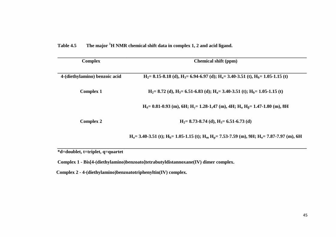
Table 4.5 The major 1H NMR chemical shift data in complex 1, 2 and acid ligand.
Complex Chemical shift (ppm)
4-(diethylamino) benzoic acid H2= 8.15-8.18 (d), H3= 6.94-6.97 (d); Ha= 3.40-3.51 (t), Hb= 1.05-1.15 (t)
Complex 1 H2= 8.72 (d), H3= 6.51-6.83 (d); Ha= 3.40-3.51 (t); Hb= 1.05-1.15 (t)
Hδ= 0.81-0.93 (m), 6H; Hγ= 1.28-1,47 (m), 4H; Hα Hβ= 1.47-1.80 (m), 8H
Complex 2 H2= 8.73-8.74 (d), H3= 6.51-6.73 (d)
Ha= 3.40-3.51 (t); Hb= 1.05-1.15 (t); Hm Hp= 7.53-7.59 (m), 9H; Ho= 7.87-7.97 (m), 6H
*d=doublet, t=triplet, q=quartet
Complex 1 - Bis[4-(diethylamino)benzoato]tetrabutyldistannoxane(IV) dimer complex.
Complex 2 - 4-(diethylamino)benzoatotriphenyltin(IV) complex.

46
4.5.2 13
C NMR Spectroscopy
Evidence of the formation of the complexes is clearly displayed in the 13
C
NMR spectrum. In the 13
C NMR data explicitly resolved the resonance of all the
distinct type of carbon atoms present in the complex. The 13
C NMR spectrum of free
ligand (Figure 4.11), complex 1 (Figure 4.12) and complex 2 (Figure 4.13) are
obtained. The downfield regions of the 13
C NMR spectrum of complex 1 show a sharp
peak carbon signal at 173.43 ppm. This signal is assigned to the δ(COO-) chemical
shift. Moreover, the 13
C NMR spectrum of complex 1 shows that the chemical shift
δ(COO-) signals is shifted downfield compared to that of the acid (173.02 ppm)
indicating the carboxylate anion is bonded to tin upon complexation. The occurrence
of six resonances in the range 109.13-150.78 ppm in the 13
C NMR spectrum of the
complex 1 and acid are due to the presence of benzene carbons. In the upfield region
of 13
C NMR spectrum, the complex 1 shows the occurrence of CH3 and CH2 in the
range 13.17-13.97 and 23.84-29.62 ppm, respectively. The butyl group of the di-n-
butyltin derivatives in CDCl3 solution exhibit four signals in the upfield region of
13.1-26.4 ppm (Yip et al., 2008). In addition, the complex 1 exhibited two sets of
butyl signals in 13
C NMR spectrum. This attributed to the butyl groups linked to the
exo- and endo-cyclic tin atom respectively (Danish et al., 1995).
The chemical shift for the aromatic carbons for the 13
C NMR spectrum of
complex 2 (in CDCl3 solution) appear in the downfield region of 116.29-151.41 ppm
(Shandu et al., 1991). The δ(COO-) signal of carboxyl carbon occur at 173.95 ppm.
The chemical shift δ(COO-) signals is shifted downfield compared to that of the acid

47
(173.02 ppm) indicating the carboxylate anion is bonded to tin upon complexation.
The carboxylate carbon being attached to two electron-withdrawing oxygen atoms, is
more deshielded and its signal appears slightly downfield in the region ( Parulekar et
al., 1990).
The phenyl group of the triphenyltin derivatives in CDCl3 solution exhibit in
the downfield region of 128.43-136.41 ppm. This occurrence is due to the effect of
anisotropic and hybridization of benzene group. The chemical shift of the δ(13
C)ipso is
found at region 137.1-143.8 ppm, whereas the chemical shift of δ(13
C)ortho, δ(13
C)meta,
δ(13
C)para are found in the region 133.9-137.2 ppm (Pruchnik et al., 2003). In the 13
C
NMR spectrum, the complex 2 shows the occurrence of δ(13
C)ipso, δ(13
C)ortho,
δ(13
C)meta and δ(13
C)para in the range 139.75 ppm, 137.07 ppm, 133.24 ppm and 128.71,
respectively. Additionally, The chemical shift of the δ(13
C)ipso is useful to determine
the coordination of tin atom, when the δ(13
C)ipso chemical shift occur at about 138
ppm and 142 ppm, hence the tin atom is said to be four and five coordinated
respectively (Pruchnik et al., 2003). Hence can conclude that the δ(13
C)ipso chemical
shift occur at 139.75 ppm the tin atom is four coordinated. The 13
C NMR data of free
ligand and complexes are determined and tabulated in Table 4.6.

48
Figure 4.11 13
C NMR spectrum of of 4-(diethylamino) benzoic acid

49
Figure 4.12 13
C NMR spectrum of bis[4-(diethylamino)benzoato]tetrabutyldistannoxane(IV) dimer complex

50
Figure 4.13 13
C NMR spectrum of 4-(diethylamino)benzoatotriphenyltin(IV) complex

51
Table 4.6 The major 13
C NMR chemical shift data in complex 1, 2 and acid ligand.
Complex Chemical shift (ppm)
4-(diethylamino) benzoic acid C1= 109.31, C2= 136.99, C3= 117.34, C4= 151.58, C7= 172.95;
Ca= 46.31, Cb= 11.99
Complex 1 C1= 109.27, C2= 136.49, C3= 119.50, C4= 152.47, C7= 173.55;
Ca= 48.49, Cb= 12.11; Cδ= 13.71-13.91; Cγ Cα Cβ= 26.09-27.29
Complex 2 C1= 110.77, C2= 135.92, C3= 117.75, C4= 152.97, C7= 173.89;
Ca= 48.72, Cb= 12.52; Cm= 128.43, Cp= 129.03, Co= 142.87, Ci= 142.87
Complex 1 - Bis[4-(diethylamino)benzoato]tetrabutyldistannoxane(IV) dimer complex.
Complex 2 - 4-(diethylamino)benzoatotriphenyltin(IV) complex

52
4.5.3 119Sn NMR Spectroscopy
It has been reported that in alkyltin carboxylates, four-coordinate tin has
δ119
Sn value raging from +200 to -60 ppm, five-coordinate tin from -90 to -190 ppm
and six-coordinate tin from -210 to -400 ppm (Mahmood et al., 2003; Tao et al., 1996;
Holecek et al., 1986). The complex 1 showed two well separated resonances of
δ(119
Sn) at -171.646 ppm and -221.432 ppm, respectively. These two well separated
resonances, respectively, are attributed to the endocyclic and exocyclic tin atoms in
the distannoxane dimer (Danish et al., 1995). The resonances of δ(119
Sn) at -171.646
ppm lie in the lower range of values for five-coordinated tin atom, whereas, the
resonances of δ(119
Sn) at -221.432 ppm lie in the upper range of values for six-
coordinated tin atom. The existence of two resonances of δ(119
Sn) is due to fast
exchange in the coordination behaviour of carboxylate groups attached to endo and
exo-cyclic tin atoms as reflected by different butyl signals (Khan et al., 2004).
Detailed assignment of each peak is shown in Figure 4.14.
On the basis of the values of the chemical shifts δ(119
Sn) of the triphenyltin(IV)
compounds examined vary over a wide range, from -44.7 ppm up to -257.2 ppm.
Although the chemical shift δ(119
Sn) lie in a broad range from -40 to -120 ppm, they
never exceed the maximum value of -128.1 ppm (tetraphenyltin) are known to form
tetrahedral molecules with a four-coordinate tin atom in the solid state (or at least in a
solution) (Zuckerman et al., 1970). The compounds with higher δ(119
Sn) values shifts
from -180 to -260 ppm involve molecules with trigonal-pyramid geometry around
five-coordinate tin atom (Carpino et al., 1980). There is a sharp and strong δ(119
Sn)

53
signal found in the 119
Sn NMR spectra of complex 2, the signal occur at chemical
shift -127.329 ppm, and hence we can conclude that the tin atom of complex 2 is a
four-coordinate tin atom. Detailed assignment of each peak is shown in Figure 4.15.
Therefore, 119
Sn NMR parameters are very useful for the determination of the
coordination number of tin and its geometry and consequently, the molecular
geometry of the complexes thus prepared (Mahmood et al., 2003). The signals of
complex 1 are given in two well separated signals peaks which confirm as endocyclic
and exocyclic tin respectively, the two signals giving two types of coordination
number of tin atom as five and six-coordinated whereas, 119
Sn NMR spectra of
complex 2 shows only a single strong peak that indicate as four-coordinated tin atom.
This indicates that only one bond between tin atom with anion carboxylate in complex
2. All of the Sn chemical shifts gained from the complexes are tabulated in Table 4.7.

54
Figure 4.14 119
Sn NMR spectrum of bis[4-(diethylamino)benzoato]tetrabutyldistannoxane(IV) dimer complex

55
Figure 4.15 119
Sn NMR spectrum of 4-(diethylamino)benzoatotriphenyltin(IV) complex

56
Table 4.7 The major 119
Sn NMR chemical shift data in bis[4-(diethylamino)benzoato]tetrabutyldistannoxane(IV)
dimer complex and 4-(diethylamino)benzoatotriphenyltin(IV) complex.
Complex
Chemical shift (ppm)
Bis[4-(diethylamino)benzoato]tetrabutyl
distannoxane(IV) dimer complex
-171.646
-221.432
4-(diethylamino)benzoatotriphenyltin(IV) complex
-127.329

57
CHAPTER 5
CONCLUSION
The bis[4-(diethylamino)benzoato]tetrabutyl distannoxane(IV) dimer complex
1 and the 4-(diethylamino)benzoatotriphenyltin(IV) Complex 2 are success
synthesised and fully charaterized.
For the complex 1 and complex 2, the calculated percentage of
carbon ,nitrogen and hydrogen compared to the data obtained from CHN analysis are
below 3.0 %. The differences between the calculated percentage and theoretical
percentage obtained from CHN analysis are not much varied. Both complex 1 and 2
giving the sharp melting point. The analysis data can deduced that the purity of the
complexe that has been successfully synthesized. Elemental analysis result of the
complex 1 and 2 were agreement with the predicted formula.
The infrared spectrum of the complex 1 and 2 showed distinct differences
from the acid ligand. The υ(OH) band which appread in the acid, was absent in the
infrared spectra of the complexes showing the occurrence of the deprotonation prior
to cooridnation of the resulting carboxylate anion in the complex formation. The
presence of the υ(Sn-O-Sn) , υ(Sn-O) and υ(Sn-C) indicates coordination of the
carboxylate group with the tin metal.

58
The differences [υΔ)], between υasym(COO) as and υsym(COO) is important to
determine the coordination of the carboxylic groups boding to the tin atom. From the
FTIR spectra of the complex 1 υasym(COO) vibraional peaks has shifted to higher
frequency which complex 2 shift to the lower frequency, whereby will causes the
electron density of the carboxyl group tend to donate to the d acceptor of the tin atom.
NMR spectroscopy provided valuable information about the structure of
complex 1 and 2. The number of protons for each fragment of the predicted molecular
structure corresponding signals in the 1H NMR spectrum implies the complex 1 and 2
synthesized fits well with the predicted molecular structure. The disapperance of the
carbxylic acid proton in 1H NMR spectra of the complex 1 and 2 is the another
confirmation to the deprotonation of carboxylic acid proton.
The chemical shift δ(COO-) signal of complex 1 and 2 is shifted downfield
compared to that of the acid indicating carboxylate anion is bonded to tin upon
complexation. In addition, the absence of any impurity peaks in the 1H NMR and
13C
NMR spectrum is indicative of an experimentally pure complexes thus prepared.
The signal of 119
Sn NMR spectra of complex 1 is given in two well separated signal
peak which confirmed as endocylic and excocylic tin respectivetely, the two signals
giving two types of cooridnation number of tin atoms as five and six-cooridnated
whereas, 119
Sn NMR spectra of complex 2 shows only a single strong peak that
indicate as four-coordinated tin atom. This indicates that only one bond between tin
atom which anion carboxylate in complex 2.

59
As a conclusion, all the value and data obtained from the qualitative and
quantitative analysis are well defined and proven that both of the complex 1 and
complex 2 were in agreement with the predicted formula and structure. The structure
of the complex 1 and 2 are shown in Figure 1 and 2.

60
Figure 5.1 The strucure of bis[4-(diethylamino)benzoato]
tetrabutyldistannoxane(IV) Dimer Complex
Figure 5.2 The structure of of 4-(diethylamino)benzoate triphenyltin(IV)
Complex


![Thermal and radiation chemistry of butyltin oxo hydroxo A ... · obtained [15]. A benefit of the organotin systems is that tin has a higher EUV absorption cross-section than many](https://img.pdfslide.us/doc/110x75/5f885a9e3655987f843d3f97/thermal-and-radiation-chemistry-of-butyltin-oxo-hydroxo-a-obtained-15-a-beneit.jpg)