Embed Size (px)
Citation preview

Chapt. 34/5
Ch. 34/5Student Learning Outcomes:• Describe how steroid hormones are from cholesterol:
• Adrenal corticol hormones include:• Cortisol, aldosterone, adrenal sex steroids
• Gonadal hormones include:• Sex steroids testosterone, estrogen
• Briefly describe structure/ function of Eicosanoids: • Prostaglandins, thromboxanes, leukotrienes

Cholesterol is precursor of steroid hormones
Cholesterol is precursor of steroid hormones:From diet, synthesized in tissues, intracellular pools, LDL• Glucocorticoids – cortisol from adremal gland,
• synthesis stimulated by ACTH (adrenocorticotropic hormone)• Mineralocorticoids – aldosterone from adrenal cortex,
secreted in response to antiotension II, III, ↑K+ levels, low Na+
• Androgens – testosterone from Leydig cells of testes; secreted in response to LH (luteinizing hormone)
• Estrogens – from ovarian follicle, corpus luteum; secretion stimulated by FSH (follicle stimulating hormone)
• Progestins – progesterone from corpus luteum, secretion stimulated by LH
Steroid hormones
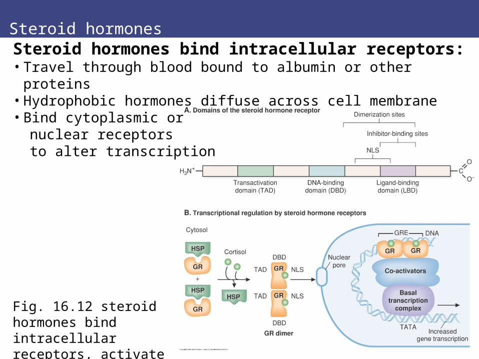
Fig. 16.12 steroid hormones bind intracellular receptors, activate transcription
Steroid hormones bind intracellular receptors:• Travel through blood bound to albumin or other proteins• Hydrophobic hormones diffuse across cell membrane• Bind cytoplasmic or nuclear receptors to alter transcription
Overview Steroid hormones
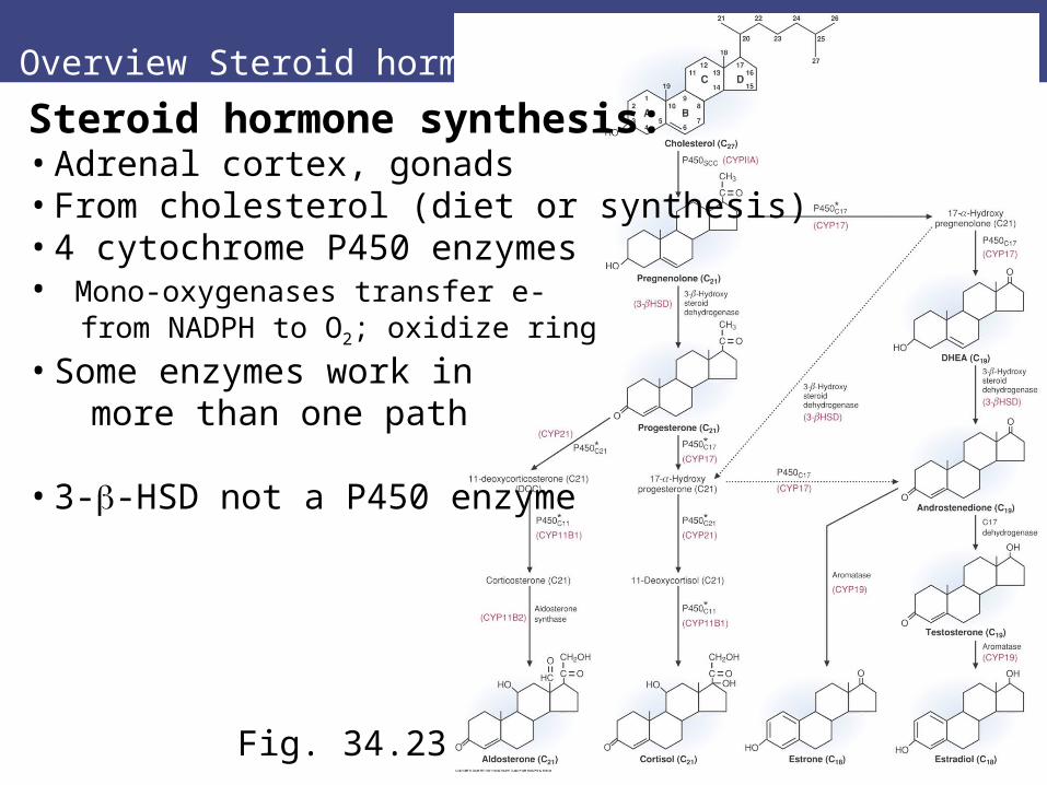
Fig. 34.23
Steroid hormone synthesis:• Adrenal cortex, gonads• From cholesterol (diet or synthesis)• 4 cytochrome P450 enzymes• Mono-oxygenases transfer e- from NADPH to O2; oxidize ring
• Some enzymes work in more than one path
• 3--HSD not a P450 enzyme
P450 enzymes
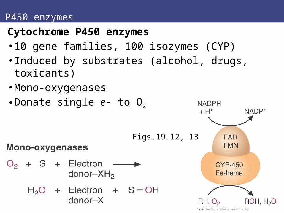
Cytochrome P450 enzymes• 10 gene families, 100 isozymes (CYP)• Induced by substrates (alcohol, drugs, toxicants)• Mono-oxygenases
• Donate single e- to O2
Figs.19.12, 13

Steroid hormones I
I. Cholesterol to progesterone:• Mitochondrial inner membrane
• Rate-limiting step P450SCC (CYPIIA)
• Cleavage of side chain• Forms pregnenolone
• Then Smooth ER for other steps• 3--HSD is not a P450 enzyme
Fig. 34.23 top
Steroid hormones
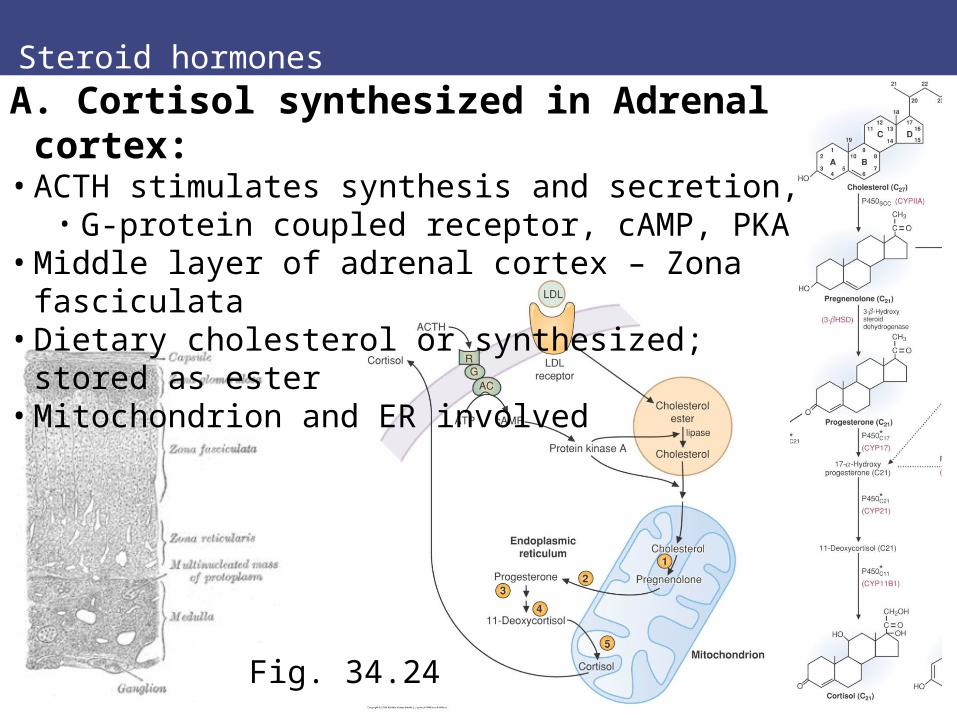
Fig. 34.24
A. Cortisol synthesized in Adrenal cortex:• ACTH stimulates synthesis and secretion,
• G-protein coupled receptor, cAMP, PKA• Middle layer of adrenal cortex – Zona fasciculata• Dietary cholesterol or synthesized; stored as ester• Mitochondrion and ER involved

Steroid hormones
Fig. 34.23 part
B. Aldosterone is synthesized in Adrenal cortex:• Zona glomerulosa layer of adrenal cortex• Cholesterol to pregesterone, then differ → DOC• Some enzymes same as for cortisol• Octapeptide angiotensin II stimulate synthesis
• Also hyperkalemia, low Na+• Stimulates Na+ uptake by kidney, ↑ extracellular fluid
Sex hormones
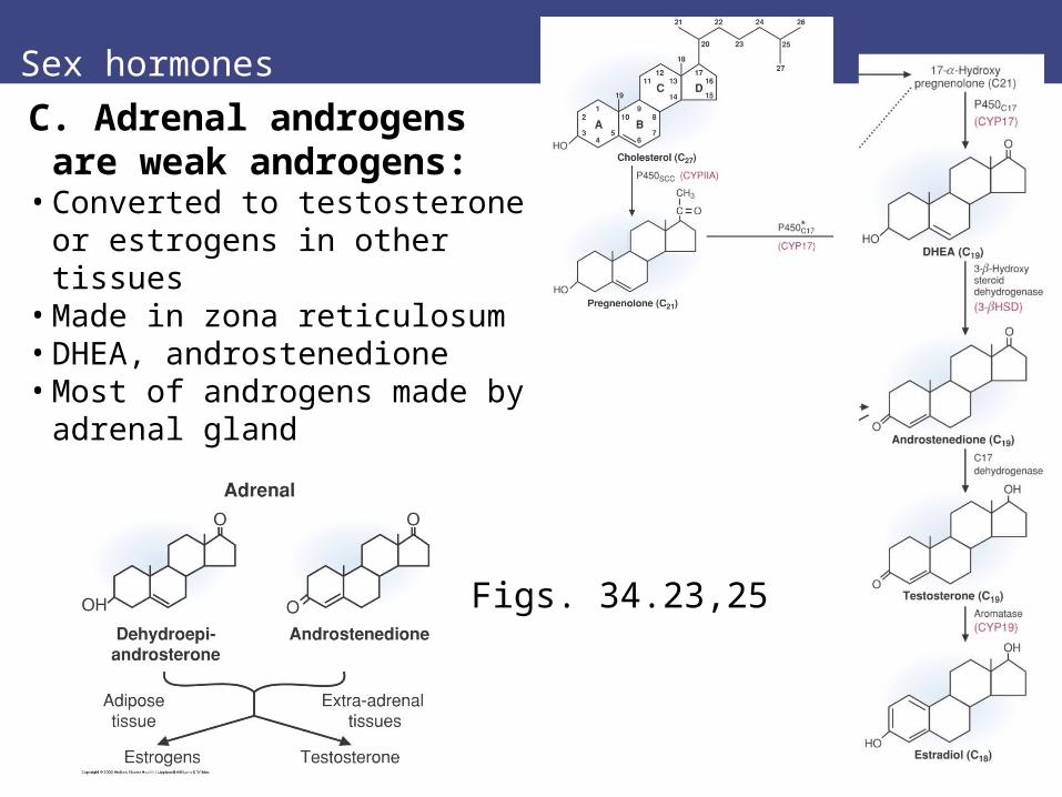
C. Adrenal androgens are weak androgens:
• Converted to testosterone or estrogens in other tissues
• Made in zona reticulosum• DHEA, androstenedione• Most of androgens made by
adrenal gland
Figs. 34.23,25
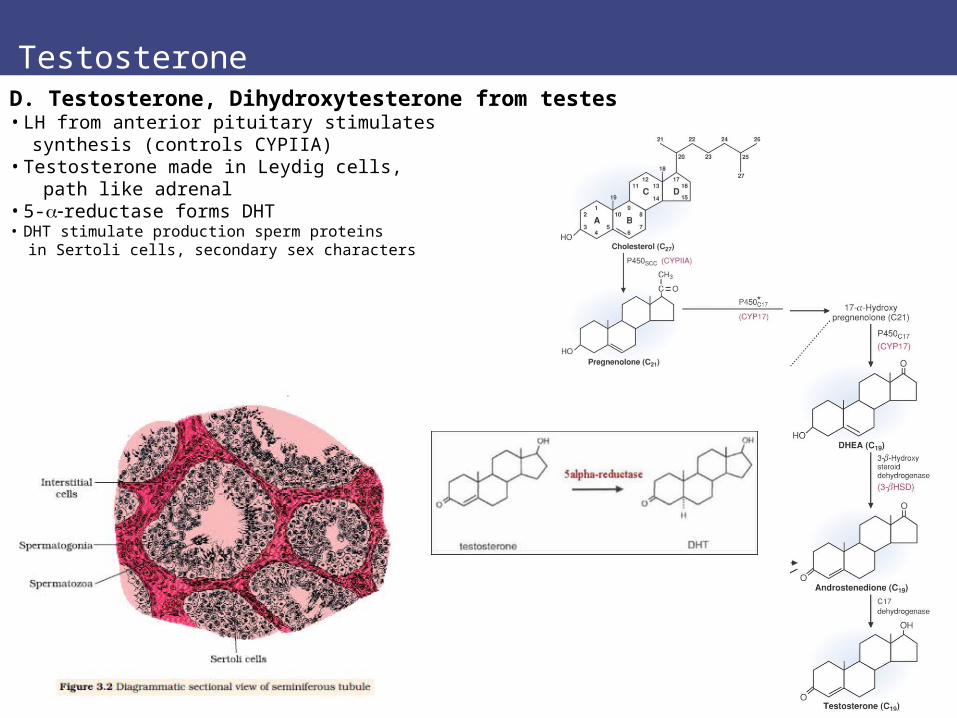
TestosteroneD. Testosterone, Dihydroxytesterone from testes• LH from anterior pituitary stimulates synthesis (controls CYPIIA)• Testosterone made in Leydig cells, path like adrenal• 5-reductase forms DHT• DHT stimulate production sperm proteins in Sertoli cells, secondary sex characters
Estrogens
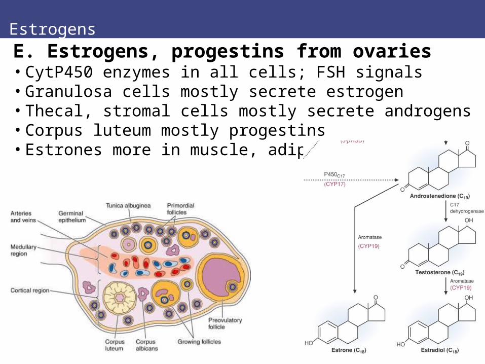
E. Estrogens, progestins from ovaries• CytP450 enzymes in all cells; FSH signals• Granulosa cells mostly secrete estrogen• Thecal, stromal cells mostly secrete androgens• Corpus luteum mostly progestins• Estrones more in muscle, adipose

Vitamin D
Fig. 34.26Vitamin Dcalcitrol
XI. Vitamin D:• Family of calciferols• Involved with Ca2+ homeostasis• From diet or synthesized in body• Skin needs UV to synthesize cholecalciferol• Liver and kidney finish• Binds nuclear hormone receptor VDR
• Activates transcription of genes
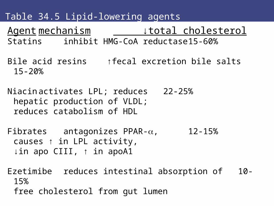
Table 34.5 Lipid-lowering agents
Agent mechanism ↓total cholesterolStatins inhibit HMG-CoA reductase 15-60%
Bile acid resins ↑fecal excretion bile salts 15-20%
Niacin activates LPL; reduces 22-25%hepatic production of VLDL;reduces catabolism of HDL
Fibratesantagonizes PPAR-, 12-15%causes ↑ in LPL activity, ↓in apo CIII, ↑ in apoA1
Ezetimibe reduces intestinal absorption of 10-15%free cholesterol from gut lumen
Sex hormones
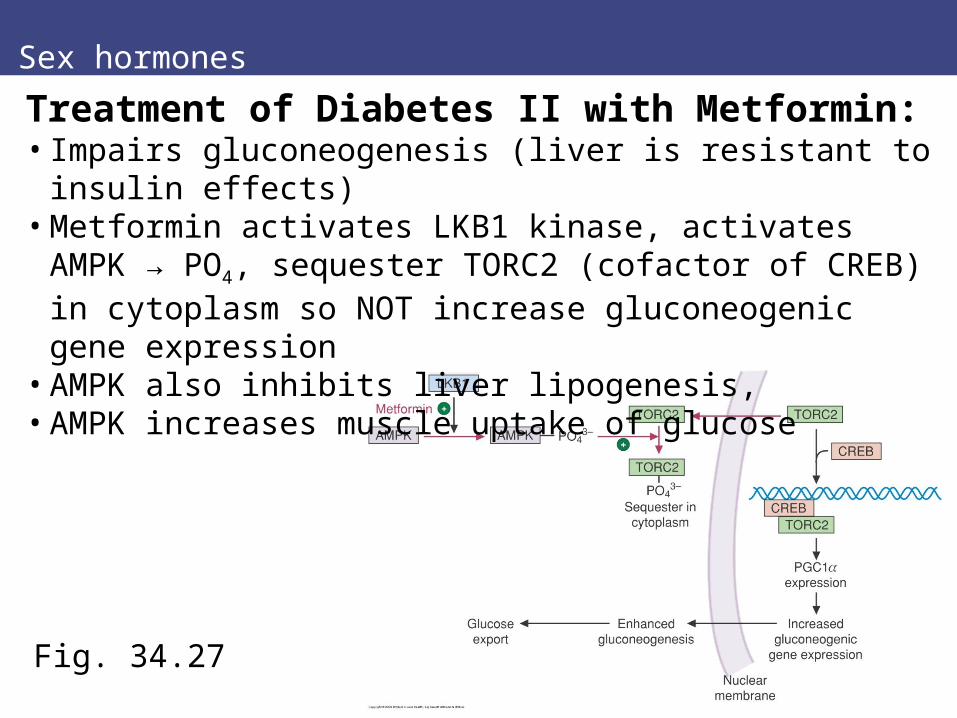
Fig. 34.27
Treatment of Diabetes II with Metformin:• Impairs gluconeogenesis (liver is resistant to insulin effects)• Metformin activates LKB1 kinase, activates AMPK → PO4,
sequester TORC2 (cofactor of CREB) in cytoplasm so NOT increase gluconeogenic gene expression
• AMPK also inhibits liver lipogenesis, • AMPK increases muscle uptake of glucose
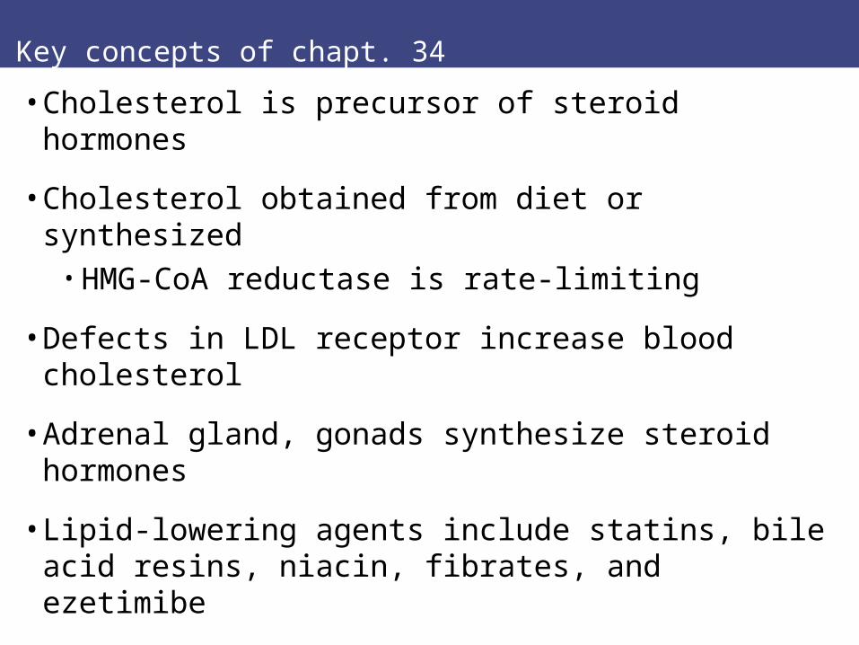
Key concepts of chapt. 34
• Cholesterol is precursor of steroid hormones
• Cholesterol obtained from diet or synthesized• HMG-CoA reductase is rate-limiting
• Defects in LDL receptor increase blood cholesterol
• Adrenal gland, gonads synthesize steroid hormones
• Lipid-lowering agents include statins, bile acid resins, niacin, fibrates, and ezetimibe
Chapter 35
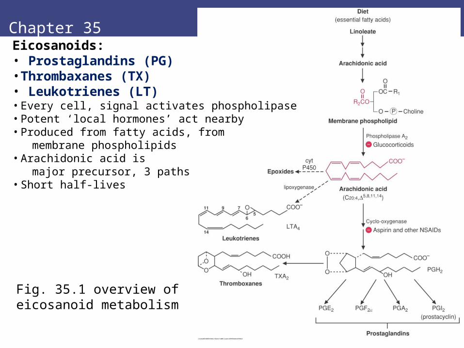
Fig. 35.1 overview of eicosanoid metabolism
Eicosanoids: • Prostaglandins (PG)• Thrombaxanes (TX)• Leukotrienes (LT)• Every cell, signal activates phospholipase• Potent ‘local hormones’ act nearby• Produced from fatty acids, from membrane phospholipids• Arachidonic acid is major precursor, 3 paths• Short half-lives

Roles of Eicosanoids
• Inflammatory response after infection, injury:• Vasodilation increases blood flow in damage area• Control bleeding through clots, activate complement• White blood cells move to injury, make cytokines • Can give symptoms pain, swelling, fever• Inappropriate response is allergy, hypersensitivity
• Smooth muscle contraction• Increase water, sodium excretion by kidney• Some constrictors of blood vessels, • Some dilators• Regulate blood pressure
Aspirin inhibits COX enzyme

Arachidonic acid precursor
Fig. 35.2
Arachidonic acid is precursor• Polynsaturated C20 fatty acid, from diet or
made from essential Linoleate fatty acid• Phospholipases cleave from phospholipid in
plasma membrane (C2 position)• Stimuli include histamines, cytokines
Fig. 33.30

II. Pathways of eicosanoids
II. Pathways for eicosanoid synthesis:
A. Cyclo-oxygenase (COX) → PG, TX
B. Lipoxygenase → Leukotrienes
C. Cytochrome P450 → epoxides• Different enzymes in different tissues gives specialization
Fig. 35.3

Cyclo-oxygenase pathway
Fig. 35.4-6 PG generic structure: C20 fatty acid;5-membered ring Fig. 35.5 PG structures
Fig. 35.6 numeral is double bonds: 2 is most common
A. Cyclo-oxygenase Pathway:Prostaglandins are large family:Letter = ring substituentsNumeral subscript = double bondsPGF also have subscriptEx. PGA1, PGF2

Cyclo-oxygenase pathway
Fig. 35.7 TX generic structure: C20 fatty acid, 6-member ring with OTXA2 has extra O C9-11
A. Cyclo-oxygenase Pathway:ThromboxanesTXA2 from platelets is most common thromboxane:
stimulates platelet aggregationcauses vasoconstriction
Formation of prostaglandins and thromboxane
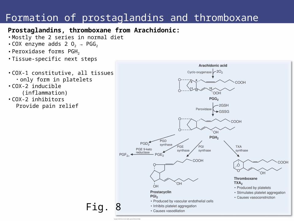
Fig. 8
Prostaglandins, thromboxane from Arachidonic:• Mostly the 2 series in normal diet• COX enzyme adds 2 O2 → PGG2
• Peroxidase forms PGH2
• Tissue-specific next steps
• COX-1 constitutive, all tissues• only form in platelets
• COX-2 inducible (inflammation)
• COX-2 inhibitorsProvide pain relief
COX inhibitors provide pain relief
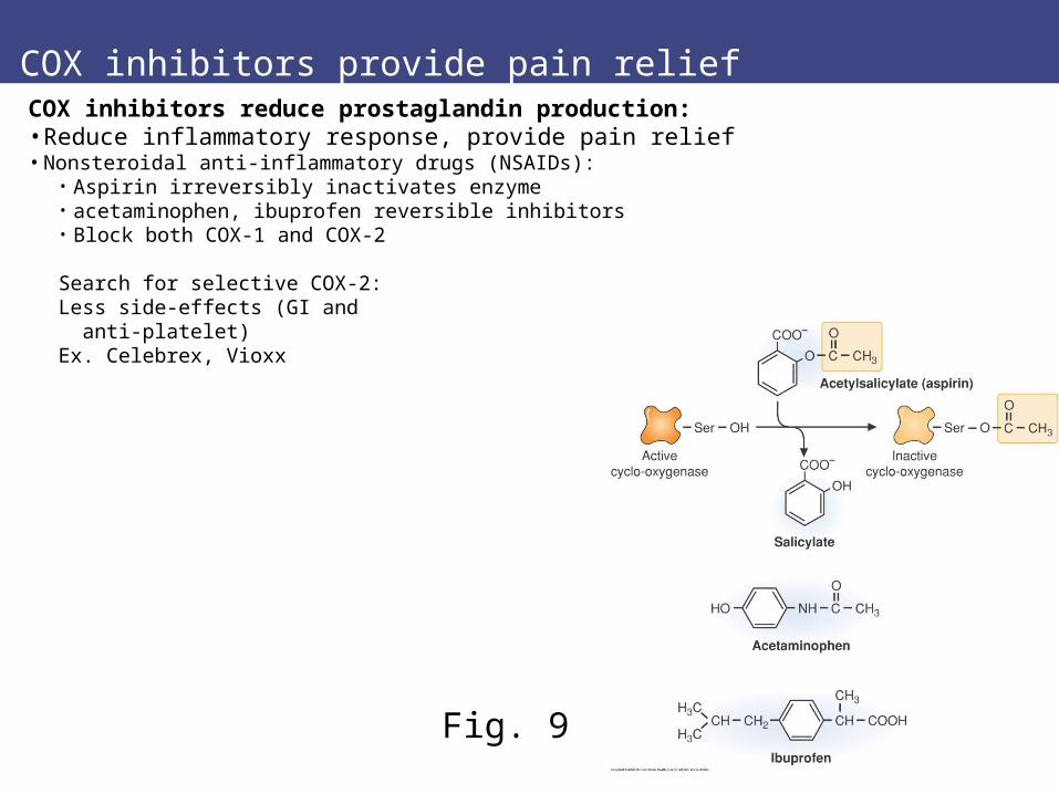
Fig. 9
COX inhibitors reduce prostaglandin production:• Reduce inflammatory response, provide pain relief • Nonsteroidal anti-inflammatory drugs (NSAIDs):
• Aspirin irreversibly inactivates enzyme • acetaminophen, ibuprofen reversible inhibitors• Block both COX-1 and COX-2
Search for selective COX-2:Less side-effects (GI and anti-platelet)Ex. Celebrex, Vioxx
Tables 35.1, 35.2 Functions of prostaglandins, thromboxanes
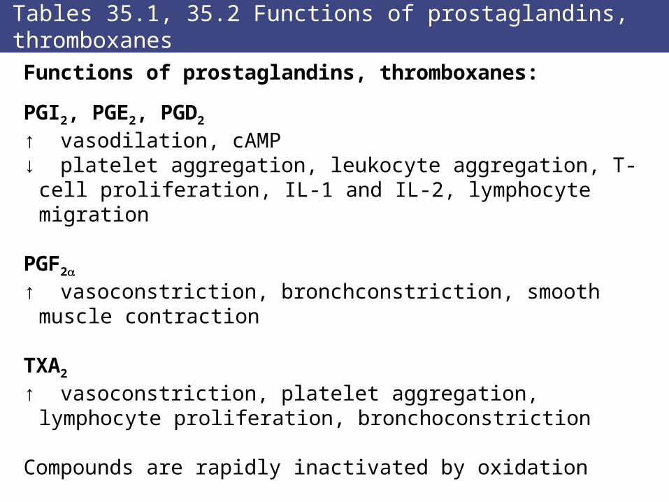
Functions of prostaglandins, thromboxanes:
PGI2, PGE2, PGD2 ↑ vasodilation, cAMP↓ platelet aggregation, leukocyte aggregation, T-cell
proliferation, IL-1 and IL-2, lymphocyte migration
PGF2
↑ vasoconstriction, bronchconstriction, smooth muscle contraction
TXA2
↑ vasoconstriction, platelet aggregation, lymphocyte proliferation, bronchoconstriction
Compounds are rapidly inactivated by oxidation
Lipoxygenase pathway: leukotrienes
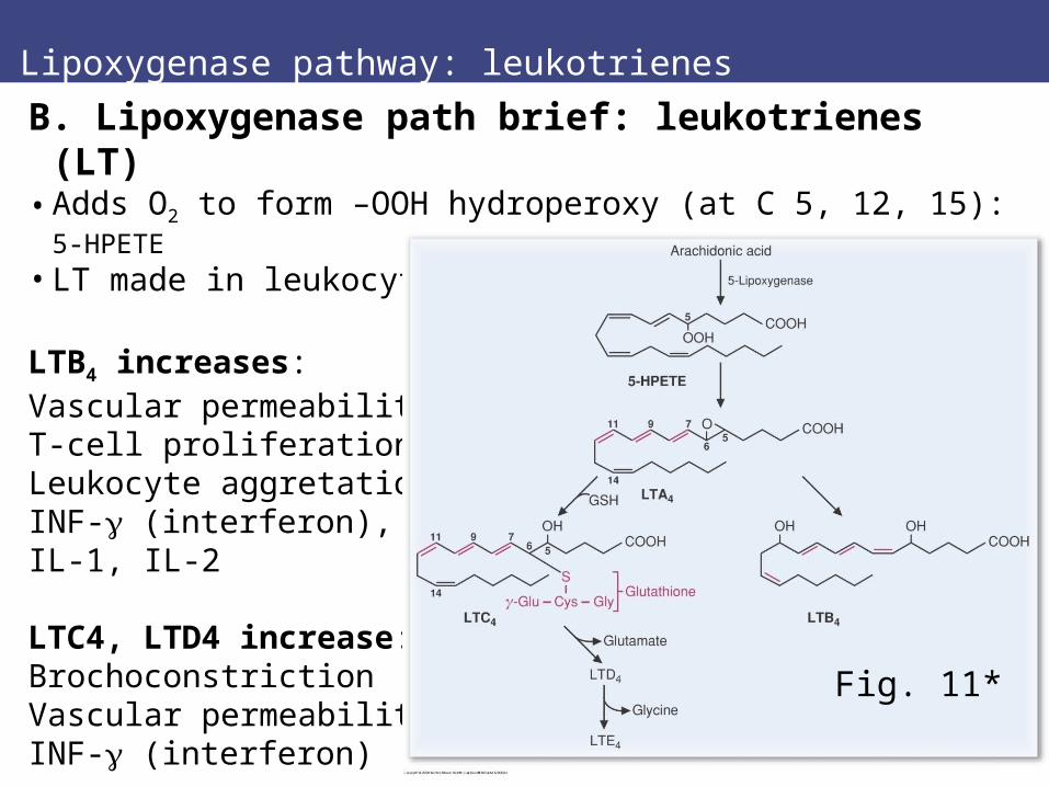
B. Lipoxygenase path brief: leukotrienes (LT)• Adds O2 to form –OOH hydroperoxy (at C 5, 12, 15): 5-HPETE
• LT made in leukocytes (triene = 3 C=C)
LTB4 increases:Vascular permeabilityT-cell proliferationLeukocyte aggretationINF- (interferon),IL-1, IL-2
LTC4, LTD4 increase:BrochoconstrictionVascular permeabilityINF- (interferon)
Fig. 11*

Table 35.4
Prostaglandin and thromboxane receptors:Specific receptors for different hormones• all are G-protein coupled• signal through increase Ca2+ in cytosol
• (PGF2, TXA2, leukotrienes)• signal through adenylate cyclase-cAMP-PKA
• (PGE, PGD, PGI series)

Key concepts
Key concepts:• Eicosanoids (postaglandins, thromboxanes, leukotrienes) are
potent regulators of cellular function)• Eicosanoids are derived from polyunsaturated fatty acid (20C)• Eicosanoids are important for inflammatory response, smooth
muscle contraction, blood pressure.• PG and T require cyclo-oxygenase (COX)• LT requires lipooxygenase
• Eicosanoids bind membrane receptors, • alter PKA or Ca2+ levels
Review question
Review question:
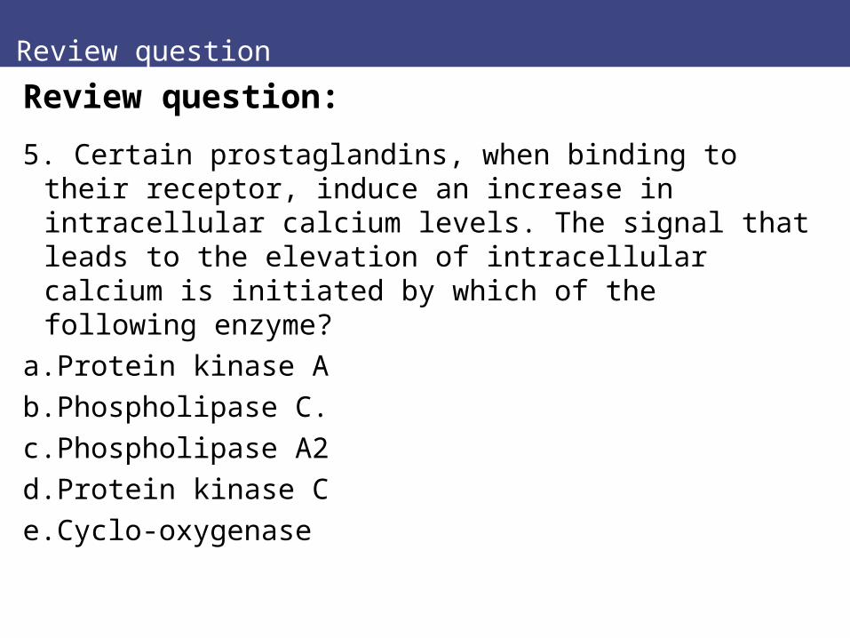
5. Certain prostaglandins, when binding to their receptor, induce an increase in intracellular calcium levels. The signal that leads to the elevation of intracellular calcium is initiated by which of the following enzyme?
a.Protein kinase A
b.Phospholipase C.
c.Phospholipase A2
d.Protein kinase C
e.Cyclo-oxygenase


![Aldosterone and dopamine receptors in the kidney: Sites for ...Aldosterone and dopamine receptors 625 with the aldosterone receptor, when measured in vitro [21, 23] (Funder and Adam,](https://img.pdfslide.us/doc/110x75/608977add019a330f10765d3/aldosterone-and-dopamine-receptors-in-the-kidney-sites-for-aldosterone-and.jpg)