Embed Size (px)
Citation preview
Journal of Cellular Biochemistry 5722-29 (1995)
Changes in the Sulfation Extent of Mem brane-Associated Proteoglycans Produced by
-
Sertoli Cells in Culture Juan Pablo Rodriguez
Unidad de Biologia Celular, INTA, Universidad de Chile, Casilla 138-1 1, Santiago, Chile
Abstract Sertoli cells in culture synthesize two different membrane-associated proteoglycans (MA-PG): a proteoglycan containing heparan sulfate (HS) and chondroitin sulfate (CS) glycosaminoglycan (GAG) chains and a CS-PG containing only CS-GAG chains. The structure of these molecules is regulated by the presence of fetal calf serum (FCS) in the culture medium. Changes in the concentration of FCS resulted in changes in the total 35S04 incorporation into MA-PG and a shift in the elution profile of each component subjected to ion-exchange chromatography. Thus, without FCS, the incorporation was low, while in 1% and 10% FCS, the uptake of the precursor was 1.7 and 4.5 times higher, respectively. MA-PG synthesized by Sertoli cells cultured in 10% FCS eluted from DEAE-Sephacel columns at higher salt concentration than the MA-PG synthesized by cells cultured in 0% or 1% FCS. Double-labeled experiments showed that the 35S04/3H-glucosamine ratio incorporated into MA-PG produced by Sertoli cells, increased from 17.6 to 23.6 and 50.9 in cells cultured at 0, 1, and 10% FCS, respectively. However, the presence of FCS affected neither the hydrodynamic size nor the chemical nature of GAG chains of MA-PG. These results show that changes in the FCS concentration promote changes in the sulfation extent of MA-PG molecules produced by Sertoli cells. 2 1995 Wiley-Liss, Inc
Key words: proteoglycans, sulfation, serum, Sertoli cells, fetal calf serum
Sertoli cells in culture synthesize different extracellular matrix components (ECM), such as fibronectin, laminin, collagen [Rodriguez et al., 1991; Skinner et al., 19851, and two proteo- glycans (PGs): a PG containing CS and HS chains and a PG containing only CS chains [Rodriguez and Minguell, 1989; Skmner and Fritz, 19851. PGs produced by Sertoli cells are located both intra- and extracellularly, as well as associated to the cell membrane (MA-PG) [Rodriguez and Minguell, 19891. Previously, we reported that MA-PG are not randomly distributed on the membrane surface. In fact, we observed a polar- ized distribution pattern [Rodriguez and Minguell, 1992al. This distribution suggests that MA-PGs play specific roles in the interactions of Sertoli cells with the cellular or the ECM envi- ronment.
The different distribution may also implies a differentially regulated synthesis for each PG. Thus, in addition to age-related changes in the
Received April 14, 1994; accepted May 17, 1994. Address reprint requests to Juan Pablo Rodriguez, Unidad de Biologia Celular, INTA, Universidad de Chile, Casilla 138-11, Santiago, Chile.
G 1995 Wiley-Liss, Inc.
amount of the MA-PG, we also reported changes of MA-PG synthesis after interaction of Sertoli cells with collagen substrate [Rodriguez and Minguell, 1989, 1992bl.
In other somatic cells subjected to different treatment in culture, it has also been shown changes in the structure of the PG molecules. The latter has been reported to occur in several cell systems, where the action of various regula- tory molecules, or the proliferation status of the cells, induce changes in the sulfation of PG [David and Van Den Berghe, 1983; Hutchison and Yasin, 1986; Robinson et al., 1984; Preston et al., 1985; Fedarko and Conrad, 19861, addi- tion of glycosaminoglycans (GAG) chains to the core protein [Nurcombe et al., 1993; Rapraeger, 19891, and increase of GAG chains length [Nur- combe et al., 19931.
In Sertoli cells, there is no information related to possible changes in the structure of these molecules as a response to regulatory molecules or in cells subjected to different culture condi- tions. Thus, this work studied whether changes in the culture conditions were able to produce changes in the structure of MA-PG produced by Sertoli cells. For these purposes, Sertoli cells
Sulfation of Membrane-Associated Proteoglycans 23
were cultured at different fetal calf serum (FCS) concentrations, generally employed in cell cul- tures as a source of different hormones and growth factors.
Results show that the MA-PG synthesized by Sertoli cells in culture change their sulfation extent. These changes are dependent on the concentration of FCS present in the culture medium. However, no difference were observed in the hydrodynamic size nor in the chemical nature of the GAG moieties of MA-PG synthe- sized by Sertoli cells under the different culture conditions used here.
MATERIALS AND METHODS Preparation and Culture Conditions of
Sertoli Cells
Sertoli cells from Wistar pubertal (40-45-day- old) rats were used in these experiments. Cells were isolated as previously described [Hutson and Stocco, 1981; Rodriguez and Minguell, 1992aI. Briefly, the testes were decapsulated and treated with a collagenase solution, 0.4 mg collagenase/ml in Medium 199 containing 0.1% bovine serum albumin (BSA), type I Worthing- ton, for 30 min at 37°C. After this enzymatic treatment, the seminiferous tubules were manu- ally isolated and treated again with the collage- nase solution for an additional 30 min at 37°C. Sertoli cells were mechanically dispersed by re- peated pipetting and resuspended in Medium 199 supplemented with 12% FCS, 12% horse serum, 0.1 FM hydrocortisone, 100 U/ml penicil- lin, and 100 kg/ml streptomycin (culture me- dium). Cells (6 x lo5 cells) were seeded into culture dishes (60 mm, Nunc) and incubated at 33°C in a humidified atmosphere of 5% COz. The culture medium was replaced by fresh culture medium every 3 days. Under these conditions, aggregates with morphological characteristics of Sertoli cells were evident in the adherent layer after day 3 in culture. Peritubular myoid-like cells and fibroblasts represented only 5% of cells in the adherent layer [Rodriguez and Minguell, 1992al.
Metabolic labeling of MA-PG
After 12 days in culture, Sertoli cells were used for PG synthesis studies. For these pur- poses, cells were incubated in culture medium supplemented with different FCS concentra- tions (O%, 1%, and 10%) and with 5 pCi/ml H-glucosamine (ICN Biomedical Inc., Irvine, CA;
40 Ci/mmol) and/or 20 kCi/ml sodium 35S- sulfate (ICN, 43 Ci/mg S) as GAG radioactive precursors. At the end of the labeling period (48 hr at 33"C), the culture medium was removed, and Sertoli cells in the adherent layer were washed twice with phosphate-buffered saline (PBS). The radioactive material present in the culture medium as well as in the washes was discarded.
Preparation and Characterization of MA-PG
MA-PG were obtained as previously described [Oohira and Wight, 19831. Briefly, Sertoli cells were exposed to a mild treatment with trypsin (0.05%, 10 min at 37°C). The released cells were collected by centrifugation and the supernatant, referred to as the membrane-associated (MA) fraction, containing the MA-PG was saved. The cells were washed two times with PBS and the washes were pooled with the MA fraction. To the MA fraction, 0.4 g/ml of solid guanidine- HC1, 1.25 mgiml of N-ethylmaleimide, and 0.18 mg/ml phenylmethylsulfonylfluoride were added [Oohira et al., 19831.
To remove the unincorporated radioisotopes, guanidine-HC1, and other chemicals, the MA fraction was applied into a Sephadex G-50 col- umn (0.8 x 15 cm) equilibrated with a buffer containing 8 M urea, 0.05 M sodium acetate, 0.15 M sodium chloride, and 0.5% Triton X-100, at pH 7.0 (buffer A).
Ion-Exchange Chromatography
To analyze the charge density, and therefore the sulfation extent of MA-PG, fractions contain- ing the excluded material (Vo) from Sephadex G-50 were pooled and loaded into a DEAE- Sephacel column (0.6 x 10 cm) equilibrated and washed with buffer A. The retained material was eluted with a linear gradient of sodium chloride (0.15-0.8 M) prepared in buffer A. Frac- tions (0.8 ml) were collected and aliquots taken for measurements of radioactivity and sodium chloride content.
Molecular Sieve Chromatography
To analyze the hydrodynamic size of MA-PG, the species eluted from the DEAE-Sephacel col- umn were pooled, dialyzed exhaustively against distilled water, and finally concentrated by lyoph- ilization to dryness. The lyophilized material was reconstituted in buffer A and subjected to rechromatography on DEAE-Sephacel. The spe-
24 Rodriguez
cies eluted from the latter DEAE-Sephacel col- umn were dialyzed against distilled water; ly- ophilized to dryness; reconstituted in a solution containing 4 M guanidine-HC1, 0.05 M sodium acetate, 0.5% Triton X-100, at pH 6.0 (buffer B); and applied to a Sepharose CL-6B column (0.5 x 100 cm) [Chang et al., 19831. After elu- tion with buffer B, fractions (0.45 ml) were collected and aliquots taken for radioactivity measurements.
Clycosam i nogl ycan Chain Characterization
The fractions containing the PG molecules eluted from the Sepharose CL-6B columns were pooled, dialyzed exhaustively against water, and lyophilized to dryness. The lyophilized material was reconstituted in water and treated with 0.1 M sodium hydroxide in the presence of sodium borohydride (75.5 mgiml), to remove the GAG chains from the core proteins [Carlson, 19681. The samples were incubated at 45°C for 48 hr; after this time, the pH was adjusted to slightly < 7 with glacial acetic acid. The GAG chains released by the alkaline borohydride treatment were analyzed by Sepharose CL-6B chromatog- raphy with buffer B as eluent. To further charac- terize the GAG moieties, the released chains were subjected to chondroitinase ABC, chon- droitinase AC 11, and nitrous acid treatments
5000
4000
3000
2000
1000
0
[Shively and Conrad, 19761. The extent of the enzymatic and chemical reactions was deter- mined by the appearance of low-molecular- weight-labeled species after Sephadex G-25 chro- matography.
RESULTS Characterization of MA-PC Synthesized by
Sertoli Cells
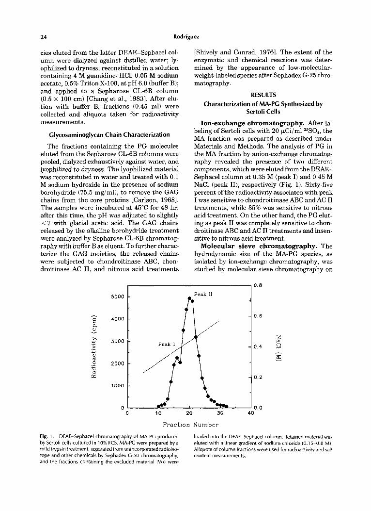
Ion-exchange chromatography. After la- beling of Sertoli cells with 20 p.Ci/ml 35S04, the MA fraction was prepared as described under Materials and Methods. The analysis of PG in the MA fraction by anion-exchange chromatog- raphy revealed the presence of two different components, which were eluted from the DEAE- Sephacel column at 0.35 M (peak I) and 0.45 M NaCl (peak 111, respectively (Fig. 1). Sixty-five percent of the radioactivity associated with peak I was sensitive to chondroitinase ABC and AC I1 treatments, while 35% was sensitive to nitrous acid treatment. On the other hand, the PG elut- ing as peak I1 was completely sensitive to chon- droitinase ABC and AC I1 treatments and insen- sitive to nitrous acid treatment.
Molecular sieve chromatography. The hydrodynamic size of the MA-PG species, as isolated by ion-exchange chromatography, was studied by molecular sieve chromatography on
0.8
0.6
0.4
0.2
0.0 0 10 20 30 40
Fraction Number
Fig. 1. DEAE-Sephacel chromatography of MA-PG produced by Sertoli cells cultured in 10% FCS. MA-PG were prepared by a mild trypsin treatment, separated from unincorporated radioiso- tope and other chemicals by Sephadex G-50 chromatography, and the fractions containing the excluded material (Vo) were
loaded into the DEAE-Sephacel column. Retained material was eluted with a linear gradient of sodium chloride (0.1 5-0.8 M). Aliquots of column fractions were used for radioactivity and salt content measurements.
Sulfation of Mem braneAssociated Proteoglycans 25
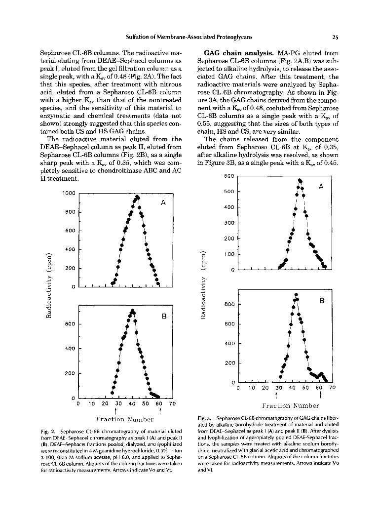
Sepharose CL-6B columns. The radioactive ma- terial eluting from DEAE-Sephacel columns as peak I, eluted from the gel filtration column as a single peak, with a KV of 0.48 (Fig. 2A). The fact that this species, after treatment with nitrous acid, eluted from a Sepharose CL-6B column with a higher K;iV than that of the nontreated species, and the sensitivity of this material to enzymatic and chemical treatments (data not shown) strongly suggested that this species con- tained both CS and HS GAG chains.
The radioactive material eluted from the DEAE-Sephacel column as peak 11, eluted from Sepharose CL-6B columns (Fig. 2B), as a single sharp peak with a KV of 0.35, which was com- pletely sensitive to chondroitinase ABC and AC I1 treatment.
1000
800
600
400
200
0
600
400
200
0 0 10 20 30 40 50 60 7 0
Fraction Number t t
Fig. 2. Sepharose CL-68 chromatography of material eluted from DEAE-Sephacel chromatography as peak I (A) and peak I1 (B). DEAE-Sephacel fractions pooled, dialyzed, and lyophilized were reconstituted in 4 M guanidine hydrochloride, 0.5% Triton X-100, 0.05 M sodium acetate, pH 6.0, and applied to Sepha- rose CL-68 column. Aliauots of the column fractions were taken
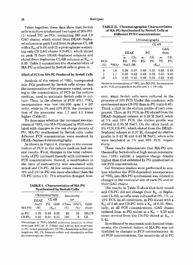
GAG chain analysis. MA-PG eluted from Sepharose CL-6B columns (Fig. 2A,B) was sub- jected to alkaline hydrolysis, to release the asso- ciated GAG chains. After this treatment, the radioactive materials were analyzed by Sepha- rose CL-6B chromatography. As shown in Fig- ure 3A, the GAG chains derived from the compo- nent with a KV of 0.48, coeluted from Sepharose CL-6B columns as a single peak with a KV of 0.55, suggesting that the sizes of both types of chain, HS and CS, are very similar.
The chains released from the component eluted from Sepharose CL-6B at KV of 0.35, after alkaline hydrolysis was resolved, as shown in Figure 3B, as a single peak with a KV of 0.45.
500
- E a 0 v
h
3
W (d 0
(d E
5 3
z
Fig. 3.
i i T t
800
600
400
200
n - 0 10 20 30 40 50 60 70
t t Fraction Number
SeDharose CL-68 chromatography of GAG chains liber- - . ated by alk'aline borohydride treatment of material and eluted from DEAE-Sephacel as peak I (A) and peak I 1 (B). After dyalisis and lyophilization of appropiately pooled DEAE-Sephacel frac- tions, the samples were treated with alkaline sodium borohy- dride, neutralized with glacial acetic acid and chromatographed on a Sepharose CL-68 column. Aliquots of the column fractions were taken for radioactivity measurements. Arrows indicate Vo
for radioactivity measurements. Arrows indicate Vo and Vt. and Vt
26 Rodriguez
Taken together, these data show that Sertoli cells in culture synthesized two types of MA-PG: (1) mixed PG (m-PG), containing HS and CS GAG chains, which eluted from DEAE-Sepha- cel columns as peak I and from Sepharose CL-6B with a K;, of 0.48; and (2) a proteoglycan contain- ing only CS GAG chains (CS-PG), which eluted as peak I1 from DEAE-Sephacel columns and eluted from Sepharose CL-6B columns at Kv = 0.35. Table I summarizes the characteristics of MA-PG synthesized by Sertoli cells is given in.
Effect of FCS on MA-PC Produced by Sertoli Cells
Analysis of the extent of 35S04 incorporated into PGs produced by Sertoli cells shows that the incorporation of the precursor varied, accord- ing to the concentration of FCS in the culture medium, used to maintain Sertoli cells in cul- ture. Thus, in the absence of FCS (O%), 35S04 incorporation was low (44,000 cpm/l x lo6 cells), while in 1% and 10% FCS, the incorpora- tion of the precursor was 1.7 and 4.5 times higher (Table 11).
To determine whether the increased incorpo- ration of 35S04 into MA-PG caused by FCS corre- lated with changes in the net charge density of PG, MA-PG synthesized by Sertoli cells under different FCS concentration were resolved by DEAE-Sephacel chromatography.
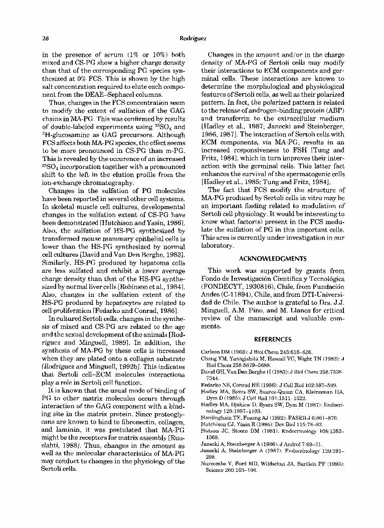
As shown in Figure 4, changes in the concen- tration of FCS in the culture medium had sev- eral results. First, changes in the total radioac- tivity of PG increased linearly with increases in FCS concentrations. Second, a modification in the ratio of radioactivity was associated with mixed and CS-PG. At low serum concentration (0% and 1%) m-PG was more abundant than the CS-PG (ratio 1.3). This situation changed, how-
TABLE I. Characteristics of MA-PG Synthesized by Sertoli Cells
C h r o m a t o ~ a ~ h ~ Sensitivity DEAE CL-6B toa NaCl PG GAG CSase HNOz GAG
MA-PG (M) (Kav) (%) (%) chain
m-PG 0.35 0.48 0.55 65 35 HS,CS CS-PG 0.45 0.35 0.55 100 0 cs aPercentage of 35S04-labeled molecules sensitive to chon- droitinase ABCiAC I1 (CSase) and nitrous acid treatment. m-PG: mixed proteoglycan; CS-PG: chondroitin sulfate pro- teoglycan; HS, CS, heparan sulfate and chondroitin sulfate glycosaminoglycan chains.
TABLE 11. Chromatographic Characteristics of MA-PG Synthesized by Sertoli Cells at
Different FCS Concentrations
Chromatomavhv CL-6B
GAG DEAE PG from
m- CS- m- CS- m- CS- FCS PG PG PG PG PG PG (%) 35S04a NaCl(M) (KaJ
0 1.0 0.30 0.37 0.48 0.35 0.55 0.45 1 1.7 0.35 0.42 0.48 0.35 0.55 0.45
10 4.5 0.35 0.45 0.48 0.35 0.55 0.45
"Relative incorporation of 35S04 into MA-PG. Incorporation at 0% FCS corresponds to 44,950 cpmil x lo6 cells.
ever, when Sertoli cells were cultured in the presence of 10% FCS. Under this condition, cells synthesized more CS-PG than m-PG (ratio 0.45). Third, a shift in the elution profile of each com- ponent. Thus, a t 0% FCS, m-PG eluted from the DEAE-Sephacel column at 0.30 M NaC1, while at 1% and 10% FCS, the elution profile was shifted to 0.35 M NaC1. On the other hand, at 0% FCS, CS-PG, which eluted from the DEAE- Sephacel column at 0.37 M, changed its elution profile to 0.42 M and 0.45 M NaC1, when cells were incubated at 1% and 10% FCS, respec- tively.
These results demonstrate that MA-PG syn- thesized by Sertoli cells at high serum concentra- tion (10%) exhibit a negative charge density higher than that exhibited by PG synthesized at low FCS concentrations.
Gel filtration studies were performed to ana- lyze whether the FCS-dependent incorporation of 35S04 into MA-PG synthesized was related to changes in the molecular size of each PG and/or their GAG chains.
The results in Table I1 show that both mixed and CS-PG did not change their KY in Sepha- rose CL-6B, as cells were cultured at 0%, 1%, or 10% FCS. In all conditions, m-PG eluted with a &, of 0.48 and CS-PG with a K,, of 0.35. Simi- larly, at all FCS concentrations, GAG chains isolated from m-PG eluted at a Kv = 0.55 and those derived from the CS-PG eluted at &, = 0.45.
As evidenced by enzymatic and chemical treat- ments, the chemical nature of MA-PG was not modified by changes in FCS concentrations. At all FCS concentrations, the sensitivity of m-PG

Sulfation of Mem brane-Associated Proteoglycans 27
E a v
x
700 , 0.8 600
500
400
300
200
100
0 700 1 , o.e
I , 0.8 5000 4000 1 i\ / 3000
2000
1000
0 0 10 20 30 40
0.4
0.2
0.0
Frac t ion Number
Fig. 4. DEAE-Sephacel chromatography of MA-PC produced by Sertoli cells at 0% (A), 1% (B), and 10% (C). After elution, aliquots of column fractions were taken for radioactivity and sodium chloride content measurements.
and CS-PG to chondroitinase ABC and nitrous acid treatments was similar to that shown in Table I.
Thus, the differences observed in the salt con- centration required to elute MA-PG synthesized at different FCS concentrations from the DEAE- Sephacel column (Fig. 4) cannot be explained in terms of changes in the hydrodynamic size and chemical nature of each PG or its constituent GAG chains. These results are suggestive of FCS-dependent changes in the sulfation extent
Na,35S04 and 3H-glucosamine double- labeling. To further substantive FCS increases,
of MA-PG.
proteoglycan sulfation, Sertoli cells, at different FCS concentrations, were double labeled with 35S04 and 3H-glucosamine, and the labeled mol- ecules were subsequently separated by DEAE- Sephacel chromatography, and the 35S04/3H ra- tio incorporated into MA-PG calculated.
The analysis of each radioactive precursor incorporated into the MA-PG from Sertoli cells cultured at different FCS concentrations is pre- sented in Table 111. The incorporation of each precursor into MA-PG increased as FCS concen- tration was modified from 0 to 10%. The ratio of radioisotopes (35S04/3H) incorporated into MA-PG increased from 17.6 in MA-PG synthe- sized at 0% FCS to 23.6 and to 50.9 in MA-PG synthesized by Sertoli cells cultured at 1% and 10% FCS, respectively.
These results demonstrate that as the FCS concentration increases, MA-PG produced by Sertoli cells become more sulfated.
DISCUSSION
This work demonstrates that MA-PG synthe- sized by Sertoli cells modify their structural characteristics according to changes in the cul- ture conditions. Indeed, changes in the FCS concentration modulate the synthesis, as well the structural characteristics of the MA-PG pro- duced by Sertoli cells in culture. In fact, when Sertoli cells are cultured in a medium supple- mented with 10% FCS, the incorporation of sulfate into both MA-PG was higher than the incorporation observed at low serum (1% or none). Under all FCS conditions neither the hydrodynamic size of each MA-PG nor the hydro- dynamic size of the GAG chains released from each MA-PG was modified.
However, the elution profiles in ion-exchange chromatography of each MA-PG varied accord- ing to changes in the FCS concentration. Thus,
TABLE 111. Double Labeling of MA-PG Produced by Sertoli Cells at Different FCS
Concentrations
Incorporation" FCS 35s04 3H-Glucosamine Ratio
(%) ( c p m i l x lo6 cells) 35S04/3H
0 44,950 2,550 17.6 1 77,900 3,300 23.6
10 203,600 4,000 50.9
aSingle isotope incorporation was obtained from the radioac- tivity recovered after DEAE-Sephacel chromatography.
28 Rodriguez
in the presence of serum (1% or 10%) both mixed and CS-PG show a higher charge density than that of the corresponding PG species syn- thesized at 0% FCS. This is shown by the high salt concentration required to elute each compo- nent from the DEAE-Sephacel columns.
Thus, changes in the FCS concentration seem to modify the extent of sulfation of the GAG chains in MA-PG. This was confirmed by results of double-labeled experiments using 35S04 and 3H-glucosamine as GAG precursors. Although FCS affects both MA-PG species, the effect seems to be more pronounced in CS-PG than m-PG. This is revealed by the occurence of an increased 35S04 incorporation together with a pronounced shift to the left in the elution profile from the ion-exchange chromatography.
Changes in the sulfation of PG molecules have been reported in several other cell systems. In skeletal muscle cell cultures, developmental changes in the sulfation extent of CS-PG have been demonstrated [Hutchison and Yasin, 19861. Also, the sulfation of HS-PG synthesized by transformed mouse mammary epithelial cells is lower than the HS-PG synthesized by normal cell cultures [David and Van Den Berghe, 19831. SimiIarIy, HS-PG produced by hepatoma cells are less sulfated and exhibit a lower average charge density than that of the HS-PG synthe- sized by normal liver cells [Robinson et al., 19841. Also, changes in the sulfation extent of the HS-PG produced by hepatocytes are related to cell proliferation [Fedarko and Conrad, 19861.
In cultured Sertoli cells, changes in the synthe- sis of mixed and CS-PG are related to the age and the sexual development of the animals [Rod- riguez and Minguell, 19891. In addition, the synthesis of MA-PG by these cells is increased when they are plated onto a collagen substrate [Rodriguez and Minguell, 1992bJ. This indicates that Sertoli cell-ECM molecules interactions play a role in Sertoli cell function.
It is known that the usual mode of binding of PG to other matrix molecules occurs through interaction of the GAG component with a bind- ing site in the matrix protein. Since proteogly- cans are known to bind to fibronectin, collagen, and laminin, it was postulated that MA-PG might be the receptors for matrix assembly [Ruo- slahti, 19881. Thus, changes in the amount as well as the molecular characteristics of MA-PG may conduct to changes in the physiology of the Sertoli cells.
Changes in the amount and/or in the charge density of MA-PG of Sertoli cells may modify their interactions to ECM components and ger- minal cells. These interactions are known to determine the morphological and physiological features of Sertoli cells, as well as their polarized pattern. In fact, the polarized pattern is related to the release of androgen-binding protein (ABP) and transferrin to the extracellular medium [Hadley et al., 1987; Janecki and Steinberger, 1986,19871. The interaction of Sertoli cells with ECM components, via MA-PG, results in an increased responsiveness to FSH [Tung and Fritz, 19841, which in turn improves their inter- action with the germinal cells. This latter fact enhances the survival of the spermatogenic cells [Hadley et al., 1985; Tung and Fritz, 19841.
The fact that FCS modify the structure of PtllA-PG produced by Sertoli cells in vitro may be an important finding related to modulation of Sertoli cell physiology. It would be interesting to know what factorb) present in the FCS modu- late the sulfation of PG in this important cells. This area is currently under investigation in our laboratory.
A C K N O W L E D G M E N T S
This work was supported by grants from Fondo de Investigacion Cientifica y Tecnologica (FONDECYT, 19308161, Chile, from Fundacion Andes (C-l1894), Chile, and from DTI-Universi- dad de Chile. The author is grateful to Drs. J.J. Minguell, A.M. Pino, and M. Llanos for critical review of the manuscript and valuable com- ments.
REFERENCES
Carlson DM (1968): J Biol Chem 243516426, Chang YM, Yanagishita M, Hascall VC, Wight TN (1983): J
David GH, Van Den Berghe H (1983): J Biol Chem 258:7338-
Fedarko NS, Conrad HE (1986): J Cell Biol102:587-599. Hadley MA, Byers SW, Suarez-Quian CA, Kleinnman HA,
Hadley MA, Djakiew D, Byers SW, Dym M (1987): Endocri-
Hardingham TE, Fosang AJ (1992): FASEB J 62361-870. Hutchison CJ, Yasin R (1986): Dev Biol 115:78-83. Hutson JC, Stocco DM (1981): Endocrinology 108:1362-
Janecki A, Steinberger A (1986): J Androl7:69-71. Janecki A, Steinberger A (1987): Endocrinology 120:291-
Nurcombe V, Ford MD, Wildschut JA, Bartlett PF (1993):
Biol Chem 2585679-5688.
7344.
Dym D (1985): J Cell Biol101:1511-1522.
nology 120:1097-1103.
1368.
298.
Science 260:103-106.
Sulfation of Membrane-Associated Proteoglycans 29
Oohira A, Wight TN, Borustein P (1983): J Biol Chem 258:2014-2021.
Preston SF, Regula CS, Sager PR, Pearson CB, Daniel LS, Brown PA, Berlin RD (1985): J Cell Biol101:108~1093.
Rapraeger A (1989): J Cell Biol109:2509-2518. Robinson J , Viti M, Hook M (1984): J Cell Biol98:946-953. Rodriguez JP, Fernandez M, Minguell JJ (1991): Cell Bio-
Rodriguez JP, Minguell JJ (1989): Cell Biochem Funct 7:293- chem Funct 9:63-67.
300.
Rodriguez JP, Minguell JJ (1992a): Eur J Cell Biol 59:348-
Rodriguez JP, MingueU JJ (1992b): J Cell Biochem 50:21-25. Ruoslahti E (1988): Annu Rev Cell Biol4:229-255. Shively JE, Conrad HD (1976): Biochemistry 15:3932-3942. Skinner MK, Fritz IB (1985): J Biol Chem 260:11874-
Skinner MK, TungPS, Fritz IB (1985): J Cell Biol100:1941-
Tung PS, Fritz IB (1984): Biol Reprod 30:213-229.
351.
11883.
1947.