Embed Size (px)
Citation preview
Anat. Embryol. 156, 103-114 (1979) Anatomy and Embryology Q by Springer-Verlag 1979
Changes in the Metrial Gland of the Rat Uterus Following Ovariectomy on Day 10 of Pregnancy
Ian Stewart and Sandra Peel Human Morphology, Faculty of Medicine, University of Southampton, Bassett Crescent East, Southampton, UK
Summary. Rats were subjected to ovariectomy or a control operation on day 10 of pregnancy and light and electron microscope studies carried out to examine the cells of the decidua basalis and the mesometrial triangle. Degeneration of the decidua basalis was rapid but proliferation in the meso- metrial triangle and differentiation of the typical granulated cells continued for some time, and resulted in the formation of a metrial gland. The earliest effect noted in the metrial gland was the appearance of inclusions in the fibroblast-like stromal cells one day after ovariectomy. During the next four days many stromal cells became packed with a variety of inclusions and it is suggested that some of these may represent phagocytosed cellular debris. Some debris appeared to be in intercellular spaces and was probably derived from granulated cells. Some granulated cells appeared to develop apparently empty vacuoles and this may be a preliminary stage in degener- ation. Although granulated cells decreased to become very few in number five days after ovariectomy, the precise mechanism of their loss and the relationship of this to the hormonal environment could not be established.
Key words: Rat metrial gland - Ovariectomy - Degeneration.
Introduction
The metrial gland of the pregnant rat uterus develops in the mesometrial triangle and forms a prominent structure at the base of the mesometrium in the latter half of pregnancy. It has been the subject of many investigations since its characteristic granulated cells were noticed by Duval (1891) and an endocrine function was proposed by Weill (1919). It has generally been supposed that the granulated cells differentiate in situ from the stromal cells which form the other main component of the metrial gland, but morphological evidence
For offprints contact: I. Stewart
0340-2061/79/0156/0103/$02.40
104 I. Stewart and S. Peel
has recently been adduced which would accord with their origin from a cell resembling a small lymphocyte (Peel and Bulmer, 1977). This, together with the immunohistological localisation of an immunoglobulin (IgG) in the granu- lated cells (Bulmer and Peel, 1977), suggests that the metrial gland may have an immunological function related to the survival of the foetus as a homograft.
It is known that impaired development of the metrial gland, with reduction of its proliferative activity, as shown by the incorporation of tritiated thymidine, occurs after ovariectomy at day 12 of pregnancy (Peel and Bulmer, 1975) and even more markedly after ovariectomy at day 10 (Bulmer and Peel, 1978). Day 10 of gestation is the time when granulated metrial gland cells first begin to appear in significant numbers in the base of the mesometrium.
A detailed histological study of the morphological changes in the metrial gland after ovariectomy on day 10 of pregnancy has therefore been carried out using both light and electron microscopy. In particular, changes in the structure of the two main cell types, the fibroblast-like stromal cells and the granulated metrial gland cells, were examined, and these were related to the results of a previous study of the normal degenerative changes in the metrial gland during the later stages of pregnancy (Peel and Stewart, 1978).
Materials and Methods
Pregnant Wistar rats were subjected to bilateral ovariectomy under ether anaesthesia on day 10 of pregnancy. Day 0 was taken as the day on which spermatozoa were detected in the vaginal smear. In the control animals, a portion of peri-ovarian fat was removed under similar conditions. Animals were killed at 8 h, 1 day, 2 days, 3 days, 4 days and 5 days after operation. The ovariectom- ised animals were examined post-mortem to confirm the absence of ovarian remnants. Tissues were prepared for light and electron microscopy as described previously (Peel and Bulmer, 1977; Stewart and Peel, 1978). Sections (lg) for light microscopy were stained with haematoxylin and eosin or with the periodic acid-Schiff (PAS) technique, with or without previous diastase digestion.
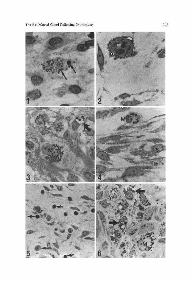
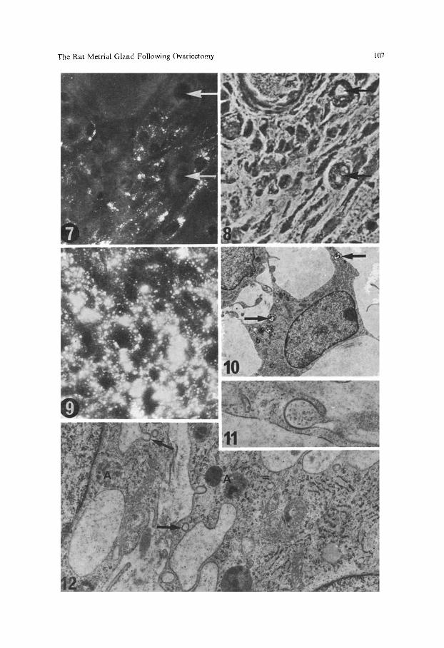
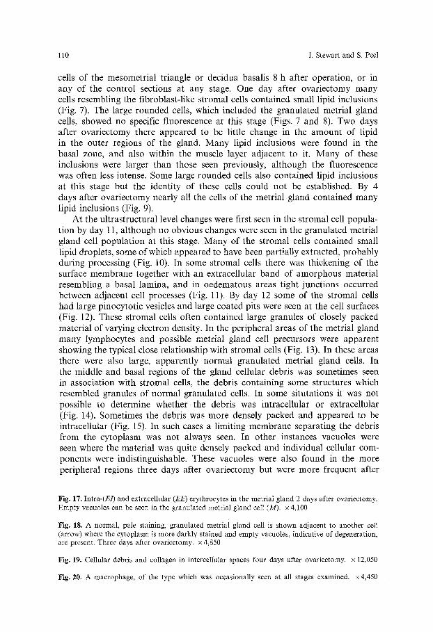
Fig. 1. A degenerate granulated metrial gland cell in the decidua basalis one day after ovariectomy. Two nuclear fragments are arrowed. Diastase, PAS and haematoxylin, x 1,200
Fig. 2. A granulated metrial gland cell in mitosis (M) in the metrial gland one day after ovariectomy. PAS and haematoxylin, x 1,400
Fig. 3. A metrial gland cell (M) showing both normal and large pale staining granules two days after ovariectomy. A vacuole (arrow) containing PAS positive inclusions can be seen in close association with a stromal cell. Diastase, PAS and haematoxylin, x 1,100
Fig. 4. Stromal cells (S) two days after ovariectomy showing both stained and unstained inclusions. The granulated metrial gland cell (M) contains a large palely stained granule. Diastase, PAS and haematoxylin, x 1,200
Fig. 5. Lymphocytes (arrows) in the peripheral region of the metrial gland two days after ovariectomy. A granulated metrial gland cell precursor can also be seen (arrowed, P). PAS and haematoxylin. x 560
Fig. 6. An area of metrial gland 5 days after ovariectomy showing rounded stromal cells with inclusions of various staining intensities (arrows). A granulated metrial gland cell (M) can also be seen. PAS and haematoxylin, x 1,100
The Rat Metrial Gland Following Ovariectomy 105
106 I. Stewart and S. Peel
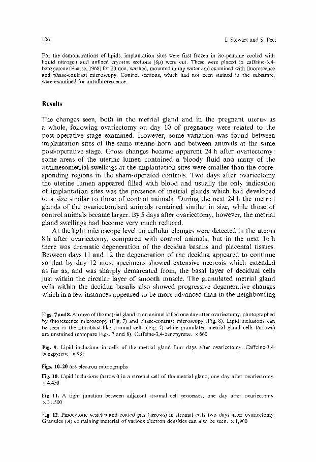
For the demonstrations of lipids, implantation sites were first frozen in iso-pentane cooled with liquid nitrogen and unfixed cryostat sections (8g) were cut. These were placed in caffeine-3,4- benzpyrene (Pearse, 1968) for 20 rain, washed, mounted in tap water and examined with fluorescence and phase-contrast microscopy. Control sections, which had not been stained in the substrate, were examined for autofluorescence.
Results
The changes seen, both in the metrial gland and in the pregnant uterus as a whole, following ovariectomy on day 10 of pregnancy were related to the post-operative stage examined. However, some variation was found between implantation sites of the same uterine horn and between animals at the same post-operative stage. Gross changes became apparent 24 h after ovariectomy: some areas of the uterine lumen contained a bloody fluid and many of the antimesometrial swellings at the implantation sites were smaller than the corre- sponding regions in the sham-operated controls. Two days after ovariectomy the uterine lumen appeared filled with blood and usually the only indication of implantation sites was the presence of metrial glands which had developed to a size similar to those of control animals. During the next 24 h the metrial glands of the ovariectomised animals remained similar in size, while those of control animals became larger. By 5 days after ovariectomy, however, the metrial gland swellings had become very much reduced.
At the light microscope level no cellular changes were detected in the uterus 8 h after ovariectomy, compared with control animals, but in the next 16 h there was dramatic degeneration of the decidua basalis and placental tissues. Between days 11 and 12 the degeneration of the decidua appeared to continue so that by day 12 most specimens showed extensive necrosis which extended as far as, and was sharply demarcated from, the basal layer of decidual cells just within the circular layer of smooth muscle. The granulated metrial gland cells within the decidua basalis also showed progressive degenerative changes which in a few instances appeared to be more advanced than in the neighbouring
Figs. 7 and 8. An area of the metrial gland in an animal killed one day after ovariectomy, photographed by fluorescence microscopy (Fig. 7) and phase-contrast microscopy (Fig. 8). Lipid inclusions can be seen in the fibroblast-like stromal cells (Fig. 7) while granulated metrial gland cells (arrows) are unstained (compare Figs. 7 and 8). Caffeine-3,4-benzpyrene. x 660
Fig. 9. Lipid inclusions in cells of the metrial gland four days after ovariectomy. Caffeine-3,4- benzpyrene, x 935
Figs. 10-20 are electron micrographs
Fig. 10. Lipid inclusions (arrows) in a stromal cell of the metrial gland, one day after ovariectomy. x 4,450
Fig. 11. A tight junction between adjacent stromal cell processes, one day after ovariectomy. x 31,500
Fig. 12. Pinocytotic vesicles and coated pits (arrows) in stromal cells two days after ovariectomy. Granules (A) containing material of various electron densities can also be seen. x 1,900
The Rat Metrial Gland Following Ovariectomy 107
108 I. Stewart and S. Peel
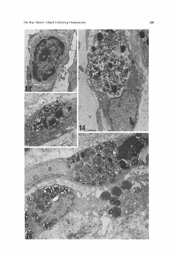
decidual cells (Fig. 1). However, at day 11, the granulated metrial gland cells within the mesometrial triangle generally appeared normal and some were found in mitosis (Fig. 2). At this stage, some of the stromal cells of the metrial gland contained small PAS positive inclusions and apparently empty circular profiles which were not seen in control tissues. Many metrial glands at this time showed oedematous changes which were usually restricted to the peripheral areas.
Two days after ovariectomy the granulated metrial gland cells showed changes which were not observed in control animals. Granulated cells, particu- larly in the basal region of the gland and within the circular muscle layer, contained relatively palely stained granules which were up to three times the diameter of the granules seen in control specimens. A few cells showed both types of granules, as well as granules which were intermediate in size and staining intensity (Fig. 3 and 4). Some PAS positive material appeared to be in vacuoles associated with stromal cells (Fig. 3). Some stromal cells, particularly in the basal zone, contained palely stained or unstained inclusions (Fig. 4) resem- bling those seen one day previously, but generally they were larger and more numerous. A relatively large number of lymphocytes and metrial gland cell precursors were apparent in the peripheral regions of many metrial glands two days after ovariectomy (Fig 5). Sometimes extravascular eosinophils were observed in the basal regions.
By three days after ovariectomy more of the granulated cells appeared to contain the large pale staining granules. Apparently disorganised granulated cells were seen with empty vacuoles in their cytoplasm. No granulated ceils were seen in mitosis at this or at later stages and the number of granulated cell precursors and lymphocytes was markedly reduced. Stromal cells throughout the gland contained large numbers of inclusions, of various sizes and staining intensities with the PAS technique and many of the stromal cells had become rounded. At four and five days after ovariectomy the number of granulated cells was reduced, and many of them showed signs of degeneration. There were very few lymphocytes or eosinophils in the intercellular spaces. The stromal cells were often large and rounded and contained many inclusions, some of which were PAS positive (Fig. 6).
Examination by fluorescence microscopy of cryostat sections stained with the caffeine-3,4-benzpyrene technique failed to demonstrate lipid either in any
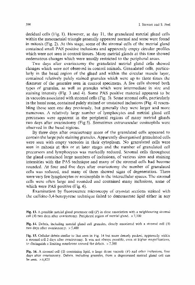
Fig. 13. A possible metrial gland precursor cell (P) in close association with a neighbouring stromal cell (53 two days after ovariectomy. Peripheral region of metrial gland, x 7,100
Fig. 14. Debris, including metrial gland cell granules, closely associated with a stromal cell (53 two days after ovariectomy, x 5,400
Fig. 15. Cellular debris similar to that seen in Fig. 14 but more densely packed, apparently within a stromal cell 2 days after ovariectomy. It was not always possible, even at higher magnifications, to distinguish a limiting membrane around the debris. • 7,300
Fig. 16. A stromal cell (53 containing lipid, a large dense vacuole (V) and other inclusions, four days after ovariectomy. Debris, including granules, from a degenerated metrial gland cell can be seen. x 6,825
The Rat Metrial Gland Following Ovariectomy 109
110 I. Stewart and S. Peel
cells of the mesometrial triangle or decidua basalis 8 h after operation, or in any of the control sections at any stage. One day after ovariectomy many cells resembling the fibroblast-like stromal cells contained small lipid inclusions (Fig. 7). The large rounded cells, which included the granulated metrial gland cells, showed no specific fluorescence at this stage (Figs. 7 and 8). Two days after ovariectomy there appeared to be little change in the amount of lipid in the outer regions of the gland. Many lipid inclusions were found in the basal zone, and also within the muscle layer adjacent to it. Many of these inclusions were larger than those seen previously, although the fluorescence was often less intense. Some large rounded cells also contained lipid inclusions at this stage but the identity of these cells could not be established. By 4 days after ovariectomy nearly all the cells of the metrial gland contained many lipid inclusions (Fig. 9).
At the ultrastructural level changes were first seen in the stromal cell popula- tion by day 11, although no obvious changes were seen in the granulated metrial gland cell population at this stage. Many of the stromal cells contained small lipid droplets, some of which appeared to have been partially extracted, probably during processing (Fig. 10). In some stromal cells there was thickening of the surface membrane together with an extracellular band of amorphous material resembling a basal lamina, and in oedematous areas tight junctions occurred between adjacent cell processes (Fig. 11). By day 12 some of the stromal cells had large pinocytotic vesicles and large coated pits were seen at the cell surfaces (Fig. 12). These stromal cells often contained large granules of closely packed material of varying electron density. In the peripheral areas of the metrial gland many lymphocytes and possible metrial gland celt precursors were apparent showing the typical close relationship with stromal cells (Fig. 13). In these areas there were also large, apparently normal granulated metrial gland cells. In the middle and basal regions of the gland cellular debris was sometimes seen in association with stromal cells, the debris containing some structures which resembled granules of normal granulated cells. In some situtations it was not possible to determine whether the debris was intracellular or extracellular (Fig. 14). Sometimes the debris was more densely packed and appeared to be intracellutar (Fig. 15). In such cases a limiting membrane separating the debris from the cytoplasm was not always seen. In other instances vacuoles were seen where the material was quite densely packed and individual cellular com- ponents were indistinguishable. These vacuoles were also found in the more peripheral regions three days after ovariectomy but were more frequent after
Fig. 17. Intra-(E/) and extracellular (EE) erythrocytes in the metrial gland 2 days after ovariectomy. Empty vacuoles can be seen in the granulated metrial gland cell (M). x 4,100
Fig. 18. A normal, pale staining, granulated metrial gland cell is shown adjacent to another cell (arrow) where the cytoplasm is more darkly stained and empty vacuoles, indicative of degeneration, are present. Three days after ovariectomy, x 4,850
Fig. 19. Cellular debris and collagen in intercellular spaces four days after ovariectomy. • 12,050
Fig. 20. A macrophage, of the type which was occasionally seen at all stages examined, x4,450
The Rat Metrial Gland Following Ovariectomy 111
112 I. Stewart and S. Peel
4 or 5 days (Fig. 16). Some of these vacuole-containing stromal cells showed the typical close association with granulated cells. In one specimen examined two days after ovariectomy, where extravasation had occurred in an area of the metrial gland, red blood cells, as well as occurring in intercellular spaces, also appeared to have been phagocytosed by stromal cells (Fig. 17). In this area some of the granulated metrial gland cells appeared to be degenerat- ing and showed large empty vacuoles in their cytoplasm (Fig. 17). Cells such as these were normally found three or more days after ovariectomy and often showed an increase in the electron density of the cytoplasm as well as a general loss in the organisation of the cellular components (Fig. 18). In some granulated metrial gland cells after ovariectomy the granules appeared larger in diameter than those seen in control animals.
Four days after ovariectomy there had been extensive necrosis of cells of the metrial gland and cellular remnants could readily be found in the intercellular spaces (Figs. 17 and 19). The stromal cells were packed with lipid inclusions as well as with granules and vesicles of various sizes, structure and electron density (Fig. 17), and many of these cells appeared rounded. At all stages exam- ined a few cells were seen more resembling a macrophage in structure than a stromal cell with phagocytic properties (Fig. 20).
Discussion
The endocrine environment after ovariectomy on day 10 of pregnancy in the rat does not allow pregnancy to proceed normally. Degenerative changes soon become apparent in the embryo and foetal placenta and are followed by similar changes in the decidua (Bulmer and Peel, 1978). In the mesometrial triangle the differentiation and proliferation of granulated metrial gland cells continues, but has been shown to be impaired. Changes were found to occur in some cells of the metrial gland after 24 h although proliferation was still continuing at this stage. Proliferation of granulated metrial gland cells was not seen after day 12. However, the presence of numerous proposed (Bulmer and Peel, 1978) metrial gland cell precursors (Fig. 5) may be related to an impairment of their differentiation in the altered endocrine environment. These changes resulted in few granulated cells being found by 5 days after ovariectomy a stage in normal pregnancy when the metrial gland cell population is well developed. The degeneration and disappearance of most of the granulated cells in the 5 days following ovariectomy showed some similarities to the normal degener- ation occurring in the metrial gland of late pregnancy (Peel and Stewart, 1978). However, the stromal cell population showed a variety of appearances, many of which were not features of normal pregnancy.
Ovariectomy during the first half of pregnancy has been reported to be followed by a rapid and dramatic degeneration of the decidua basalis (Deanesly, 1973). In this study degeneration of the decidua was also rapid and dramatic and was usually accompanied by degeneration of the granulated metrial gland cells within the decidua. Occasionally the granulated cells showed more advanced degenerative changes than surrounding decidual cells. Such degeneration, asso-
The Rat Metrial Gland Following Ovariectomy 113
ciated with nuclear fragmentation, was not seen in granulated cells in the decidua basalis or mesometrial triangle of controls or in the metrial gland after ovariec- tomy. The immediate cause of the decidual degeneration after ovariectomy has been ascribed to vascular changes (Deanesly, 1973). There may be some difference in the micro-environment between the decidua basalis and the meso- metrial triangle, possibly due to a difference in blood supply, producing a dramatic degeneration of granulated metrial gland cells in the decidua basalis whilst allowing their continued proliferation and survival in the mesometrial triangle. Ovariectomy was, however, followed by vascular changes which affected the mesometrial triangle. Oedema was observed in many specimens after oper- ation, occasionally there was extravasation, and erythrocytes as well as eosinophils were presented in intercellular spaces (Fig. 17).
The first changes in the mesometrial triangle after ovariectomy on day 10 of pregnancy occurred in the fibroblast-like stromal cells on day 11 and in gen- eral changes first became apparent in the basal regions of the gland. Lipid positive inclusions, not present in control specimens, were seen at both light and electron microscope levels. Many of the inclusions detected in the stromal cell population at day 11 and 12, particularly in peripheral regions, were not in areas where extracellular debris or cell degeneration were apparent (Figs. 10 and 12). Some inclusions (Fig. 12) may be associated with the extensive pinocy- totic activity which occurred at these stages, particularly in areas where oedema was present, and which was not seen in control animals. Other inclusions (Fig. 10) possibly represented an early interruption in the normal metabolic activity of the stromal cells. With increasing time after ovariectomy the inclusions in the stromal cells became larger and more numerous and were seen in most areas of the mesometrial triangle.
Degeneration of granulated metrial gland cells and the appearance of ex- tracellular debris occurred 2 days after ovariectomy. Some of the metrial gland cells had appearances similar to those occurring in late stages of normal pregnancy (Figs. 17 and 18). Some contained granules larger than normal which showed a variety of staining intensities with the PAS technique (Figs. 3 and 4). It is unclear whether this could represent a change in the function of these cells following ovariectomy or whether it is an early indication of the premature degeneration of the granulated cells.
Extracellular debris, apparently the result of cell lysis, was not extensive until day 14 but even then granules typical of granulated metrial gland cells (Fig. 16) were not often seen. The apparent absence of extracellular debris in the earlier stages after ovariectomy and the scarcity of extracellular granules at all stages could be accounted for by the phagocytosis of this material by the stromal cells with which they were closely associated. This would result in a stromal cell being associated with structures which could not categorically be said to be intracellular (Figs. 3 and 14), without any extracellular debris being obvious. It may be that the inclusions with the appearance of large, pleomorphic, densely stained structures (Fig. 17) represent a later stage in the stromal cell activity. It has been suggested that some stromal cells in normal pregnancy are capable of phagocytosis (Sharma and Peel, 1978) and ultrastruc- tural studies have indicated that endocytosis occurs, particularly in the later
114 I. Stewart and S. Peel
s tages o f p r e g n a n c y ( L a r k i n a n d F l i ck inge r , 1969, Peel a n d S tewar t , 1978).
A l t h o u g h s o m e cells were seen in th is s tudy w h i c h m o r e r e s e m b l e d typ ica l
m a c r o p h a g e s , these w e r e few in n u m b e r a n d they d id n o t gene ra l ly s h o w inclu-
s ions (Fig. 20) w h i c h c o u l d r ead i ly be iden t i f i ed as debr i s f r o m d e g e n e r a t e d
g r a n u l a t e d m e t r i a l g l a n d cells. T h e prec ise d i s t i nc t ion b e t w e e n a m o n o c y t e de r i ved
m a c r o p h a g e a n d a f i b r o b l a s t w h i c h is c a p a b l e o f p h a g o c y t o s i s r e m a i n s to be es tab l i shed .
Acknowledgments. We are grateful to Professor D. Bulmer for his advice and constructive criticism and to Vicki, Sue and Louise for their assistance in preparing the illustrations. This work was supported by the Wellcome Trust.
References
Bnlmer, D., Peel, S. : The demonstration of immunoglobulin in the metrial gland cells of the rat placenta. J. Reprod. Fertil. 49, 143-145 (1977)
Bulmer, D., Peel, S.: The effects on the rat uterus and placenta of ovareictomy at day 10 of pregnancy. J. Anat. 128, 185-194 (1979)
Deanesly, R. : Termination of early pregnancy in rats after ovariectomy is due to immediate collapse of the progesterone-dependent decidua. J. Reprod. Fertil. 35, 183-186 (1973)
Duval, M. : Le placenta des rongeurs. III Le placenta de la souris et du rat. J. Anat. Physiol., Paris 27, 24~96, (1891)
Larkin, L.H., Flickinger, C.J.: Ultrastructure of the metrial gland cell in the pregnant rat. Amer. J. Anat. 126, 337-354 (1969)
Pearse, A.G.E. : Histochemistry, theoretical and applied. London: Churchill 1960 Peel, S., Bulmer, D. : The effects of late ovariectomy on the proliferation and differentiation of
the uterus of the pregnant rat. J. Anat. 119, 569-578 (1975) Peel, S., Bulmer, D. : The fine structure of the rat metrial gland in relation to the origin of
the granulated cells. J. Anat. 123, 687~96 (1977) Peel, S., Stewart, I. : Ultrastructural changes in the rat metrial gland in the latter half of pregnancy.
Anat. Embryol. 155, 209-219 (1979) Sharma, R.P., Peel, S.: The uptake of proteins by the uterus and placenta of the pregnant rat.
J. Anat. 126, 410411 (1978) Stewart, I., Peel, S. : The differentiation of the decidua and the distribution of metrial gland cells
in the pregnant mouse uterus. Cell Tissue Res. 187, 167-179 (1978) Weill, P. : Glande myom6triale endocrine dans l'uterus de la rate gestante. C. r. S6anc. Soc. Biol.
82, 1433-1435 (1919)
Accepted February 5, 1979