Embed Size (px)
Citation preview
UvA-DARE is a service provided by the library of the University of Amsterdam (http://dare.uva.nl)
UvA-DARE (Digital Academic Repository)
Changes in spore chemistry and appearance with increasing maturity
Fraser, W.T.; Watson, J.S.; Sephton, M.A.; Lomax, B.H.; Harrington, G.; Gosling, W.D.; Self,S.Published in:Review of Palaeobotany and Palynology
DOI:10.1016/j.revpalbo.2013.11.001
Link to publication
Citation for published version (APA):Fraser, W. T., Watson, J. S., Sephton, M. A., Lomax, B. H., Harrington, G., Gosling, W. D., & Self, S. (2014).Changes in spore chemistry and appearance with increasing maturity. Review of Palaeobotany and Palynology,201, 41-46. https://doi.org/10.1016/j.revpalbo.2013.11.001
General rightsIt is not permitted to download or to forward/distribute the text or part of it without the consent of the author(s) and/or copyright holder(s),other than for strictly personal, individual use, unless the work is under an open content license (like Creative Commons).
Disclaimer/Complaints regulationsIf you believe that digital publication of certain material infringes any of your rights or (privacy) interests, please let the Library know, statingyour reasons. In case of a legitimate complaint, the Library will make the material inaccessible and/or remove it from the website. Please Askthe Library: https://uba.uva.nl/en/contact, or a letter to: Library of the University of Amsterdam, Secretariat, Singel 425, 1012 WP Amsterdam,The Netherlands. You will be contacted as soon as possible.
Download date: 01 Apr 2020
Review of Palaeobotany and Palynology 201 (2014) 41–46
Contents lists available at ScienceDirect
Review of Palaeobotany and Palynology
j ourna l homepage: www.e lsev ie r .com/ locate / revpa lbo
Research paper
Changes in spore chemistry and appearance with increasing maturity
Wesley T. Fraser a,⁎, Jonathan S. Watson b, Mark A. Sephton b, Barry H. Lomax c, Guy Harrington d,William D. Gosling a, Stephen Self a
a Department Environment, Earth & Ecosystems, Centre for Earth, Planetary, Space & Astronomical Research (CEPSAR), The Open University, Milton Keynes MK7 6AA, UKb Department of Earth Science & Engineering, Imperial College, London SW7 2AZ, UKc School of Biological Sciences, The University of Nottingham, Nottingham LE12 5RD, UKd School of Geography, Earth & Environmental Sciences, The University of Birmingham, Birmingham B15 2TT, UK
⁎ Corresponding author at: Geography, Faculty of HuOxford Brookes University, Gipsey Lane Campus, Oxford483863; fax: +44 1865 483937.
E-mail addresses: [email protected] (W.T. [email protected] (J.S. Watson), m.a.sephto(M.A. Sephton), [email protected] (B.H. [email protected] (G. Harrington), Willam(W.D. Gosling), [email protected] (S. Self).
0034-6667/$ – see front matter © 2013 Elsevier B.V. All rihttp://dx.doi.org/10.1016/j.revpalbo.2013.11.001
a b s t r a c t
a r t i c l e i n f oArticle history:Received 24 December 2012Received in revised form 21 August 2013Accepted 9 November 2013Available online 19 November 2013
Keywords:sporopolleninthermal maturationpalynologypalynomorphsporeheating
Sporopollenin is the primary biopolymer found in thewalls of pollen and spores; duringmaturation sporopollen-in undergoes a number of discrete chemical changes, despite maintaining identifiable morphological featureswhich can be exploited for palynological study. Here we report the results of heating experiments performedusing Lycopodium clavatum spores designed to investigate the changes that occur within sporopollenin acrossa wide range of temperatures (0–350 °C) to simulate different degrees of maturation. Changes in sporopolleninfunctionality were assessed using Fourier transform infrared (FTIR) microspectroscopy. Our analyses show thatthe chemical structure of sporopollenin remains relatively stable over awide range of simulatedmaturation con-ditions, until a threshold of 250–300 °C is reached, at which point a reorganisation of chemical structure begins.Comparison of these artificiallymatured spores with fossil material obtained from a Carboniferous-age section inthe United Kingdom shows a strong chemical resemblance, suggesting that our experimental procedure accu-rately reflects the process of maturation and provides an insight into the chemical stability of sporopollenin inthe geosphere.
© 2013 Elsevier B.V. All rights reserved.
1. Introduction
The outerwall of spores and pollen is primarily constructed from thebiopolymer sporopollenin, a chemically, physically and biologicallyinert biomacromolecule. The recalcitrant nature of sporopollenin is like-ly a key factor in the high preservation potential of palynomorphs, asobserved in the geological record. This inert chemical nature providesa wealth of evidence on key palaeobotanical events in, for example,the timing and diversification of the earliest plants (Wellman et al.,2003), vascular plants (Steemans et al., 2009), and the diversificationof early angiosperms (Lupia, 1999). Paradoxically it is the incrediblyinert character of sporopollenin that has prevented a definitive chemicalstructure from being elucidated, with the exact structure and mono-meric composition still debated. However, it may be that the structureof sporopollenin does yield to some chemical changes given sufficienttime and suitable maturation conditions.
manities and Social Sciences,OX3 0BP, UK. Tel.: +44 1865
),[email protected]),[email protected]
ghts reserved.
Recently a body of work has been produced establishing sporopol-lenin as an effective biogeochemical proxy for historical UV-B flux(Rozema et al., 1997, 1999, 2001a,b; Blokker et al., 2005; Watsonet al., 2007; Lomax et al., 2008, 2012; Willis et al., 2011; Fraser et al.,2011, 2012). To date the longest sporopollenin-based proxy spans c.5,000 years into the past (Willis et al., 2011). One potential limitationto the applicability of a sporopollenin-based proxy is its longevity inthe geological record, with increasing maturity as a confounding factor,potentially altering the chemical functionality used to construct theproxy in the above work. Thus, it is of importance to establish the geo-logical longevity of this geochemical proxy. The discovery of chemicallyunaltered, Carboniferous (Early Pennsylvanian, ~310 Ma) sporopollen-in from the cave infill deposits within Central Quarry, Kendall County,Illinois, USA (Lat. N 41.47669, Long. W 88.43788 Plotnick et al., 2009)has already been achieved (Fraser et al., 2012); however, only in isolat-ed individual specimens. The recognition of relatively unalteredpalynomorphs and a fuller understanding of the fate of primary plantbiochemistry following death and burialwould open the door for recon-struction of deep-time UV-B flux.
The underlying basis of the UV-B proxy described by Rozema et al.(1997), Watson et al. (2007), Lomax et al. (2008, 2012) and Fraseret al. (2011) is the abundance of aromatic-based moieties within thestructure of sporopollenin. However, the investigation of fossil sporesand pollen has revealed that fossilised sporopollenin appears chemical-ly very different to sporopollenin found in present day plants. Recent
42 W.T. Fraser et al. / Review of Palaeobotany and Palynology 201 (2014) 41–46
work by Steemans et al. (2010) has compared naturally occurringfossilised biopolymers from a number of sources using FTIR spectrosco-py. The spectra obtained from fossilised sporopollenin differ from thatof modern day Lycopodium spores reported in the literature (Watsonet al., 2007; Lomax et al., 2008; Zimmerman, 2010; Fraser et al., 2011).Overall, fossil sporopollenin FTIR spectra appear to be less complexwith fewer prominent peaks thanmodern counterparts. This simplifica-tion reflects a reduction in hydroxyl groups, loss of carbonyl groups, anda relative increase in aliphatic abundance (Watson et al., 2012). The dis-parity betweenmodern and fossil material has hindered the elucidationof the geological fate of sporopollenin.
Differences between modern and fossil sporopollenin could beexplained by a number of hypotheses: 1) Sporopollenin chemical com-position is fundamentally different in modern flora compared with thatof ancient flora, 2) More than one type of sporopollenin has existed inthe past, and multiple forms may continue to exist today, or 3) Post-depositional and burial processes (broadly low-grade diagenesisthrough to catagenesis)may have altered sporopollenin over geologicaltime.
Thefirst hypothesis, related to the change in sporopollenin chemistrythrough time, is regarded as unlikely because there are many examplesof ancient flora with modern-day analogues. To achieve a fundamentalchange in chemical composition through time in these plant groups itwould be expected that other changes would also be manifest, e.g. inthe form and structure of these plants. However, it is evident that withinsome groups of plants (i.e. ‘lower plants’) there have been only very lim-ited changes through time. In addition, recent work by Fraser et al.(2012) has indicated that sporopollenin within Carboniferous-age,exquisitely preserved, megaspores from cave deposits in Illinois have anear-identical sporopollenin chemical composition to modern-dayanalogues.
The second hypothesis, related to multiple forms of sporopollenin isbased on the comparison between extant and ancient sporopollenincomposition (De Leeuw et al., 2006; Vandenbroucke and Largeau,2007). However, difficulties in identifying if the chemical compositionof the fossil sporopollenin has been altered after the production ofthe pollen grains mean that the argument eventually collapses tocircularity.
The third hypothesis, that of low-grade maturity changes takingplace, seems the most plausible as this process has been previouslyrecognised in other types of organic matter (Gupta et al., 2007a,b,c).The testing of this third hypothesis is therefore the focus of the experi-ments presented in this manuscript.
Insight gained from our work presented here into the stability ofsporopollenin chemistry within the geological record could help extendthe applicability of the recently developed proxy for UV-B radiation, andcould be applied to help answer a number of questions of a similar veinto those of Watson et al. (2007) and Lomax et al. (2008). The use of ageological scale UV-B proxy could act as a gateway to understandingpast environments, where UV-B radiation varied in relation to the pre-vailing environmental conditions, such as shifts in vegetation patterns(Fraser et al., 2011), long-term surface elevation changes (Lomaxet al., 2012) or globally significant stratospheric ozone depletion events(perhaps due to catastrophic volcanic eruptions) which have beenlinked to the end Permian extinction event (Visscher et al., 2004;Beerling et al., 2007; Svensen et al., 2009).
To test the validity of the diagenesis/catagenesis sporopollenin alter-ation hypothesis (hypothesis three above) we have subjected modernsporopollenin to artificial geological maturation processes using in-creasing temperature as an analogue for increasing maturity over geo-logical time. Yule et al. (2000) attempted such an approach, butacknowledge that their experimentswere conducted under oxygenatedconditions, thus oxidation of samples during artificial maturation mayhave contributed to the observed chemical changes; geological sampleswill have been subject to elevated temperature and pressure underessentially anoxic conditions. Therefore, to model anoxic geological
conditions, such experiments should be conducted under non-oxidising conditions, i.e. in an inert gas atmosphere or under vacuum.
The work presented here reports on artificial maturation simula-tions by heating of Lycopodium clavatum spores throughout a range oftemperatures. This included fresh unheated material through to sporessubjected to temperatures of 350 °C under vacuum conditions. The pri-mary objective of this work is to explore themanner inwhich sporopol-lenin changes when subjected to elevated temperature conditionscommensurate with burial in geological environments. Palynologicalanalysis is used widely in the oil and gas industry for assessing the hy-drocarbon source potential of sedimentary rocks, and for evaluatingthermal maturity (Hopping, 1967; Staplin, 1977; Lis et al., 2005), thusan understanding of how the chemical composition in visually identifi-able palynomorphs changes will help improve maturation studies. Fur-thermore, this work will provide insight into the potential stability ofsporopollenin chemistry for use in palaeoproxy work on reconstructingultraviolet radiation in the past.
2. Materials and methods
2.1. Fossil samples
Fossil material used in the study was isolated from archive materialcollected from the Duckmanton Railway Cutting (53°13′44″N, 001°21′58″W), located 6 km East of Chesterfield, Derbyshire, UK. Specimensare late Early Pennsylvanian (~312 Ma Upper Bashkirian, equivalenttoWestphalian B). Samples were processed following standard palyno-logical techniques at the University of Sheffield (Traverse, 2007); how-ever processing was halted prior to oxidation of the spore material topreserve the primary biogeochemical signature of the fossil sporopol-lenin. Macerated residues were then picked and isolated individualmegaspores identified and collected for geochemical analysis.Palaeobotanical data presented in this study are from Tuberculatisporitesmamillarius, which have been previously recovered in situ fromsigillarian cones (e.g. Chaloner, 1967), consequently they are a memberof the Lycophyta most closely related to extant Isoetales.
2.2. Experimental sample preparation and artificial maturation
Lycopodium clavatum spores (Sigma-Aldrich, UK) were soxhletextracted for a period of 24 h in a dichloromethane:methanol mixture(97:3 v/v). Soxhlet extraction results in the removal of ‘free’ com-pounds, leaving the sporewall biopolymer for subsequent biogeochem-ical analysis (Watson et al., 2012). Clean 100 mm borosilicate tubes(6 mm o.d., 4 mm i.d.) were then loaded with ~14 mg of solvent-extracted spores and flame sealed under vacuum (~0.2 Pa) to ensureinert, non-oxidising conditions existed in the tubes. These tubes werethen placed in an oven heated to the desired temperature and held atthis temperature for a period of 48 h. Temperatures of 0, 100, 150,200, 250, 300, and 350 °C were used to simulate varying intensities ofmaturity conditions from immature to thermally mature. After heating,the spores were solvent extracted, as described inWatson et al. (2012).
2.3. Light microscopy
Artificially matured spores were placed into distilled water andmounted on coverslips, and allowed to dry. Images were photographedon a Zeiss axiotope (sic) microscope using a Canon 50D EOS.
2.4. FTIR
All geochemical investigations reported here were performed usinga Continuum IR-enabled microscope fitted with a 15× reflechromatobjective lens and nitrogen-cooled MCT-A detector operating in trans-mission mode. This was interfaced with a Thermo Nicolet Nexus (Ther-mo Fisher Scientific, USA) bench unit to providemicroscopic analysis of
43W.T. Fraser et al. / Review of Palaeobotany and Palynology 201 (2014) 41–46
spores. Analysis was conducted using a microscope aperture of100 × 100 μm at 500 scans per sample; a background spectrum wascollected and automatically subtracted from the sample spectrumafter each individual analytical run. Analyses were replicated fivetimes per sample. Sample material was dry-mounted on a NaCl IR-transparent disc. Background spectra were collected from an area ofthe disc that contained no spore sample.
Post-capture interrogation of FTIR spectra follows the method ofSteemans et al. (2010). Briefly, the method of Steemans et al. (2010)is as follows. Relative abundances of the various functional groups rep-resented within the spectra are compared across different samples byusing ratios; this alleviates any complications arising due to the propor-tional nature of FTIR analysis, where a longer pathlength of IR beamthrough a sample increases the absorbance at a particularwavenumber.In particular, the following functional group ratios are used (Table 1):aliphatic (vasCHn, 2925 cm−1)/aromatica (vC_C, 1600 cm−1), car-boxyl (vC_O, 1710 cm−1)/aromatica (vC_C, 1600 cm−1), aliphatic(vasCHn, 2925 cm−1)/carboxyl (vC_O, 1710 cm−1), aliphatic(vasCHn, 2925 cm−1)/aromaticb (vC_C, 1500 cm−1), and carboxyl(vC_O, 1700 cm−1)/aromatica (vC_C, 1600 cm−1). Here we excludeone ratio reported by Steemans et al. (2010); that of 2928/2854 cm−1.We exclude this particular ratio from our analyses because it does notprovide an estimate of aliphatic chain length, as suggested. By using ra-tios any relative shifts in abundance of the particular functional groupspresent within sporopollenin can be assessed.
3. Results and discussion
3.1. Light microscopy
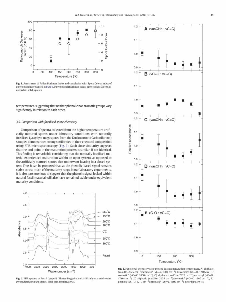
Light microscopy (Plate 1) demonstrates a typical colour shift inartificially matured Lycopodium, as would be expected from the well-defined spore colour index (SCI), with increasingly mature samplesshowing colours through yellow (SCI 3), to orange (SCI 6), and darkbrown (SCI 8) to black (SCI 9/10) at greatest maturity (Marshall,1991; Marshall and Yule, 1999). A quantitative measure of spore colourhas recently been developed by Goodhue and Clayton (2010); thePalynomorph Darkness Index (PDI%). Comparison of PDI% analysis ofpalynomorphs presented in Plate 1 with spore colour index values isshown in Fig. 1.
3.2. FTIR microspectroscopy
A strong absorbance band occurs centred at 3300 cm−1, and de-creases in intensity with increasing maturity in both modern and fossilspores (Fig. 2). A pair of peaks is found at 2925 cm−1 and 2850 cm−1,which increase in apparent intensity with increasing maturation level.At mid-rangematurity conditions a peak at 1710 cm−1 is well resolved,but appears only as a shoulder at low and high maturity levels. A peakoccurring at 1610 cm−1 increases prominence with increasing temper-atures. In the region of 1520–1510 cm−1 a sharp well defined peak canbe seen to become less pronounced with elevated maturity levels.Throughout thematurity range the peak centred at 1450 cm−1 remainsstable. Finally, in the region 1400–1000 cm−1, detail becomes increas-ingly poorly resolved with increasing maturity (Fig. 2).
Table 1Band assignment of observed absorbance bands present in FTIR spectra.
Wavenumber (cm−1) Band assignment
2925 vasCHn
2850 vsCHn
1710 vC_O1600 vC_C1500 vC_C1210 vC\O
3.3. Impact of artificial maturation on spore colour
The results from our light microscopy study confirm only minorchanges in appearance, primarily colour, under milder maturation con-ditions (0–200 °C). The strongest colour changes are seen at higherexperimental temperatures of N250 °C (Fig. 1), as is commonly seen inspore colour index analysis (Marshall, 1991). Further reassurance thatinterpretations are correct is provided by the correlation of Spore ColourIndex with the recently developed Palynomorph Darkness Index(Goodhue and Clayton, 2010); both of which correspond well to ourgeochemical data (Figs. 1, 3). By combining our geochemical data withthese colour change observations our data show that the chemical com-position of sporopollenin remains stable across a broad range of tem-peratures, relating to a wide maturity window. Functional groupbreakdown is initiated at temperatures in excess of 250 °C when a sig-nificant colour change is also observed. The broad latitude of chemicalstability under simulated maturation conditions suggests the possibilityof applying a biochemical proxy to geological material for investigatingpast variations in UV-B flux.
3.4. Impact of artificial maturation on spore chemistry
The method presented by Steemans et al. (2010) for interrogatingFTIR spectra is followed here in order to provide quantification of chem-ical changes across different samples. The broad absorbance band at3000 cm−1 is due to hydroxyl (OH) groups,most likely hydroxyl groupsthat are involved in hydrogen bonding given the broad andwide natureof this band (Fig. 3). Absorbance at 2925 cm−1 and 2850 cm−1 are bothdue to stretching vibrations of CHn groups (vasCHn, and vsCHn, respec-tively). The sharp band present at 1740 cm−1 is due to C_O bonds,most likely relating to ester linkages within the primary structure ofsporopollenin. The final bands of interest here occur in the regions1605 cm−1 and 1520–1510 cm−1; these are attributed to the presenceof aromatic ring structureswithin sporopollenin, specifically as phenolicmoieties contributing to the composition of sporopollenin. This inter-pretation is consistent with data presented previously by Rouxhetet al. (1980), Watson et al. (2007), Lomax et al. (2008), Steemanset al. (2010) and Zimmerman (2010).
Closer inspection of Fig. 3 reveals that thephenolic peak at 1510 cm−1
decreases in size, and shifts position to lowerwavenumberswith increas-ingmaturation conditions, becoming indistinct from the adjacent peak atlower wavenumbers once an experimental temperature of 300 °C isreached. In fact, a distinct change in peak character is observable from200 to 250 °C. We interpret this to indicate a change in bonding patternon the aromatic ring of the phenolic groups. The two identified phenoliccomponents present in sporopollenin, ferulic acid and para-coumaricacid share similar bonding patterns, with 1,3,4 and 1,4 substitutionaround the ring structure, respectively. We suggest that at the highestmaturation temperatures, a reorganisation around the ring structureis initiated, resulting in this shift in infrared absorbance band posi-tion. Such an interpretation is consistent with the observations ofother studies (Lis et al., 2005; Gupta et al., 2007a,b,c; Watson et al.,2012) where clear defunctionalisation and repolymerisation occursunder diagenetic conditions, resulting in an altered chemical struc-ture. However, at lower experimental temperatures (b200 °C), i.e.lower maturity, such changes do not appear to occur, with the aro-matic components remaining unaltered.
Both indicators of aliphatic to aromatic balance (i.e. 2925/1600 cm−1
and 2925/1500 cm−1) show a similar trend, with a reduction in ratiovalue to 200 °C, favouring an interpretation of apparent reduced aliphaticand/or increased aromatic contribution with increasing maturity. Com-parison of aliphatic and aromatic ratios against carbonyl groups withinester linkages suggests a slight increase in aromatic components above250 °C, however, aliphatic components remain fairly stable across thematuration range in relation to ester linkages (Fig. 3). The C–O/phenolic(1210/1600 cm−1) ratio is stable across the range of maturation
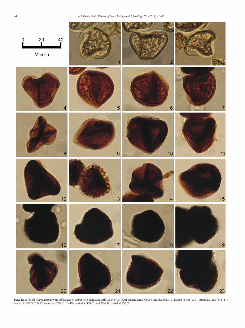
Plate 1. Spores of Lycopodium showingdifferences in colourwith increasing artificial thermalmaturation taken at×500magnification. 1–3) heated to 100 °C, 4–7) treated at 150 °C, 8–11)treated to 200 °C, 12–15) treated at 250 °C, 16–19) treated at 300 °C, and 20–23) treated at 350 °C.
44 W.T. Fraser et al. / Review of Palaeobotany and Palynology 201 (2014) 41–46
Temperature (oC)
0 50 100 150 200 250 300 350
Pal
ynom
orph
Dar
knes
s In
dex
(PD
I %)
0
20
40
60
80
100
Spo
re C
olou
r In
dex
0
2
4
6
8
10
Fig. 1. Assessment of Pollen Darkness Index and correlation with Spore Colour Index ofpalynomorphs presented in Plate 1. Palynomorph Darkness Index, open circles; Spore Col-our Index, solid squares.
0.9
1.0
1.1
1.2
1.0
1.1
1.2
A (vasCHn : vC=C)
B (vC=O : vC=C)
45W.T. Fraser et al. / Review of Palaeobotany and Palynology 201 (2014) 41–46
temperatures, suggesting that neither phenolic nor aromatic groups varysignificantly in relation to each other.
0.9
Rea
ltive
abo
sorb
ance
0.9
1.0
1.1
1.2
1.1
1.2 D (vasCHn : vC=C)
C (vasCHn : vC=O)
3.5. Comparison with fossilised spore chemistryComparison of spectra collected from the higher temperature artifi-cially matured spores under laboratory conditions with naturallyfossilised Lycophytamegaspores from the Duckmanton (Carboniferous)samples demonstrates strong similarities in their chemical compositionusing FTIR microspectroscopy (Fig. 2). Such close similarity suggeststhat the end point in the maturation process is similar, if not identical.This finding is remarkable considering that the naturally fossilised ma-terial experienced maturation within an open system, as opposed tothe artificially matured spores that underwent heating in a closed sys-tem. Thus it can be proposed that, as the phenolic-based signal remainsstable acrossmuch of thematurity range in our laboratory experiments,it is also parsimonious to suggest that the phenolic signal locked withinnatural fossil material will also have remained stable under equivalentmaturity conditions.
Wavenumber (cm-1)
5001000150020002500300035004000
Abs
orba
nce
0.0
0.5
1.0
1.5
2.0
2.5
3.0
Fossil
300ºC
350ºC
250ºC
150ºC
100ºC
0ºC
200ºC
Fig. 2. FTIR spectra of Fossil Lycopsid (Blogiga bloggsis) and artificially matured extantLycopodium clavatum spores. Black line, fossil material.
Temperature (oC)
0.9
1.0
0 100 200 300
0.9
1.0
1.1
1.2E (C-O : vC=C)
Fig. 3. Functional chemistry ratio plotted against maturation temperature. A) aliphatic(vasCHn, 2925 cm−1)/aromatica (vC_C, 1600 cm−1), B) carboxyl (vC_O, 1710 cm−1)/aromatica (vC_C, 1600 cm−1), C) aliphatic (vasCHn, 2925 cm−1)/carboxyl (vC_O,1710 cm−1), D) aliphatic (vasCHn, 2925 cm−1)/aromaticb (vC_C, 1500 cm−1), E)phenolic (vC\O, 1210 cm−1)/aromatica (vC_C, 1600 cm−1). Error bars are 1σ.
46 W.T. Fraser et al. / Review of Palaeobotany and Palynology 201 (2014) 41–46
4. Conclusions
From the evidence presented here it is shown that sporopolleninchemistry remains relatively stable across the lower half of a range ofsimulated diagenetic conditions, only beginning to alter substantially athigher temperatures, i.e. above 200 °C, approximating to greatermatura-tion. This new chemical information provides evidence of an underlyingchemical basis for the preservation of spores and pollen in the geologicalrecord. Further to this, such broad apparent chemical stability lendsweight to the notion that chemical analyses combinedwith palynologicalstudy may extend the applicability of a new palaeoclimatic proxy(Watson et al., 2007; Lomax et al., 2008, 2012) to deeper geological time.
Acknowledgements
Duckmanton Railway cutting samples used in this study were col-lected by Dr Edwin (Ted) G. Spinner during his career at the Universityof Sheffield and now form part of the University of Sheffield Palynologyarchive. This researchwas initially funded via financial support througha Natural Environment Research Council (NERC) studentship and CASEaward to W.T.F. from the Thermo Fisher Scientific, a NERC standardgrant (NER/A/S/2002/00865) and a Leverhulme Trust Early Career Fel-lowship to B.H.L. (ECF/2006/0492).
References
Beerling, D.J., Harfoot, M., Lomax, B.H., Pyle, J.A., 2007. The stability of the stratosphericozone layer during the end-Permian eruption of the Siberian Traps. Philos. Trans. R.Soc. A 365 (1856), 1843–1866.
Blokker, P., Yeloff, D., Boelen, P., Broekman, N.V.J., Rozema, J., 2005. Development of aproxy for past surface UV-B irradiation: A thermally assisted hydrolysis and methyl-ation py-GC/MS method for the analysis of pollen and spores. Analytical Chemistry77, 6026–6031.
Chaloner, W.G., 1967. Lycophyta. In: Boureau, E. (Ed.), Traité de Paléobotanique, II,Bryophyta, Psilophyta, Lycophyta. Masson, Paris, pp. 435–802.
De Leeuw, J.W., Versteegh, G.J.M., Van Bergen, P.F., 2006. Biomacromolecules of algae andplants and their fossil analogues. Plant Ecol. 182, 209–233.
Fraser, W.T., Sephton, M.A., Watson, J.S., Self, S., Lomax, B.H., James, D.I., Wellman, C.H.,Callaghan, T.V., Beerling, D.J., 2011. UV-B absorbing pigments in spores: biochemicalresponses to shade in a high-latitude birch forest and implications for sporopollenin-based proxies of past environmental change. Polar Res. 30. http://dx.doi.org/10.3402/polar.v30o0.8312.
Fraser, W.T., Scott, A.C., Forbes, A.E.S., Glasspool, I.J., Plotnick, R.E., Kenig, F., Lomax, B.H.,2012. Evolutionary stasis of sporopollenin biochemistry revealed by unaltered Car-boniferous spores. New Phytol. 196 (2), 397–401.
Goodhue, R., Clayton, G., 2010. Palynomorph Darkness Index (PDI)— a new technique forassessing thermal maturity. Palynology 34 (2), 147–156.
Gupta, N.S., Briggs, D.E.G., Collinson, M.E., Evershed, R.P., Michels, R., Pancost, R.D., 2007a.Molecular preservation of plant and insect cuticles from the Oligocene Enspel Forma-tion, Germany: evidence against derivation of aliphatic polymer from sediment. Org.Geochem. 38, 404–418.
Gupta, N.S., Briggs, D.E.G., Collinson, M.E., Evershed, R.P., Michels, R., Jack, K.S., Pancost,R.D., 2007b. Evidence for the in-situ polymerisation of labile aliphatic organiccompounds during the preservation of fossil leaves: implications for organic matterpreservation. Org. Geochem. 38, 499–522.
Gupta, N.S., Michels, R., Briggs, D.E.G., Collinson, M.E., Evershed, R.P., Pancost, R.D., 2007c.Experimental evidence for the formation of geomacromolecules from plant leaflipids. Org. Geochem. 38, 28–36.
Hopping, C.A., 1967. Palynology and the oil industry. Rev. Palaeobot. Palynol. 2 (1–4), 23–48.Lis, G.P., Mastalerz, M., Schimmelmann, A., Lewan, M.D., Stankiewicz, B.A., 2005. FTIR ab-
sorption indices for thermal maturity in comparison with vitrinite reflectance R0 intype-II kerogens from Devonian black shales. Org. Geochem. 36, 1533–1552.
Lomax, B.H., Fraser, W.T., Sephton, M.A., Callaghan, T.V., Self, S., Harfoot, M., Pyle, J.A.,Wellman, C.H., Beerling, D.J., 2008. Plant spore walls as a record of long-term changesin ultraviolet-B radiation. Nat. Geosci. 1, 592–596.
Lomax, B.H., Fraser, W.T., Harrington, G.J., Blackmore, S., Sephton, M.A., Harris, N.B.W.,2012. A novel palaeoalimetry proxy based on spore and pollen wall chemistry.Earth Planet. Sci. Lett. 353–354, 22–28.
Lupia, R., 1999. Discordant morphological disparity and taxonomic diversity during theCretaceous angiosperm radiation; North American pollen record. Paleobiology 25(1), 1–28.
Marshall, J.E.A., 1991. Quantitative spore colour. J. Geol. Soc. Lond. 148, 223–233.Marshall, J.E.A., Yule, B.L., 1999. Spore colour measurement. In: Jones, T.P., Rowe, N.P.
(Eds.), Fossil Plants and Spores: Modern Techniques. Geological Society, London,pp. 165–168.
Plotnick, R.E., Kenig, F., Scott, A.C., Glasspool, I.J., Eble, C.F., Lang, W., 2009. Pennsylvanianpaleokarst and cave fills from Northern Illinois, U.S.A.: a window into Late Carbonif-erous environments and landscapes. Palaios 24, 627–637.
Rouxhet, P.G., Robin, P.L., Nicaise, G.B., 1980. Characterisation of kerogens and their evo-lution by infrared spectroscopy. In: Durand, B. (Ed.), Kerogen. Editions Techniq, Paris,pp. 163–190.
Rozema, J., Van de Staaij, J., Björn, L.-O., Caldwell, M., 1997. UV-V as an environmental fac-tor in plant life: stress and regulation. Trends Ecol. Evol. 12, 22–28.
Rozema, J., Van de Staaij, J., Björn, L.-O., de Bakker, N., 1999. Depletion of stratosphericozone and solar UV-B radiation: evolution of landplants, UV-screens and functionsof polyphenolics. In: Rozema, J. (Ed.), Stratospheric ozone depletion: the effects of en-hanced UV-B radiation on terrestrial ecosystems. Backhuys publishers, Leiden, TheNetherlands, pp. 1–19.
Rozema, J., Broekman, R.A., Blokker, P., Meijkamp, B.M., de Bakker, N., Van de Staaij, J., VanBeem, A., Ariese, F., Kars, S.M., 2001a. UV-B absorbance and UV-B absorbing com-pounds (para-coumaric acid) in pollen and sporopollenin: the perspective to trackhistoric UV-B levels. J. Photochem. Photobiol. B Biol. 62, 108–117.
Rozema, J., Noordijk, A.J., Broekman, R.A., Van Beem, A., Meijkamp, B.M., de Bakker, N.V.J.,Van de Staaij, J.W.M., Stoetenga, M., Bohncke, S.J.P., Konert, M., Kars, S., Peat, H., Smith,R.I.L., Convey, P., 2001b. (Poly)phenolic compounds in pollen and spores of Antarcticplants as indicators of solar UV-B: a new proxy for the reconstruction of past solarUV-B? Plant Ecol. 154, 11–26.
Staplin, F.L., 1977. Thermal history from color of particulate organic matter: a review. Pal-ynology 1, 9–18.
Steemans, P., Le Hérissé, A., Melvin, J., Miller, M.A., Paris, F., Verniers, J., Wellman,C.H., 2009. Origin and radiation of the earliest vascular land plants. Science324, 353.
Steemans, P., Lepot, K., Marshall, C.P., Le Herisse, A., Javaux, E.J., 2010. FTIR characterisa-tion of the chemical composition of Silurian miospores (cryptospore and triletespores) from Gotland, Sweden. Rev. Palaeobot. Palynol. 162, 577–590.
Svensen, H., Planke, S., Polozov, A.G., Schmidbauer, N., Corfu, F., Podladchikov, Y.Y.,Jamtveit, B., 2009. Siberian gas venting and the end-Permian environmental crisis.Earth Planet. Sci. Lett. 277 (3–4), 490–500.
Traverse, A., 2007. Paleopalynology, 2nd edition. Springer, Dordrecht, Germany.Vandenbroucke, M., Largeau, C., 2007. Kerogen origin, evolution and structure. Org.
Geochem. 38, 719–833.Visscher, H., Looy, C.V., Collinson, M.E., Brinkhuis, H., Cittert, J.H.A.V.K.V., Kurschner, W.M.,
Sephton, M.A., 2004. Environmental mutagenesis during the end-Permian ecologicalcrisis. Proc. Natl. Acad. Sci. U.S.A. 101 (35), 12952–12956.
Watson, J.S., Sephton, M.A., Sephton, S.V., Beerling, D.J., Self, S., Fraser, W.T., Lomax,B.H., Gilmour, I., Wellman, C.H., 2007. Rapid determination of spore chemistryusing thermochemolysis gas chromatography–mass spectrometry and micro-Fourier transform infrared spectroscopy. Photochem. Photobiol. Sci. 6,689–694.
Watson, J.S., Fraser, W.T., Sephton, M.A., 2012. Formation of a polyalkyl macromoleculefrom the hydrolysable component within sporopollenin during heating/pyrolysis ex-periments with Lycopodium spores. J. Anal. Appl. Pyrol. 95, 138–144.
Wellman, C.H., Osterloff, P.L., Mohiuddin, U., 2003. Fragments of the earliest land plants.Nature 425, 282–285.
Willis, K.J., Feurdean, A., Birks, H.J.B., Bjune, A.E., Breman, E., Broekman, R., Grytnes, J.-A.,New, M., Singarayer, J.S., Rozema, J., 2011. Quantification of UV-B flux through timeusing UV-B-absorbing compounds contained in fossil Pinus sporopollenin. NewPhytol. 192, 553–560.
Yule, B.L., Roberts, S., Marshall, J.E.A., 2000. The thermal evolution of sporopollenin. Org.Geochem. 31, 859–870.
Zimmerman, B., 2010. Characterisation of pollen by vibrational spectroscopy. Appl.Spectrosc. 64, 1364–1373.