Embed Size (px)
Citation preview

Gen Physiol Biophys (1999), 18, 165—180 165
Changes in Passive Electric Parameters of Human Erythrocyte Membrane During Hyperthermia: Role of Spectrin Phosphorylation
I T IVANOV
Thracian University, Stara Zagora, Medical Institute, Dept of Physics and Biophysics, Stara Zagora 6000, Bulgaria
Abstrac t . In prefixed by 1 mmol/1 OSO4 human erythrocytes, the discocyte shape was preserved upon heating to temperatures which include the denaturat ion temperature of the m a m peripheral protem spectrin Nevertheless, the suspension of fixed cells displayed threshold decrease in its capacitance and resistance at the tern perature range where spectrin denaturates The same changes were established using mtact cells and their resealed ghosts For packed cells (ghosts), the capacitance and resistance decreased about 17% (31%) and 30% (19%) These da ta indicate a decrease m the be ta dispersion of erythrocyte membrane associated, according to a previous study (Ivanov 1997), with the heat denaturat ion of spectrin at 49 5°C The amplitude of the 49 5 °C decrease in beta dispersion was reversibly reduced m intact erythrocytes and white ghosts following reversible decrease in the phosphorylation of their membrane proteins It was fully eliminated in ghosts following their re-sealmg with alkaline phosphatase (0 1 mg/ml) which dephosphorylated membrane proteins These findings are discussed m relation to similar changes found in normal and tumour tissues and cells during hyperthermia
K e y words: Erythrocyte membrane — Electric properties — Beta-dispersion — Spectrm phosphorylation
In troduct ion
The electrical charges of tissues spend the energy of endogenous electric fields in
different ways the free ones move overcoming the active resistance of medium while
Correspodence to I T Ivanov, Dept Physics and Biophysics, Medical Institute, Thracian University, Armeiska Str 11, Stara Zagora 6000, Bulgaria E-mail vmiszObgcict acad bg

166 Ivanov
the bound ones accumulate potential energy of displacement. The latter dielectric polarisation produces an additional dispersed electric resistivity. Both the dielectric polarisation and related dispersed resistivity fall within the frequency domain of 0.5-10 MHz, that is the beta dispersion, which originates from the presence of biomembranes and is sensitive to changes in their structure (Bonincontro et al. 1989) . The beta dispersion is explained by the Maxwell-Vagner effect at the interface between the membrane and the surrounding highly conductive media. After hyperthermic exposure, the amplitude of beta dispersion has been found decreased in some normal and tumour tissues (McRae and Estrick 1992, 1993) and in Ehrlich tumour cell suspension (Polivoda 1985). So far, the involvement of particular cellular proteins has not been pointed out in this response of cells to hyperthermia.
The aim of this study was to investigate spectrin as a possible participant in the beta dispersion and, consequently, passive electric properties of human erythrocytes. Several authors (Ivanov and Lyutskanov 1987; Bao et al. 1992) have studied the temperature dependence of these parameters up to 50 °C without paying attention to this problem. Spectrin is a major peripheral protein attached to the inner surface of the erythrocyte membrane. In spite of its morphogenic role, the spectrin network strongly impacts the vertical distribution of lipids and the lateral movement of lipids and membrane proteins (Carraway and Carraway 1989). The interaction of spectrin with the membrane has been revealed to include electrostatic (Kahana et al. 1991) as well as hydrophobic forces (Streichman et al. 1991). The enzymatic destruction of spectrin network has induced leakness to nonelectrolytes (Klonk and Deuticke 1992). Due to the high degree of phosphorylation, the surface charge of spectrin has quite a distinctive value that strongly contributes to the attachment and the functions of the network (Heinrich et al. 1982).
Materials and Methods
Erythrocytes
Human erythrocytes were separated from freshly drawn blood or transfusion blood bank and washed in NaCl saline. White ghosts and one-step resealed ghosts were prepared as described previously (Ivanov 1993).
Chemicals
Alkaline phosphatase (EC 3.1.3.1) type I-S, 5.8 units/mg; Ca ionophore A 23187; osmium tetroxide OSO4 and glutaraldehyde, adenosine and adenine, all purchased from Sigma Chemical Co. Adenosine-5'-triphosphoric acid disodium salt was from Reanal (Hungary).

Spectrin and Erythrocyte Membrane Electric Properties 167
Measurements
The measurements of passive electric properties, capacitance and resistance, of intact erythrocytes and their ghosts was carried out at 200 kHz by RX-bridge as described elsewhere (Pauly and Schwan 1966). The voltage of the generator was fed into the RX-bridge which included a sample cavity and a series add of variable capacitance and resistance both placed in a symmetrical way. The balance of the bridge was established by oscilloscope. The sample cavity was of 0.6 ml and contained two silver electrodes. Filled by NaCl saline, its capacitance and resistance were 18 pF and 75 ohms respectively. Prior to the measurement, portions of packed erythrocytes and ghosts were exposed to the indicated temperature for 4 min, without preheating, immediately cooled down to room temperature and used.
During transient heating of cell and ghost suspension, the suspension conductivity underwent sharp changes around certain temperatures (Ivanov 1993). These changes were conveniently detected by recording the first derivative of the suspension conductivity on chart. Intact cells or resealed ghosts (inside content 150 mmol/1 NaCl) were suspended (hematocrit 0.07) in an isotonic 50 mmol/1 NaCl/sucrose medium, thus imposing an outward ion concentration gradient across the membranes. The suspension was heated at constant heating rate (2°C/min), and the output signal Us of conductometer was fed into a differentiating amplifier, the output voltage U of which was recorded (derivative conductivity thermogram).
The dependence of Us on the increase in temperature, AT, is closely linear:
Us=Uso-(l + Ks-AT),
where Ks is the temperature coefficient of Us. When T increases at a steady-rate, V, U can be expressed as
U = 1/(R • C) • dU,/dt = l/(R • C) • Uso-Ks • V,
where 1/(R • C) is the amplification coefficient. During the heating, only Ks was allowed to change, which sensed the possible change in suspension conductivity. The time differentiation was applied in order to compensate for the strong Boltz-mann dependence of suspension conductivity on the temperature. At a steady-rate heating, the thermogram appeared as a horizontal line, unless the suspension conductivity increased, this causing a sharp peak around the inducing temperature Tm. In multiple assays, carried out in identical conditions (heating rate, hematocrit, amplification and Uso) the reproducibility of Tm was within ±0.2°C.
The passive electric properties of human erythrocytes can be described by the double shell model (Asami et al. 1980). The electric properties of their membranes can be represented in terms of paralleled conductance and capacitance (Asami et al. 1980). At low frequency, the conductivity of erythrocyte suspension is greatly

168 Ivanov
influenced by the erythrocyte membrane, which acts as an insulator (Bothwell and Schwan 1956). Above 50 kHz, the membrane capacitance short-circuits the membrane and the cells behave as dispersed particles of uniform resistivity (Schwan 1965; Lassen 1977). Consequently, at low frequency the applied technique was sensitive to changes in both active resistance and dispersed resistivity of membranes, while at frequencies over 100 kHz it was assumed to be mainly sensitive to changes in dispersed resistivity.
This technique is assumed not to be sensitive to changes in the cell shape in particular. This statement is supported by the report that conductivity of a suspension containing disorderly oriented erythrocytes was not affected by reversible changes in the shape of cells (Ivanov and Popov 1993). The validity of this statement was here ascertained using cells subjected to limited fixation that was able to arrest the shape of cells but not to prevent changes in their resistance and capacitance during heating. Fixation was carried out in NaCl saline containing the indicated concentration of Os04 at room temperature for 30 min and 0.05 hematocrit. The morphological changes of intact and fixed cells during transient heating were monitored by light microscopy. The fixed cells were further used after three washings in excess NaCl saline.
Reversible A TP depletion of erythrocytes
To deplete the ATP pool of cells and to dephosphorylate the membrane proteins, cells were washed in excess NaCl saline and stored at 4°C for 3 days in the same media. Cells from a transfusion blood bank that had been stored at 4°C for 20 days were also used. To replenish ATP and to restore the normal phosphorylation of membrane proteins, these cells were incubated at 37°C for 2 h in ATP restoring medium containing 130 mmol/1 NaCl, 10 mmol/1 KC1, 10 mmol/1 phosphate buffer, pH 7.4, glucose 10 mmol/1, adenosine 5 mmol/1 and adenine 1 mmol/1, as described elsewhere (Whittam and Wiley 1967; Gazitt et all. 1976). In both cases, ghosts produced from these cells were used immediately.
Phosphorylation of erythrocyte ghosts
Phosphorylation of the membrane proteins of white ghosts was done according to Guthrow et al. (1972). White ghosts produced from cells depleted by ATP were suspended in a medium containing 1 mmol/1 MgCb, 3 mmol/1 Tris-HCl, pH 8.0 with or without 0.2 mmol/1 ATP. In parallel experiments Mg was replaced for the indicated divalent cations. After restoration of isotonicity by NaCl, the membranes were resealed at 37°C for 20 min. The membrane proteins were phosphorylated during the same time period provided ATP was present in the medium. The phosphorylated and dephosphorylated (control) membranes were separated by centrifugation and used immediately.

Spectrin and Erythrocyte Membrane Electric Properties 169
Dephosphorylation of erythrocyte ghosts
Alkaline phosphatase, present in the cytosole, rapidly dephosphorylates the membrane proteins, including spectrin. To allow this, one-step ghosts resealed in isotonic NaCl saline were hemolysed in 4°C cold media, dilution 1:15, containing 3 mmol/1 Tris-HCl, pH 7.4, 1 mmol/1 MgCi2 and 0.1 mg/ml alkaline phosphatase. After iso-tonicity restoration by NaCl, the suspension was placed at 37°C for 0.5 h to reseal the membranes and to allow the enzymatic dephosphorylation. The control membranes were treated in the same way, except the alkaline phosphatase was omitted. The membranes obtained were separated, washed in NaCl saline and studied. The difference in the mean volume of control and alkaline phosphatase treated ghosts was assessed by the microhematocrit capillaries technique.
Results and Discussion
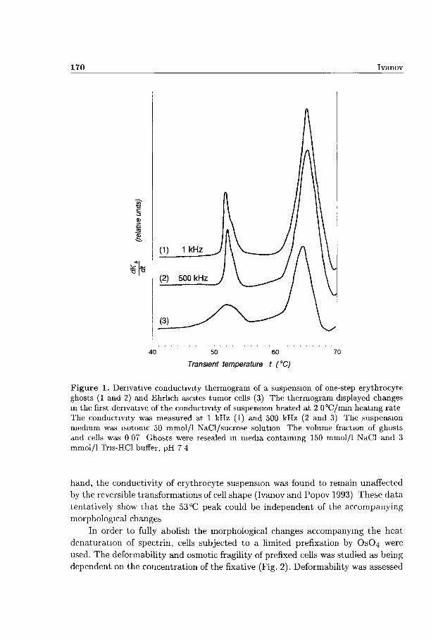
In Fig. 1, curve 1, the derivative conductivity thermogram of a suspension of resealed ghosts is shown demonstrating two threshold changes in suspension conductivity at about 53 °C and 66 °C. The corresponding thermogram of intact erythrocytes had the same shape (not shown). The 66°C peak has been shown to reveal the thermal poration of the erythrocyte membrane induced at 62 °C (Ivanov and Benov 1992). Using different modifications of the spectrin network such as low-salt extraction, denaturation by urea, and selected cleavage by trypsin, we were able to relate the 53 °C peak to the heat denaturation of spectrin (Ivanov 1997) known to take place at 49.5 °C (Brandts at al. 1977). In respect to the inducing temperatures of the related membrane events, these peaks were shifted to the right due to the heating rate applied. At lower heating rates, the top temperature of every peak was also decreased extrapolating the inducing temperature of the respective membrane event (not shown). The area under the first peak, measured by the weight method, was highly reproducible and corresponded to the amplitude of the decrease in suspension resistivity.
The denaturation of spectrin by heat has been associated with the concomitant spherization of human erythrocytes and their ghosts (Ponder 1961) and with the partial fragmentation and vesiculation of erythrocytes from many species (Coakley et al. 1979). The mechanical properties, filterability and deformability, also changed during spectrin denaturation (Rakow and Hochmuth 1975; Nash and Meiselman 1985). Consequently, the change in suspension conductivity occurring at 53 °C should be interpreted with care as it could originate from change in cell resistivity but, in general, the above mentioned shape transformations could also contribute. A number of erythrocyte modifications known to inhibit the shape transformations about 49.5X1 (Coakley et al. 1980; Herrman et al. 1985) have been recently applied without any effect on the 53 °C peak area (Ivanov 1993). On the other
170 Ivanov
50 60
Transient temperature t (°C)
70
Figure 1. Derivative conductivity thermogram of a suspension of one-step erythrocyte ghosts (1 and 2) and Ehrlich ascites tumor cells (3) The thermogram displayed changes in the first derivative of the conductivity of suspension heated at 2 0°C/mm heating rate The conductivity was measured at 1 kHz (1) and 500 kHz (2 and 3) The suspension medium was isotonic 50 mmol/1 NaCl/sucrose solution The volume fraction of ghosts and cells was 0 07 Ghosts were resealed in media containing 150 mmol/1 NaCl and 3 mmol/1 Tris-HCl buffer, pH 7 4
hand, the conductivity of erythrocyte suspension was found to remain unaffected
by the reversible transformations of cell shape (Ivanov and Popov 1993) These da ta
tentatively show tha t the 53 °C peak could be independent of the accompanying
morphological changes
In order to fully abolish the morphological changes accompanying the heat
denaturation of spectrin, cells subjected to a limited prefixation by OSO4 were
used. The deformability and osmotic fragility of prefixed cells was studied as being
dependent on the concentration of the fixative (Fig. 2). Deformability was assessed

Spectrin and Erythrocyte Membrane Electric Properties 171
1 6
u S 12 E 53 | 08
3š (? 04
-
_ _
"
r*~~~ f ^1
0— -B
/ /
-
1
1 6 ^
E
0 1 10 Concentration of Os On (mmol/1)
Figure 2. Changes in the deformability (•) and osmotic fragility ( • ) of fixed human erythrocytes The fixation was carried out at room temperature, at 0 05 hematocrit for 15 mm with the indicated concentration of Os04 Deformability was assessed by the increase in the apparent hematocrit measured by the microhematocrit capillaries technique at 16,000 rpm for 10 mm Osmotic fragility was assessed by hemolysis (optical density at 650 nm) measured 30 mm after 50 fú of the initial suspension was injected into 2 ml H2O
by the ratio of hematocrit values after and before the fixation Osmotic fragility was expressed as the percentage hemolysis obtained by mixing 50 fú of fixed cell suspension with 2 ml distilled water Hemolysis was measured as optical density at 650 nm This double check indicated tha t OSO4, in concentrations between 0 5 and 1 mmol/1, produced the least detectable fixation, which was chosen for further studies
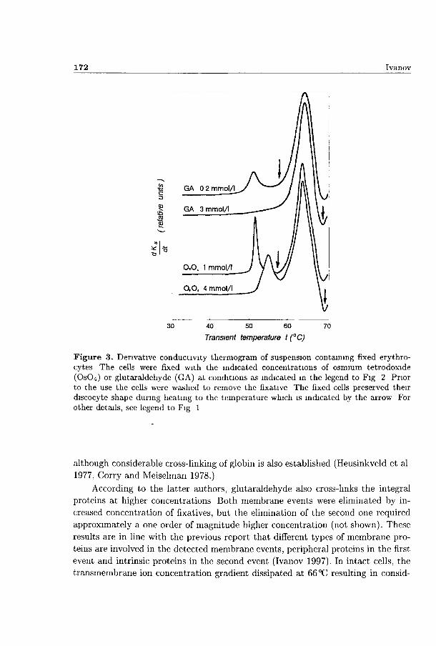
It was established microscopically tha t the biconcave shape of cells prefixed with 1 mmol/1 OSO4, was totally preserved upon heating up to 57°C, and it was also preserved in most cells heated up to 70°C (not shown) Fig 3 shows tha t the 53°C peak appeared, however, unchanged on the theimogram for the suspension of prefixed cells despite the lack of any morphological changes at this temperature Moreover, the conductivity change occurring at 53 °C was not eliminated even in cells prefixed by OSO4 at concentrations several times higher (Fig 3) although all cells remained biconcave up to 70 °C Thus, the possibility to arrest the shape of cells without affecting the changes in suspension conductivity at 53°C unambiguously demonstiated tha t this peak ansed solely from a change in the resistivity of the cells
Similar elimination of shape transformations at 53°C was also obtained m cells prefixed with 0 2 mmol/1 glutaraldehyde, howe\er, the amplitude of the 53°C peak was reduced by a factor of 2 in these cells (Fig 2) Apparently, OSO4 was more specific than glutaraldehyde in preventing the shape changes during spectrin denaturation This was possibly due to the ability of OSO4 to cross-link membrane lipids, whereas glutaraldehyde, at these concentrations, mainly cross-links spectrin
172 Ivanov
40 50 60
Transient temperature t (°C)
70
Figure 3. Derivative conductivity thermogram of suspension containing fixed erythrocytes The cells were fixed with the indicated concentrations of osmium tetrodoxide (OsCU) or glutaraldehyde (GA) at conditions as indicated in the legend to Fig 2 Prior to the use the cells were washed to remove the fixative The fixed cells preserved their discocyte shape during heating to the temperature which is indicated by the arrow For other details, see legend to Fig 1
although considerable cross-linking of globin is also established (Heusinkveld et al 1977, Corry and Meiselman 1978.)
According to the latter authors, glutaraldehyde also cross-links the integral proteins at higher concentrations Both membrane events were eliminated by increased concentration of fixatives, but the elimination of the second one required approximately a one order of magnitude higher concentration (not shown). These results are in line with the previous report that different types of membrane proteins are involved in the detected membrane events, peripheral proteins in the first event and intrinsic proteins in the second event (Ivanov 1997). In intact cells, the transmembrane ion concentration gradient dissipated at 66 °C resulting in consid-

Spectrin and Erythrocyte Membrane Electric Properties 173
erable volume changes and egress of hemoglobin (Ivanov and Benov 1992). In the prefixed cells, the same collapse of the ion concentration gradient was detected conductometrically (Fig. 3), although no volume changes and egress of hemoglobin were observed (not shown).
The decrease in homogeneous resistivity at 53 °C had the same amplitude in intact cells and their ghosts (Fig. 1). Consequently, it could involve a decrease in resistance or, alternatively, polarizability of cell membranes. The contributions of both these factors to the 53 °C conductivity change could be distinguished by comparing the thermograms obtained at low (1 kHz) and high (500 kHz) frequencies. Thus, with low and high frequency currents a uniform conductometric change in cells and ghosts was obtained at 53 °C (Fig. 1, curves 1 and 2). This finding evidenced that the decrease in membrane polarizability was the main factor involved, although a change in membrane resistance could also have happened, according to Klonk and Deuticke (1992). This conclusion is supported by the reports that, in contrast to the change at 66 °C, the 53 °C change in suspension conductivity does not practically depend on the ion concentration gradient imposed across the membranes of cells and their ghosts (Ivanov and Benov 1992; Ivanov 1993).
Had the dispersed resistivity of cells and ghosts actually changed at the denaturation temperature of spectrin, it could have been directly detected by capacity measurements. Fig. 4 shows the effect of short time heat exposure of cells and ghosts on their capacity and resistivity. The erythrocytes were prefixed by 1 mmol/1 OSO4 in order to eliminate shape transformations. The exposure to the temperature of spectrin denaturation induced irreversible decrease in both capacity and resistivity of the prefixed cells (Fig. AA), intact cells (not shown) and their ghosts (Fig. AB). The concomitant diminutions in capacity and resistivity had similar magnitudes, moreover, the relative change in capacity of ghosts was twice that in their resistivity. These results indicate that during spectrin denaturation, the capacity of cells and ghosts decreased resulting in an increase in the inside electric field which could account for the related decrease in homogeneous resistivity. Therefore, the 53 °C decrease in the suspension resistivity could be taken as a measure of the related decrease in the beta-dispersion of cells. Since the change in cell resistivity was better detectable and conveniently recorded in comparison to the corresponding change in cell capacity, the first variable was only foccused on in the following experiments.
In contrast to the lipid bilayer, the spectrin network itself could not function as a friction barrier for the electric current. Spectrin is, however, a heavily phosphorylated fibrilar protein with extremely high linear density of charges producing strong potential barrier under the membrane (Heinrich et al. 1982). The electrostatic barrier of this network can be expected to cause accumulation of dipolar charges on the opposite ends of the cytoplasm thus contributing to the beta-dispersion of cells and ghosts. According, to Coakley and Deeley (1980), long time incubation of cells at 46 °C does not impact on the degree of spectrin phosphorylation whereas their

174 Ivanov
•c ej
<u o c-to
8í
o c-<11
s-</>
800
750
700
650
600
550
500
*~- " ^ - — * ,
-
-
"
A
\
800
750
700
650
600
bbO
500
.Q.
< • >
C
TO
c-o to u: bj-J
CO
25 30 35 40 45 50 55
Temperature - t(°C)
"5ŕ P S H) n c m to to S i.-o (O Dl O to J3
CO
750
700
650
600
550
500
*~~
-
~~~-u~
^
B 750
700
650
600
550
500
25 30 35 40 45 50 55
Temperature t (°C )
u? ,a.
o c o TO & O C o c tu
8-co
Figure 4. Temperature dependence of capacitance (•) and resistance (A) of fixed erythrocytes (A) and ghosts prepared from intact cells (B) The suspensions were maximally packed, incubated at the indicated temperature for 4 mm and rapidly cooled down The passive electric properties were measured at room temperature with RX-bridge at 200 kHz The erythrocytes were prefixed by 1 mmol/1 Os04 to abolish the morphological transformations associated with the thermal denaturation of spectrin at 49 5 °C The ghosts were resealed in medium containing 1 mmol/1 Mg and 30 mmol/1 phosphate buffer, pH 7 4
short-time exposure to 49 5 °C is accompanied by a rapid and complete elimination of spectrin phosphorylation Thus, the dephosphorylation of spectrin that accom-

Spectrin and Erythrocyte Membrane Electric Properties 175
Table 1. Amplitude of the spectnn-related decrease in beta dispersion of human erythrocyte membrane as affected by the phosphorylation of its proteins
Ghost preparation
1 Ghosts prepared from ATP-depleted cells The cells were metabohcally depleted by incubation at 4°C in NaCl saline for 3 days
2 Ghosts prepared from metabohcally restored cells The cells were first ATP depleted (as above) and then restored by incubation m ATP restoring medium
3 Ghosts prepared from fresh cells pre-treated with 1 mmol/1 NaN3 and subjected to metabolic starvation at 4°C in NaCl saline for 5 days
Area under the 53 CC peak (decrease in ghost resistivity) % difference from fresh cells
15
95
90
4 Three-step ghosts with dephosphorylated membrane less than 5 proteins (control ghosts) They were prepared from ATP depleted cells from 20 day old blood bank and finally resealed in 150 mmol/1 NaCl, 1 mmol/1 Mg and 3mmol/l Tris-HCl, pH 8 0
5 Three-step ghosts with rephosphorylated membrane 150 proteins They were prepared and resealed as the control ones (as under 4) except for the reseahng medium contained 0 3 mmol/1 ATP in addition
It was measured as the area under the 53 °C peak on the derivative conductivity thermogram of erythrocyte ghosts, and expressed in respect to the same area in fresh erythrocytes Different levels of membrane protein phosphorylation were obtained in various ghost preparations as indicated bellow Each result represents the mean value of at least three experiments
panied its thermal denaturation could be involved in the 53 °C decrease in beta dispersion of cells and ghosts.
According to these considerations, the amplitude of 53°C decrease in beta
dispersion of cells and ghosts could be influenced by the initial surface charge
of spectrin network, especially tha t due to phosphate groups Results relating the
amplitude of 53 °C peak to the phosphorylation of membrane proteins are displayed
in Table 1 The phosphorylation of membrane proteins was reversibly changed by
changing the ATP pool m cells, since, according to Gazitt et al. (1976), both
176 Ivanov
these variables are interrelated. During the metabolic depletion of cells, the area of the 53 °C peak progressively diminished in cells and ghosts prepared from them (Table 1). After the renewal of ATP pool in cells, the area of the 53 °C peak restored most of its value in comparison to that in fresh cells. In cells pre-treated with 0.5 mmol/1 NaN3, an inhibitor of most ATP-ases (Daggett et al. 1985), the metabolic starvation of cells did not significantly affect the 53 °C peak (Table 1), possibly due to the conservation of ATP level and, consequently, spectrin phosphorylation.
Similar results were obtained using three-step ghosts prepared from ATP-depleted cells in which the 53°C peak was absent. These membranes were finally resealed in the presence of both ATP and Mg to allow the transfer of phosphate from ATP to the membrane proteins (Laris and Letchworth 1976). The presence of Mg - ATP in these conditions allowed rephosphorylation of membrane proteins (Guthrow et al. 1972), predominantly spectrin (Johnson et al. 1982). The rephosphorylation reaction resulted in the reappearance of the 53 °C conductivity peak with an amplitude exceeding that in intact cells (Table 1). The 53 °C peak did not reappeare if the ghosts were resealed without ATP and/or Mg. Apart from Mg other divalent ions (Co, Ba and Mn to a lesser extent) also helped the restoration of the 53°C peak in concentrations up to 2 mmol/1 (not shown). In contrast to these cations, Ca fully inhibited the 53°C peak in concentrations higher than 0.1 mmol/1 (not shown). 0.5 mmol/1 Ca eliminated the 53°C peak also in one-step ghosts, prepared from fresh cells (not shown) and in intact fresh cells treated with 0.2 mmol/1 Ca and the Ca - ionophore A 23187 (not shown). These findings are m accordance with the results of Guthrow et al. (1972) that Mg, Co, Mn and also Ba to a lesser extent, support the phosphorylation of membrane proteins in ghosts, while Ca inhibits it.
Using hypotonic test it was found that 2 mmol/1 Ba and Mn, resealed within one-step ghosts, arrested their shape, possibly through cross-linking of their membrane proteins. In these ghosts, the shape of the 53°C peak actually changed, however, its area was reduced insignificantly (not shown). This also indicates that the 53 °C threshold decrease in suspension resistivity reflected a change in the structure of the membranes, rather that in the shape of the cells.
The dependence of the 53 °C peak amplitude on the degree of spectrin phosphorylation was also demonstrated using ghosts prepared from fresh cells the proteins of which were dephosphorylated by the application of alkaline phosphatase. The mean volume of ghosts subjected to enzymatic dephosphorylation was reduced by about 30% as compared to the volume of untreated (control) ghosts. This could be explained by the subsequent shrinkage of spectrin network and membranes due to the reduction of the negative charges and mutual repulsion of spectrin filaments (Kozlov and Markin 1986). As expected, the 53 °C peak was totally inhibited in membranes resealed with alkaline phosphatase whereas it was normal in the control ghosts (Fig. 5, curves 1 and 2).

Spectrin and Erythrocyte Membrane Electric Properties 177
40 50 60
Transient temperature t (°C)
70
Figure 5. Derivative conductivity thermogram of ghost suspension after enzymatic dephosphorylation of membrane proteins Two-step ghosts were resealed in medium containing 150 mmol/1 NaCl, 1 mmol/1 Mg, 3 mmol/1 Tris-HCl buffer, pH 7 4 with (curve 2) or without (curve 1) 0 1 mg/ml type I-S alkaline phosphatase The ghosts were resealed at 37 °C for 0 5 h allowing the dephosphorylation of membrane proteins by alkaline phosphatase For other details, see the legend to Fig 1
The unfolding of spectrin by heat should induce, apart of its dephosphorylation, an impairment of the permeability barrier. Both these effects could impact on suspension resistivity: the dephosphorylation as a dispersive component and the barrier insult as a change in active resistance. In ghosts resealed with alkaline phosphatase the dispersive change in resistivity should be eliminated allowing the detection of the barrier insult alone. In such ghosts, the 53°C peak was, however, not detectable (Fig. 5, the curve 2) as compared to the control ones (Fig 5, the curve 1). This finding supported the conclusion mentioned above that the dispersive component of cellular resistivity strongly prevailed over the active one in changing

178 Ivanov
the suspension resistivity during spectrin denaturation This conclusion is in line with the previous report (Ivanov and Benov 1992) that thermohemolysis is induced as colloid-osmotic lysis due to barrier damage associated with the 62 °C event in the membranes Although the 62 °C event was more distant on the temperature axis, it apparently exercised a greater impact on the permeability barrier compared to the 49 5 °C event
As in human erythrocytes, a similar threshold decrease m resistivity has been registered in heated erythrocytes of mammals and birds in which spectrin is also present (Ivanov 1993) Due to similarity in structure, the latter effect could also involve spectrm-related decrease in beta-dispersion It was induced between 53 55 °C in mammalian and 57-59X1 in bird cells that well correlated to the known 3 to 4°C higher body temperature in birds compared to mammals This data possi bly indicate an increased thermostability of spectrin in bird compared to that in mammal cells In man and other mammals, the temperature of spectrm denatura tion is only about 13 °C above their body temperature (Brandts et al 1977) and the same temperature difference is also apparent for avian erythrocytes Probably, the increased thermostability of spectrin in birds reflects the necessity to avoid its thermal unfolding during the life-span of the erythrocytes in blood circulation
Although in smaller amounts, spectnn-hke proteins are also found in other animal cells including muscle cells and the Ehrhch ascites tumoi cells (Carraway and Carraway 1989) In hyperthermic conditions, the denaturation of these proteins should induce a similar decrease in beta-dispersion of tissues and isolated cells as that observed by many investigators (Pohvoda 1985, McRae and Estrick 1992, 1993) A threshold decrease in dispersed resistivity was also detected in heated Ehnch ascites tumor cells (Fig 1, curve 3) It was similar in amplitude and inducing temperature to that found in human erythrocytes which possibly indicates the in volvement of a spectnn-hke protein in it Therefore, the decrease in the membrane related dielectric polarisation appears to be a common element in the response of animal cells to hyperthermic challenge
According to Foster and Schwan (1989), plasma membiane shields the interior of the cell, including the cell nucleus, the contractile apparatus etc , from incident electric fields with frequencies below the beta-dispersion The results presented indicate that spectrin, spectnn-hke proteins, and other membrane proteins as well could significantly contribute to that shielding Furthermore, the shielding effect of membrane proteins could be regulated through changing their phosphorylation status, which might have physiological implications
References
Asami K , Hanai T Koizumi N (1980) Dielectric approach to suspensions of ellipsoidal particles covered with a shell in particular reference to biological cells Jpn J Appl Physiol 19, 359—365

Spectrin and Erythrocyte Membrane Electric Properties 179
Bao J Z , Davis C C , Schmucler R E (1992) Frequency domain impedance measurements of erythrocytes Constant phase angle impedance characteristics and a phase transition Biophys J 61, 1427—1434
Bonmcontro A , Cametti C , Sportelly L (1989) Electrical parameters of erythrocyte membranes deduced from radiowave conductivity measurements J Membrane Science 41, 345—352
Bothwell T P , Schwan H P (1956) Electrical properties of the plasma membrane of erythrocytes at low frequencies Nature 178, 265—266
Brandts J F , Enckson L , Lysko K , Schwartz A T , Taverna R D (1977) Calorimetnc studies of the structural transitions of the human erythrocyte membrane The involvement of spectrin in the A transition Biochemistry USA 16, 3450—3454
Carraway K L , Carraway C A C (1989) Membrane-cytoskeleton interactions in animal cells Biochim Biophys Acta 988, 147—171
Coakley W T , Bater A J , Crum L A , Deeley J O (1979) Morphological changes, haemolysis and microvesiculation of heated human erythrocytes J Therm Biol 4, 85—95
Coakley W T , Deeley J O (1980) Effects of ionic strength, serum protein and surface charge on membrane movements and vesicle production m heated erythrocytes Biochim Biophys Acta 602, 355—375
Corry W D , Meiselman H J (1978) Modification of erythrocyte membrane physio-chemical properties by millimolar concentrations of glutaraldehyde Blood Cells 4, 465—480
Daggett S G , Tomaszek T A , Schuster S M (1985) Interaction of azide with beef heart mitochondrial ATPase Arch Biochem Biophys 236, 815—824
Foster K R , Schwan H P (1989) Dielectric properties of tissues and biological materials Cnt Rev Biomed Eng 17, 25—104
Gazitt Y , Ohad I , Loyter A (1976) Phosphorylation and dephosphorylation of membrane proteins as a possible mechanism for structural rearrangements of membrane components Biochim Biophys Acta 436, 1—14
Guthrow C E , Allen J E , Rasmussen H (1972) Phosphorylation of endogenous membrane protein by an endogenous, membrane-associated cyclic adenosine 3',5'-mono-phosphate-dependent protein kinase m human erythrocyte ghosts J Biol Chem 247, 8145—8153
Hemrich R , Gaestel M , Glaser R (1982) The electric potential profile across the erythrocyte membrane J Theor Biol 96, 211—231
Herrman A , Leutzsh P , Lassman G , Donath E (1985) Spectroscopic characterization and vesicle formation of heated human erythrocytes and the influence of the antiviral agent amantidme Biochim Biophys Acta 812, 277—285
Heusmkveld R S , Goldstein D A , Weed R I , Lacelle P L (1977) Effect of protein modification on erythrocyte membrane mechanical properties, Blood Cells 3, 175— 182
Ivanovi T (1993) Investigation of the barrier function disturbance m erythrocyte membranes during transient heating Biol Membrány 10, 160—169 (m Russian)
Ivanovi T (1997) Involvement of erythrocyte membrane proteins m temperature-induced disturbances of the permeability barrier Membr Cell Biol 11 (1), 45—56
Ivanov I T , Benov L C (1992) Thermohaemolysis of human erythrocytes m isotonic NaCl/sucrose media during transient heating J Therm Biol 17, 381—389

180 Ivanov
Ivanov I T , Lyutskanov V G (1987) Thermotropic behaviour of mtact human erythrocyte membranes revealed by differential scanning conductometry Mol Cryst Liq Cryst 152, 327—322
Ivanov I T , Popov B K (1993) Do changes m cell shape affect suspension conductivity7
Gen Physiol Biophys 12, 311—315 Johnson R M , McGowan M W , Morse P D , Dzandu J K (1982) Proteolytic analysis
of the topological arrangement of red cell phosphoproteins Biochemistry USA 21, 3599—3604
Kahana E , Streichman S , Silver B L (1991) The role of electrostatic forces in the interaction between the membrane and cytoskeleton of human erythrocytes Biochim Biophys Acta 1066, 1—5
Klonk S , Deuticke B (1992) Involvement of cytoskeletal proteins in the barrier function of the human erythrocyte membrane II Formation of membrane leaks in ghost membranes after limited proteolysis of skeletal proteins by trypsin Biochim Biophys Acta 1106, 137—142
Kozlov M M , Markm V S (1986) A model for skeleton of a red blood cell description of electrical and mechanical properties Biol Membrány 3, 404—421 (in Russian)
Larís Ph C , Letchworth P E (1976) Characteristics of an adenosine triphosphatase in erythrocyte membranes, stimulated by 2,4-dimtrophenol J Cell Physiol 69, 143—150
Lassen U V (1977) Electrical potential and conductance of the red cell membrane In Membrane Transport m Red Cells (Eds J Ellory and V Lew), pp 137—174, Academic Press, New York
McRae D A , Estrick M A (1992) The dielectric parameters of excised EMT-6 tumours and their changes during hyperthermia Phys Med Biol 37, 2045—2058
McRae D A , Estrick M A (1993) Changes in electrical impedance of skeletal muscle measured during hyperthermia Int J Hyperther 9, 247—261
Nash G B , Meiselman H J (1985) Alteration of red cell membrane viscoelasticity by heat treatment effect on cell deformability and suspension viscosity Biorheology 22, 73—74
Pauly H , Schwan H P (1966) Dielectric properties and ion mobility in erythrocytes Biophys J 6, 621—639
Pohvoda B I (1985) Relationship between K-dependent cell swelling and the state of cellular membranes Biofizika 29, 685—686 (in Russian)
Ponder E (1961) Various types of ghosts derived from human red cells heat, fragmentation and phase optics studies J Exp Biol 29, 605—619
Rakow A L , Hochmuth R M (1975) Thermal transition in the human erythrocyte membrane effect on elasticity Biorheology 12, 1—3
Schwan H P (1965) Determination of biological impedance In Physical Technique in Biological Research (Ed W L Nastur), pp 323—407, Academic Press, New York
Streichman S , Kahana E , Silver B L (1991) EPR study of the hydrophobic interaction of spectrin with fatty acids Biochim Biophys Acta 1066, 9—13
Whittam R , Wiley J S (1967) Potassium transport and nucleoside metabolism m human red cells J Physiol (London) 191, 663—652
Final version accepted June 15, 1999