Embed Size (px)
Citation preview

CASE REPORT Open Access
Changes in oncogenic protein levels inperi-implant oral malignancy: a case reportMi Hyun Seo1 , Hoon Myoung1 , Jong Ho Lee1 , Soung Min Kim1* and Suk Keun Lee2*
Abstract
Background: Oral squamous cell carcinoma (OSCC) constitutes a group of tumors that exhibit heterogeneousbiology, histopathology, and clinical behaviors.
Case presentation: A 73-year-old male had a whitish leukoplakia-like lesion around inflamed peri-implant area(#42, #43, and #44), and this lesion had transformed to OSCC within 3 years. He underwent mass resection, selectiveneck dissection, and reconstructive surgery. To detect any carcinogenesis progression, we examined the removedtumor tissue as well as the patient’s preoperative and postoperative sera to identify causative oncogenic proteinsusing immunoprecipitation high-performance liquid chromatography (IP-HPLC).
Conclusions: The protein expression levels of p53, E-cadherin, β-catenin, MMP-10, HER2, NRAS, Met, HER2, and ERbwere significantly lower in the serum collected on postoperative day 10 than in the preoperative serum, and ifthese proteins are consistently not elevated in the serum 3 months after surgery compared with the preoperativeserum, these proteins can be potential oncogenic proteins. However, we also found that the serum extracted3 months after the operation had elevated levels of oncogenic proteins compared with that of the preoperativeand 10-day postoperative serum indicating the possibility of tumor recurrence. At postoperative follow-up period,ipsilateral neck metastasis and second primary lesion were found and additional surgery was performed to thepatient. IP-HPLC using the patient’s serum shows the possibility of oncogenic protein detection. However, follow-upIP-HPLC data is needed to find out patient-specific prognostic factors.
Keywords: Oral squamous cell carcinoma (OSCC), Immunoprecipitation high-performance liquid chromatography(IP-HPLC), Oncogenic protein, Peri-implant oral malignancy (PIOM)
BackgroundSquamous cell carcinoma (SCC) is the most seriousmalignant tumor that frequently invades adjacent orofa-cial structures and spreads to cervical lymph nodes.Clinical and pathological behaviors of oral squamous cellcarcinoma (OSCC) are highly variable in terms of oralulceration, bone destruction, infiltrative growth, andtumor metastasis [1–3]. Treatment modalities can be de-cided based on clinical staging, pathologic diagnosis,general status of patient, or anatomic region of tumorpresence. Clinical staging can be determined during theinitial work-up, including a clinical exam, radiological
exam with dental panorama, ultrasonography, computedtomography (CT), magnetic resonance imaging (MRI),and positron emission tomography-computed tomog-raphy (PET-CT). One of the primary treatments forOSCC is radical excision with or without adjuvantchemoradiotherapy, which has proven to be effective forlocoregional control. Nevertheless, biomarkers involvedin tumor recurrence and prognosis have not been identi-fied for OSCC [4, 5]. Clinically, detection of tumormarkers in the serum is easy and non-invasive. Squa-mous cell carcinoma antigen (SCC-Ag), cytokeratin, arewell-known tumor-related markers [6]. We assumed thatprotein markers produced by the tumor can be reducedby tumor ablative surgery. The serum of cancer patientwas used for protein analysis.Immunoprecipitation high-performance liquid chro-
matography (IP-HPLC) is a type of protein detectionmethod that is based on real antigen-antibody reaction
© The Author(s). 2019 Open Access This article is distributed under the terms of the Creative Commons Attribution 4.0International License (http://creativecommons.org/licenses/by/4.0/), which permits unrestricted use, distribution, andreproduction in any medium, provided you give appropriate credit to the original author(s) and the source, provide a link tothe Creative Commons license, and indicate if changes were made.
* Correspondence: [email protected]; [email protected];[email protected] of Oral and Maxillofacial Surgery, Dental Research Institute,School of Dentistry, Seoul National University, 101 Daehak-ro, Jongno-gu,Seoul 110-768, South Korea2Department of Oral Pathology, College of Dentistry, Gangneung-WonjuNational University, 7, Jukheon-gil, Gangneung-si, Gangwon-do, South Korea
Maxillofacial Plastic andReconstructive Surgery
Seo et al. Maxillofacial Plastic and Reconstructive Surgery (2019) 41:46 https://doi.org/10.1186/s40902-019-0235-z

in a phosphate-buffered saline (PBS) solution, followedby purification using protein A/G-conjugated agarosebeads. Although its procedures are simple and easy toapply to most biological samples, IP-HPLC can yield aminimum error range by using micro-beads instead ofsmall wells to mimic the enzyme-linked immunosorbentassay (ELISA) [7, 8]. The patient’s serum was collectedbefore his operation, 10 days after the operation, and3 months after the operation. Each sample was analyzedvia IP-HPLC, which has been improved in terms of dataaccuracy and statistical analysis.
Case presentationA 73-year-old male patient was referred from his generaldentist for further evaluation of whitish lesion on theattached gingiva and associated peri-implantitis. A pano-ramic view shows generalized alveolar bone loss andcalculi deposition in the peri-implant region (#42, #43,#44), and the right mandibular anterior and premolararea showed peri-implant crestal bone loss. Laboratoryfindings were within normal limits. Other severaloncogenic protein elevation situations, including tobaccoand/or alcohol use, no nutritional deficiencies, nofindings of ionizing radiation exposure, no immunodefi-ciency or immunosuppressant, and other removal pros-thesis irritations, were all excluded. A whitish lesion wasexcised, and the specimen was sent to an oral patholo-gist. The contaminated implant surface was treated witha laser. The pathologic diagnosis was confirmed as oralcandidiasis. The patient underwent laser treatment threetimes to treat the peri-implantitis lesion. One year later,his implant of #42, #43, and #44 area were removed dueto peri-implantitis at the local clinic. From the referral
letter of local clinic, the implant system was internal fric-tional connection type having a SLA surface, which wasinstalled for more than 10 years.About 3 years later after the laser operation, a bulging
mass was identified on the lingual side of #43 and #44area. An incisional biopsy was performed and wasdiagnosed as a SCC (Fig. 1). Further work-up was per-formed including lab, chest X-ray, ECG, MRI, contrastCT, PET-CT, and neck sonography. The patient wasdiagnosed with cT4aN2cM0 stage IVA according to theTNM staging system proposed by the American JointCommittee on Cancer (AJCC, 2018). He was immedi-ately scheduled for an operation that included selectiveneck dissection, mass resection with marginal mandibu-lectomy, and reconstruction with a radial forearm freeflap. The final pathologic report was well-differentiatedsquamous cell carcinoma, with a 1.5 × 2.0-cm size oftumor, no metastasis to any of the 27 regional lymphnodes, and clear surgical resection margin. Vascular andperineural invasions were not observed; thus, he wasdiagnosed as pT1N0M0, stage I. Especially, rather thanon the interface between the implant and the bone,tumor cells occurred on the surface of the mucosal softtissue first and penetrated deeply along the implantthreads. Neither adjuvant radiotherapy nor chemother-apy was not administered to the patient.A metastatic lymph node was found at the right
ipsilateral level IV on enhanced CT taken 4 monthspostoperatively. Selective neck dissection, including rightlevel IV, was performed, and a newly developed suspi-cious for malignant lesion was found on the rightmaxilla. The patient’s maxilla lesion was confirmed forSCC by incisional biopsy; therefore, he underwent an
Fig. 1 Clinical photos of the patient. a Initial visit; b peri-implantitis treatment with laser; c after 3-times laser treatment; d 5 months after initialvisit; e 3 years after initial visit, the lesion was confirmed OSCC by incisional biopsy; f preoperative view show bulging tumor mass to lingual side
Seo et al. Maxillofacial Plastic and Reconstructive Surgery (2019) 41:46 Page 2 of 9

additional surgery 13 days after second selective neckdissection (Fig. 2). The final pathologic report on themaxillary lesion was well-differentiated SCC with a 2.0 ×1.4-cm size of tumor; depth of invasion was 0.7 cm.Involvement of underlying bone was present. Surgicalresection margins were clear.The surgically removed specimens were fixed in 10%
neutral buffered formalin, processed following a routineprotocol, and serial micro-sections with different anti-sera were also prepared for immunohistochemical stain-ing. All data files of the patient were selected from thefiles of the Department of Oral and MaxillofacialSurgery, Seoul National University Dental Hospitalunder the approval of the institutional review board ofSeoul National University (S-D20170026).
IP-HPLC analysis for the protein extract obtained fromthe serum of patientsThe patient’s blood was collected preoperatively, 10 dayspostoperatively, and 3 months postoperatively. Afterprecipitation at room temperature, the samples werecentrifuged at 4000 rpm for 20min. Only the supernantswere collected and mixed with lysis buffer and used forIP-HPLC. We applied 100 μg of each protein extract tothe immunoprecipitation procedure using a protein A/Gagarose column (Amicogen Co., Korea). The protein A/G agarose columns were separately pre-incubated with1 μg of each of the 20 different antisera, including p53,muc1, muc4, TGF-β1, survivin, Wnt1, E-cadherin, β-catenin, matrix metalloproteinase (MMP)-3, MMP-10,TNFα, HER1, HER2, PAI-1, NRAS, KRAS, CEA, Met,FASL, and ERb. Briefly, the protein samples were mixedwith 5 ml of binding buffer (150 mM NaCl, 10 mM TrispH 7.4, 1 mM EDTA, 1 mM EGTA, 0.2 mM sodiumvanadate, 0.2 mM PMSF, and 0.5% NP-40) and incu-bated in the protein A/G agarose columns at 10 °C for 1h. The columns were placed on a rotating stirrer during
the incubation. After washing each column with asufficient amount of PBS solution (pH 7.3, 137 mMNaCl, 2.7 mM KCl, 43 mM Na2HPO4-7H2O, and 1.4mM KH2PO4), the target protein was eluted with 150 μlof IgG elution buffer (Pierce Co., USA). The immuno-precipitated proteins were analyzed by HPLC (1100series®, Agilent, USA) using a reverse-phase column andmicro-analytical detector system (SG Hightech Co.,Korea), operated with a 0.15-M NaCl and 20% aceto-nitrile solution at 0.4 mL/min for 30 min, and analyzedvia UV spectroscopy at 280 nm. In the IP-HPLC results,the sample protein peak areas obtained from the HPLCanalysis in the negative control were used to eliminatethe antibody peak area (mAU*s) [7–9]. To comparepreoperative and postoperative serum protein, theprotein peak area values were proportionally normalizedby the α-tubulin value and plotted as a bar.
IP-HPLC analysis of extracted tumor proteinProtein extracts were prepared from tumor tissue,100 μg each protein extract to the immunoprecipitationprocedure using a protein A/G agarose column. Theprotein A/G agarose columns were individually pre-incubated with 1 μg of each of the 9 different antisera,including TNFα, NRAS, HER2, Met, E-cadherin, p53,survivin, TGF-β1, and NFκB. Briefly, the protein sampleswere mixed with 5 ml of binding buffer (150 mM NaCl,10 mM Tris pH 7.4, 1 mM EDTA, 1 mM EGTA, 0.2 mMsodium vanadate, 0.2 mM PMSF, and 0.5% NP-40) andincubated in the protein A/G agarose columns at 10 °Cfor 1 h. To compare the normal gingiva and SCC tissueprotein, the protein peak area values were proportionallynormalized by the α-tubulin value and plotted as a bar.
Laboratory analysisRoutine laboratory results were collected, includingcomplete blood cell counts (CBC) with differential
Fig. 2 Clinical signs showed the recurrence of tumor. a Newly developed maxillary peri-implant oral malignancy lesion, b CT findings suspectedof metastatic lymph node
Seo et al. Maxillofacial Plastic and Reconstructive Surgery (2019) 41:46 Page 3 of 9

count, C-reactive protein (CRP) levels, and erythrocytesedimentation rate (ESR). A modest elevation in theplasma CRP in the range observed in apparently healthyindividuals is a strong predictor of future vascular events[10]. Chen et al. [11] reported the presence of elevatedpreoperative serum CRP level (> 5.0 mg/L) is an inde-pendent prognostic indicator of oral cancer. The resultsfrom the blood sample tests were compared from thefirst visit, preoperatively, postoperatively, and at the timeof recurrence.
IP-HPLC analysis from the serum of patientsThe IP-HPLC analyses revealed that p53, E-cadherin, β-catenin, MMP-10, HER2, NRAS, Met, and ERb haddecreased at postoperative day 10. Other protein
markers related to oncogenic signaling were increased atpostoperative day 10. This suggests that oncogenicproteins, such as p53, E-cadherin, β-catenin, MMP-10,HER2, NRAS, Met, and ERb were released from theprimary tumor; therefore, serum levels of these onco-genic proteins decreased after tumor-ablative surgery(Fig. 3). In 3 months postoperatively using the patient’sserum, all oncogenic protein markers were elevated andtumor recurrence or metastasis was suspected (Fig. 4).
IP-HPLC analysis from extracted tumor proteinIP-HPLC analyses revealed that NRAS, Met, p53, andNFκB were overexpressed in the SCC tissue in the com-parison of oncogenic protein levels between normal gin-giva and SCC tissue (Fig. 5).
Fig. 3 An upper bar graph and a lower line graph comparing oncogenic protein expression profiles between preoperative and postoperative day10 serum
Seo et al. Maxillofacial Plastic and Reconstructive Surgery (2019) 41:46 Page 4 of 9

Laboratory findingsInflammatory markers such as white blood cell count(WBC), absolute neutrophil count (ANC), ESR, and CRPwere elevated immediately postoperatively. CRP was notconducted preoperatively, and so, that data cannot becompared to other time points; however, CRP levels hadnot changed significantly before and after the secondoperation (right level IV selective neck dissection,Additional file 1: Figure S1). Notably, ESR counts wereelevated in the preoperative period, as determined whena malignant lesion was confirmed by incisional biopsy
and compared with a sample taking at the first visit.That indicates that some inflammatory reactions canaffect the potential of malignant transformation.Perioperative changes in the RBCs, hemoglobin,
hematocrit showed that the levels were decreased during thepost-surgery, but tended to recover over time (Add-itional file 2: Figure S2). The proportion of segmented neu-trophils increased immediately after surgery, which arecharacteristic cells of acute inflammatory reactions, becausethey moved to surgical wound immediately after trauma inseveral minutes. The graphed curves of lymphocyte,
Fig. 4 An upper bar graph and a lower line graph comparing oncogenic protein expression profiles between preoperative, postoperative 10 days,and postoperative 3 months after first ablation surgery
Seo et al. Maxillofacial Plastic and Reconstructive Surgery (2019) 41:46 Page 5 of 9

monocyte, eosinophil, and basophil levels showed oppositetrends from the segmented neutrophil levels (Add-itional file 3: Figure S3).
DiscussionThe peri-implant leukoplakia lesion and peri-implantitistransformed to SCC over several years in this patient;oral cancers may be preceded by potentially malignantdisorders, recognizable mucosal diseases such as local-ized leukoplakia or erythroplakia, or other wide-spreadconditions, all of which harbor a considerably increasedrisk for SCC [12].Peri-implantitis is a common long-term complication
of dental implant. Clinical characteristics of peri-implantitis include swelling, erythema, and often suppur-ation. Marginal bone loss and pocket formation aretypical. The etiology is multifactorial and not fullyunderstood, with bacterial factors, surgical factors, im-plant surface characteristics, prosthetic design, andgenetic predisposition suggested as contributing factors.A review of several small case series included analyses ofbiopsy material from peri-implantitis, reported hyperpla-sia, and ulceration of pocket epithelium and presence ofa mixed population of inflammatory cells [13]. A studyof 117 biopsies from peri-implantitis cases reported thatnearly 50% of cases did not exhibit simple inflammatorychanges. Instead, other potentially aggressive lesions,such as pyogenic granuloma, giant cell granuloma, orActinomyces-related inflammation were diagnosed [14].A literature review of retrospective analyses of casesfrom 2000 to 2016 revealed 47 reported cases of oralmalignancy involving dental implants. Peri-implant ma-lignancies were primary tumors in 29 cases (61.7%), re-current or second primary tumors in 11 cases (23.4%),and metastasis from distant tumors in 4 cases (8.5%).
Recognized risk factors for oral cancer included poten-tially malignant conditions (erythroplakia, leukoplakia,oral lichen planus, or proliferative verrucous leukoplakia)in 21 cases (44.7%). There is very little evidence thatlinks dental implant and cancer. Regarding orthopedicstainless-steel plates, an annual prevalence increase of0.12% per year for osteosarcoma has been reported andis suggested to be a result of corrosion caused by surfaceimperfections and chronic inflammation [15]. No suchdata exist regarding titanium dental implants. Only asingle in vitro experiment found that exposure to titan-ium particles yielded a dose-responsive induction ofchromosomal instability in human fibroblasts, similar tothat induced by heavy metal and low-dose radiation ex-posure. There are conflicting results in the literature,with no clear evidence that corrosion material (as op-posed to titanium oxide nanoparticles) can play a role incarcinogenesis [16, 17]. However, peri-implant malig-nancy is not as rare as currently reported and may beresponsible for 1.5% of oral malignancy cases [18].Numerous candidate biomarkers have been evaluated in
blood, tumor, and saliva specimens, which yields hopeselucidating disease pathways and improved prognosis pre-diction. Several classes of molecules of particular interesthave been evaluated, including MMPs, interleukins,proangiogenic and antiangiogenic factors, growth factors,and tumor factors [19]. In this study, we evaluated theoncogenic proteins using preoperative and postoperativeserum and tumor tissue via IP-HPLC. Serum sampleswere collected preoperatively, 10 days postoperatively (be-fore recurrence detection), and 3 months postoperatively(between the first tumor ablation surgery and the seconddissection of metastatic lymph node). p53, E-cadherin, β-catenin, MMP-10, HER2, NRAS, Met, and ERb were lowerin the serum collected on the postoperative 10 days.
Fig. 5 A line graph (a) and a bar graph (b) comparing oncogenic protein expression profiles between normal gingiva and SCC tissue from thefirst ablation surgical specimen
Seo et al. Maxillofacial Plastic and Reconstructive Surgery (2019) 41:46 Page 6 of 9
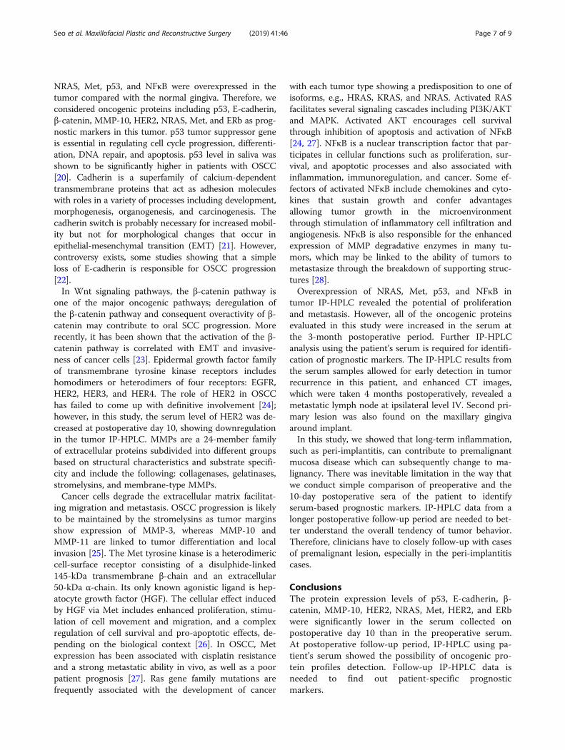
NRAS, Met, p53, and NFκB were overexpressed in thetumor compared with the normal gingiva. Therefore, weconsidered oncogenic proteins including p53, E-cadherin,β-catenin, MMP-10, HER2, NRAS, Met, and ERb as prog-nostic markers in this tumor. p53 tumor suppressor geneis essential in regulating cell cycle progression, differenti-ation, DNA repair, and apoptosis. p53 level in saliva wasshown to be significantly higher in patients with OSCC[20]. Cadherin is a superfamily of calcium-dependenttransmembrane proteins that act as adhesion moleculeswith roles in a variety of processes including development,morphogenesis, organogenesis, and carcinogenesis. Thecadherin switch is probably necessary for increased mobil-ity but not for morphological changes that occur inepithelial-mesenchymal transition (EMT) [21]. However,controversy exists, some studies showing that a simpleloss of E-cadherin is responsible for OSCC progression[22].In Wnt signaling pathways, the β-catenin pathway is
one of the major oncogenic pathways; deregulation ofthe β-catenin pathway and consequent overactivity of β-catenin may contribute to oral SCC progression. Morerecently, it has been shown that the activation of the β-catenin pathway is correlated with EMT and invasive-ness of cancer cells [23]. Epidermal growth factor familyof transmembrane tyrosine kinase receptors includeshomodimers or heterodimers of four receptors: EGFR,HER2, HER3, and HER4. The role of HER2 in OSCChas failed to come up with definitive involvement [24];however, in this study, the serum level of HER2 was de-creased at postoperative day 10, showing downregulationin the tumor IP-HPLC. MMPs are a 24-member familyof extracellular proteins subdivided into different groupsbased on structural characteristics and substrate specifi-city and include the following: collagenases, gelatinases,stromelysins, and membrane-type MMPs.Cancer cells degrade the extracellular matrix facilitat-
ing migration and metastasis. OSCC progression is likelyto be maintained by the stromelysins as tumor marginsshow expression of MMP-3, whereas MMP-10 andMMP-11 are linked to tumor differentiation and localinvasion [25]. The Met tyrosine kinase is a heterodimericcell-surface receptor consisting of a disulphide-linked145-kDa transmembrane β-chain and an extracellular50-kDa α-chain. Its only known agonistic ligand is hep-atocyte growth factor (HGF). The cellular effect inducedby HGF via Met includes enhanced proliferation, stimu-lation of cell movement and migration, and a complexregulation of cell survival and pro-apoptotic effects, de-pending on the biological context [26]. In OSCC, Metexpression has been associated with cisplatin resistanceand a strong metastatic ability in vivo, as well as a poorpatient prognosis [27]. Ras gene family mutations arefrequently associated with the development of cancer
with each tumor type showing a predisposition to one ofisoforms, e.g., HRAS, KRAS, and NRAS. Activated RASfacilitates several signaling cascades including PI3K/AKTand MAPK. Activated AKT encourages cell survivalthrough inhibition of apoptosis and activation of NFκB[24, 27]. NFκB is a nuclear transcription factor that par-ticipates in cellular functions such as proliferation, sur-vival, and apoptotic processes and also associated withinflammation, immunoregulation, and cancer. Some ef-fectors of activated NFκB include chemokines and cyto-kines that sustain growth and confer advantagesallowing tumor growth in the microenvironmentthrough stimulation of inflammatory cell infiltration andangiogenesis. NFκB is also responsible for the enhancedexpression of MMP degradative enzymes in many tu-mors, which may be linked to the ability of tumors tometastasize through the breakdown of supporting struc-tures [28].Overexpression of NRAS, Met, p53, and NFκB in
tumor IP-HPLC revealed the potential of proliferationand metastasis. However, all of the oncogenic proteinsevaluated in this study were increased in the serum atthe 3-month postoperative period. Further IP-HPLCanalysis using the patient’s serum is required for identifi-cation of prognostic markers. The IP-HPLC results fromthe serum samples allowed for early detection in tumorrecurrence in this patient, and enhanced CT images,which were taken 4 months postoperatively, revealed ametastatic lymph node at ipsilateral level IV. Second pri-mary lesion was also found on the maxillary gingivaaround implant.In this study, we showed that long-term inflammation,
such as peri-implantitis, can contribute to premalignantmucosa disease which can subsequently change to ma-lignancy. There was inevitable limitation in the way thatwe conduct simple comparison of preoperative and the10-day postoperative sera of the patient to identifyserum-based prognostic markers. IP-HPLC data from alonger postoperative follow-up period are needed to bet-ter understand the overall tendency of tumor behavior.Therefore, clinicians have to closely follow-up with casesof premalignant lesion, especially in the peri-implantitiscases.
ConclusionsThe protein expression levels of p53, E-cadherin, β-catenin, MMP-10, HER2, NRAS, Met, HER2, and ERbwere significantly lower in the serum collected onpostoperative day 10 than in the preoperative serum.At postoperative follow-up period, IP-HPLC using pa-tient’s serum showed the possibility of oncogenic pro-tein profiles detection. Follow-up IP-HPLC data isneeded to find out patient-specific prognosticmarkers.
Seo et al. Maxillofacial Plastic and Reconstructive Surgery (2019) 41:46 Page 7 of 9

Supplementary informationSupplementary information accompanies this paper at https://doi.org/10.1186/s40902-019-0235-z.
Additional file 1: Figure S1. Changes in the serum markers which canbe used for evaluation of degree of inflammation showing WBC (A), ANC(B), ESR (C), and hs-CRP (D). IP-HPLC 1 indicated the timing of the serumcollected for IP-HPLC analysis at postoperative 10 days. IP-HPLC 2 indi-cated the timing of the serum collected for IP-HPLC analysis at postoper-ative 3 months. The result of IP-HPLC 2 described the recurrence oftumor, and after enhanced CT taking, the selective neck dissection wasperformed to remove of metastatic lymph node.
Additional file 2: Figure S2. Changes of component of completeblood cell counts showing RBC (A), Hb (B), Hct (C), and PLT (D).
Additional file 3: Figure S3. Changes of differential count of CBCshowing segmental neutrophil (A), lymphocyte (B), monocyte (C),eosinophil (D), and basophil (E).
AbbreviationsANC: Absolute neutrophil count; CBC: Complete blood cell counts; CRP: C-reactive protein; CT: Computed tomography; ELISA: Enzyme-linkedimmunosorbent assay; EMT: Epithelial-mesenchymal transition;ESR: Erythrocyte sedimentation rate; HGF: Hepatocyte growth factor; IP-HPLC: Immunoprecipitation high-performance liquid chromatography;MMPs: Matrix metalloproteinases; MRI: Magnetic resonance imaging;OSCC: Oral squamous cell carcinoma; PBS: Phosphate-buffered saline; PET-CT: Positron emission tomography-computed tomography; PIOM: Peri-implant oral malignancy; WBC: White blood cell count
AcknowledgementsThis study was supported by a grant of the Korean Health Technology R&DProject, Ministry of Health & Welfare, Republic of Korea (HI15C0689), and bythe National Research Foundation of Korea (2017R1D1A1B04029339).
Authors’ contributionsAll authors read and approved the final manuscript. MH read and wrote theentire manuscript, HM revised and corrected the manuscript, JH and SMprepared the patient data and prepared for journal submission, and SKdesigned the pathologic data.
FundingThere is no funding related to this article.
Availability of data and materialsData sharing is not applicable to this article as no data sets were generatedor analyzed during the current study.
Ethics approval and consent to participateThe study protocol and access to patient records were approved by theInstitutional Review Board of Seoul National University (S-D20170026).
Consent for publicationWritten informed consent was obtained from the patient for publication ofthis case report and accompanying images.
Competing interestsThe authors declare that they have no competing interests.
Received: 9 July 2019 Accepted: 16 October 2019
References1. Shibuya Y, Hasegawa T, Akashi M et al (2013) Oral squamous cell
carcinoma with multiple neck metastases--cases with more than tenpathologically positive lymph nodes in the unilateral side. J OralMaxillofac Surg 71:793–797
2. Suslu N, Hosal AS, Aslan T et al (2013) Carcinoma of the oral tongue: a caseseries analysis of prognostic factors and surgical outcomes. J Oral MaxillofacSurg 71:1283–1290
3. Grimm M (2012) Prognostic value of clinicopathological parameters andoutcome in 484 patients with oral squamous cell carcinoma: microvascularinvasion (V+) is an independent prognostic factor for OSCC. Clin TranslOncol 14:870–880
4. Liao CT, Wang HM, Ng SH et al (2006) Good tumor control andsurvivals of squamous cell carcinoma of buccal mucosa treated withradical surgery with or without neck dissection in Taiwan. Oral Oncol42:800–809
5. Chen IH, Liao CT, Wang HM et al (2014) Using SCC antigen and CRP levelsas prognostic biomarkers in recurrent oral cavity squamous cell carcinoma.PLoS One 9:e103265
6. DE Paz D, Young CK, Chien HT et al (2019) Prognostic roles of SCC antigen,CRP and CYFRA 21-2 in oral cavity squamous cell carcinoma. Anticancer Res39:2025–2033
7. Kim YS (2015) Protein expression changes induced by cisplatin in anoral cancer cell line as determined by immunoprecipitation-based highperformance liquid chromatography. Kor J Oral Maxillofac Pathol 39:567–582
8. Kim YS, Lee SK (2015) IP-HPLC analysis of human salivary protein complexes.Kor J Oral Maxillofac Pathol 39:615–622
9. Kim SM, Jeong D, Kim MK et al (2017) Two different protein expressionprofiles of oral squamous cell carcinoma analyzed byimmunoprecipitation high-performance liquid chromatography. World JSurg Oncol 15:151
10. Hashimoto H, Kitagawa K, Hougaku H et al (2011) C-reactive protein is anindependent predictor of the rate of increase in early carotidatherosclerosis. Circulation 104:63–67
11. Chen HH, Chen IH, Liao CT et al (2011) Preoperative circulating C-reactive protein levels predict pathological aggressiveness in oralsquamous cell carcinoma: a retrospective clinical study. Clin Otolaryngol36:147–153
12. van der Waal I (2009) Potentially malignant disorders of the oral andoropharyngeal mucosa; terminology, classification and present concepts ofmanagement. Oral Oncol 45:317–323
13. Berglundh T, Zitzmann NU, Donati M (2011) Are peri-implantitislesions different from periodontitis lesions? J Clin Periodontol 38:188–202
14. Kaplan I, Hirshberg A, Shlomi B et al (2015) The importance ofhistopathological diagnosis in the management of lesions presenting asperi-implantitis. Clin Implant Dent Relat Res 17:e126–e133
15. Boudrieau RJ, McCarthy RJ, Sisson RD Jr (2005) Sarcoma of the proximalportion of the tibia in a dog 5.5 years after tibial plateau levelingosteotomy. J Am Vet Med Assoc 227:1613–1617
16. Coen N, Kadhim MA, Wright EG et al (2003) Particulate debris from atitanium metal prosthesis induces genomic instability in primary humanfibroblast cells. Br J Cancer 88:548–552
17. Matsumoto M, Filho HN, Ferrari R et al (2014) Genotoxicity ofendosseous implants using two cellular lineages in vitro. J OralImplantol 40:25–29
18. Kaplan I, Zeevi I, Rosenfeld E et al (2017) Clinicopathological evaluation ofmalignancy adjacent to dental implants. Oral Surg Oral Med Oral PatholOral Radiol 123:103–112
19. Lessing AAD, Joseph AM, Lindgren BR et al (2017) Association of oralcavity and oropharyngeal cancer biomarkers in surgical drain fluidwith patient outcomes. JAMA Otolaryngol Head Neck Surg 143:670–678
20. Agha-Hosseini F, Mirzaii-Dizgah I, Miri-Zarandi NS (2015) Unstimulatedsalivary p53 in patients with oral lichen planus and squamous cellcarcinoma. Acta Med Iran 53:439–443
21. Maeda M, Johnson KR, Wheelock MJ (2005) Cadherin switching: essential forbehavioral but not morphological changes during and epithelium-to-mesenchyme transition. J Cell Sci 118:873–887
22. Ukpo DC, Thorstad WL, Zhang Q et al (2012) Lack of association of cadherinexpression and histopathologic type, metastasis, or patient outcome inoropharyngeal squamous cell carcinoma: a tissue microarray study. HeadNeck Pathol 6:38–47
23. Iwai S, Yonekawa A, Harada C et al (2010) Involvement of the Wnt-β-cateninpathway in invasion and migration of oral squamous carcinoma cells. Int JOncol 37:1095–1103
24. Sinevici N, O’Sullivan J (2016) Oral cancer: deregulated molecular eventsand their use as biomarkers. Oral Oncol 16:12–18
Seo et al. Maxillofacial Plastic and Reconstructive Surgery (2019) 41:46 Page 8 of 9
25. Brusevold IJ, Søland TM, Khuu C et al (2010) Nuclear and cytoplasmicexpression of Met in oral squamous cell carcinoma and in an organotypicoral cancer model. Eur J Oral Sci 118:342–349
26. Cho YA, Kim EK, Heo SJ et al (2016) Alteration status and prognosticvalue of MET in head and neck squamous cell carcinoma. J Cancer 7:2197–2206
27. Murugan AK, Munirajan AK, Tsuchida N (2012) Ras oncogenes in oral cancer:the past 20 years. Oral Oncol 48:383–392
28. Chen Z, Yan B, van Waves C (2008) The role of the NF-kappa Btranscriptome and proteome as biomarkers in human head and necksquamous cell carcinomas. Biomark Med 2:409–426
Publisher’s NoteSpringer Nature remains neutral with regard to jurisdictional claims inpublished maps and institutional affiliations.
Seo et al. Maxillofacial Plastic and Reconstructive Surgery (2019) 41:46 Page 9 of 9