Embed Size (px)
Citation preview
RESEARCH Open Access
Changes in HSP gene and protein expression innatural scrapie with brain damageCarmen Serrano1, Rosa Bolea2, Jaber Lyahyai1, Hicham Filali2, Luis Varona3, Ane Marcos-Carcavilla4, Cristina Acín2,Jorge H Calvo5, Magdalena Serrano4, Juan J Badiola2, Pilar Zaragoza1, Inmaculada Martín-Burriel1*
Abstract
Heat shock proteins (Hsp) perform cytoprotective functions such as apoptosis regulation and inflammatoryresponse control. These proteins can also be secreted to the extracellular medium, acting as inflammatorymediators, and their chaperone activity permits correct folding of proteins and avoids the aggregation ofanomalous isoforms. Several studies have proposed the implication of Hsp in prion diseases. We analysed the geneexpression and protein distribution of different members of the Hsp27, Hsp70, and Hsp90 families in the centralnervous system of sheep naturally infected with scrapie. Different expression profiles were observed in the areasanalysed. Whereas changes in transcript levels were not observed in the cerebellum or medulla oblongata, asignificant decrease in HSP27 and HSP90 was detected in the prefrontal cortex. In contrast, HSP73 was over-expressed in diencephalons of scrapie animals. Western blotting did not reveal significant differences in Hsp90 andHsp70 protein expression between scrapie and control animals. Expression rates identified by real-time RT-PCR andwestern blotting were compared with the extent of classical scrapie lesions using stepwise regression. Changes inHsp gene and protein expression were associated with prion protein deposition, gliosis and spongiosis rather thanwith apoptosis. Finally, immunohistochemistry revealed intense Hsp70 and Hsp90 immunolabelling in Purkinje cellsof scrapie sheep. In contrast, controls displayed little or no staining in these cells. The observed differences in geneexpression and protein distribution suggest that the heat shock proteins analysed play a role in the natural form ofthe disease.
IntroductionTransmissible spongiform encephalopathies (TSE) arefatal neurodegenerative diseases that include scrapie insheep, bovine spongiform encephalopathy in cattle, andseveral human neuropathies such as Creutzfeldt-Jakobdisease or fatal familial insomnia. These diseases arecharacterised by the accumulation in the central nervoussystem of an abnormally folded version (PrPSc) of a nor-mal cellular protein, PrPC. The related neurologicallesions include spongiform changes in grey matter,intraneuronal vacuoles in particular nuclei of the brainstem, gliosis, and neuronal degeneration [1-5]. Althoughvarious mechanisms have been proposed to explain neu-ronal death in prion diseases, apoptosis and autophagyare the types of cell death considered most likely to beinvolved [6].
Our group has been investigating the molecularmechanisms underlying neuronal apoptosis in ovinenaturally infected with scrapie. Despite the induction ofBax in scrapie brains, neurons suffering this type of Pro-grammed Cell Death (PCD) were not observed in nat-ural scrapie, suggesting either an extremely low numberof cells undergoing apoptosis or the existence of neuro-protective mechanisms [7,8]. Expression profiles ofother genes involved in different apoptotic pathwaysindicate that both pro and anti-apoptotic mechanismsare activated in prion-infected tissues [9]. Nevertheless,other factors could be involved in the regulation of celldeath in natural scrapie.It has been shown that over-expression of an indivi-
dual heat shock protein (Hsp) can provide a protectiveeffect against damaging stimuli in the same manner ascan a mild Hsp-inducing stress [10,11]. For example,culture neurons derived from the central and peripheralnervous systems as well as different neuronal cell lines
* Correspondence: [email protected] de Genética Bioquímica (LAGENBIO), Facultad de Veterinaria,Universidad de Zaragoza, Miguel Servet 177, 50013 Zaragoza, SpainFull list of author information is available at the end of the article
Serrano et al. Veterinary Research 2011, 42:13http://www.veterinaryresearch.org/content/42/1/13 VETERINARY RESEARCH
© 2011 Serrano et al; licensee BioMed Central Ltd. This is an Open Access article distributed under the terms of the Creative CommonsAttribution License (http://creativecommons.org/licenses/by/2.0), which permits unrestricted use, distribution, and reproduction inany medium, provided the original work is properly cited.
are protected against thermal or ischemic stress byover-expression of Hsp [12,13].Hsp27, Hsp70, and Hsp90 (each named according to
its mass in kilodaltons) are three of the best-characterised Hsp families. The anti-apoptotic role ofthese Hsp is mediated by different mechanisms. Hsp27inhibits the activation of caspase-3, induced after cyto-chrome-c release from mitochondria and after the acti-vation of death receptors such as Fas [14,15]. The othertwo chaperones (Hsp70 and Hsp90) have also beenreported to target the mitochondrial pathway at differ-ent stages. Hsp90 binds to Apaf-1 and prevents its inter-action with cytochrome-c [16], and Hsp70 preventscytochrome-c/Apaf-1 complex formation by interactingwith caspase-9 [17,18]. Hsp70 also antagonizes thecaspase-independent mitochondrial pathway by bind-ing directly to AIF [19] and inhibiting its nucleartranslocation [20].Detailed studies of individual Hsp also suggest that
they play a role in ensuring the correct protein foldingof other proteins within the cells, acting as so-called“molecular chaperones” [21]. Most neurodegenerativediseases are associated with events of protein misfolding.The accumulation of misfolded protein aggregates canoverwhelm the ubiquitin-proteasomal system, inducingapoptosis and increasing neuronal vulnerability to subse-quent insults [22]. Due to their anti-apoptotic and cha-perone roles, Hsp are being considered for therapeuticuse in neurodegenerative diseases such as Alzheimer,disease, Parkinson disease and stroke (reviewed in [23]).In addition to the functions mentioned, potential invol-vement of Hsp in the conversion of prion protein to itsprotease-resistant form and propagation of the infec-tious protein is being investigated (reviewed by [24]).Analysis of the expression patterns of genes and pro-
teins is a good tool to identify the possible implicationof these proteins in TSE. Comparison of gene expressionprofiles with the distribution of scrapie related lesions inthe brain could reveal the role of different factors in theneuropathology of prion diseases. A recent report fromour group identified the association between activationof the mitochondrial apoptosis pathway and early stagesof PrPSc deposition, whereas induction of the extrinsicpathway was related to reactive gliosis and neuronal loss[9]. During prion infection, gene and protein expression,principally of Hsp70 family members, is increased in dif-ferent prion disease models [25-29].At present, little is known about the expression and
distribution of Hsp in a model of natural infection suchas naturally scrapie-infected sheep. Only recently,HSP90AA1 has been analysed as a possible candidategene involved in scrapie susceptibility in sheep [30],probably by modulating the incubation period of thisdisease [31]. We present here the first gene expression
analysis of four members of the HSP27 (HSPB1), HSP70(the constitutive form HSP72/HSPA1A and the inducibleHSP73/HSPA8), and HSP90 (the inducible form,HSP90AA1) gene families, their protein expression pro-files, and their distribution in the central nervous sys-tems of sheep naturally infected with clinical-stagescrapie. The association between gene and proteinexpression data and scrapie histopathological lesions hasalso been investigated. These heat shock proteins havebeen chosen for their known roles as chaperones andapoptosis modulators, as well as their possible functionsin prion diseases.
Materials and methodsAnimals and sample collectionThirteen females of the Rasa Aragonesa sheep breedwere included in a recent study that focused on the ana-lysis of the molecular mechanism of apoptosis in scrapie[9]. The same RNA and tissue samples were used in thepresent work. Briefly, scrapie was diagnosed in vivo ineight of these sheep [32] and confirmed after animalswere sacrificed [33]. When these animals were sacri-ficed, all exhibited clinical signs of terminal-stage scra-pie. The five control animals were selected from adifferent flock of the same breed, in which no scrapiecases have been reported to date. All animals analysedwere 3 to 6 years old and displayed the ARQ/ARQ gen-otype for the PRNP gene, which is the most susceptiblegenotype in this ovine breed. Care and handling of theanimals were performed according to the rules estab-lished by the National Research Council.Immediately after sacrifice, samples from the medulla
oblongata, diencephalon, cerebellum, and prefrontal cor-tex were harvested from each sheep. The bilateral natureof scrapie lesions calls for half-trimming of the brain.One half was placed in RNAlater® (Ambion, Austin,TX, USA) for 24 h at 4°C and then frozen at -80°C untilRNA extraction; the other half was placed in formalin-fixative and paraffin-embedded for further histopatholo-gical analysis. Sections adjacent to the sampled areaswere frozen in liquid nitrogen and stored at -80°C forprotein extraction. The FASTH system (Prionics AG,Zurich, Switzerland) was used to homogenise the tissuesamples collected for RNA and protein extraction.
Gene expressionTotal RNA was isolated from tissue samples using theRNeasy Lipid Mini Kit (Qiagen, Hilden, Germany) aspreviously described [9]. Complementary DNA for eachanimal and central nervous system (CNS) region wassynthesised from 2 μg of total RNA using random hex-amers and the Superscript First Strand Synthesis Systemfor RT-PCR (Invitrogen, Carlsbad, CA, USA). Theexpression rates of four genes involved in the heat
Serrano et al. Veterinary Research 2011, 42:13http://www.veterinaryresearch.org/content/42/1/13
Page 2 of 12
shock response were evaluated using quantitative real-time RT-PCR. Primer express 2.0 software (PE AppliedBiosystems, Foster City, CA, USA) was used to designprimers and probes based on known bovine sequencesfor HSP72 (HSPA1A), HSP73 (HSPA8), and HSP27(HSPB1) (Table 1). The primers corresponding to theHSP90 (HSP90AA1) gene were previously described inovine species [30]. The identity of the PCR productswas confirmed by sequencing and BLAST comparisonwith the GenBank database. The length of the RT-PCRproducts is shown in Table 1.Gene expression was analysed using SYBR® Green (PE
Applied Biosystems) assays. A dissociation curve proto-col was run after every real-time RT-PCR reaction inorder to identify the presence of spurious PCR bands orhigh levels of primer dimers. The appropriate primersused for amplification of all genes, as well as their con-centrations, are shown in Table 1.Real-time RT-PCR amplifications were performed in
an ABI-Prism 7000 Sequence Detection System (PEApplied Biosystems). All real-time RT-PCR reactionswere run in triplicate in a total reaction volume of10 μL using 10-20 ng of cDNA as template. Amplifica-tion of cDNA by PCR was achieved using universalcycling conditions with an initial 10 min activation anddenaturation step at 95°C, followed by 40 cycles of 15 sat 95°C and 30 s at the suitable annealing temperature(Table 1). The levels of gene expression were deter-mined using the comparative Ct method. A normalisa-tion factor (NF) was used to determine the expressionlevel of each gene in each sample as described [7,34].
Protein extraction and Western blot analysisAt least 0.2 g of the brain frozen sections were homoge-nised in 2 mL of Prionics® Check Western homogenisa-tion buffer (Prionics AG, Zurich, Switzerland) and
centrifuged twice at 10 000 × g for 10 min at 4°C.Supernatants containing total protein extracts wererecovered and protein concentrations measured by BCA(bicinchoninic acid) protein assay (Sigma-Aldrich,St. Louis, MO, USA). After denaturation at 95°C for5 min, protein extracts (50 μg of total protein) weresubjected to SDS/PAGE (8% polyacrylamide) at 120 Vfor 1 h 30 min and transferred to PVDF membranes(GE Healthcare, Little Chalfont Buckinghamshire, UK)at 100 V for 2 h using a Mini-PROTEAN 3 system (Bio-Rad, Hercules, CA, USA).The PVDF membranes were treated with blocking
solution (TBS buffer, 0.5% Tween 20 and 5% non-fatmilk) at 4°C overnight, then incubated for 1 h with theappropriate primary antibody diluted in blocking buffer(1:1000 for mouse monoclonal anti-Hsp70, Santa CruzBiotechnology, Santa Cruz, CA, USA; 1:1000 for mousemonoclonal anti-Hsp90, Stressgen, Ann Arbor, MI,USA; 1:500 for mouse monoclonal anti-Hsp27, SantaCruz Biotechnology). Antibodies against Hsp70 andHsp90 react with both the constitutive (Hsp72and Hsp90b, respectively) and the inducible (Hsp73 andHsp90a, respectively) forms of these proteins. Anti-GAPDH antibody (rabbit polyclonal IgG; Santa CruzBiotechnology) diluted 1:4000 in blocking buffer wasused for normalisation of the protein expression values.Next, the membranes were incubated for 1 h with HRP-conjugated secondary antibody diluted 1:4000 in block-ing buffer (goat anti-mouse IgG-HRP for anti-Hsp orgoat anti-rabbit IgG-HRP for anti-GAPDH; Santa CruzBiotechnology). Three washes with TBS-0.5% Tween 20were performed between incubation periods. Westernblots were developed using the ECL Plus Western Blot-ting system (GE Healthcare, Little Chalfont Buckin-ghamshire, UK) and exposed to X-ray films. Theantibody used for Hsp27 immunodetection did not
Table 1 Genes analysed and real time RT-PCR conditions
Genes GenBank1 Species2 Primers (5’® 3’)3 bp4 PCR conditions5
Ta [nM] r2 Slope
HSP27 BT021550 Bovine F: TGGCGCGTGTCCCTGGA 80 60 300 0.990 -3.33
R: GTGATCTCCACCACGCC 300
HSP72 AY662497 Bovine F: ACCCGCAGAACACGGTGTT 119 60 900 0.995 -3.33
R: AGGCTTGTCTCCGTCGTTGA 900
HSP73 NM_174345 Bovine F: CAACCTGCTTGGCAAGTTTGA 108 60 900 0.990 -3.20
R: GAAACATTGAGGATGCCATTGG 900
HSP90 [30] Ovine F: AGTCTGGAGGATCCCCAGACA 78 60 300 0.994 -3.32
R: GGGTCATCCTCGTCAATACCA 3001 GenBank accession numbers of the sequences used for primer design.2 Species of origin of the sequences.3 Primers (F: Forward and R: Reverse) used for the gene amplification.4 Length of the amplicon in base pairs (bp).5 Real-time RT-PCR conditions for gene expression analyses: annealing temperature (Ta), primer concentration ([nM]), correlation coefficient (r2) and slope of thestandard curve.
Serrano et al. Veterinary Research 2011, 42:13http://www.veterinaryresearch.org/content/42/1/13
Page 3 of 12
reveal any specific band in the CNS protein extractsunder the conditions employed in this work. Therefore,the quantification analysis was performed based on theHsp70 and Hsp90 bands, using the spot density datacalculated with the AlphaEase FC software (Alpha Inno-tech, San Leandro, CA, USA).Additionally, in order to check the linearity of the
band densities obtained by the X-ray methodology, weperformed a Western blot assay with a dilution series(12.5 μg, 25 μg, 50 μg, 75 μg and 100 μg) of a proteinextract of CNS from a negative sheep.
Immunohistochemical detection of HspFive-micrometer-thick sections from the medulla oblon-gata (MO), diencephalon (D), cerebellum (C), and pre-frontal cortex (PFC) from scrapie-infected and controlsheep were processed for immunochemical detection ofHsp27, Hsp70, and Hsp90 proteins with the antibodiesused for Western blot. The unmasking protocol usedincluded a steam heat procedure. In brief, rehydratedslices were immersed in preheated antigen retrievalsolution (citrate buffer 1×, DAKO), exposed to heat in apressure cooker for 10 min, and cooled slowly for20 min. After treatment, the sections were incubatedwith blocking reagent (DAKO, Glostrup, Denmark) for10 min to block endogenous peroxidase activity. Next,sections were incubated for 1 h at room temperature(RT) with the primary antibodies used for western blot-ting (see above): Hsp70 (1:150), Hsp90 (1:300), andHsp27 (1:50, 1:100, and 1:150). The enzyme-conjugatedpolymer Envision (DAKO; 30 min) was used as thevisualisation system and DAB (DAKO; 10 min) wasused as the chromogen. Sections were counterstained bytreatment with haematoxylin.The reactivity of antibodies was confirmed in sheep
breast carcinoma tissue (positive controls for Hsp70 andHsp90) and in normal breast tissue (positive control forHsp27). The specificity of immunoreactivity was demon-strated by Western blot and by the elimination of label-ling in all cases when primary antibodies were omitted(see above). In routine immunoreactions, the omissionof primary antibodies served as a negative control,whereas breast carcinoma lymph node tissue sectionswere included as positive controls.
Histopathology, prion, and caspase-3 detectionHistopathological study of the medulla oblongata, dience-phalon, cerebellum, and prefrontal cortex has been pre-viously reported [9]. Neuronal vacuolation and neuropilspongiosis were evaluated in hematoxylin-eosin-stainedslices and PrPSc detection was performed for adjacent sec-tions, as previously described [35]. Astrogliosis was evalu-ated on the basis of glial fibrillary acidic protein (GFAP)immunoreactivity, and induction of caspase-dependent
apoptosis was evaluated by immunohistochemical detec-tion of the active form of caspase-3. To visualise microglialcells, affinity histochemistry was applied using biotinylatedLectin I (Isolectin B4) from Bandeiraea (Griffonia) simpli-cifolia (LGS) (Vector Laboratories, Peterborough,UK), following the previously described protocol [36].Briefly, dewaxed sections with an initial block of endogen-ous peroxidase activity were incubated overnight with thislectin. The washing buffer was supplemented with CaCl2,MgCl2·6H2O, and MnCl2·4H2O (1 mM). Binding wasvisualised with the avidin-biotin complex (ABC Complex;Thermo Scientific Pierce Labs, Rockford, IL, USA) and 3,3’-diaminobenzidine.Global quantification of scrapie lesions (spongiosis and
neuronal vacuolation), PrPSc, GFAP, and caspase-3immunoreactivity, as well as lectin staining, were scoredon a scale ranging from 0 to 5. The subjective scoreswere determined by two observers.
Statistical analysisQuantitative results obtained from real-time RT-PCRassays were expressed as mean ± standard error of themean (SEM) values. Gene expression means for the con-trol group were arbitrarily standardised to 1.0, and datafor the scrapie-infected group were compared based onthis calibrator. The Student’s t-test analysis was appliedto determine whether the differences observed betweengroups were statistically significant (p < 0.05). Differ-ences in normalised data obtained from Western blotwere also evaluated using the Student’s t-test.We studied the relationship between the gene and
protein expression profiles of each chaperone and histo-pathological lesions by correlation. Further, in order todetermine joint effects from several lesions we per-formed a stepwise regression with probabilities for for-ward selection and backward elimination set at p < 0.05.The model used for analysis of each combination ofgene/protein and tissue lesion was
y b pr b nv b sp b as b mi b cp ei i i i i i i i= + + + + + + + 1 2 3 4 5 6
where yi is the gene or protein expression profile ofthe ith individual; pri, nvi, spi, asi, mii and cpi representnumerical scores of the ith individual for PrPSc, neuro-nal vacuolisation, spongiosis, astrogliosis, microgliosis,and activation of caspase-3, respectively; b1, b2, b3, b4,b5and b6 are the slopes of the multiple regression asso-ciated with each variable; and ei is the residual.Finally, to confirm the differences in Hsp70 and
Hsp90 immunolabelling in Purkinje cells, stained andnon-stained Purkinje cells were counted in five micro-scope areas (20× magnification) for each animal. Percen-tages were converted to arcsin values before applyingthe Student’s t-test.
Serrano et al. Veterinary Research 2011, 42:13http://www.veterinaryresearch.org/content/42/1/13
Page 4 of 12
Statistical analyses were performed using either SPSS(Chicago, IL, USA) or STATISTIX (Analytical Software,Tallahassee, FL, USA).
ResultsHSP gene expressionUntreated samples and samples treated with reversetranscriptase showed differences of more than 6 cyclesfor every gene, indicating that genomic DNA was suc-cessfully removed (data not shown). Standard curves forall factors analysed displayed appropriate slopes and cor-relation values (Table 1).The expression of the four HSP genes was analysed in
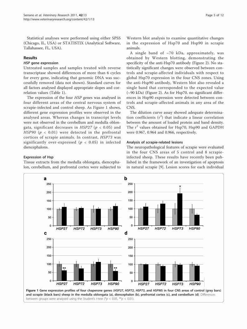
four different areas of the central nervous system ofscrapie-infected and control sheep. As Figure 1 shows,different gene expression profiles were observed in theanalysed areas. Whereas changes in transcript levelswere not observed in the cerebellum and medulla oblon-gata, significant decreases in HSP27 (p < 0.05) andHSP90 (p < 0.01) were detected in the prefrontalcortices of scrapie animals. In contrast, HSP73 wassignificantly over-expressed (p < 0.05) in infecteddiencephalons.
Expression of HspTissue extracts from the medulla oblongata, diencepha-lon, cerebellum, and prefrontal cortex were subjected to
Western blot analysis to examine quantitative changesin the expression of Hsp70 and Hsp90 in scrapieanimals.A single band of ~70 kDa, approximately, was
obtained by Western blotting, demonstrating thespecificity of the anti-Hsp70 antibody (Figure 2). No sta-tistically significant changes were observed between con-trols and scrapie-affected individuals with respect toglobal Hsp70 expression in the four CNS zones. Usingthe anti-Hsp90 antibody, Western blot also revealed asingle band that corresponded to the expected value(~90 kDa) (Figure 2). As for Hsp70, no significant differ-ences in Hsp90 expression were detected between con-trols and scrapie-affected animals in any area of theCNS.The dilution curve assay showed adequate determina-
tion coefficients (r2) that indicate a linear correlationbetween the amount of loaded protein and band density.The r2 values obtained for Hsp70, Hsp90 and GAPDHwere 0.967, 0.964 and 0.966, respectively.
Analysis of scrapie-related lesionsThe neuropathological features of scrapie were evaluatedin the four CNS areas of 5 control and 8 scrapie-infected sheep. These results have recently been pub-lished in the framework of an investigation of apoptosisin natural scrapie [9]. Lesion scores for each individual
Figure 1 Gene expression profiles of four chaperone genes (HSP27, HSP72, HSP73, and HSP90) in four CNS areas of control (grey bars)and scrapie (black bars) sheep in the medulla oblongata (a), diencephalon (b), prefrontal cortex (c), and cerebellum (d). Differencesbetween groups were analysed using the Student’s t-test (*p < 0.05, **p < 0.01).
Serrano et al. Veterinary Research 2011, 42:13http://www.veterinaryresearch.org/content/42/1/13
Page 5 of 12
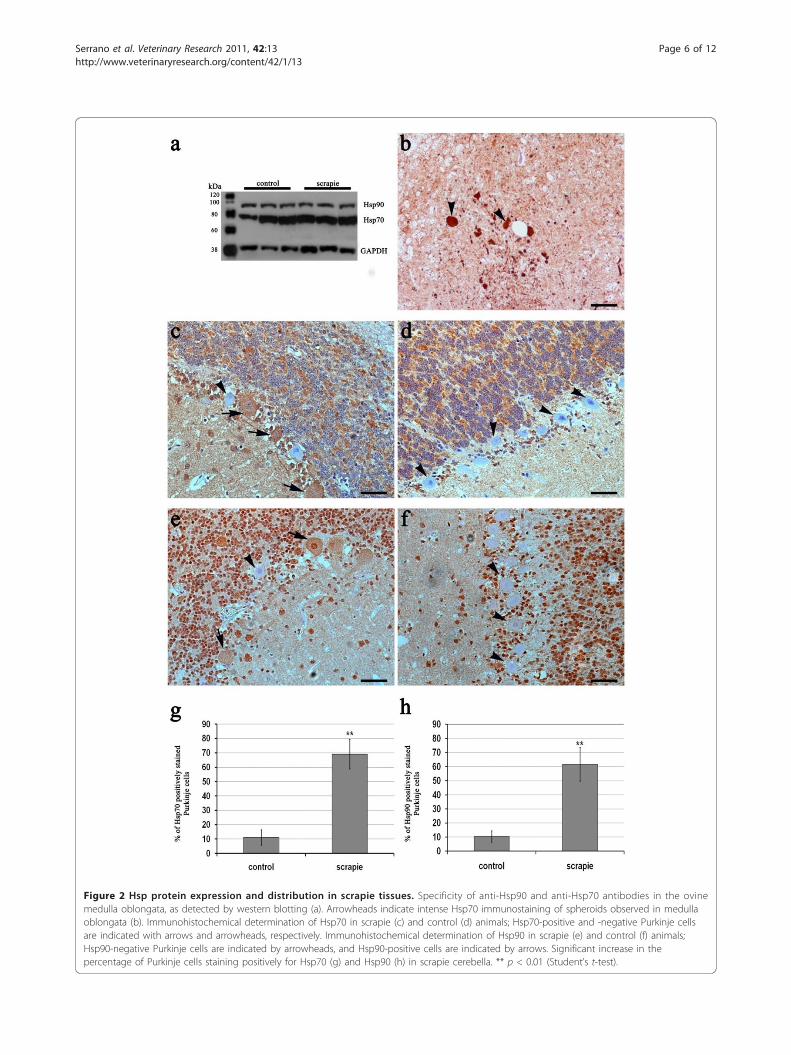
Figure 2 Hsp protein expression and distribution in scrapie tissues. Specificity of anti-Hsp90 and anti-Hsp70 antibodies in the ovinemedulla oblongata, as detected by western blotting (a). Arrowheads indicate intense Hsp70 immunostaining of spheroids observed in medullaoblongata (b). Immunohistochemical determination of Hsp70 in scrapie (c) and control (d) animals; Hsp70-positive and -negative Purkinje cellsare indicated with arrows and arrowheads, respectively. Immunohistochemical determination of Hsp90 in scrapie (e) and control (f) animals;Hsp90-negative Purkinje cells are indicated by arrowheads, and Hsp90-positive cells are indicated by arrows. Significant increase in thepercentage of Purkinje cells staining positively for Hsp70 (g) and Hsp90 (h) in scrapie cerebella. ** p < 0.01 (Student’s t-test).
Serrano et al. Veterinary Research 2011, 42:13http://www.veterinaryresearch.org/content/42/1/13
Page 6 of 12
are presented as Additional file 1. Briefly, neuronalvacuolisation was detected only in affected animals, andthe highest lesion levels were observed in the medullaoblongata and diencephalon. Neuropil spongiosis wasobserved in both groups of animals; however, theincreased rate of this lesion in scrapie animals was sta-tistically significant in all areas except the cerebellum.Prion protein deposition intensity varied by area; theorigin of this difference was the low level of PrPSc
immunolabelling observed in the prefrontal cortex.Astrogliosis was evaluated by GFAP detection. A gen-
eralised increase in the astroglial marker GFAP wasnoticed in the scrapie-affected cases. Apparent hyperpla-sia and hypertrophy of stellate-shaped astroglial cellswere observed in both the grey and white matter whencompared to control brains, indicative of reactive astro-gliosis. Differences between areas were not observed ineither control or infected sheep.Histochemical labelling with LGS revealed an increase
in the number of activated microglial cells in the fouranalysed areas in infected animals. In both control andscrapie-affected animals, there was diffuse immunolabel-ling of the neuropil in the molecular layer of thecerebellar grey matter, as described by Vidal et al. [36].Similar labelling of the perineuronal area of the cerebel-lar nuclei was observed. The morphology of theimmunolabelled microglial cells was either ramified withan elongated rod-shaped nucleus or amoeboid with amore rounded nucleus, indicating activation.Finally, cell death was also analysed by immunodetec-
tion of caspase-3, using an antibody that preferentiallyreacts with the activated form of this protein. In general,very weak staining was observed in the different CNSsections; the intensity and distribution of staining didnot differ significantly between the scrapie and controlgroups.
Relationship between gene and protein expressionprofiles and histopathological lesionsSingle correlations were performed between every lesionand the gene and protein expression in the four ana-lysed tissues. Significant negative correlations weredetected between prion protein deposition and theexpression of HSP27 (-0.677, p = 0.011), HSP72 (-0.672,p = 0.023) and HSP90 (-0.582, p = 0.047) genes; betweenspongiosis and HSP27 gene (-0.592, p = 0.033) andHsp90 protein (-0.614, p = 0.034) expression andbetween vacuolation and Hsp90 protein expression(-0.694, p = 0.012) in the prefrontal cortex. Further anegative correlation was also detected between HSP27gene expression and Caspase3 (-0.728, p = 0.017) in thecerebellum.Stepwise regression was carried out to determine pos-
sible association between gene or protein expression
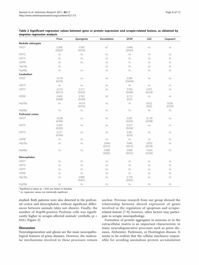
profiles and joint effects of prion-related lesions in thefour areas analysed. Table 2 shows the significantregression values obtained. Increases on HSP geneexpression in the prefrontal cortex (HSP27, HSP72 andHSP73) and in the cerebellum (HSP27, HSP73 andHSP90) were associated with decreases of PrPSc (nega-tive regression) and increases of GFAP (positive regres-sion) immunolabelling. Inversed associations wereobserved in the medulla oblongata, but only for HSP27.Microglia activation (LGS) was positively associated
with HSP73 gene and Hsp70 protein expression in thecerebellum and negatively with HSP27 gene and Hsp70and Hsp90 protein expression in the prefrontal cortex.High HSP73 and HSP90 gene expression rates showed
association with an increase of spongiosis in the cerebel-lum. However, low degrees of spongiosis were associatedto higher HSP27 gene expression rates in the medullaoblongata and higher Hsp70 protein expression levels inthe cerebellum and diencephalon. Increases on vacuoli-sation were associated with slight decreases of Hsp70and Hsp90 protein expression in the prefrontal cortex.Finally, slight increases on Hsp70 protein expressionwere associated with the appearance of active caspase-3immunoreactivity.
Protein distributionCellular localisation of Hsp70 and Hsp90 was revealedby immunohistochemistry. Anti-Hsp27 antibody did notallow the immunohistochemical detection of ovineHsp27 under the conditions employed here.For Hsp70, immunohistochemistry images of the ana-
lysed areas showed a diffuse protein distribution withstained neuronal bodies, mild labelling of the neuropile,and granular labelling of glial cell cytoplasmic processes(Figure 2). No marked differences in staining intensitywere detected between tissues samples from control andinfected sheep. High variability within groups wasobserved. However, Hsp70 was over-expressed in Pur-kinje cells of most of the scrapie-infected animals (Fig-ure 2). Moreover, in one infected sheep showing a largespongiform change, the expression of Hsp70 was lowfor all areas studied. In addition, positively labelled“spheroids” were noted in the brains of some affectedsheep (Figure 2), most likely corresponding to foci ofneuronal degeneration [36,37].The antibody used for the detection of Hsp90 resulted
in specific cytoplasmic staining in the carcinoma mam-mary gland sections (positive controls) as well as inCNS sections of the animals tested. The negative con-trols did not show any immunohistochemical reaction.High variability in Hsp90 immunoreactivity wasobserved in the medulla oblongata in scrapie and con-trol animals. Intense staining was observed in either theneuronal bodies or neuropile, depending on the animal
Serrano et al. Veterinary Research 2011, 42:13http://www.veterinaryresearch.org/content/42/1/13
Page 7 of 12
studied. Both patterns were also detected in the prefron-tal cortex and diencephalon, without significant differ-ences between animals (data not shown). Finally, thenumber of Hsp90-positive Purkinje cells was signifi-cantly higher in scrapie-affected animals’ cerebella (p <0.01) (Figure 2).
DiscussionNeurodegeneration and gliosis are the main neuropatho-logical features of prion diseases. However, the molecu-lar mechanisms involved in these processes remain
unclear. Previous research from our group showed therelationship between altered expression of genesinvolved in the regulation of apoptosis and scrapie-related lesions [7-9]; however, other factors may partici-pate in scrapie neuropathology.Formation of protein aggregates in neurons or in the
extracellular matrix is an important characteristic inmany neurodegenerative processes such as prion dis-eases, Alzheimer, Parkinson, or Huntington disease. Itseems to be evident that the cellular machinery respon-sible for avoiding anomalous protein accumulation
Table 2 Significant regression values between gene or protein expression and scrapie-related lesions, as obtained bystepwise regression analysis
Prion Spongiosis Vacuolation GFAP LGS Caspase3
Medulla oblongata
HSP27 0.399(0.003)1
-0.381(0.016)
ns2 -0.443(0.043)
ns ns
HSP72 ns ns ns ns ns ns
HSP73 ns ns ns ns ns ns
HSP90 ns ns ns ns ns ns
Hsp70p ns ns ns ns ns
Hsp90p ns ns ns ns ns ns
Cerebellum
HSP27 -0.178(0.032)
ns ns 0.294(0.0430)
ns ns
HSP72 ns ns ns ns ns ns
HSP73 -0.719(0.015)
0.317(0.023)
ns 0.783(0.034)
0.555(0.018)
ns
HSP90 -0.465(0.008)
0.182(0.039)
ns 0.712(0.01)
ns ns
Hsp70p ns -0.014(0.010)
ns ns 0.022(0.02)
0.026(0.010)
Hsp90p ns ns ns ns ns ns
Prefrontal cortex
HSP27 -0.228(0.004)
ns ns 0.267(0.025)
-0.129(0.038)
ns
HSP72 -0.347(0.003)
ns ns 0.277(0.034)
ns ns
HSP73 -0.271(0.036)
ns ns 0.381(0.03)
ns ns
HSP90 ns ns ns ns ns ns
Hsp70p ns ns -0.049(0.015)
0.045(0.013)
-0.023(0.018)
ns
Hsp90p ns ns -0.080(0.002)
0.046(0.015)
-0.024(0.020)
ns
Diencephalon
HSP27 ns ns ns ns ns ns
HSP72 ns ns ns ns ns ns
HSP73 ns ns ns ns ns ns
HSP90 ns ns ns ns ns ns
Hsp70p ns -0.805(0.001)
ns 0.778(0.002)
ns ns
Hsp90p ns ns ns ns ns ns1Significant p-values (p < 0.05) are shown in brackets.2 ns: regression values not statistically significant.
Serrano et al. Veterinary Research 2011, 42:13http://www.veterinaryresearch.org/content/42/1/13
Page 8 of 12
would be implicated in these pathologies. Therefore,chaperones could play an important role in neuronaldegeneration [38]. In this context, little is known aboutnatural models of TSE. The aim of this study was toanalyse the gene and protein expression of four Hspchaperones in the CNS of sheep naturally infected withscrapie; the chaperones analysed were chosen accordingto their known roles in apoptosis modulation or theirpossible functions in prion diseases.Over-expression of HSP70 family genes has been
reported in brain samples from scrapie-infected mice[25,27]. Our expression analysis revealed significantover-expression of HSP73 transcripts in scrapie dience-phalons. Hsp70 prevents the accumulation and/or pro-motes the degradation of specific PrP conformers in flymodels and protects against PrP neurotoxicity throughunknown mechanisms [39]. In our study, HSP73 expres-sion in diencephalons was independent of PrPSc immu-nolabelling (Table 2) suggesting an alternative role forthis chaperone. We reported a lack of apoptosis induc-tion despite the significant increase of BAX [8] and BAK[9] in scrapie diencephalons. Joining these results, wehypothesize that HSP73 could counteract the inductionof apoptosis by these mitochondrial factors.In contrast, in the prefrontal cortex, the expression of
HSP90 and HSP27 genes decreased in the scrapie group.This HSP90 pattern is not in agreement with the Hsp90up-regulation observed in murine models of BovineSpongiform Encephalopathy (BSE) [28]. It is possiblethat mRNA levels do not correspond to protein expres-sion. It has been suggested that under inflammation orischemic processes, Hsp synthesis results from a post-transcriptional regulatory mechanism that involvesmRNA stabilisation [40]. This is why we analysed theexpression of Hsp proteins by Western blot. Interest-ingly, no significant differences were detected betweencontrols and scrapie animals. An association analysisestablished differential distribution of certain poly-morphisms in the 5’ and 3’ regions of the ovineHSP90AA1 gene in scrapie-susceptible animals [30].Previously, other authors observed an important role ofthe 3’UTR region in post-transcriptional regulation ofHsp70 upon heat shock [41,42]. Thus, the decrease ofthe HSP90 gene expression observed in the prefrontalcortex could be caused by variation at transcriptional orpost-transcriptional levels. Perhaps these mutations donot affect HSP90 translation, or, alternately, Westernblot technique is not sensitive enough to detect certainchanges in protein expression.In addition, interindividual variability has been
reported for natural scrapie [7-9,43]. This may masksmall differences in gene and protein expressionbetween healthy and scrapie-affected animals. For thisreason, we performed an additional statistical analysis to
detect whether a relationship exists between the expres-sion pattern and the histopathological changes observedin scrapie animals. This methodology allows the analysisof the molecular mechanisms underlying these processesin the natural form of the disease. Although we areaware that the histopathological valuation does not cor-respond to a real scale, our procedure can reliably detectthe association among different parameters. However,the lack of statistical power due to the limited numberof individuals analysed could prevent the detection ofadditional associations.Although stepwise regression results were different in
the four areas analysed, it is worth mentioning that weidentified a negative association between prion proteindeposition and the expression of the HSP genes in thecerebellum (HSP27, HSP73 and HSP90) and prefrontalcortex (HSP27, HSP72 and HSP73). It has been demon-strated that Hsp70 members avoid protein aggregation,which leads to neurodegenerative diseases [44-46]. Thus,down-regulation of chaperones could contribute to theaccumulation of anomalous proteins and finally to cellstress. A decrease of Hsp27 has been described in earlystages of in vitro models of spinocerebellar ataxia type 3[47] and type 7 [48], which are neurodegenerative disor-ders associated with an accumulation of polyglutamine-containing proteins.Glial reaction is the other classical sign of natural
scrapie. We previously reported a positive associationbetween astrogliosis and the extrinsic pathway of apop-tosis in the medulla oblongata and prefrontal cortex. Inaddition to apoptosis regulation, factors involved in thispathway can participate in inflammation [49], reactivegliosis and neuroinflammation [50,51]. Hsp are alsosecreted as inflammatory mediators that bind surfacereceptors and induce the production of nitric oxide,cytokines, and other immunoregulatory molecules.Levels of the small heat shock protein Hsp25 increasedduring reactive astrogliosis in a murine model of BSE[52], and Hsp70 expression has been reported in reac-tive astrocytes following ischemia [53,54]. Microglialactivation by Hsp proteins, such as Hsp70 and Hsp90,results in clearance of amyloid peptide aggregates[55,56]. In agreement with this, in the present workGFAP immunolabelling was positively associated withthe expression of several HSP genes and proteins in theareas where these genes were negatively associated withprion protein immunolabelling (cerebellum and prefron-tal cortex). Then, high levels of HSP gene or proteinexpression would be associated to the prevention ordegradation of prion protein aggregates and the pre-sence of reactive astrocytosis in natural scrapie.Although a strong positive association was foundbetween HSP73 gene expression and microglia stainingin the cerebellum, our association results are not
Serrano et al. Veterinary Research 2011, 42:13http://www.veterinaryresearch.org/content/42/1/13
Page 9 of 12
conclusive with respect to the role of HSP in microgliaactivation.The Hsp analysed in this work were selected due to
their known role in apoptosis regulation. In a previousstudy, searching for possible evidence of cell deathmechanisms in natural scrapie by immunodetection ofactivated caspase-3 [9], we observed weak staining in allCNS sections without finding significant differencesbetween control and scrapie animals. However, a positiveassociation was found between caspase-3 and the mito-chondrial pathway of apoptosis [9]. Since Hsp work asanti-apoptotic factors [15,17,19] we analysed the relation-ship between caspase-3 expression and Hsp gene andprotein expression. Only Hsp70 protein expression in thecerebellum showed a positive correlation with caspase-3.The absence of an association in the remaining areasmay reflect that, in general, Hsp are not involved in theregulation of neuronal death during natural disease, aswas observed in the acute phase of epilepsy [57].Finally, when cases of natural disease are studied,
changes detected in certain cell populations are fre-quently not correlated with variations in gene or proteinexpression in the final tissue mixture, as we observedfor Bax protein determination in the CNS of scrapiesheep [8]. For this reason, in order to confirm the pre-vious results and elucidate the cell type involved in pos-sible expression variability, an immunohistochemicalassay of Hsp was performed in each area of the CNS. Ingeneral, diffuse and highly variable staining for Hsp70and Hsp90 was observed in the four CNS areas of bothgroups of animals. Nevertheless, the over-expression ofHsp in Purkinje cells of scrapie cerebella was remark-able. Intense staining for Hsp72 in this cell type wasalso observed in Creutzfeldt-Jakob disease (CJD) patients[58]; accumulation of this protein in Purkinje cells sup-ports the proposed neuroprotective role of Hsp in thenatural form of the disease. Similarly, although caspase-3 immunostaining was present in cerebellar granularcells, we did not observe this staining in Purkinje cells,in either scrapie-affected or control sheep [8,9]. To ourknowledge, there is no clear evidence of prion proteintoxicity in Purkinje cells. Only over-expression of theDoppel gene in Prnp-deficient mice has been correlatedwith Purkinje cell loss [59,60]. Our results support thepossible protective function of Hsp70 in Purkinje cellsand suggest a role of Hsp90 in this mechanism. Furtheranalysis would be necessary to clarify the role of Hspover-expression in these cells in prion diseases.In CJD [58], the regions with severe spongiosis showed
low immunoreactivity for Hsp72. In agreement with thisreport, we detected low expression of Hsp70 protein in oneinfected animal showing the greatest degree of spongiformalteration. In fact, the stepwise regression analysis revealeda negative association of Hsp70 protein with spongiosis in
the cerebellum and diencephalon, suggesting that thereduction in the cytoprotective effect of Hsp70 could beinvolved in neuronal loss, at least in these two areas. How-ever, the results of correlation and stepwise regressionmust be taken with caution due to the reduced amount ofavailable data. Nevertheless, they illustrate the joint effectof several lesions of the gene or protein expression on thetissues analysed and pointed out the complexity of causesof modifications of gene and protein expression.In conclusion, Hsp seem to be involved in gliosis and
inflammatory reactions rather than in anti-apoptoticprocesses in natural scrapie. However, the intenseimmunoreactivity of Purkinje cells for Hsp70 andHsp90, as well as the negative association of Hsp70 withprion protein immunolabelling and spongiosis, suggestsa neuroprotective effect in this cell type against thestress related to ovine encephalopathy.
Additional material
Additional file 1: Individual scoring for scrapie lesions. Individualscoring for prion deposition, spongiosis, neuronal vacuolization, GFAPimmunostaining, LGS staining and activated caspase-3 immunoreactivityfor control (C) and scrapie (Sc)-infected sheep in the different areasanalysed.
AcknowledgementsThe authors thank Silvia Ruiz for her technical assistance. This work wasperformed as part of the EET2003-09890 project (CICYT/FEDER) andAGL2008-02506 (MICINN) projects and was partially financed by theGobierno de Aragón (Grupos de Excelencia and Departamento deAgricultura). H. Filali was supported by a doctoral grant from the Ministeriode Asuntos Exteriores (MAE/AECI).
Author details1Laboratorio de Genética Bioquímica (LAGENBIO), Facultad de Veterinaria,Universidad de Zaragoza, Miguel Servet 177, 50013 Zaragoza, Spain. 2Centrode Investigación en Encefalopatías y Enfermedades TransmisiblesEmergentes, Universidad de Zaragoza, Miguel Servet 177, 50013 Zaragoza,Spain. 3Unidad de Genética Cuantitativa y Mejora Animal, Facultad deVeterinaria, Universidad de Zaragoza, Miguel Servet 177, 50013 Zaragoza,Spain. 4Departamento de Mejora Genética Animal, INIA, Ctra La Coruña Km7.5, 28040 Madrid, Spain. 5Unidad de Tecnología en Producción Animal,CITA, Av. de Montañana, 930, 50059 Zaragoza, Spain.
Authors’ contributionsCS carried out the gene and protein expression studies and wrote themanuscript. RB conceived and carried out the histopathological andimmunohistochemical analysis. JL, AM-C, JHC and PZ participated in geneexpression analysis and the interpretation of data. LV performed thestatistical association studies. MS participated in the statistical analysis,interpreted the data and critically revised the manuscript. HF, CA and JJBparticipated in the histopathological analysis and evaluation. IM-B conceivedof the study, participated in its design and coordination and wrote thepaper. All authors discussed the results and implications, read and approvedthe final manuscript.
Competing interestsThe authors declare that they have no competing interests.
Received: 4 March 2010 Accepted: 21 October 2010Published: 24 January 2011
Serrano et al. Veterinary Research 2011, 42:13http://www.veterinaryresearch.org/content/42/1/13
Page 10 of 12
References1. Budka H, Aguzzi A, Brown P, Brucher JM, Bugiani O, Gullotta F,
Haltia M, Hauw JJ, Ironside JW, Jellinger K, Kretzschmar HA, Lantos PL,Masullo C, Schlote W, Tateishi J, Weller RO: Neuropathologicaldiagnostic criteria for Creutzfeldt-Jakob disease (CJD) and otherhuman spongiform encephalopathies (prion diseases). Brain Pathol1995, 5:459-466.
2. Kim YS, Carp RI, Callahan S, Wisniewski HM: Incubation periods andhistopathological changes in mice injected stereotaxically in differentbrain areas with the 87V scrapie strain. Acta Neuropathol 1990,80:388-392.
3. Wells GA, Hancock RD, Cooley WA, Richards MS, Higgins RJ, David GP:Bovine spongiform encephalopathy: diagnostic significance of vacuolarchanges in selected nuclei of the medulla oblongata. Vet Rec 1989,125:521-524.
4. Wood JL, McGill IS, Done SH, Bradley R: Neuropathology of scrapie: astudy of the distribution patterns of brain lesions in 222 cases of naturalscrapie in sheep, 1982-1991. Vet Rec 1997, 140:167-174.
5. Gubler E, Hilbe M, Ehrensperger F: Lesion profiles and gliosis in thebrainstem of 135 Swiss cows with bovine spongiform encephalopathy(BSE). Schweiz Arch Tierheilkd 2007, 149:111-122.
6. Liberski PP, Sikorska B, Bratosiewicz-Wasik J, Gajdusek DC, Brown P:Neuronal cell death in transmissible spongiform encephalopathies(prion diseases) revisited: from apoptosis to autophagy. Int J Biochem CellBiol 2004, 36:2473-2490.
7. Lyahyai J, Bolea R, Serrano C, Monleon E, Moreno C, Osta R, Zaragoza P,Badiola JJ, Martin-Burriel I: Correlation between Bax overexpression andprion deposition in medulla oblongata from natural scrapie withoutevidence of apoptosis. Acta Neuropathol 2006, 112:451-460.
8. Lyahyai J, Bolea R, Serrano C, Vidal E, Pumarola M, Badiola JJ, Zaragoza P,Martin-Burriel I: Differential expression and protein distribution of Bax innatural scrapie. Brain Res 2007, 1180:111-120.
9. Serrano C, Lyahyai J, Bolea R, Varona L, Monleon E, Badiola JJ, Zaragoza P,Martin-Burriel I: Distinct spatial activation of intrinsic and extrinsicapoptosis pathways in natural scrapie: association with prion-relatedlesions. Vet Res 2009, 40:42.
10. Kitagawa K, Matsumoto M, Tagaya M, Hata R, Ueda H, Niinobe M, Handa N,Fukunaga R, Kimura K, Mikoshiba K, Kamada T: ’Ischemic tolerance’phenomenon found in the brain. Brain Res 1990, 528:21-24.
11. Chopp M, Chen H, Dereski MO, Garcia JH: Mild hypothermic interventionafter graded ischemic stress in rats. Stroke 1991, 22:37-43.
12. Uney JB, Kew JN, Staley K, Tyers P, Sofroniew MV: Transfection-mediated expression of human Hsp70i protects rat dorsal rootganglian neurones and glia from severe heat stress. FEBS Lett 1993,334:313-316.
13. Mailhos C, Howard MK, Latchman DS: Heat shock proteins hsp90 andhsp70 protect neuronal cells from thermal stress but not fromprogrammed cell death. J Neurochem 1994, 63:1787-1795.
14. Garrido C, Bruey JM, Fromentin A, Hammann A, Arrigo AP, Solary E: HSP27inhibits cytochrome c-dependent activation of procaspase-9. FASEB J1999, 13:2061-2070.
15. Bruey JM, Ducasse C, Bonniaud P, Ravagnan L, Susin SA, Diaz-Latoud C,Gurbuxani S, Arrigo AP, Kroemer G, Solary E, Garrido C: Hsp27 negativelyregulates cell death by interacting with cytochrome c. Nat Cell Biol 2000,2:645-652.
16. Pandey P, Saleh A, Nakazawa A, Kumar S, Srinivasula SM, Kumar V,Weichselbaum R, Nalin C, Alnemri ES, Kufe D, Kharbanda S: Negativeregulation of cytochrome c-mediated oligomerization of Apaf-1 andactivation of procaspase-9 by heat shock protein 90. EMBO J 2000,19:4310-4322.
17. Beere HM, Wolf BB, Cain K, Mosser DD, Mahboubi A, Kuwana T, Tailor P,Morimoto RI, Cohen GM, Green DR: Heat-shock protein 70 inhibitsapoptosis by preventing recruitment of procaspase-9 to the Apaf-1apoptosome. Nat Cell Biol 2000, 2:469-475.
18. Saleh A, Srinivasula SM, Balkir L, Robbins PD, Alnemri ES: Negativeregulation of the Apaf-1 apoptosome by Hsp70. Nat Cell Biol 2000,2:476-483.
19. Ravagnan L, Gurbuxani S, Susin SA, Maisse C, Daugas E, Zamzami N,Mak T, Jaattela M, Penninger JM, Garrido C, Kroemer G: Heat-shockprotein 70 antagonizes apoptosis-inducing factor. Nat Cell Biol 2001,3:839-843.
20. Gurbuxani S, Schmitt E, Cande C, Parcellier A, Hammann A, Daugas E,Kouranti I, Spahr C, Pance A, Kroemer G, Garrido C: Heat shock protein 70binding inhibits the nuclear import of apoptosis-inducing factor.Oncogene 2003, 22:6669-6678.
21. Ellis RJ: The molecular chaperone concept. Semin Cell Biol 1990, 1:1-9.22. Kang SC, Brown DR, Whiteman M, Li R, Pan T, Perry G, Wisniewski T, Sy MS,
Wong BS: Prion protein is ubiquitinated after developing proteaseresistance in the brains of scrapie-infected mice. J Pathol 2004,203:603-608.
23. Latchman DS: HSP27 and cell survival in neurones. Int J Hyperthermia2005, 21:393-402.
24. Chernoff YO: Stress and prions: lessons from the yeast model. FEBS Lett2007, 581:3695-3701.
25. Booth S, Bowman C, Baumgartner R, Dolenko B, Sorensen G, Robertson C,Coulthart M, Phillipson C, Somorjai R: Molecular classification of scrapiestrains in mice using gene expression profiling. Biochem Biophys ResCommun 2004, 325:1339-1345.
26. Diedrich JF, Carp RI, Haase AT: Increased expression of heat shockprotein, transferrin, and beta 2-microglobulin in astrocytes duringscrapie. Microb Pathog 1993, 15:1-6.
27. Kenward N, Hope J, Landon M, Mayer RJ: Expression of polyubiquitin andheat-shock protein 70 genes increases in the later stages of diseaseprogression in scrapie-infected mouse brain. J Neurochem 1994,62:1870-1877.
28. Sawiris GP, Becker KG, Elliott EJ, Moulden R, Rohwer RG: Molecular analysisof bovine spongiform encephalopathy infection by cDNA arrays. J GenVirol 2007, 88:1356-1362.
29. Tatzelt J, Voellmy R, Welch WJ: Abnormalities in stress proteins in priondiseases. Cell Mol Neurobiol 1998, 18:721-729.
30. Marcos-Carcavilla A, Calvo JH, Gonzalez C, Moazami-Goudarzi K, Laurent P,Bertaud M, Hayes H, Beattie AE, Serrano C, Lyahyai J, Martin-Burriel I,Serrano M: Structural and functional analysis of the HSP90AA1 gene:distribution of polymorphisms among sheep with different responses toscrapie. Cell Stress Chaperones 2008, 13:19-29.
31. Marcos-Carcavilla A, Moreno C, Serrano M, Laurent P, Cribiu EP,Andreoletti O, Ruesche J, Weisbecker JL, Calvo JH, Moazami-Goudarzi K:Polymorphisms in the HSP90AA1 5’ flanking region are associated withscrapie incubation period in sheep. Cell Stress Chaperones 2010,15:343-349.
32. Vargas F, Lujan L, Bolea R, Monleon E, Martin-Burriel I, Fernandez A, DeBlas I, Badiola JJ: Detection and clinical evolution of scrapie in sheep by3rd eyelid biopsy. J Vet Intern Med 2006, 20:187-193.
33. Bolea R, Monleon E, Schiller I, Raeber AJ, Acin C, Monzon M, Martin-Burriel I,Struckmeyer T, Oesch B, Badiola JJ: Comparison of immunohistochemistryand two rapid tests for detection of abnormal prion protein in differentbrain regions of sheep with typical scrapie. J Vet Diagn Invest 2005,17:467-469.
34. Lyahyai J, Serrano C, Ranera B, Badiola JJ, Zaragoza P, Martin-Burriel I: Effectof scrapie on the stability of housekeeping genes. Anim Biotechnol 2010,21:1-13.
35. Hardt M, Baron T, Groschup MH: A comparative study ofimmunohistochemical methods for detecting abnormal prion proteinwith monoclonal and polyclonal antibodies. J Comp Pathol 2000,122:43-53.
36. Vidal E, Acin C, Foradada L, Monzon M, Marquez M, Monleon E,Pumarola M, Badiola JJ, Bolea R: Immunohistochemical characterisation ofclassical scrapie neuropathology in sheep. J Comp Pathol 2009,141:135-146.
37. Siso S, Puig B, Varea R, Vidal E, Acin C, Prinz M, Montrasio F, Badiola J,Aguzzi A, Pumarola M, Ferrer I: Abnormal synaptic protein expression andcell death in murine scrapie. Acta Neuropathol 2002, 103:615-626.
38. Muchowski PJ: Protein misfolding, amyloid formation, andneurodegeneration: a critical role for molecular chaperones? Neuron2002, 35:9-12.
39. Fernandez-Funez P, Casas-Tinto S, Zhang Y, Gomez-Velazquez M, Morales-Garza MA, Cepeda-Nieto AC, Castilla J, Soto C, Rincon-Limas DE: In vivogeneration of neurotoxic prion protein: role for hsp70 in accumulationof misfolded isoforms. PLoS Genet 2009, 5:e1000507.
40. Jacquier-Sarlin MR, Jornot L, Polla BS: Differential expression andregulation of hsp70 and hsp90 by phorbol esters and heat shock. J BiolChem 1995, 270:14094-14099.
Serrano et al. Veterinary Research 2011, 42:13http://www.veterinaryresearch.org/content/42/1/13
Page 11 of 12
41. Lee MG: The 3’ untranslated region of the hsp 70 genes maintains thelevel of steady state mRNA in Trypanosoma brucei upon heat shock.Nucleic Acids Res 1998, 26:4025-4033.
42. Moseley PL, Wallen ES, McCafferty JD, Flanagan S, Kern JA: Heat stressregulates the human 70-kDa heat-shock gene through the 3’-untranslated region. Am J Physiol 1993, 264:L533-537.
43. Vidal E, Bolea R, Tortosa R, Costa C, Domenech A, Monleon E, Vargas A,Badiola JJ, Pumarola M: Assessment of calcium-binding proteins(Parvalbumin and Calbindin D-28K) and perineuronal nets in normal andscrapie-affected adult sheep brains. J Virol Methods 2006, 136:137-146.
44. Dedmon MM, Christodoulou J, Wilson MR, Dobson CM: Heat shock protein70 inhibits alpha-synuclein fibril formation via preferential binding toprefibrillar species. J Biol Chem 2005, 280:14733-14740.
45. Muchowski PJ, Schaffar G, Sittler A, Wanker EE, Hayer-Hartl MK, Hartl FU:Hsp70 and hsp40 chaperones can inhibit self-assembly of polyglutamineproteins into amyloid-like fibrils. Proc Natl Acad Sci USA 2000, 97:7841-7846.
46. Evans CG, Wisen S, Gestwicki JE: Heat shock proteins 70 and 90 inhibitearly stages of amyloid beta-(1-42) aggregation in vitro. J Biol Chem2006, 281:33182-33191.
47. Wen FC, Li YH, Tsai HF, Lin CH, Li C, Liu CS, Lii CK, Nukina N, Hsieh M:Down-regulation of heat shock protein 27 in neuronal cells and non-neuronal cells expressing mutant ataxin-3. FEBS Lett 2003, 546:307-314.
48. Tsai HF, Lin SJ, Li C, Hsieh M: Decreased expression of Hsp27 and Hsp70in transformed lymphoblastoid cells from patients with spinocerebellarataxia type 7. Biochem Biophys Res Commun 2005, 334:1279-1286.
49. Sawada M: Brain cytokine network and novel characteristics of microglia.Nihon Shinkei Seishin Yakurigaku Zasshi 1999, 19:151-154, in Japanese.
50. Ferrer I, Puig B, Krupinsk J, Carmona M, Blanco R: Fas and Fas ligandexpression in Alzheimer’s disease. Acta Neuropathol 2001, 102:121-131.
51. Martinez M, Fernandez-Vivancos E, Frank A, De la Fuente M, Hernanz A:Increased cerebrospinal fluid fas (Apo-1) levels in Alzheimer’s disease.Relationship with IL-6 concentrations. Brain Res 2000, 869:216-219.
52. Tortosa R, Vidal E, Costa C, Alamillo E, Torres JM, Ferrer I, Pumarola M:Stress response in the central nervous system of a transgenic mousemodel of bovine spongiform encephalopathy. Vet J 2008, 178:126-129.
53. Giffard RG, Xu L, Zhao H, Carrico W, Ouyang Y, Qiao Y, Sapolsky R,Steinberg G, Hu B, Yenari MA: Chaperones, protein aggregation, andbrain protection from hypoxic/ischemic injury. J Exp Biol 2004,207:3213-3220.
54. Renolleau S, Benjelloun N, Ben-Ari Y, Charriaut-Marlangue C: Regulation ofapoptosis-associated proteins in cell death following transient focalischemia in rat pups. Apoptosis 1997, 2:368-376.
55. Takata K, Kitamura Y, Tsuchiya D, Kawasaki T, Taniguchi T, Shimohama S:Heat shock protein-90-induced microglial clearance of exogenousamyloid-beta1-42 in rat hippocampus in vivo. Neurosci Lett 2003,344:87-90.
56. Kakimura J, Kitamura Y, Takata K, Umeki M, Suzuki S, Shibagaki K,Taniguchi T, Nomura Y, Gebicke-Haerter PJ, Smith MA, Perry G,Shimohama S: Microglial activation and amyloid-beta clearance inducedby exogenous heat-shock proteins. FASEB J 2002, 16:601-603.
57. Yang T, Hsu C, Liao W, Chuang JS: Heat shock protein 70 expression inepilepsy suggests stress rather than protection. Acta Neuropathol 2008,115:219-230.
58. Kovacs GG, Kurucz I, Budka H, Adori C, Muller F, Acs P, Kloppel S,Schatzl HM, Mayer RJ, Laszlo L: Prominent stress response of Purkinjecells in Creutzfeldt-Jakob disease. Neurobiol Dis 2001, 8:881-889.
59. Sakaguchi S, Katamine S, Nishida N, Moriuchi R, Shigematsu K, Sugimoto T,Nakatani A, Kataoka Y, Houtani T, Shirabe S, Okada H, Hasegawa S,Miyamoto T, Noda T: Loss of cerebellar Purkinje cells in aged micehomozygous for a disrupted PrP gene. Nature 1996, 380:528-531.
60. Moore RC, Mastrangelo P, Bouzamondo E, Heinrich C, Legname G,Prusiner SB, Hood L, Westaway D, DeArmond SJ, Tremblay P: Doppel-induced cerebellar degeneration in transgenic mice. Proc Natl Acad SciUSA 2001, 98:15288-15293.
doi:10.1186/1297-9716-42-13Cite this article as: Serrano et al.: Changes in HSP gene and proteinexpression in natural scrapie with brain damage. Veterinary Research2011 42:13.
Submit your next manuscript to BioMed Centraland take full advantage of:
• Convenient online submission
• Thorough peer review
• No space constraints or color figure charges
• Immediate publication on acceptance
• Inclusion in PubMed, CAS, Scopus and Google Scholar
• Research which is freely available for redistribution
Submit your manuscript at www.biomedcentral.com/submit
Serrano et al. Veterinary Research 2011, 42:13http://www.veterinaryresearch.org/content/42/1/13
Page 12 of 12