Embed Size (px)
Citation preview

8/31/2016
1
Patient Management: High Risk Lesions
Deborah Thames R.T. (R)(M)(QM)
What are High Risk Lesions?
– Is pre‐cancer such a word?
– High risk factor
– High risk lesions need to have special attention
– Surveillance
– Medications
– Preventative surgery
What are High Risk Lesions
– Hyperplasia with and without atypia
– Hyperplasia is defined as enlargement of tissue caused by an increase in the reproduction rate of the cells.
– In the breast, this could be
– Atypical Ductal Hyperplasia
– Atypical Lobular Hyperplasia
– Flat Epithelial Hyperplasia with atypia
– Lobular carcinoma in‐situ
– Radial scar/complex sclerosing lesion
– Atypical columnar cell hyperplasia

8/31/2016
2
Surveillance
– Breast surveillance requires shorter term imaging
– Usual management for surveillance is
– Mammography with 6 month follow up for the first two years, with ultrasound
every other 6 months and possible MRI once a year.
– Make sure patient gets a clinical breast exam twice a year
– Make sure patient is conducting breast self examination
Medication/Drug controlTamoxifen
– Prevention medicine for high risk lesions
– Tamoxifen approved by FDA in 1998
– Nolvadex (Pill) and soltamox (liquid) brand names
– Pill taken once a day
– Estrogen modulator to prevent breast cancer in women with high risk
– Estrogen receptor antagonist antineoplastic agent
– Women having periods and haven’t reached menopause yet
– Reduces the risk by 30‐50 percent if taken for 5‐10 years
– Hormone Therapy

8/31/2016
3
Tamoxifen
– Selective Estrogen Regulator modulator (SERM)
– Selectively blocks or activates estrogen’s action on specific cells.
– Sits in the estrogen receptors in the breast cells. If a SERM is in the estrogen
receptor, there is no room for estrogen to attach to the cell, therefore the cell
doesn’t receive estrogens signals to grow and multiply.
– While tamoxifen blocks estrogen’s action on the breast cells, it activates
estrogen’s action in bone and liver cells to help stop bone loos after menopause
– Lower cholesterol levels
– Hormone Therapy SERM
Tamoxifen
– Side effects…
– Abnormal vaginal bleeding
– Pain and/or pressure in the pelvis area
– Leg swelling and/or tenderness
– Chest pain/shortness of breast
– Weakness, tingling, or numbness in the face, arm, and leg
– Vision problems/dizziness and sudden severe headaches
– Weight gain/mood swings/nausea/hot flashes/hair thinning
– Loss of libido

8/31/2016
4
Evista
– Evista chemical name is Raloxifene approved by FDA in 1997
– A type of SERM
– Decreases low density lipoprotein (LDL) cholesterol.
– Unlike Raloxifene, evista does not increase high density lipoprotein (HDL)
Fareston
– Fareston chemical name is toremifene approved by FDA in 1997
– A type of SERM
– ERDs (Estrogen Receptor Downregulators):
– Faslodex (chemical name: fulvestrant)

8/31/2016
5
– Hormone Therapy
– ERDs (Estrogen Receptor Downregulators):
– Block the effects of estrogen in breast tissue. ERDs work in a similar way to
SERMs. ERDs sit in the estrogen receptors in breast cells, so the cell doesn't
receive estrogen's signals to grow and multiply.
– ERDS also reduce the number of estrogen receptors and changes the shape
of the breast cell estrogen receptors, so they don't work as well
– Unlike SERMS, ERDs don't activate estrogen receptors in other parts of the
body, such as the bones or the uterus. ERDs only block and destroy estrogen
receptors.
– Hormone Therapy
– ERD’s Effects
– Nausea
– Vomiting
– Hot flashes
– Headache
– Constipation
– Diarrhea
– Sore throat
– Back/stomach/abdominal pain
– Injection site pain
– Before there was medication to prevent the growth of tumors from hormones,
doctors relied on the removal of the endocrine organs.
– Oophorectomy: the removal of the ovaries.
– Adrenalectomy: the removal of the adrenal glands.
– Hypophysectomies: removal of the pituitary glands.

8/31/2016
6
– Biologic Therapy
– A drug treatment that helps the body’s immune system fight cancer.
– Biologic Therapy
– What does HER2/neu stand for?
– Biologic Therapy
– Human Epidermal growth factor Receptor 2

8/31/2016
7
– Biologic Therapy
– What is HER2/neu?
– Biologic Therapy
– HER2/neu are receptors on the cell membrane that when triggered, transport a
signal to cause cell growth.
– Who in the class wants to be HER2 Positive raise your hand?
– Biologic Therapy

8/31/2016
8
– Hormone Therapy
Pt before serms Same pt on tamoxifen one year later
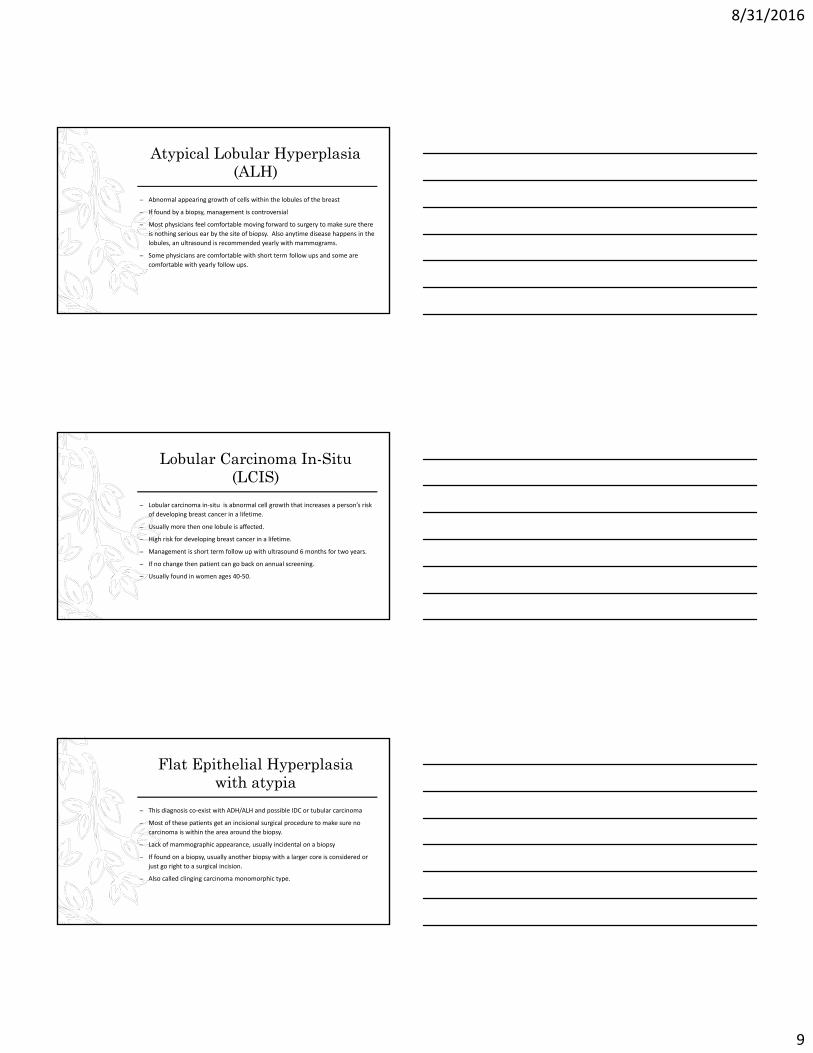
Atypical Ductal Hyperplasia(ADH)
– Happens within the walls of the ducts in the breast
– ADH is a high risk lesion/calcifications
– Marker for increased risk for developing breast cancer
– 4‐5 times higher risk added on to patients with first degree relative
– If found on a biopsy, and a large area of disease, a segmental mastectomy is
recommended, if a small area, most are comfortable with 6 month follow up.
8/31/2016
9
Atypical Lobular Hyperplasia(ALH)
– Abnormal appearing growth of cells within the lobules of the breast
– If found by a biopsy, management is controversial
– Most physicians feel comfortable moving forward to surgery to make sure there
is nothing serious ear by the site of biopsy. Also anytime disease happens in the
lobules, an ultrasound is recommended yearly with mammograms.
– Some physicians are comfortable with short term follow ups and some are
comfortable with yearly follow ups.
Lobular Carcinoma In-Situ(LCIS)
– Lobular carcinoma in‐situ is abnormal cell growth that increases a person’s risk
of developing breast cancer in a lifetime.
– Usually more then one lobule is affected.
– High risk for developing breast cancer in a lifetime.
– Management is short term follow up with ultrasound 6 months for two years.
– If no change then patient can go back on annual screening.
– Usually found in women ages 40‐50.
Flat Epithelial Hyperplasia with atypia
– This diagnosis co‐exist with ADH/ALH and possible IDC or tubular carcinoma
– Most of these patients get an incisional surgical procedure to make sure no
carcinoma is within the area around the biopsy.
– Lack of mammographic appearance, usually incidental on a biopsy
– If found on a biopsy, usually another biopsy with a larger core is considered or
just go right to a surgical incision.
– Also called clinging carcinoma monomorphic type.

8/31/2016
10
Atypical Columnar Cell Hyperplasia
– Also called blunt duct adenosis and columnar alterations.
– Wide range of histologic changes.
– May represent early morphologically identifiable low grade carcinoma
– Most cases are just an annual following with mammograms.
– If a large area is seen, possible short term follow up.
Radial Scar
– Form of Complex sclerosing hyperplasia lesion.
– Characteristics are translucent, circular, or oval region in the center.
– Fat is usually centered around this area.
– Between stromal structures of the breast.
– Just a moderate increase risk for breast cancer development over a lifetime.
– Most are hard to see on an ultrasound, better seen mammographically
– Management is usually surgical incision.
Path for high risk pathology

8/31/2016
11
BREAST, LEFT, STEREOTACTIC GUIDED COREBIOPSYAT 4 O’CLOCK, 9 CM FROM NIPPLE:Columnar cell change with single focus of borderline flatepithelial atypia.Ductal hyperplasia, focally florid.Lymphohistiocytic inflammation suggestive of prior cystrupture.Microcalcification identified in single lobular unit.See Comment.(B) BREAST, LEFT, STEREOTACTIC GUIDED COREBIOPSYAT 2 O’CLOCK, 10 CM FROM NIPPLE:Columnar cell change with hyperplasia, negative foratypia.Ductal hyperplasia, focally florid.Apocrine metaplasia.Microcalcifications not identified.See Comment.