Embed Size (px)
Citation preview
CH8
Insane in the Membrane

CHAPTER 8MEMBRANE STUCTURE AND
FUNCTION
Copyright © 2002 Pearson Education, Inc., publishing as Benjamin Cummings
Section A: Membrane Structure
1. Membrane models have evolved to fit new data
2. Membranes are fluid
3. Membranes are mosaics of structure and function
4. Membrane carbohydrates are important for cell-cell recognition

Plasma Membrane – An Introduction
• the plasma membrane is selectively permeable, allowing some substances to cross more easily than others.
• The main macromolecules in membranes are lipids and proteins, but include some carbohydrates, also included is cholesterol.
• The most abundant lipids are phospholipids.• Phospholipids and most other membrane
constituents are amphipathic molecules.– Amphipathic molecules have both hydrophobic
regions and hydrophilic regions.
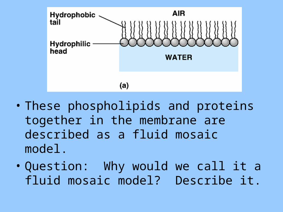
• These phospholipids and proteins together in the membrane are described as a fluid mosaic model.
• Question: Why would we call it a fluid mosaic model? Describe it.
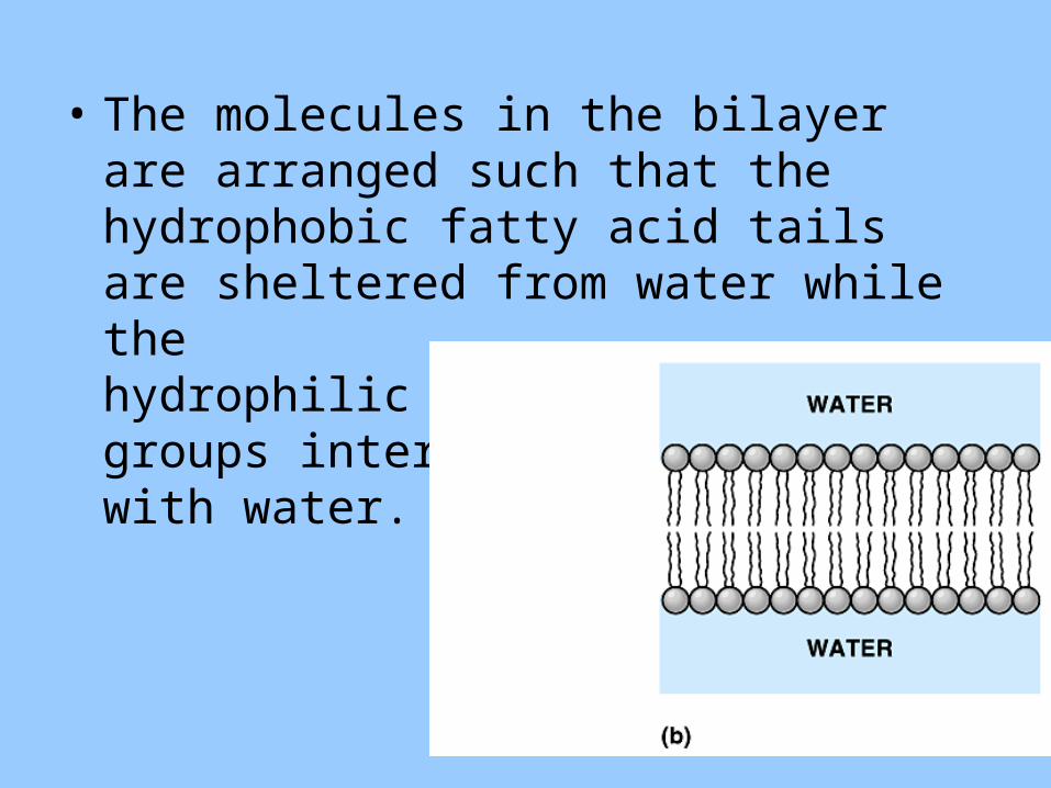
• The molecules in the bilayer are arranged such that the hydrophobic fatty acid tails are sheltered from water while the hydrophilic phosphate groups interact with water.
Want to learn more about how the fluid mosaic model was discovered?
• Check out the research in your book or online.
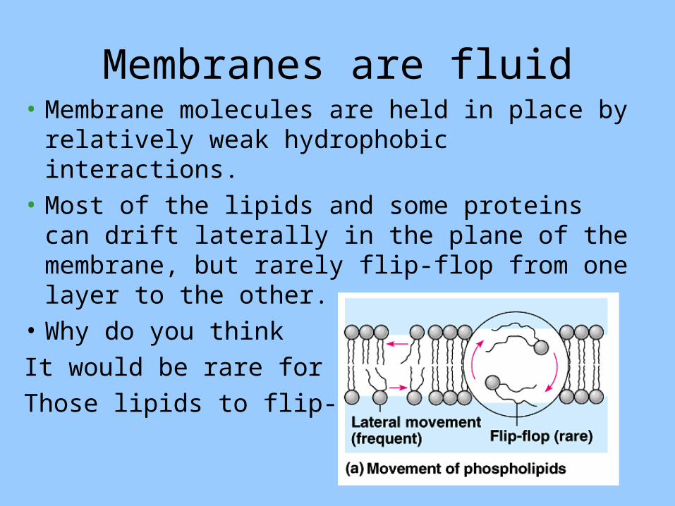
Membranes are fluid• Membrane molecules are held in place by
relatively weak hydrophobic interactions.• Most of the lipids and some proteins can drift
laterally in the plane of the membrane, but rarely flip-flop from one layer to the other.
• Why do you thinkIt would be rare forThose lipids to flip-flop?

• The lateral movements of phospholipids are rapid, about 2 microns per second.
• Many larger membrane proteins move more slowly but do drift.– Some proteins move in very directed manner,
perhaps guided/driven by the motor proteins attached to the cytoskeleton.
– Other proteins never move, anchored by the cytoskeleton.
Copyright © 2002 Pearson Education, Inc., publishing as Benjamin Cummings
Fig. 8.5

• Membrane fluidity is influenced by temperature and by its constituents.
• As temperatures cool, membranes switch from a fluid state to a solid state as the phospholipids are more closely packed.
• Membranes rich in unsaturated fatty acids are more fluid that those dominated by saturated fatty acids because the kinks in the unsaturated fatty acid tails prevent tight packing.
Copyright © 2002 Pearson Education, Inc., publishing as Benjamin Cummings
Fig. 8.4b

• The steroid cholesterol is wedged between phospholipid molecules in the plasma membrane of animals cells.
• At warm temperatures, it restrains the movement of phospholipids and reduces fluidity.
• At cool temperatures, it maintains fluidity by preventing tight packing.
Copyright © 2002 Pearson Education, Inc., publishing as Benjamin Cummings
Fig. 8.4c
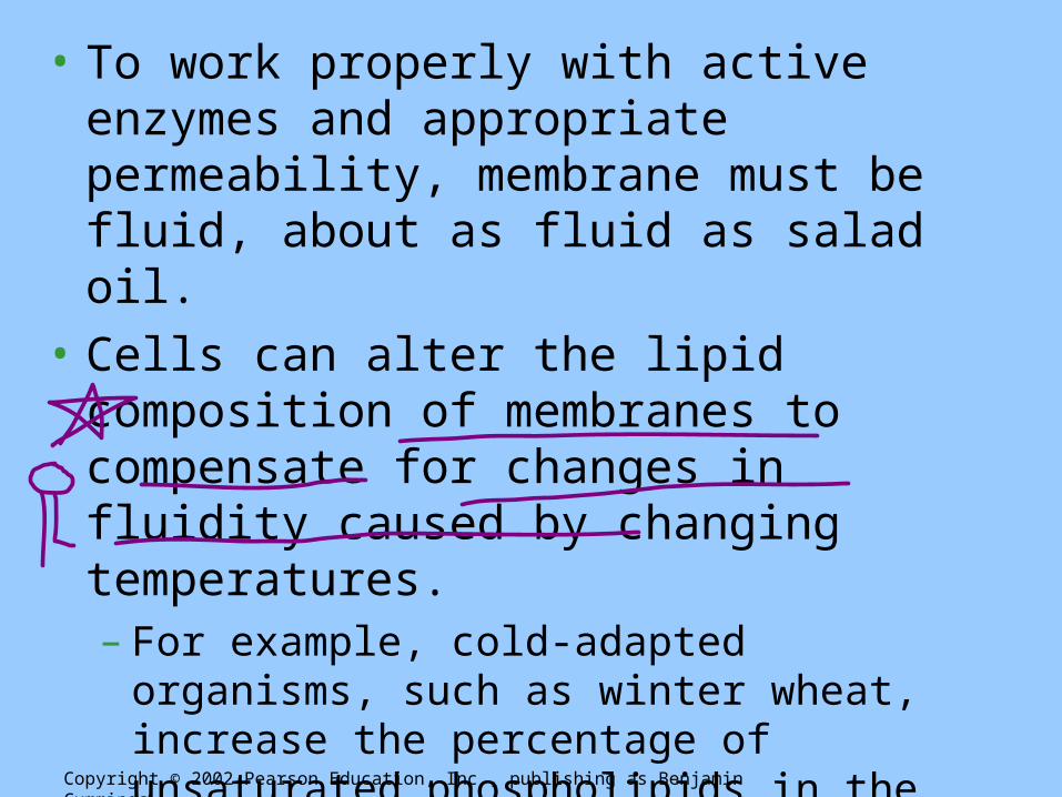
• To work properly with active enzymes and appropriate permeability, membrane must be fluid, about as fluid as salad oil.
• Cells can alter the lipid composition of membranes to compensate for changes in fluidity caused by changing temperatures.– For example, cold-adapted organisms, such as
winter wheat, increase the percentage of unsaturated phospholipids in the autumn.
– This allows these organisms to prevent their membranes from solidifying during winter.
Copyright © 2002 Pearson Education, Inc., publishing as Benjamin Cummings
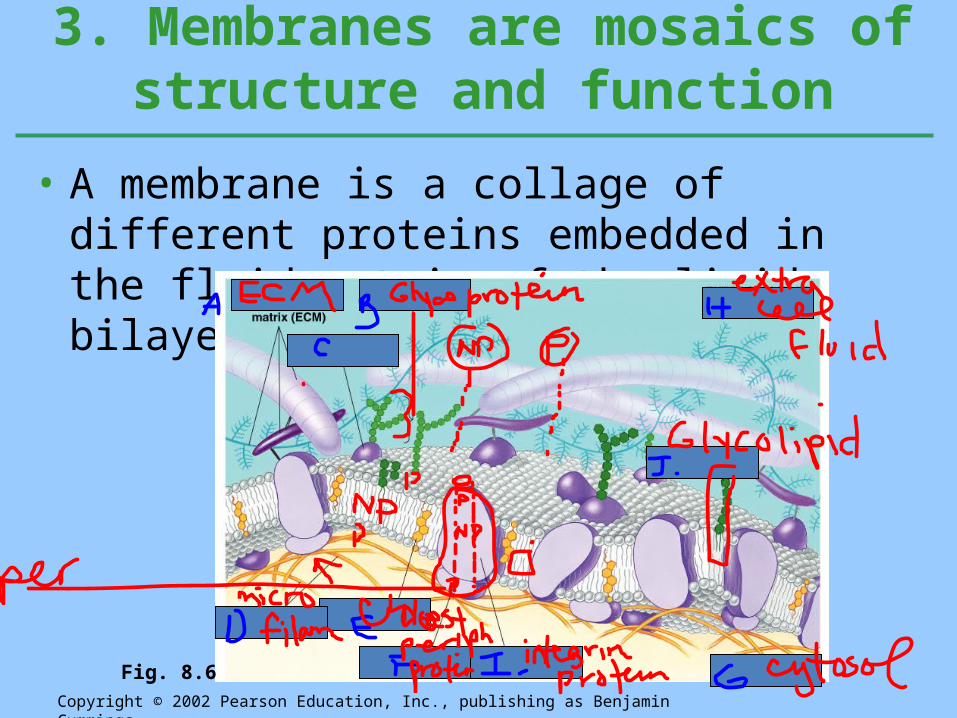
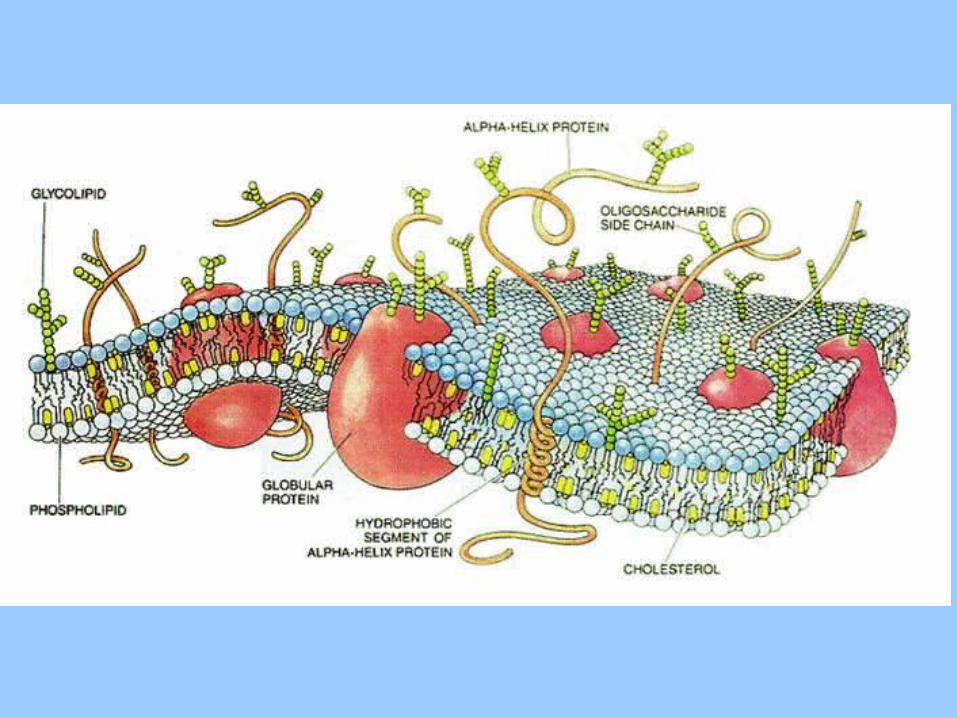
• A membrane is a collage of different proteins embedded in the fluid matrix of the lipid bilayer.
3. Membranes are mosaics of structure and function
Copyright © 2002 Pearson Education, Inc., publishing as Benjamin Cummings
Fig. 8.6
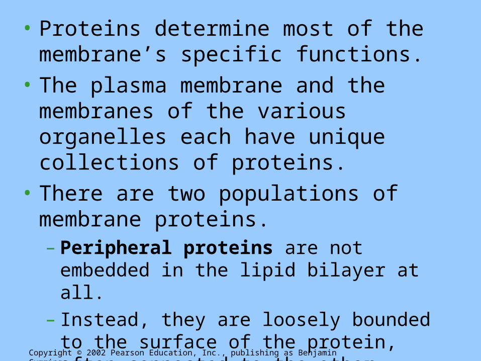
• Proteins determine most of the membrane’s specific functions.
• The plasma membrane and the membranes of the various organelles each have unique collections of proteins.
• There are two populations of membrane proteins.– Peripheral proteins are not embedded in the lipid
bilayer at all.– Instead, they are loosely bounded to the surface of
the protein, often connected to the other population of membrane proteins.
Copyright © 2002 Pearson Education, Inc., publishing as Benjamin Cummings
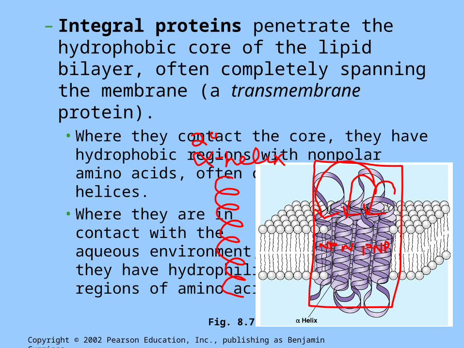
– Integral proteins penetrate the hydrophobic core of the lipid bilayer, often completely spanning the membrane (a transmembrane protein).• Where they contact the core, they have hydrophobic
regions with nonpolar amino acids, often coiled into alpha helices.• Where they are in
contact with the aqueous environment, they have hydrophilic regions of amino acids.
Copyright © 2002 Pearson Education, Inc., publishing as Benjamin Cummings
Fig. 8.7

Okay, break time
• Take a few minutes and reflect on what we just went over.
• Any Questions?
• One role of membrane proteins is to reinforce the shape of a cell and provide a strong framework.– On the cytoplasmic side, some membrane proteins
connect to the cytoskeleton.– On the exterior side, some membrane proteins
attach to the fibers of the extracellular matrix.
Copyright © 2002 Pearson Education, Inc., publishing as Benjamin Cummings

• The proteins in the plasma membrane may provide a variety of major cell functions.
Copyright © 2002 Pearson Education, Inc., publishing as Benjamin Cummings
Fig. 8.9

Stop• Break out time: • Each lab table will be given a different protein
function. Find this information in the book on page 144.– In your group, describe the primary function of your
protein channel– What do they look like? Do they have a special
configuration?– You will be explaining it to the class, so make sure you
know what you are talking about.
• The membrane plays the key role in cell-cell recognition.– Cell-cell recognition is the ability of a cell to
distinguish one type of neighboring cell from another.– This attribute is important in cell sorting and
organization as tissues and organs in development.– It is also the basis for rejection of foreign cells by the immune
system.– Cells recognize other cells by keying on surface molecules,
often carbohydrates, on the plasma membrane.
4. Membrane carbohydrates are important for cell-cell recognition
Copyright © 2002 Pearson Education, Inc., publishing as Benjamin Cummings

• Membrane carbohydrates are usually branched oligosaccharides with fewer than 15 sugar units.
• They may be covalently bonded either to lipids, forming glycolipids, or, more commonly, to proteins, forming glycoproteins.
• The oligosaccharides on the external side of the plasma membrane vary from species to species, individual to individual, and even from cell type to cell type within the same individual.– This variation marks each cell type as distinct.– The four human blood groups (A, B, AB, and O) differ in the
external carbohydrates on red blood cells.
CHAPTER 8MEMBRANE STRUCTURE AND
FUNCTION
Copyright © 2002 Pearson Education, Inc., publishing as Benjamin Cummings
Section B: Traffic Across Membranes1. A membrane’s molecular organization results in selective permeability
2. Passive transport is diffusion across a membrane
3. Osmosis is the passive transport of water
4. Cell survival depends on balancing water uptake and loss
5. Specific proteins facilitate the passive transport of water and selected solutes: a closer look
6. Active transport is the pumping of solutes against their gradients
7. Some ion pumps generate voltage across membranes
8. In cotransport, a membrane protein couples the transport of two solutes
9. Exocytosis and endocytosis transport large molecules
1. A membrane’s molecular organization results in selective
permeability (this should be a review)• A steady traffic of small molecules and ions
moves across the plasma membrane in both directions.– For example, sugars, amino acids, and other
nutrients enter a muscle cell and metabolic waste products leave.
– The cell absorbs oxygen and expels carbon dioxide.
– It also regulates concentrations of inorganic ions, like Na+, K+, Ca2+, and Cl-, by shuttling them across the membrane.
• However, substances do not move across the barrier indiscriminately; membranes are selectively permeable.
• Permeability of a molecule through a membrane depends on the interaction of that molecule with the hydrophobic core of the membrane.– Hydrophobic molecules, like hydrocarbons, CO2, and
O2, can dissolve in the lipid bilayer and cross easily.– Ions and polar molecules pass through with difficulty.• This includes small molecules, like water, and larger critical
molecules, like glucose and other sugars.– Proteins can assist and regulate the transport of ions
and polar molecules.
• Specific ions and polar molecules can cross the lipid bilayer by passing through transport proteins that span the membrane.– Some transport proteins have a hydrophilic channel
that certain molecules or ions can use as a tunnel through the membrane.
– Others bind to these molecules and carry their passengers across the membrane physically.
• Each transport protein is specific as to the substances that it will translocate (move).– For example, the glucose transport protein in the liver will
carry glucose from the blood to the cytoplasm, but not fructose, its structural isomer.
Copyright © 2002 Pearson Education, Inc., publishing as Benjamin Cummings

Tell me what diffusion is
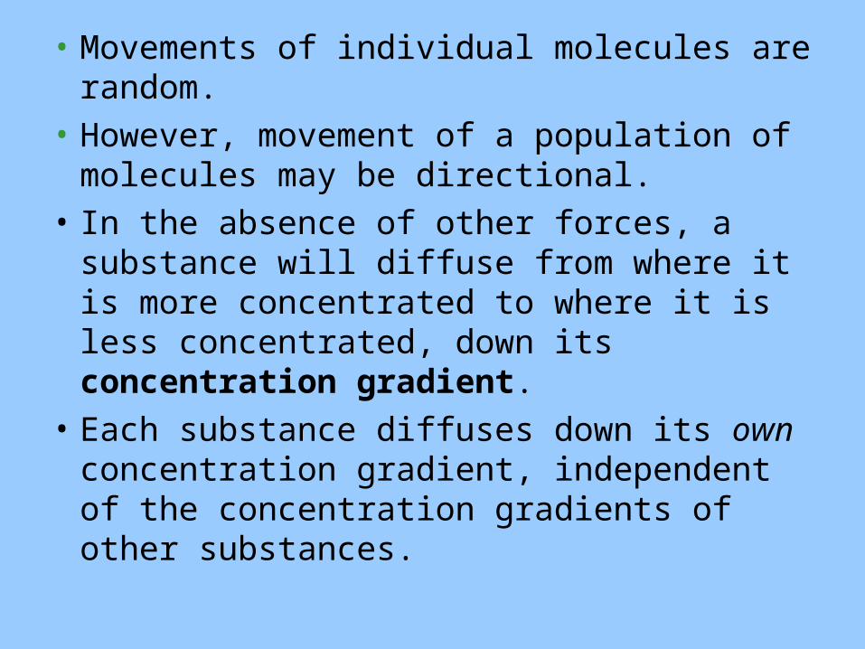
• Movements of individual molecules are random.
• However, movement of a population of molecules may be directional.
• In the absence of other forces, a substance will diffuse from where it is more concentrated to where it is less concentrated, down its concentration gradient.
• Each substance diffuses down its own concentration gradient, independent of the concentration gradients of other substances.


• The diffusion of a substance across a biological membrane is passive transport because it requires no energy from the cell to make it happen.– The concentration gradient represents potential
energy and drives diffusion.
• However, because membranes are selectively permeable, the interactions of the molecules with the membrane play a role in the diffusion rate.
• Diffusion of molecules with limited permeability through the lipid bilayer may be assisted by transport proteins.Copyright © 2002 Pearson Education, Inc., publishing as Benjamin Cummings
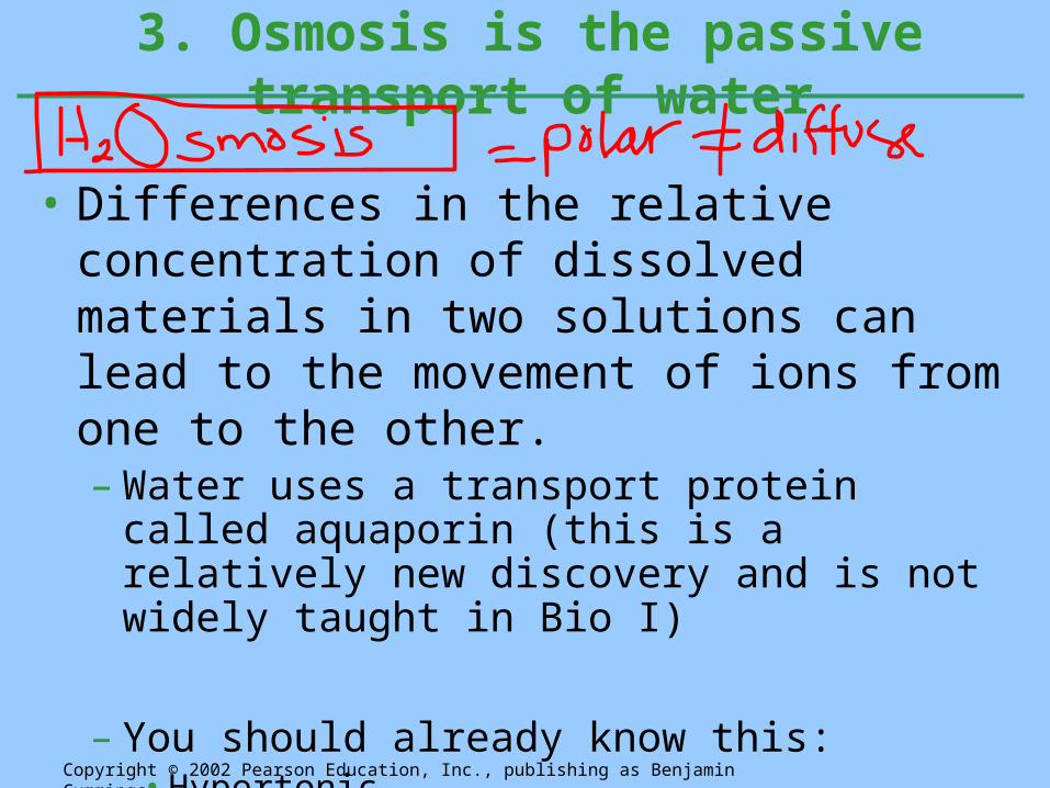
• Differences in the relative concentration of dissolved materials in two solutions can lead to the movement of ions from one to the other.– Water uses a transport protein called aquaporin (this
is a relatively new discovery and is not widely taught in Bio I)
– You should already know this:• Hypertonic• Hypotonic• Isotonic
3. Osmosis is the passive transport of water
Copyright © 2002 Pearson Education, Inc., publishing as Benjamin Cummings

Example Time

• The direction of osmosis is determined only by a difference in total solute concentration.– The kinds of solutes in the solutions do not matter.– This makes sense because the total solute
concentration is an indicator of the abundance of bound water molecules (and therefore of free water molecules).
• When two solutions are isotonic, water molecules move at equal rates from one to the other, with no net osmosis.
Copyright © 2002 Pearson Education, Inc., publishing as Benjamin Cummings
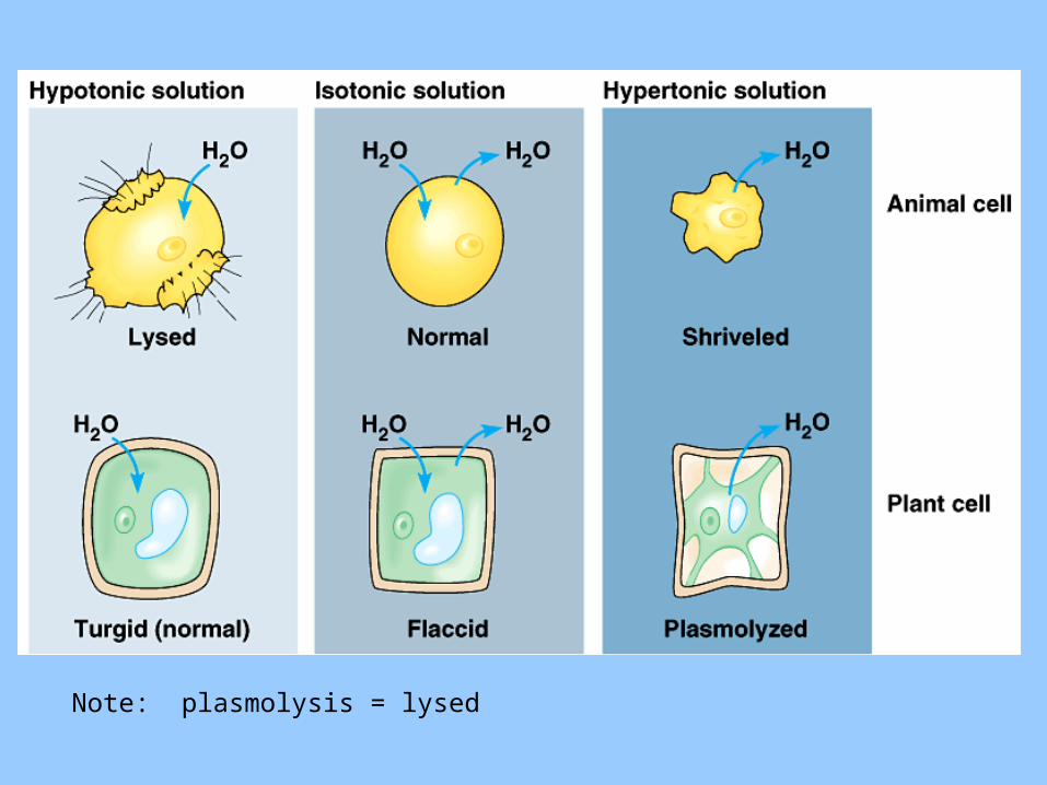
Note: plasmolysis = lysed
• For a cell living in an isotonic environment (for example, many marine invertebrates) osmosis is not a problem.– Similarly, the cells of most land animals are bathed
in an extracellular fluid that is isotonic to the cells.
• Organisms without rigid walls have osmotic problems in either a hypertonic or hypotonic environment and must have adaptations for osmoregulation to maintain their internal environment.
Copyright © 2002 Pearson Education, Inc., publishing as Benjamin Cummings
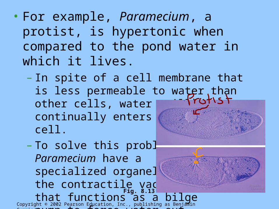
• For example, Paramecium, a protist, is hypertonic when compared to the pond water in which it lives.– In spite of a cell membrane that is less permeable
to water than other cells, water still continually enters the Paramecium cell.
– To solve this problem, Paramecium have a specialized organelle, the contractile vacuole, that functions as a bilge pump to force water out of the cell.
Copyright © 2002 Pearson Education, Inc., publishing as Benjamin Cummings
Fig. 8.13

Let’s talk about facillitated diffusion
• What do you already know?

• Transport proteins have much in common with enzymes.– They may have specific binding sites for the solute.– Transport proteins can become saturated when they
are translocating passengers as fast as they can.– Transport proteins can be inhibited by molecules that
resemble the normal “substrate.”• When these bind to the transport proteins, they outcompete
the normal substrate for transport.– While transport proteins do not usually catalyze chemical
reactions, they do catalyze a physical process, transporting a molecule across a membrane that would otherwise be relatively impermeable to the substrate.
Copyright © 2002 Pearson Education, Inc., publishing as Benjamin Cummings
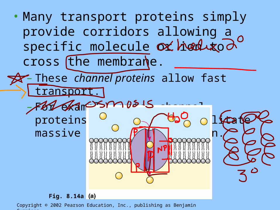
• Many transport proteins simply provide corridors allowing a specific molecule or ion to cross the membrane.– These channel proteins allow fast transport.– For example, water channel proteins, aquaprorins,
facilitate massive amounts of diffusion.
Copyright © 2002 Pearson Education, Inc., publishing as Benjamin Cummings
Fig. 8.14a

• Some channel proteins, gated channels, open or close depending on the presence or absence of a physical or chemical stimulus.
• The chemical stimulus is usually different from the transported molecule.• For example, when neurotransmitters bind to specific
gated channels on the receiving neuron, these channels open.– This allows sodium ions into a nerve cell.– When the neurotransmitters are not present, the channels are
closed.
Copyright © 2002 Pearson Education, Inc., publishing as Benjamin Cummings
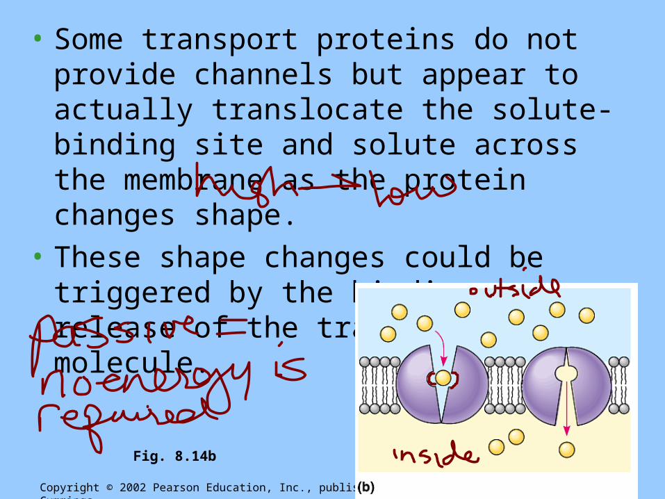
• Some transport proteins do not provide channels but appear to actually translocate the solute-binding site and solute across the membrane as the protein changes shape.
• These shape changes could be triggered by the binding and release of the transported molecule.
Copyright © 2002 Pearson Education, Inc., publishing as Benjamin Cummings
Fig. 8.14b
What is active transport???
• Active transport is performed by specific proteins embedded in the membranes.
• ATP supplies the energy for most active transport.– Often, ATP powers active transport by shifting a
phosphate group from ATP (forming ADP) to the transport protein.
– This may induce a conformational change in the transport protein that translocates the solute across the membrane.
Copyright © 2002 Pearson Education, Inc., publishing as Benjamin Cummings

• The sodium-potassium pump actively maintains the gradient of sodium (Na+) and potassium ions (K+) across the membrane.– Typically, an animal cell has higher concentrations
of K+ and lower concentrations of Na+ inside the cell (think keep K).
– The sodium-potassium pump uses the energy of one ATP to pump three Na+ ions out and two K+ ions in.
Copyright © 2002 Pearson Education, Inc., publishing as Benjamin Cummings

Copyright © 2002 Pearson Education, Inc., publishing as Benjamin Cummings
Fig. 8.15

Copyright © 2002 Pearson Education, Inc., publishing as Benjamin Cummings
Fig. 8.16 Both diffusion and facilitated diffusion are forms of passive transport of molecules down their concentration gradient, while active transport requires an investment of energy to move molecules against their concentration gradient.

• All cells maintain a voltage across their plasma membranes.– The cytoplasm of a cell is negative in charge
compared to the extracellular fluid because of an unequal distribution of cations and anions on opposite sides of the membrane.
– This voltage, the membrane potential, ranges from -50 to -200 millivolts.
7. Some ion pumps generate voltage across membranes
Copyright © 2002 Pearson Education, Inc., publishing as Benjamin Cummings
• The membrane potential acts like a battery.• The membrane potential favors the passive
transport of cations into the cell and anions out of the cell.
• Two combined forces, collectively called the electrochemical gradient, drive the diffusion of ions across a membrane: – a chemical force based in an ion’s concentration
gradient – an electrical force based on the effect of the
membrane potential on the ion’s movement.
Copyright © 2002 Pearson Education, Inc., publishing as Benjamin Cummings

• Ions diffuse not simply down its concentration gradient, but diffuses down its electrochemical gradient.– For example, before stimulation there is a higher
concentration of Na+ outside a resting nerve cell.– When stimulated, a gated channel opens and Na+ diffuse into the
cell down the electrochemical gradient.
• Special transport proteins, electrogenic pumps, generate the voltage gradients across a membrane– The sodium-potassium pump in animals restores the
electrochemical gradient not only by the active transport of Na+ and K+, but because it pumps two K+ ions inside for every three Na+ ions that it moves out.
Copyright © 2002 Pearson Education, Inc., publishing as Benjamin Cummings
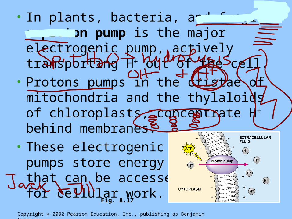
• In plants, bacteria, and fungi, a proton pump is the major electrogenic pump, actively transporting H+ out of the cell.
• Protons pumps in the cristae of mitochondria and the thylaloids of chloroplasts, concentrate H+ behind membranes.
• These electrogenic pumps store energy that can be accessed for cellular work.
Copyright © 2002 Pearson Education, Inc., publishing as Benjamin Cummings
Fig. 8.17
• A single ATP-powered pump that transports one solute can indirectly drive the active transport of several other solutes through cotransport via a different protein.
• As the solute that has been actively transported diffuses back passively through a transport protein, its movement can be coupled with the active transport of another substance against its concentration gradient.
8. In cotransport, a membrane protein couples the transport of two solutes
Copyright © 2002 Pearson Education, Inc., publishing as Benjamin Cummings

• Plants commonly use the gradient of hydrogen ions that is generated by proton pumps to drive the active transport of amino acids, sugars, and other nutrients into the cell.– The high concentration of H+ on one side of the
membrane, created by the proton pump, leads to the facilitated diffusion of protons back, but only if another molecule, like sucrose, travels with the hydrogen ion.
Copyright © 2002 Pearson Education, Inc., publishing as Benjamin Cummings
Fig. 8.18

• Small molecules and water enter or leave the cell through the lipid bilayer or by transport proteins.
• Large molecules, such as polysaccharides and proteins, cross the membrane via vesicles.
• During exocytosis, a transport vesicle budded from the Golgi apparatus is moved by the cytoskeleton to the plasma membrane.
• When the two membranes come in contact, the bilayers fuse and spill the contents to the outside.
9. Exocytosis and endocytosis transport large molecules
Copyright © 2002 Pearson Education, Inc., publishing as Benjamin Cummings

• During endocytosis, a cell brings in macromolecules and particulate matter by forming new vesicles from the plasma membrane.
• Endocytosis is a reversal of exocytosis.– A small area of the palsma membrane sinks inward
to form a pocket– As the pocket into the plasma membrane deepens,
it pinches in, forming a vesicle containing the material that had been outside the cell
Copyright © 2002 Pearson Education, Inc., publishing as Benjamin Cummings

• One type of endocytosis is phagocytosis, “cellular eating”.
• In phagocytosis, the cell engulfs a particle by extending pseudopodia around it and packaging it in a large vacuole.
• The contents of the vacuole are digested when the vacuole fuses with a lysosome.
Copyright © 2002 Pearson Education, Inc., publishing as Benjamin Cummings
Fig. 8.19a

• In pinocytosis, “cellular drinking”, a cell creates a vesicle around a droplet of extracellular fluid.– This is a non-specific process.
Copyright © 2002 Pearson Education, Inc., publishing as Benjamin Cummings
Fig. 8.19b
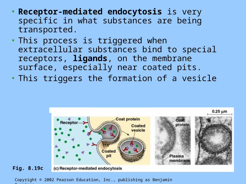
• Receptor-mediated endocytosis is very specific in what substances are being transported.
• This process is triggered when extracellular substances bind to special receptors, ligands, on the membrane surface, especially near coated pits.
• This triggers the formation of a vesicle
Copyright © 2002 Pearson Education, Inc., publishing as Benjamin Cummings
Fig. 8.19c

• Receptor-mediated endocytosis enables a cell to acquire bulk quantities of specific materials that may be in low concentrations in the environment.– Human cells use this process to absorb cholesterol.– Cholesterol travels in the blood in low-density
lipoproteins (LDL), complexes of protein and lipid.– These lipoproteins bind to LDL receptors and enter the
cell by endocytosis.– In familial hypercholesterolemia, an inherited disease, the LDL
receptors are defective, leading to an accumulation of LDL and cholesterol in the blood.
– This contributes to early atherosclerosis.
Copyright © 2002 Pearson Education, Inc., publishing as Benjamin Cummings

WOW!!! That was a lot of info!
• Now what do you remember????