Embed Size (px)
Citation preview
C H A P T E Rz z z z z z z z z z z z z z z z z z z z z z z z z z z22
Enhancement of Skeletal RepairEnhancement of Skeletal Repair
z z z z z z z z z z z z z z z z z z z z z z z z z z z z z z z z z z z z z z z z z z z z z z z z z z z z z z z z z z z z z z z z z z z z z z z z z z z z z z z z z z z z z z z z z z z z z z z z z z z z z z z z z z z z z z
Calin S. Moucha, M.D.Thomas A. Einhorn, M.D.
The process of skeletal repair is considered to bebiologically optimal under most clinical conditions. How-ever, of the 6.2 million fractures that occur annually in theUnited States, 5% to 10% go on to nonunion or delayedunion.210 In many instances, the cause of impaired healingis unknown; however, surgical and nonsurgical interven-tions can interfere with healing and may cause delayedunion or nonunion.26 Technical errors during surgery,53
systemic status of the patient,78, 156, 165, 226, 274 and thenature of the traumatic injury itself66, 189, 268 are just someof the many factors that may influence fracture healing.Even when some or all of these risk factors are avoided,many fractures fail to heal.77 Moreover, specific parts of theskeleton are known to be at increased risk for impairedfracture healing. At these sites, there may be problemsrelated to peculiarities of local blood supply or difficultiesin controlling the mechanical strain environment. Ex-amples include the neck of the talus, the neck of the femur,and the carpal scaphoid.32 Therefore, although mostfractures heal uneventfully, clinical scenarios exist in whichenhancement of fracture healing would be of benefit toensure rapid restoration of skeletal function. This chapterreviews some of the methods that have been shown tostimulate skeletal repair.
PHYSICAL METHODS OFENHANCEMENTz z z z z z z z z z z z z z z z z z z z z z z z z z z z z z z z z z z z z z z z z z z z z z z z z z z z z z z z z z z
A fracture’s mechanical environment can play an importantrole in its healing. For example, unstable fixation causesexcessive interfragmentary movement and may retard therepair process. On the other hand, controlled micromotionor controlled, rhythmic distraction of a fracture site canenhance fracture healing.109, 128, 142, 175 Knowledge ofhow strategic alteration of the mechanical environment ofa fracture influences its healing is based on resultsobtained from clinical studies using different operative
treatments such as rigid internal fixation, external fixation,or intramedullary fixation. To develop new methods forthe stimulation of fracture healing by mechanical means, itis necessary to gain a fundamental understanding of theways by which mechanical forces are transduced intocellular and molecular signals.
Methods for enhancing fracture repair using biophysi-cal techniques include electrical or electromagnetic stim-ulation of nonunions and ultrasound stimulation of freshfractures. Each of these techniques has shown substantialefficacy in well-controlled clinical trials and, as we discussin this chapter, there is abundant cellular and molecularknowledge that exists on which to base hypotheticalexplanations for the observed effects in fractures. Treat-ment of a nonunion, however, may differ from that of afresh fracture, and the successful use of a biophysicalmodality in one setting may not transfer to another.
Mechanical Enhancement
The quality and the quantity of the callus formed by ahealing fracture can be greatly influenced by the operativeor nonoperative method used to treat it. Generally, motionat the site of a fracture causes callus formation.204 Thecallus functions to decrease the initial interfragmentarymotion sufficiently enough to produce an environmentsuitable for fracture union. This goal is attained byimproving the structure of the fracture site (by increasingcross-sectional area) and by recruiting cells that arenecessary for bony regeneration. If the motion becomesexcessive, however, a hypertrophic nonunion may result.
As early as the 19th century, physicians disagreed asto the implications of weight bearing on a healingbone. Nicholas Andre (1659–1742) and Just Lucas-Championniere (1843–1913) believed that early con-trolled activity promoted healing of tissues, whereas JohnHunter (1728–1793), John Hilton (1807–1878), andHugh Owen Thomas (1834–1878) did not.31 In 1892,Julius Wolff280 suggested that the structure of bone adapts
639
#6818 @ l 1/ id/CLS b k /GRP d /JOB b /DIV h22 8/29/02 12 P 1/22 P 639 COLOR T#6818 @ l 1/ id/CLS b k /GRP d /JOB b /DIV h22 8/29/02 12 P 1/22 P 639 BLACK T
to changes in its stress environment. This concept hascome to be known as Wolff’s law. Several investigators haveattempted to delineate the molecular and cellular mecha-nisms that govern the ability of bone to respond toloading.* The conclusion drawn from these studies is thattissue loading influences cell shape, gene expression,protein synthesis, and proliferation of several cell typesfound in the fracture callus.
Controlling the weight-bearing status of a limb is onemethod of clinically altering the stress environment at afracture site. Reports of the effects of weight bearing,however, have been conflicting. Sarmiento and associ-ates236 found that weight bearing improved fracturehealing in the femurs of normal rats. Kirchen andcolleagues147 suggested that microgravity, such as duringspace flight, may influence fracture healing. Aro andassociates9 showed no difference in fracture healing,regardless of weight bearing, in paraplegic rats. Similarly,Riggins and co-workers218 reported that weight bearinghad no effect on fracture healing in chickens. Meadowsand colleagues177 evaluated the effect of weight bearing onthe healing characteristics of a cortical defect in caninetibias. They demonstrated an increase in the amount ofwoven bone formed in defects of weight-bearing tibiascompared with that in non–weight-bearing tibias. Thisfinding appeared to reflect a disuse response in theunderloaded bones as opposed to the formation of morebone in those that bore weight. The investigators con-cluded that weight bearing was a permissive factor for theformation of woven bone in tibial defects and that it mayincrease the formation of bone during the process ofskeletal repair.
The two factors that have received the most investiga-tive attention with respect to the ways in which mechan-ical treatment influences fracture healing are blood flowand interfragmentary strain. Smith and associates251
studied the blood flow to cortical bone in canine tibias thathad been subjected to an experimental osteotomy anddifferent modes of fixation. They showed that when nointernal fixation was used after the osteotomy, blood flowto the canine tibial diaphysis was reduced. Four hourslater, blood flow to the fracture site was further reduced byan additional 50%. Reaming of the tibia and insertion of atight-fitting intramedullary nail reduced the blood floweven further. On the other hand, fractures that weretreated with either internal or external fixation did notexperience such dramatic reductions in blood flow.197
Whereas early stability appears to have a beneficial effecton blood flow, instability and reaming of the endostealbone have deleterious effects. It is possible, however, thatthe development of collateral vessels overcomes the lattereffect.
The theory of interfragmentary strain suggests that it isthe balance that develops between the degree of localinterfragmentary strain and the ability of the callus towithstand the strain that determines the type of healing.Granulation tissue can tolerate 100% strain; fibrous tissueand cartilage withstand lower amounts. Therefore, granu-lation tissue is best able to tolerate the changes in
interfragmentary motion that occur during early reparativestages of fracture healing. As the fracture becomes morestable, the presence of cartilage and new bone reduces thestrain and allows fracture healing to proceed. Strain isusually proportional to the size of the fracture gap, but asmall gap, such as that which is present in a plated femurin which the fracture ends are not actually apposed, has ahigh strain. As a result, resorption occurs, allowinggranulation tissue and callus to form and produce a lowerstrain environment.
The influences of different mechanical loading condi-tions on tissue differentiation have been studied in avariety of skeletal settings. These investigations have led toa theory that relates mechanical loading history to tissuedifferentiation in the process of endochondral bonerepair. It identifies tissue vascularity and two key mechan-ical parameters, cyclic hydrostatic stress (pressure) andcyclic tensile strain, as important determinants of tissuedifferentiation. Using an osteotomized long bone as theexperimental system, Blenman19 and Carter40 and theirco-workers performed two-dimensional finite elementanalyses to model idealized fracture callus, includingperiosteal, endosteal, and fracture gap (interfragmentary)callus regions. The stress and strain histories at eachlocation within the callus were calculated. High levels ofcompressive hydrostatic stress occurred within the fracturegap. At the middle of the gap, high levels of strain werepresent in the radial and circumferential directions. Incontrast, at the periosteal and endosteal callus regionsremote from the interfragmentary gap, low levels ofhydrostatic stress and tensile strain were observed. Thesefindings suggest that an association exists among (1) in-termittent compressive hydrostatic stress and chondrogen-esis, (2) intermittent strain and fibrogenesis, and (3) lowlevels of mechanical stimulation and osteogenesis (withgood vascularity) or chondrogenesis (with poor vascular-ity). In addition, these studies showed that after cartilageforms within callus, moderate levels of cyclic tensile strain(or distortional strain) accelerate endochondral ossifica-tion. Local cyclic hydrostatic stress delays endochondralossification, however, possibly via an inhibition of revas-cularization. More recent research by Claes and associ-ates54 supports similar concepts.
A few investigators have attempted to design systems inwhich mechanical input would stimulate fracture repair.Goodship and Kenwright109 studied the influence ofcontrolled micromotion on fracture healing in two groupsof sheep in which tibial diaphyseal fractures had beencreated. The tibial fractures in one group were treated withrigid external fixation, and those in the other group weresubjected to a regimen of controlled axial micromotion(500 cycles at 0.5 Hz) for 17 min/day. Enhanced fracturerepair was demonstrated by radiographic, histologic, andbiomechanical analyses in the group having micromotion.In a subsequent clinical, prospective randomized con-trolled trial, Kenwright and colleagues142 compared theeffects of controlled axial micromotion on tibial diaphysealfracture healing in patients who were treated with externalfixation and stratified according to fracture severity gradeand extent of soft tissue injury. The tibias that were treatedwith induced micromotion were subjected to controlledlongitudinal displacement and loading of the transfixion
*See references 36, 47, 113, 114, 116, 129, 138, 145, 172, 198,227, 264.
640 SECTION I • General Principles
#6818 @ l 1/ id/CLS b k /GRP d /JOB b /DIV h22 8/29/02 12 P 2/22 P 640 COLOR T#6818 @ l 1/ id/CLS b k /GRP d /JOB b /DIV h22 8/29/02 12 P 2/22 P 640 BLACK T
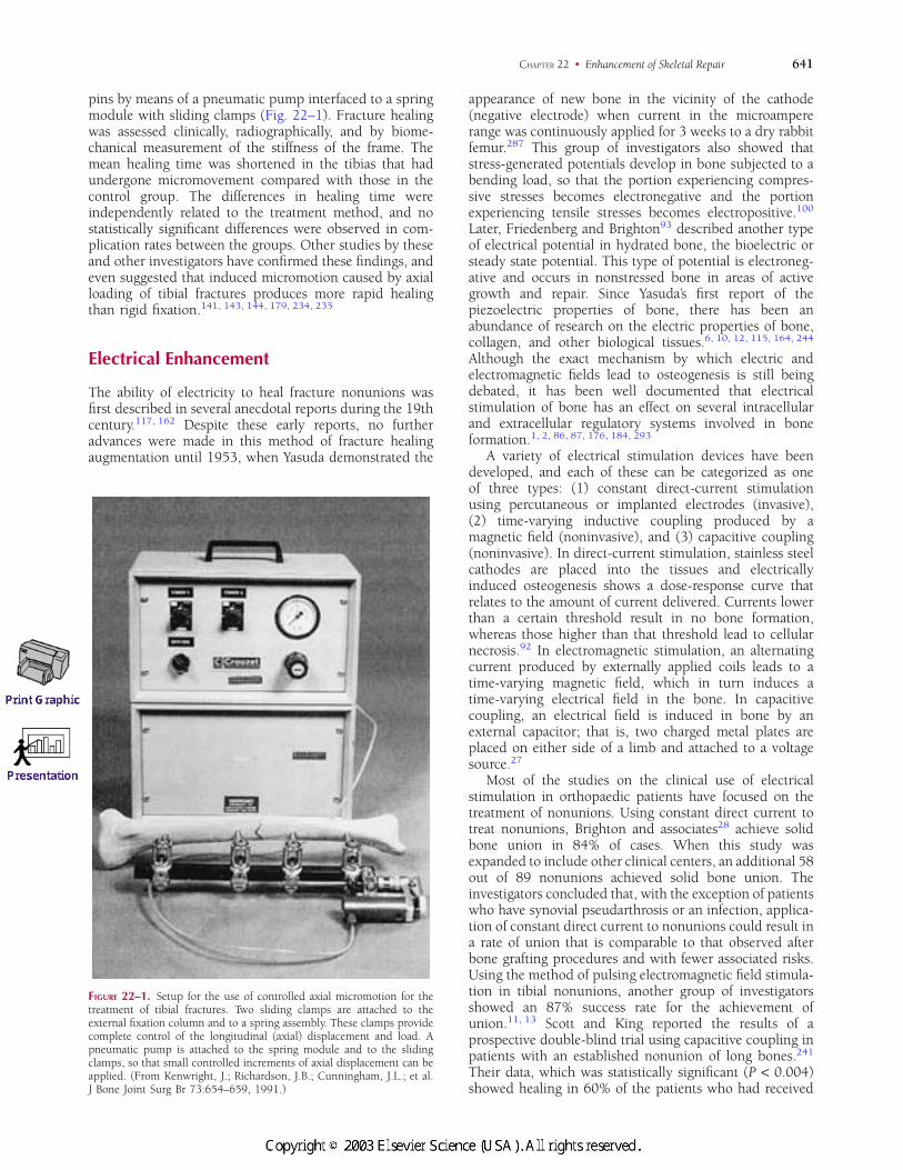
pins by means of a pneumatic pump interfaced to a springmodule with sliding clamps (Fig. 22–1). Fracture healingwas assessed clinically, radiographically, and by biome-chanical measurement of the stiffness of the frame. Themean healing time was shortened in the tibias that hadundergone micromovement compared with those in thecontrol group. The differences in healing time wereindependently related to the treatment method, and nostatistically significant differences were observed in com-plication rates between the groups. Other studies by theseand other investigators have confirmed these findings, andeven suggested that induced micromotion caused by axialloading of tibial fractures produces more rapid healingthan rigid fixation.141, 143, 144, 179, 234, 235
Electrical Enhancement
The ability of electricity to heal fracture nonunions wasfirst described in several anecdotal reports during the 19thcentury.117, 162 Despite these early reports, no furtheradvances were made in this method of fracture healingaugmentation until 1953, when Yasuda demonstrated the
appearance of new bone in the vicinity of the cathode(negative electrode) when current in the microampererange was continuously applied for 3 weeks to a dry rabbitfemur.287 This group of investigators also showed thatstress-generated potentials develop in bone subjected to abending load, so that the portion experiencing compres-sive stresses becomes electronegative and the portionexperiencing tensile stresses becomes electropositive.100
Later, Friedenberg and Brighton93 described another typeof electrical potential in hydrated bone, the bioelectric orsteady state potential. This type of potential is electroneg-ative and occurs in nonstressed bone in areas of activegrowth and repair. Since Yasuda’s first report of thepiezoelectric properties of bone, there has been anabundance of research on the electric properties of bone,collagen, and other biological tissues.6, 10, 12, 115, 164, 244
Although the exact mechanism by which electric andelectromagnetic fields lead to osteogenesis is still beingdebated, it has been well documented that electricalstimulation of bone has an effect on several intracellularand extracellular regulatory systems involved in boneformation.1, 2, 86, 87, 176, 184, 293
A variety of electrical stimulation devices have beendeveloped, and each of these can be categorized as oneof three types: (1) constant direct-current stimulationusing percutaneous or implanted electrodes (invasive),(2) time-varying inductive coupling produced by amagnetic field (noninvasive), and (3) capacitive coupling(noninvasive). In direct-current stimulation, stainless steelcathodes are placed into the tissues and electricallyinduced osteogenesis shows a dose-response curve thatrelates to the amount of current delivered. Currents lowerthan a certain threshold result in no bone formation,whereas those higher than that threshold lead to cellularnecrosis.92 In electromagnetic stimulation, an alternatingcurrent produced by externally applied coils leads to atime-varying magnetic field, which in turn induces atime-varying electrical field in the bone. In capacitivecoupling, an electrical field is induced in bone by anexternal capacitor; that is, two charged metal plates areplaced on either side of a limb and attached to a voltagesource.27
Most of the studies on the clinical use of electricalstimulation in orthopaedic patients have focused on thetreatment of nonunions. Using constant direct current totreat nonunions, Brighton and associates28 achieve solidbone union in 84% of cases. When this study wasexpanded to include other clinical centers, an additional 58out of 89 nonunions achieved solid bone union. Theinvestigators concluded that, with the exception of patientswho have synovial pseudarthrosis or an infection, applica-tion of constant direct current to nonunions could result ina rate of union that is comparable to that observed afterbone grafting procedures and with fewer associated risks.Using the method of pulsing electromagnetic field stimula-tion in tibial nonunions, another group of investigatorsshowed an 87% success rate for the achievement ofunion.11, 13 Scott and King reported the results of aprospective double-blind trial using capacitive coupling inpatients with an established nonunion of long bones.241
Their data, which was statistically significant (P < 0.004)showed healing in 60% of the patients who had received
FIGURE 22–1. Setup for the use of controlled axial micromotion for thetreatment of tibial fractures. Two sliding clamps are attached to theexternal fixation column and to a spring assembly. These clamps providecomplete control of the longitudinal (axial) displacement and load. Apneumatic pump is attached to the spring module and to the slidingclamps, so that small controlled increments of axial displacement can beapplied. (From Kenwright, J.; Richardson, J.B.; Cunningham, J.L.; et al.J Bone Joint Surg Br 73:654–659, 1991.)
641CHAPTER 22 • Enhancement of Skeletal Repair
#6818 @ l 1/ id/CLS b k /GRP d /JOB b /DIV h22 8/29/02 12 P 3/22 P 641 COLOR T#6818 @ l 1/ id/CLS b k /GRP d /JOB b /DIV h22 8/29/02 12 P 3/22 P 641 BLACK T
electrical stimulation but in none of the patients who hadbeen managed with a placebo unit. More recently, Good-win and colleagues110 performed a multicenter random-ized double-blind prospective comparison to evaluate theeffect of noninvasive capacitively coupled electrical stimu-lation on the success rate of lumbar spine fusion surgery.For the 179 patients who completed treatment and evalua-tion, the overall protocol success rate was 84.7% for theactive patients and 64.9% for the placebo patients. Accord-ing to the Yates corrected chi-square test, these results werealso statistically significant (P = 0.0043). However, thestudy had a 12% dropout rate due to noncompliance, thepatients had varying degrees of instability of the spine, andthe differences among the various surgeons with regard toexperience performing internal fixation of the spine werenot taken into account in the study.
Although these reports show that electrical stimulationmay be successful in the treatment of nonunions and, in aselect number of patients, spinal fusion, the application ofthis technology to the treatment of fresh fractures has notbeen clearly demonstrated. Two studies have shown thatpulsed electromagnetic fields enhance bone regenerationin fresh osteotomies and fracture animal models.91, 233 Inthe more recent of these studies, Fredericks and colleaguesperformed tibial osteotomies stabilized by external fixationin New Zealand white rabbits. One day after surgery, theanimals were randomly assigned to receive either noexposure, 30 minutes, or 60 minutes per day of alow-frequency, low-amplitude pulsed electromagnetic field(PEMF). Specimens were examined biomechanically andradiographically, and the results indicated that normalintact torsional strength was achieved by 14 days in the60-minute PEMF group, by 21 days in the 30-minutePEMF group, and by 28 days in the sham controls. Inaddition, the 60-minute PEMF-treated osteotomies hadsignificantly higher torsional strength than did shamcontrols at 14 and 21 days postoperatively. The 30-minutePEMF-treated osteotomies were significantly stronger thanthose in the sham controls only after 21 days. Lastly,maximum fracture callus area correlated with the time toreach normal torsional strength. Others have failed toreproduce these results.4, 159 To our knowledge, nopublished clinical study has shown that electrical stimu-lation enhances the repair of fresh fractures in humans.
Another potential application of electrical stimulation isin the treatment of fractures that show delayed union.Sharrard247 conducted a double-blind, multicenter trial ofthe use of PEMFs in patients who had a delayed union ofa tibial fracture. Forty-five tibial fractures that had notunited for more than 16 but less than 32 weeks weretreated with immobilization in a plaster cast incorporatingthe coils of an electromagnetic stimulation unit. The unitwas activated in 20 of these fractures. The results showedradiographic evidence of union in nine of the fractures thathad undergone active electromagnetic stimulation and inonly three of the fractures in the control group.
Although there has been an abundance of research onthe use of electrical enhancement of fracture healing,numerous questions still abound. More studies are neces-sary to further understand the use of this modality at boththe basic science and clinical levels, especially with regardto its use in fresh fractures.
Ultrasonic Enhancement
Low-intensity pulsed ultrasound has been known forsome time to stimulate fresh fracture healing in experi-mental animals and healing of nonunions in hu-mans.74, 173, 206, 275, 282 Its use in enhancing the healing offresh fractures in humans, however, is more controversial.Heckman and co-workers,123 in a prospective, random-ized, double-blind evaluation, examined the use of a newultrasound stimulating device as an adjunct to conven-tional treatment with a cast of 67 closed or grade-I openfractures of the tibial shaft. Thirty-three fractures weretreated with the active device and 34 with a placebo controldevice. At the end of the treatment, there was a statisticallysignificant decrease in the time to clinical healing (86 ± 5.8days in the active-treatment group compared with 114 ±10.4 days in the control group) (P = 0.01) and a significantdecrease in the time to overall (clinical and radiographic)healing (96 ± 4.9 days in the active-treatment groupcompared with 154 ± 13.7 days in the control group) (P =0.0001). The patients’ compliance with the use of thedevice was excellent, and no serious complications werereported. This study confirmed earlier studies that demon-strated the efficacy of low-intensity ultrasound stimulationin the acceleration of the normal fracture repair process.Kristiansen showed similar findings in an investigation ofpatients with fresh distal radius fractures.152
Cook and associates60 investigated the ability oflow-intensity ultrasound to accelerate the healing of tibialand distal radius fractures in smokers. The usual healingtime for tibial fractures in smokers is 175 ± 27 days, butwith ultrasound treatment the healing time was reduced by41% to 103 ± 8.3 days. Smokers with distal radiusfractures had a healing time of 98 ± 30 days, which wasreduced by 51% to 48 ± 5.1 days with ultrasoundtreatment. Treatment with the active ultrasound devicealso substantially reduced the incidence of delayed unionsin tibias in smokers and nonsmokers. These results haveoptimistic implications because they suggest that ultra-sound can mitigate the delayed healing effects of smoking,which is a common risk factor associated with nonunionsand delayed unions. Moreover, this study is especiallynoteworthy because it provides insight into the mechanismthat mediates this fracture-healing modality. Althoughseveral theories of how ultrasound enhances fracturehealing have been proposed,200, 285 by showing thatultrasound facilitates bony regeneration in a host that hasa systemic reduction in generalized healing due to pooroxygen transport,248 one can hypothesize that ultrasoundmay elicit its effects, at least in part, by enhancing thedelivery of oxygen. This theory is supported by an in vitrostudy on the effect of ultrasound on human mandibularosteoblasts, gingival fibroblasts, and monocytes.216 Theauthors of this study showed that ultrasound stimulatesthese cells to produce angiogenic factors such asinterleukin-8 (IL-8), fibroblast growth factor (FGF), andvascular endothelial growth factor, therefore suggestingthat the effects of ultrasound treatment are mediated bystimulation of angiogenesis, which ultimately enhances thehealing environment caused by local hypoxia.
Although these studies give credence to the value ofultrasound in enhancing healing of fresh human fractures
642 SECTION I • General Principles
#6818 @ l 1/ id/CLS b k /GRP d /JOB b /DIV h22 8/29/02 12 P 4/22 P 642 COLOR T#6818 @ l 1/ id/CLS b k /GRP d /JOB b /DIV h22 8/29/02 12 P 4/22 P 642 BLACK T
treated nonoperatively, a study by Emami and co-workers84 showed no effect of low-intensity ultrasound onhealing time of fresh tibial fractures treated with a reamedand statically locked intramedullary rod. Therefore, todate, it appears that the clinical use of ultrasound forenhancing fracture healing has been shown to be beneficialonly in delayed unions and nonunions and in freshfractures in smokers. At least one type of ultrasound devicehas been approved for marketing in the United States, andit is anticipated that more clinical data will be available onthe use of this biophysical signal in the near future.
BIOLOGIC METHODSOF ENHANCEMENTz z z z z z z z z z z z z z z z z z z z z z z z z z z z z z z z z z z z z z z z z z z z z z z z z z z z z z z z z z z
Knowledge of the cellular and molecular biology of themusculoskeletal system has led to a better understandingof the basic processes that regulate repair of skeletaltissues. To apply this new information to the enhancementof skeletal repair, the specificity of a particular stimulus forits receptor or targeted pathway must first be determined.Local stimulation of skeletal repair involves not only thedevelopment of specific molecules to stimulate discretecomponents of the healing process but also the design ofdelivery systems to optimize the effect of a stimulatingfactor. The development of systemic methods for enhance-ment of skeletal repair is attractive, but the introduction ofa systemic agent that targets these processes requires a highdegree of specificity, and this approach needs moreextensive investigation. Local factors for the stimulation ofskeletal healing have been evaluated, and data are availablefrom both clinical and experimental studies.
Local Enhancement
Local methods for the enhancement of skeletal repair canbe categorized into osteogenic, osteoconductive, andosteoinductive approaches. Osteogenesis is the process ofnew bone formation. Osteogenic approaches to fracturehealing enhancement include the use of naturally occur-ring materials that have been shown to induce orsupport bone formation, such as autologous bone mar-row grafts55, 102, 246 and autologous or allogeneic bonegrafts.33, 35, 96 Experience with bone grafting dates back tothe early 1900s,46 and it has been estimated that there aremore than 250,000 bone grafts performed annually in theUnited States.182 Osteoconduction is the process by whichfibrovascular tissue and osteoprogenitor cells invade aporous structure that acts as a temporary scaffold andreplace it with newly formed bone. The most widelystudied osteoconductive substances are those composedof hydroxyapatite, calcium phosphate or calcium sul-fate composites, and the bioactive glasses.157 Lastly, theprocess that promotes mitogenesis of undifferentiatedmesenchymal cells, leading to formation of osteoprogeni-tor cells that have osteogenic capacity, is known asosteoinduction. Urist made the first observation thatimplantation of demineralized lyophilized segments ofbone matrix either subcutaneously or intramuscularly in
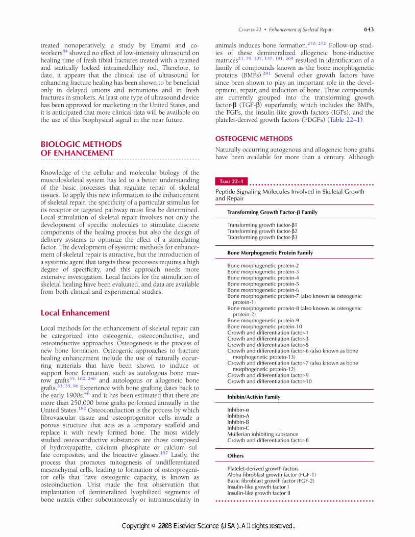
animals induces bone formation.270, 272 Follow-up stud-ies of these demineralized allogeneic bone-inductivematrices21, 79, 107, 137, 181, 269 resulted in identification of afamily of compounds known as the bone morphogeneticproteins (BMPs).281 Several other growth factors havesince been shown to play an important role in the devel-opment, repair, and induction of bone. These compoundsare currently grouped into the transforming growthfactor-β (TGF-β) superfamily, which includes the BMPs,the FGFs, the insulin-like growth factors (IGFs), and theplatelet-derived growth factors (PDGFs) (Table 22–1).
OSTEOGENIC METHODS
Naturally occurring autogenous and allogeneic bone graftshave been available for more than a century. Although
TABLE 22–1z z z z z z z z z z z z z z z z z z z z z z z z z z z z z z z z z z z z z z z z z z
Peptide Signaling Molecules Involved in Skeletal Growthand Repair
Transforming Growth Factor-� Family
Transforming growth factor-β1Transforming growth factor-β2Transforming growth factor-β3
Bone Morphogenetic Protein Family
Bone morphogenetic protein-2Bone morphogenetic protein-3Bone morphogenetic protein-4Bone morphogenetic protein-5Bone morphogenetic protein-6Bone morphogenetic protein-7 (also known as osteogenic
protein-1)Bone morphogenetic protein-8 (also known as osteogenic
protein-2)Bone morphogenetic protein-9Bone morphogenetic protein-10Growth and differentiation factor-1Growth and differentiation factor-3Growth and differentiation factor-5Growth and differentiation factor-6 (also known as bone
morphogenetic protein-13)Growth and differentiation factor-7 (also known as bone
morphogenetic protein-12)Growth and differentiation factor-9Growth and differentiation factor-10
Inhibin/Activin Family
Inhibin-αInhibin-AInhibin-BInhibin-CMullerian inhibiting substanceGrowth and differentiation factor-8
Others
Platelet-derived growth factorsAlpha fibroblast growth factor (FGF-1)Basic fibroblast growth factor (FGF-2)Insulin-like growth factor IInsulin-like growth factor II
z z z z z z z z z z z z z z z z z z z z z z z z z z z z z z z z z z z z z z z z z z z z z z z z z z z z z
643CHAPTER 22 • Enhancement of Skeletal Repair
#6818 @ l 1/ id/CLS b k /GRP d /JOB b /DIV h22 8/29/02 12 P 5/22 P 643 COLOR T#6818 @ l 1/ id/CLS b k /GRP d /JOB b /DIV h22 8/29/02 12 P 5/22 P 643 BLACK T
much has been written about the use of bone grafts inskeletal reconstruction, relatively little attention has beendevoted to the specific application of bone grafts in thehealing of fresh fractures. The scientific principles ofautogenous cortical and cancellous bone graft responsessuggest that the process begins with the formation of ahematoma around the implanted bone. The hematomamay release bioactive molecules such as growth factors andcytokines from degranulated platelets.20 Necrosis of thegraft follows, and a local inflammatory response isstimulated. Within days, a fibrovascular stroma developsin which host-derived blood vessels and osteogenicprecursor cells migrate toward the graft. Eventually thegraft is penetrated by osteoclasts, which initiate theresorptive phase of incorporation. Because only a few cellsfrom the graft survive the transplantation, the majorcontributions of the graft to the process of fracture healingare its osteoconductive properties. The elaboration of anyosteoinductive factors from the graft during resorption andthe stimulation of an inflammatory response accompaniedby cytokines can also contribute to fracture healing.81 Ithas been suggested that the host response to cancellousbone grafts differs from the response to cortical bone graftsin terms of the rate and completeness of repair. The moreporous nature of the cancellous tissue may permit a morerapid revascularization and lead to a more completeincorporation than that which occurs with cortical bonegrafts. Moreover, although the present understanding ofthe biology of cancellous bone grafts suggests that theprocess of graft resorption precedes the osteoblastic boneformation response, the reverse may be true with autoge-nous cortical bone grafts.124 Although the resorptive phasein the incorporation of cancellous grafts appears to besmall, transient, and often difficult to observe radiograph-ically, it leads to a stimulation or triggering of new boneformation, a process involving the elaboration of factorsthat are specifically mitogenic for osteoprogenitor cells.
Initially, cancellous bone grafts have minimal structuralintegrity. However, this changes rapidly during the processof osteointegration (new bone formation and incorpora-tion) within preexisting osseous elements. Conversely,Enneking and colleagues85 demonstrated that corticalgrafts initially provide structural strength before theprocess of osteointegration begins and that while the graftis being remodeled and resorbed by osteoclastic activity itcan lose up to one third of its strength over 6 to 18months.
As reconstructive efforts at limb-sparing surgery have
expanded, free vascularized cortical grafts have been usedmore frequently. The most common grafts involve thefibula, although the ribs, iliac crest, and other bones havealso been used. With vascularized grafts, there is nosignificant cell necrosis, and biomechanical studies haveshown that they are superior to cortical grafts for the first6 months of incorporation. After this initial period, nodemonstrable difference exists between cortical and vas-cularized bone grafts as measured by torque, bending, andtension tests. In addition, when bone grafts are used tobridge gaps greater than 12 cm, vascularized grafts aresuperior. Vascularized grafts have a reported stress fracturerate of 25% compared with 50% for nonvascularizedcortical grafts.104 Because the procedure is technicallydemanding, it appears that so far the use of vascularizedgrafting for fracture healing enhancement has had limitedindications such as for nonunions involving irradiatedtissue,75 or those involving the carpal scaphoid73, 101, 291
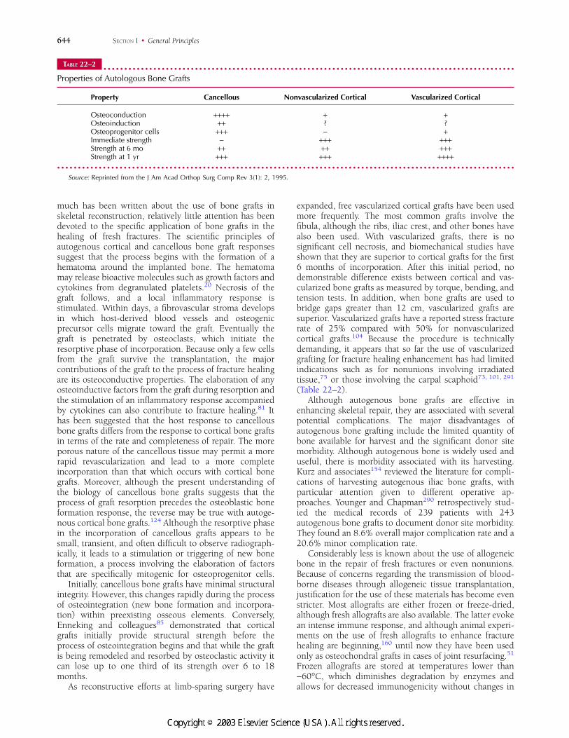
(Table 22–2).Although autogenous bone grafts are effective in
enhancing skeletal repair, they are associated with severalpotential complications. The major disadvantages ofautogenous bone grafting include the limited quantity ofbone available for harvest and the significant donor sitemorbidity. Although autogenous bone is widely used anduseful, there is morbidity associated with its harvesting.Kurz and associates154 reviewed the literature for compli-cations of harvesting autogenous iliac bone grafts, withparticular attention given to different operative ap-proaches. Younger and Chapman290 retrospectively stud-ied the medical records of 239 patients with 243autogenous bone grafts to document donor site morbidity.They found an 8.6% overall major complication rate and a20.6% minor complication rate.
Considerably less is known about the use of allogeneicbone in the repair of fresh fractures or even nonunions.Because of concerns regarding the transmission of blood-borne diseases through allogeneic tissue transplantation,justification for the use of these materials has become evenstricter. Most allografts are either frozen or freeze-dried,although fresh allografts are also available. The latter evokean intense immune response, and although animal experi-ments on the use of fresh allografts to enhance fracturehealing are beginning,160 until now they have been usedonly as osteochondral grafts in cases of joint resurfacing.51
Frozen allografts are stored at temperatures lower than−60°C, which diminishes degradation by enzymes andallows for decreased immunogenicity without changes in
TABLE 22–2z z z z z z z z z z z z z z z z z z z z z z z z z z z z z z z z z z z z z z z z z z z z z z z z z z z z z z z z z z z z z z z z z z z z z z z z z z z z z z z z z z z z z z z z z z z z z z z z z z z
Properties of Autologous Bone Grafts
Property Cancellous Nonvascularized Cortical Vascularized Cortical
Osteoconduction ++++ + +Osteoinduction ++ ? ?Osteoprogenitor cells +++ − +Immediate strength − +++ +++Strength at 6 mo ++ ++ +++Strength at 1 yr +++ +++ ++++
z z z z z z z z z z z z z z z z z z z z z z z z z z z z z z z z z z z z z z z z z z z z z z z z z z z z z z z z z z z z z z z z z z z z z z z z z z z z z z z z z z z z z z z z z z z z z z z z z z z z z z z z z z z z z zSource: Reprinted from the J Am Acad Orthop Surg Comp Rev 3(1): 2, 1995.
644 SECTION I • General Principles
#6818 @ l 1/ id/CLS b k /GRP d /JOB b /DIV h22 8/29/02 12 P 6/22 P 644 COLOR T#6818 @ l 1/ id/CLS b k /GRP d /JOB b /DIV h22 8/29/02 12 P 6/22 P 644 BLACK T
biomechanical properties. Freeze-drying (lyophilization)involves the removal of water and vacuum packing offrozen tissue. Although the freeze-drying of allograftsresults in a reduction in their immunogenicity,97 thistreatment also affects the mechanical integrity of the graft,resulting in a reduction in its load-bearing capacity.202
Moreover, because the donor’s cells die in the process ofallograft preparation and preservation, whatever limitedosteogenic contribution could have been provided by cellsis lost with allogeneic bone grafts.
Allografts can be used for structural and nonstructuralpurposes. Morcellation of cancellous and cortical bone hasbeen used to reconstruct defects after curettage of bonecysts and benign neoplasms and to reconstruct periarticu-lar defects during arthroplasty. Some surgeons have mixedallograft bone with autogenous bone graft or bone marrowin an effort to enhance osteogenesis and osteoinduction.
Structurally, allografts can be used to reconstructdiaphyseal defects as intercalary segments or in arthrode-ses about the ankle, hip, and spine. In addition, largesegments can be conformed to replace acetabular defects.Complications with large structural allografts, however,can be numerous and can include nonunion (10%),fracture (5%–15%), and infection (10%–15%).249 Inaddition to local infections, major concerns arise regardingthe potential for transmission of hepatitis and the humanimmunodeficiency virus. The American Association ofTissue Banks has set strict standards that have decreasedthe risk of disease transmission. Their records indicate thatof the 3 million tissue transplantations performed since theidentification of the human immunodeficiency virus, onlytwo donors’ tissues have been linked with documentedtransmission. Both cases involved unprocessed, fresh-frozen allografts,3 and in one of these cases, other samplesfrom the same donor that were lyophilized and irradiateddid not transmit the virus. This suggests that lyophilizationand irradiation may destroy the human immunodeficiencyvirus. Although the risk of transmission of an infectiousagent is always possible, it is important to realize thatowing to strict donor screening practices, procurementtechniques, and serologic testing, allografts are used dailyin orthopaedic practices without reports of significantassociated morbidities.
A number of reports have suggested that autoge-nous bone marrow alone is an effective osteogenicgraft.55, 102, 242, 246 This concept is based on the fact thatautogenous bone marrow contains osteogenic precursorsthat could contribute to bone formation.71, 94, 199 Autoge-nous marrow has been used clinically to augment theosteogenic response to implanted allografts35 and xenoge-neic bone.208, 229, 230 However, autogenous bone marrowused alone may also be an effective graft for stimulatingbone formation and skeletal repair. Connolly and associ-ates55 investigated the osteogenic capacity of autogenousbone marrow in a controlled study in rabbits. They foundthat osteogenesis was accelerated in diffusion chambersloaded with centrifuged and concentrated bone marrowcells that had been implanted into the peritoneal cavity(ectopic site) in a delayed union model (orthotopic site).Garg and colleagues102 treated long bone fractures inpatients with plaster cast immobilization and percutaneousinjection of autogenous bone marrow. Patients were kept
from weight bearing for 6 weeks after the operation, afterwhich protected weight bearing was allowed until unionwas achieved. The investigators reported clinical andradiographic healing in 17 of 20 fractures.
The use of autogenous bone marrow preparations forthe treatment of fractures may be refined by the develop-ment of better methods for the isolation, purification, andcultural expansion of marrow-derived mesenchymalcells.37, 38 Selective adhesion methods can be used toisolate cells with osteogenic potential from the marrow cellpopulation of hemopoietic origin. Once isolated, thesecells may be added to a culture medium containing factorsthat stimulate cell replication but not differentiation, toultimately yield a supply of cells that are highly osteogenic.A series of animal studies have already shown that cellsprepared in this manner may be combined with a calciumphosphate ceramic delivery system to regenerate bone orenhance the repair of skeletal defects.29, 111
OSTEOCONDUCTIVE METHODS
Osteoconductive materials used as bone graft substitutesare designed to provide an optimal setting for ingrowth ofsprouting capillaries, perivascular tissues, and osteopro-genitor cells from the recipient host bed.14, 64, 271 Archi-tectural characteristics of the material (e.g., pore size andpore density), as well as its biologic properties such as celladherence capabilities, are just several factors that influ-ence its mechanical strength and ability for osteoconduc-tion.70, 90, 153 The most common osteoconductive bonegraft substitutes used to date are porous ceramics com-posed of hydroxyapatite, calcium phosphate, calciumcarbonate, calcium sulfate, bioactive glass, and bovinebone. Ceramics are made by sintering, a process by whichmineral salts are heated to temperatures above 1000°C.Sintering has been shown to reduce the amount ofcarbonated apatite, an unstable and weakly soluble form ofhydroxyapatite that allows significant osteoclastic remod-eling. Because of this process, many of the ceramicscurrently in use provide a stable biologic construct forosteoconduction to occur.83, 273 Chiroff and colleagues50
were the first to describe the fact that corals made bymarine invertebrates have a structure similar to that ofcortical and cancellous bone and therefore may have a roleas bone graft substitutes. The compounds reviewed beloware those that have been the most extensively studied inbasic science and clinical investigations.
Collagraft (Zimmer, Inc., Warsaw, IN), a mixture ofhydroxyapatite, tricalcium phosphate, and bovine colla-gen, was designed for use with autogenous bone marrowand acts as a nonstructural bone graft substitute.63 Twomajor prospective clinical trials have been reported andboth have concluded that Collagraft is both efficacious andsafe.44, 65 The more recent of these two studies, whichcompared the safety and efficacy of autogenous bone graftobtained from the iliac crest with those of Collagraft, wasa prospective, randomized investigation conducted con-currently at 18 medical centers. Two hundred thirteenpatients (249 fractures) were followed for a minimum of24 months to monitor healing and the occurrence ofcomplications. The results showed no significant differ-ences between the two treatment groups with respect to
645CHAPTER 22 • Enhancement of Skeletal Repair
#6818 @ l 1/ id/CLS b k /GRP d /JOB b /DIV h22 8/29/02 12 P 7/22 P 645 COLOR T#6818 @ l 1/ id/CLS b k /GRP d /JOB b /DIV h22 8/29/02 12 P 7/22 P 645 BLACK T
rates of union (P = 0.94, power = 88%) and functionalmeasures (use of analgesics, pain with activities of dailyliving, and impairment in activities of daily living; P >0.10). The prevalence of complications did not differbetween the treatment groups except for the rate ofinfection, which was higher in the patients who weremanaged with an autogenous graft. Twelve patients whowere managed with a synthetic graft had a positiveantibody titer to bovine collagen; seven of them agreed tohave intradermal challenge with bovine collagen. Onepatient had a positive skin response to the challenge buthad no complications with regard to healing of thefracture. Despite these two optimistic clinical trials,clinicians need to consider (1) the graft’s lack of structuralsupport; (2) the need to combine the collagen-mineralcomposite with the patient’s bone marrow, risking compli-cations from another procedure; and (3) the potentialimmunogenicity and risk of disease transmission with theuse of bovine collagen.
Pro Osteon (Interpore Cross International, Inc., Irvine,CA) is produced by harvesting tricalcium phosphate frommarine coral exoskeletons and converting it into hydroxy-apatite. The coralline material is different from otherhydroxyapatite implants in that it is structurally similar tocancellous bone. Owing to this architectural similarity,osteoconduction is optimized. In addition, the materialcan be cut to fit the area being grafted, and it has beenshown to have good strength in compression. Two studiesin particular have confirmed the clinical advantages of thisbone graft substitute, one in distal radius fractures279 andanother in tibial plateau fractures.30 Some of the concernsthat have been expressed by clinicians with this materialinclude variable quality and strength, undefined resorp-tion rates,132 and persistent radiopaqueness, making itdifficult to estimate fracture healing.238 Nevertheless,coralline hydroxyapatite is a very popular bone graftsubstitute with well-documented clinical benefits.
Norian SRS skeletal replacement system (Norian Corp.,Cupertino, CA) is a paste consisting of powdered calciumphosphate and calcium carbonate mixed with a solution ofcalcium phosphate. The material can be injected into afracture, where within about 10 minutes, it hardens owingto the formation of the mineral dahllite. After 12 hours, thedahllite formation is almost complete, giving the materialan ultimate compressive strength of 55 MPa. As a result ofthese properties, treatment of certain fractures with NorianSRS can augment the fixation that is achieved with a castor with operative means. Animal studies have shown thatNorian SRS is in many cases extensively resorbed andreplaced by host bone.56, 89 Several investigators haveconfirmed the efficacy of Norian in fractures of the distalradius,136, 150, 231, 288 the calcaneus,239 and the hip.82, 108
This and other calcium phosphate composites will cer-tainly have great potential as bone graft substitutes. Aswith other types of cement, more research is requiredconcerning resorption rates and long-term consequencesof extrusion of the material into the soft tissues.
Osteoset (Wright Medical Technology, Inc., Arlington,TN) is a bone graft substitute composed of calcium sulfatepellets. The preparation is mostly resorbed by as early as 6to 8 weeks, a property that has been criticized by some. Infact, the manufacturer states that the material does not
provide structural support and that it should not be asubstitute for internal or external fixation. To our knowl-edge, no controlled studies have been published to dateregarding the clinical efficacy of this product. This is inpart because Osteoset was marketed in the United Statesbefore the institution of the current Food and DrugAdministration approval process. In an effort to introducea bone graft substitute with osteoconductive and osteoin-ductive properties, the manufacturer of Osteoset has alsointroduced Allomatrix Injectable Putty, a combination ofAlloGro demineralized bone matrix and Osteoset. To ourknowledge, no clinical trials with this product have beenreported. Although this and other previously describedbone graft substitutes are relatively safe with respect toinducing an immune response or transmitting an infec-tious disease, there has been a report of three inflammatoryreactions associated with the use of Osteoset followingresection of bone tumors.222 Well-designed clinical trialson the use of Osteoset in humans are still needed to betterunderstand the safety and efficacy of this product.
Bioactive glass and bovine bone–derived ceramics aretwo types of bone graft substitutes that have not yetundergone enough testing to make them clinically usefulproducts. To our knowledge, Novabone and Biogran arethe two bioactive glass products that have been developedto act as bone graft substitutes. Neither of these productshas been studied extensively, although animal studies haveproven bioglass materials to be promising.195 Althoughnonphysiologic and offering little structural support, thesetwo products need to be further studied. Endobon, abovine cancellous bone–derived ceramic, is marketed inEurope as a bone graft substitute. Although anecdotalreports of its success exist, to our knowledge, no clinicaltrials on this product have been performed.
OSTEOINDUCTIVE METHODS
Demineralized Allograft Bone MatrixTo our knowledge, four commercially available productscontaining demineralized allograft bone matrix exist.Grafton DBM (Osteotech, Inc., Eatontown, NJ) andDynaGraft (GenSci Regeneration Sciences, Inc., Missis-sauga, Ontario, Canada) contain only demineralizedhuman bone matrix. Osteofil (Sofamor Danek Group, Inc.,Memphis, TN) and Opteform (Exactech, Inc., Gainesville,FL) contain demineralized human bone matrix mixed witheither porcine gelatin or compacted corticocancelloushuman bone chips, respectively. All of these products haveosteoinductive effects. The last two have the advantage ofhardening at body temperature and the potential forremodeling to normal bone. To date, Grafton DBM andDynaGraft are indicated primarily for nonunions anddelayed unions and for patients with potentially poorhealing potential such as smokers and diabetics. Althoughall have the potential for disease transmission, themanufacturers claim that this has not yet occurred.Although Grafton is manufactured with glycerol, whichhas been suggested to be neurotoxic, no reports of neuraltoxicity have been made to date. Because of their ability toharden, Osteofil and Opteform have the potential foroffering structural integrity to fractures and other bonydefects.
646 SECTION I • General Principles
#6818 @ l 1/ id/CLS b k /GRP d /JOB b /DIV h22 8/29/02 12 P 8/22 P 646 COLOR T#6818 @ l 1/ id/CLS b k /GRP d /JOB b /DIV h22 8/29/02 12 P 8/22 P 646 BLACK T
Bone Morphogenetic Proteins and OtherGrowth FactorsThe TGF-β superfamily of proteins, which includes theBMPs, FGFs, IGFs, and PDGFs, elicit their actions bybinding to transmembrane receptors that are linked togene sequences in the nucleus of various cells by acascade of chemical reactions.171, 265 Because these cas-cades activate several genes at once, specific growth factorsgenerate multiple effects, both within a single cell typeas well as in different cell types.161, 243 Osteoinductionhas been described as occurring in three major phases:chemotaxis, mitosis, and differentiation.215 The aforemen-tioned growth factors, all polypeptide molecules, provide amechanism for stimulative and regulative effects on thesephases.
Transforming Growth Factor-β. TGF-β, a peptidefirst identified by its ability to cause phenotypic transfor-mation of rat fibroblasts,220 has been shown to be afundamental, multifunctional, regulatory protein that caneither stimulate or inhibit several critical processes of cellfunction.255 Since then, five different isomers of TGF-βhave been identified (three of these are found in humans),as have several other related polypeptide growth factors.These are now all a part of the TGF-β superfamily, whichalso includes other ubiquitous compounds such as theBMPs, activins, inhibins, and growth and differentiationfactors. The largest source of TGF-β in the body is theextracellular matrix of bone, and platelets probablyrepresent the second largest reservoir for this pep-tide.42, 251
All the TGF-βs are disulfide-linked dimers compris-ing 12- to 18-kD subunits.146 Most are homodimers(TGF-β1, TGF-β2, and TGF-β3), but some are het-erodimers (TGF-β1.2 and TGF-β2.3).193 TGF-βs aresecreted in a latent propeptide form that requires activa-tion by extracellular proteolytic activity. In bone, it isthought that this occurs within the acidic microenviron-ment formed by the sealing zone directly beneathbone-resorbing osteoclasts.196 Chondrocytes and osteo-blasts have been shown to produce TGF-β,135, 221 whichitself affects protein synthesis in these cell lines.223
Joyce and associates134 were the first to investigate theendogenous expression of TGF-β in organ cultures offracture callus. Using immunohistochemical and recombi-nant DNA techniques, they analyzed fresh femur fracturesmade in male rats at four distinct histologic stages:immediately after the injury, during intramembranousbone formation, during chondrogenesis, and duringendochondral ossification. Using immunolocalization,TGF-β was found to persist for up to 10 days after thefracture was created. During intramembranous boneformation, TGF-β was localized both intracellularly inosteoblasts and proliferating mesenchymal cells, as well asextracellularly. TGF-β was localized to mesenchymal cells,immature chondrocytes, and mature chondrocytes duringchondrogenesis, as well as to the extracellular matrixsurrounding chondrocyte precursors. During endochon-dral ossification, ossified matrix on the bone side of theossification front no longer stained for TGF-β, whereas theextracellular matrix surrounding the hypertrophic chon-drocytes that bordered the ossification front stainedintensely for TGF-β. Gene expression of TGF-β was
evaluated by Northern blot analysis from eight pooledfracture calluses, microdissected into soft (fibrous andcartilaginous) and hard (osseous) callus, at 3-day intervals.TGF-β messenger RNA (mRNA) levels peaked in the softcallus 13 days after fracture, corresponding to thehistologic progression of chondrogenesis. TGF-β mRNAlevels in the hard callus were highest at 5 and 15 days afterfracture, corresponding to intramembranous bone forma-tion and endochondral ossification, respectively. Sincethen, several other investigators have added information tothis original report.7, 224 Some studies have shown thatTGF-β decreases rat osteoblast differentiation and miner-alization.260 Overall, however, the in vitro studies indicatethat TGF-β increases the expression of osteoblast differen-tiation markers such as alkaline phosphatase, type Icollagen, and osteonectin, and acts in synergy with1,25-dihydroxyvitamin D3 to increase alkaline phospha-tase levels.130, 277
To our knowledge, Mustoe and co-workers183 were thefirst to show that TGF-β applied exogenously enhanceshealing of tissues. They applied this peptide directly tolinear incisions made through the dorsal skin of rats anddemonstrated a 220% increase in maximal wound strengthafter 5 days and acceleration in the rate of healing by atleast 3 days. Numerous investigations on the effect ofexogenously introduced TGF-β into injured bone havealso been performed.15, 16, 120, 205 We focus on the threepublished reports available to date that have evaluated theeffect of TGF-β using fracture healing models.67, 163, 190
Critchilow and associates studied the effect of TGF-β2on rat tibial fracture healing. The tibiae were fractured andimmobilized with either a six-hole stainless steel dynamiccompression plate (stable mechanical conditions) or aplastic plate designed to leave a 0.5-mm gap at the fracturesite (unstable mechanical conditions). TGF-β2 was in-jected into the fracture site as a one-time dose (either 60 or600 ng) 4 days after the injury was produced. Thefractures were examined at 5, 7, 10, and 14 days after thefracture. The callus of fractures healing under stablemechanical conditions consisted almost entirely of bone,whereas those of the fractures healing under unstablemechanical conditions had a large area of cartilage over thefracture site with bone on each side. Under stablemechanical conditions, 60 ng of TGF-β2 had an insignif-icant effect on callus development, whereas the higherdose of 600 ng led to a larger callus. Under unstablemechanical conditions, the quantity of tissue componentschanged, but the size of the callus remained unaffected. Atthe lower dose of 60 ng of TGF-β2, the callus containedmore fibrous tissue and less bone and cartilage. Theamounts of bone, cartilage, and fibrous tissue in callustreated with 600 ng of TGF-β2 were similar to those in thecontrol group, although the lack of bone between thecartilage and periosteum indicated that the callus is lessmature. The investigators concluded that TGF-β2 does notenhance fracture healing.
Lind and colleagues163 studied the effect of TGF-βadministered continuously using an osmotic minipump tounilaterally plated adult rabbit tibial osteotomies. For 6weeks, the experimental groups received either 1 or 10 µgper day, and the control group received injections withoutTGF-β. At 6 weeks, fracture healing was evaluated by
647CHAPTER 22 • Enhancement of Skeletal Repair
#6818 @ l 1/ id/CLS b k /GRP d /JOB b /DIV h22 9/16/02 13 P 9/22 P 647 COLOR T#6818 @ l 1/ id/CLS b k /GRP d /JOB b /DIV h22 9/16/02 13 P 9/22 P 647 BLACK T
mechanical tests, histomorphometry, and densitometry.Markedly increased callus volume and statistically signifi-cant maximal bending strengths were demonstrated in thegroups receiving 1 µg of TGF-β per day. In the groupadministered 10 µg of TGF-β per day, there was nostatistically significant increase in bending strength, al-though the callus volume persisted to be greater than thatin the control group. There was no statistically significanteffect in any of the experimental groups on bendingstiffness, bone mineral content, cortical thickness, orHaversian canal diameter. The investigators concluded thatexogenous administration of TGF-β might enhance frac-ture healing in rabbits by increasing callus size but that thecallus created may be too immature to enhance themechanical strength of the osteotomy.
Nielson and associates190 studied the effect of TGF-βadministered locally around the fracture line of healing rattibial fractures stabilized with an intramedullary pin.TGF-β was injected at a dose of either 4 ng or 40 ng everyother day for 40 days. The strength, stiffness, energyabsorption, and deflection of the fractures were measured.Biomechanical testing showed an increase in load to failureand callus diameter in the group treated with higher (40ng) dose of TGF-β. The researchers concluded that TGF-βincreases callus formation and strength in rat tibialfractures after 40 days of healing.
Comparing these and other studies and making clini-cally relevant conclusions is difficult owing to differencesin models, dose regimens, delivery systems, and isoformsof TGF-β used.41 Overall, TGF-β appears to have someefficacy in augmenting fracture healing if the fracture isstable. More research using validated and consistentmodels is needed to further assess the role of TGF-β onenhancement of normal fracture healing.
Bone Morphogenetic Proteins. The BMPs are asubfamily of the TGF-β superfamily of polypeptides. BMPsare distinguished from other members of the superfamilyby having, in general, seven rather than nine conservedcysteines in the mature region.228 Several known membersof this family of osteoinductive growth factors can besubdivided into several classes based on structure.127
BMPs play crucial roles in growth, differentiation, andapoptosis in a variety of cells during development,including chondrocytes and osteoblasts. Compared withTGF-β, however, BMPs have been shown to have moreselective and powerful effects on bone healing in animalmodels.
During fracture repair, endogenously expressed BMPsinclude BMP-2, BMP-3 (osteogenin), BMP-4, and BMP-7(osteogenic protein, OP-1). In humans, BMP-2 and BMP-7have been the most extensively studied, and they havebeen isolated, sequenced, and manufactured using recom-binant DNA technology. The importance of BMPs duringbone healing has been demonstrated using numerous invitro48, 263, 284 and in vivo models.23, 119, 131, 186, 194, 289
Several investigators have also studied the ability ofexogenously administered BMPs to promote bone regen-eration in osseous locations. BMP-2,106, 148, 149, 240, 286
BMP-7,58, 59, 61 and BMP-3257 have all been shown topromote fracture healing of critical-sized defects. Acritical-sized defect may be defined as the smallestintraosseous wound that would not heal by bone forma-
tion in the lifetime of the animal. Critical-sized diaphysealdefect models mimic a clinical situation in which so muchbone is lost that even normal mechanisms cannot repair it,such as a result of trauma or bone resection formusculoskeletal tumors. Although these defects do notheal without intervention, they are not truly models ofnormal or impaired bone healing, as it is the inherent sizeof the defect that leads to failed healing rather than—as inthe case of a delayed union or nonunion—the hostcharacteristics or the local fracture environment. Althoughsignificant developments in fracture repair enhancementhave been achieved using critical-sized diaphyseal defectmodels, they do not simulate the more common clinicalsituation in which the cause of a nonunion is a compro-mised healing environment other than massive bone loss.Therefore, we provide a review of some of the literaturethat has supported the use of BMPs to enhance the healingof fractures.
Einhorn and co-workers investigated the effects ofpercutaneously injecting recombinant BMP-2 into stan-dardized, closed mid-diaphyseal femur fractures in rats 6hours after injury.80 First, 278 male rats were divided intothree groups of 96 animals, each receiving either noinjection at all, injection of an aqueous buffer, or injectionof the buffer plus 80 µg of rhBMP-2. Animals in each ofthese groups were then further sub-divided into fourgroups and were sacrificed at 7, 14, 21, and 28 days afterfracture. At the conclusion of the experiment, 18 femorafrom each subgroup were tested biomechanically and 6were analyzed histologically. A statistically significantincrease in stiffness in the rhBMP-2–treated fractures wasobserved by day 14 and continued at 21 and 28 days afterfracture compared with that in the other two groups. Therewas also a significant increase in strength in the rhBMP-2–treated fractures at day 28. A robust subperiostealmembranous bone response, greater than that seen ineither of the control groups, was demonstrated histologi-cally in the fractures treated with rhBMP-2. In addition,compared with that in controls, there was relativematuration of osteochondrogenic cells in the rhBMP-2treated fractures. Bridging callus appeared earlier in therhBMP-2–treated groups, and relatively increased periph-eral woven bone was seen in these groups as well. Theinvestigators concluded that local percutaneous injectionof rhBMP-2 into fresh fractures might accelerate the rate ofnormal fracture healing. Bostrom and Camacho22 andTurek and co-workers266 studied BMP-2 combined with anabsorbable collagen sponge and applied as an onlay graftin a rabbit ulnar osteotomy and confirmed its effects on thehealing of fresh fractures described by Einhorn andassociates.
Exogenously administered BMP-7 has been evaluated inanimal non–critical-sized defect models by three groups ofinvestigators. Using a closed diaphyseal tibial fracturemodel in the goat, den Boer and colleagues69 investigatedthe effect of BMP-7 introduced into a fresh fracture gapand concluded that injections of BMP-7 solution in thesefractures accelerates their healing during the first 2 weeks.Cook57 and Poplich and colleagues209 created bilateral3-mm non–critical-sized defects in the midulna of 35 adultmale dogs and also showed that BMP-7 enhanced fracturehealing as measured by several parameters.
648 SECTION I • General Principles
#6818 @ l 1/ id/CLS b k /GRP d /JOB b /DIV h22 8/29/02 12 P 10/22 P 648 COLOR T#6818 @ l 1/ id/CLS b k /GRP d /JOB b /DIV h22 8/29/02 12 P 10/22 P 648 BLACK T
Although the effect of exogenously administered BMPinto acute bony defects has been studied for some time, ithas not been until recently that investigators have studiedthe effect of BMPs on the treatment of nonunions. Whereasin acute defects, the species specificity of a BMP does notappear to be important,292 its effect on the ability of a BMPto enhance healing of a nonunion has been shown in acanine model.121 Heckman and associates122 performedstandardized nonunions in the midportion of the radialdiaphysis in 30 mature mongrel dogs. The nonunion wastreated with implantation of a carrier consisting of poly(DL-lactic acid) and polyglycolic acid polymer (50:50polylactic acid–polyglycolic acid [PLG50]) containingcanine-purified BMP or TGF-β1, or both, or the carrierwithout BMP or TGF-β1. Five groups, consisting of sixdogs each, were treated with (1) implantation of the carrieralone, (2) implantation of the carrier with 15 mg ofBMP, (3) implantation of the carrier with 1.5 mg of BMP,(4) implantation of the carrier with 15 mg of BMP and 10ng of TGF-β1, or (5) implantation of the carrier with 10 ngof TGF-β1. The specimens were examined radiographi-cally and histomorphometrically 12 weeks after implanta-tion. The radii treated with either 1.5 mg or 15 mg of BMPshowed significantly increased periosteal and endostealbone formation. No significant radiographic or histomor-phometric evidence of healing was observed after implan-tation of the polylactic acid–polyglycolic acid carrier aloneor in combination with 10 ng TGF-β1. The investigatorsconcluded that species-specific BMP incorporated into apolylactic acid–polyglycolic acid carrier implanted at thesite of an ununited diaphyseal fracture increases boneformation. In addition, TGF-β1 at the dose used in thestudy did not have a similar effect and did not potentiatethe effect of BMP. The investigators suggested that thebiodegradable implant containing BMP that was used intheir study was an effective bone-graft substitute. Thisstudy confirmed the biocompatibility of polylactic acid–polyglycolic acid composites, the bioavailability of BMPand TGF-β1 released from this implant, and, mostimportant, the capability of BMP to augment bone healingin chronic nonunions.
Clinical experience with exogenously administeredBMPs in bony defects or fresh fractures is somewhatlimited. To date, only preliminary results exist on theeffects of BMP-2 and BMP-7 to enhance bony regenerationin humans. Recently described is an open label safety andfeasibility trial using BMP-2 with an absorbable collagensponge carrier.217 Twelve patients with Gustillo grade II,IIIA, or IIIB open tibia fractures from four major traumacenters were treated with 3.4 or 6.8 mg of BMP-2. Eight ofthe 12 patients were treated with a nonreamed tibial rod,and 4 were treated with an external fixator. At the time ofdefinitive wound closure (median, 4 days postoperatively),one or two Helistat (Colla-tec, Plainsboro, NJ) absorbablecollagen sponges soaked with 0.43 mg/mL of BMP-2 wereapplied to the fracture site. Independent radiologists andorthopaedic surgeons reviewed radiographs of the frac-tures 4 months postoperatively. Nine (75%) of thefractures healed without additional intervention, and three(25%) required a secondary bone graft procedure. Asidefrom two patients in whom transient serum antibodiesagainst BMP-2 developed, no significant complications
were encountered. Although these results showed that theuse of BMP-2 in these types of injuries is safe, feasible, andprobably efficacious, the final results of a larger prospec-tive randomized clinical trial are not yet complete.
Friedlaender reported preliminary results of a multicen-ter clinical trial using NOVOS (Styrker Biotech, Natick,MA), a form of BMP-7 associated with a bovine-derivedcollagen carrier.95 In this trial, 124 tibial nonunions in 122patients were treated in 18 centers. Inclusion criteria werenonunion of at least 9 months and the surgeon’s choice ofintramedullary fixation with bone graft as the mostappropriate method of treatment. None of these non-unions was expected to heal if left untreated. Patientsreceived either autograft or NOVOS. A successful resultwas defined as a return to full weight bearing, reduction inpain, and radiographic union by 9 months. Preliminaryresults of this trial indicated that NOVOS was comparableto autogenous grafting in achieving success. The healingrate, however, was not 100% in either group.
Geesink and colleagues investigated the osteogenicpotential of BMP-7 in a critically sized human bony defectmodel.105 Twenty-four patients undergoing high tibialosteotomy for osteoarthritis of the knee were divided intofour groups. In the first group the fibular osteotomy hadbeen left untreated, and in the second group demineralizedbone had been used to fill the defect. Radiologic anddual-energy x-ray absorptiometry parameters measuredduring the first postoperative year showed no evidence ofbony changes in the untreated group, whereas matrixformation of new bone was observed from 6 weeks onwardin the group treated with demineralized bone. The thirdgroup received 2.5 mg of recombinant BMP-7 combinedwith a collagen type I carrier, and the fourth groupreceived collagen type I carrier only. The results of this partof the study showed that all but one of the patients treatedwith the BMP-7 exhibited formation of new bone from 6weeks on as compared with insignificant formation of newbone observed in those who received the collagen carrieralone. The investigators concluded that recombinanthuman BMP-7 is effective in healing human critical-sizedbony defects.
Fibroblast Growth Factors. The FGF family, to ourknowledge, currently includes 19 members.283 The mostabundant types in normal adult tissues are acidic fibroblastgrowth factor (aFGF) and basic fibroblast growth factor(bFGF), also named FGF-1 and FGF-2. Both are heparin-binding polypeptides that have been shown to bind to thesame receptor.188 These molecules are best known for theireffects on endothelial cell replication and neovasculariza-tion.34 The expression of FGFs during fracture repair,however, has been well documented,25, 232 and their rolein fracture healing and its enhancement has been investi-gated in several animal studies.24
Although one report18 has questioned the ability ofFGF to enhance fracture healing, the results of mostexperiments to date have been positive with respect totheir effects. Jingushi and associates, using a rat bilateralfemoral fracture model, explored the effect of exogenousaFGF on normal fracture healing.133 Compared withcontrols, the animals that had received injections of aFGFshowed enlarged calluses in the cartilage formation stage ofhealing, and these calluses remained enlarged until 4
649CHAPTER 22 • Enhancement of Skeletal Repair
#6818 @ l 1/ id/CLS b k /GRP d /JOB b /DIV h22 8/29/02 12 P 11/22 P 649 COLOR T#6818 @ l 1/ id/CLS b k /GRP d /JOB b /DIV h22 8/29/02 12 P 11/22 P 649 BLACK T
weeks after fracture. Nakamura and associates tested theeffects of bFGF on healing tibial fractures in dogs185 andconcluded that bFGF promotes fracture healing in dogs bythe stimulation of bone remodeling. Radomsky andco-workers, first in rabbits212 and more recently inbaboons,211 showed evidence that FGF-2, delivered in ahyaluronan gel, accelerates fracture healing. In the latterstudy, FGF-2 (4 mg/mL) and hyaluronan (20 mg/mL) werecombined into a viscous gel formulation and percutane-ously injected as a one-time dose into a 1-mm gaposteotomy that was surgically created in the fibulae ofbaboons. Radiographically, this combination led to astatistically significant increase in callus area at the treatedsite. Histologic analysis revealed a significantly greatercallus size, periosteal reaction, vascularity, and cellularityin the treated groups compared with those in the untreatedcontrols. Furthermore, specimens treated with 0.1, 0.25,and 0.75 mL of hyaluronan/FGF-2 demonstrated a 48%,50%, and 34% greater average load at failure and an 82%,104%, and 66% greater energy to failure than theuntreated controls, respectively. Although the aforemen-tioned studies on the effects of exogenously delivered FGFare promising, it does not appear that this class ofmolecules is as specific or as potent as the BMPs. Inaddition, only minimal research has been reported on theside effects of high doses of FGF.174
Platelet-Derived Growth Factor. PDGF is the majormesenchymal cell mitogen present in serum.225 PDGF is adimeric molecule consisting of disulfide-bonded A- andB-polypeptide chains. Both homodimeric (PDGF-AA andPDGF-BB) and heterodimeric (PDGF-AB) forms exist.125 APDGF-like peptide has been found in bovine bone,118 andit has been shown to have in vitro effects on several linesof osteoblastic cells.43, 112, 261 Early in the course offracture healing, it has been shown to be released bydegranulating platelets in the fracture hematoma, possiblyacting as a chemotactic agent.8 Later in fracture repair,PDGF protein is detectable in both young and maturehypertrophic chondrocytes and osteoblasts.20
The effects of exogenously administered PDGF onfracture healing are controversial. Marden and colleaguesshowed that PDGF inhibits the bone regeneration inducedby osteogenin in rat craniotomy defects.167 Nash andassociates tested the effects of exogenously administeredPDGF on bone healing using a rabbit tibial osteotomymodel.187 Although histologically the treated groupsappeared to have increased callus density and volume,three-point bending to failure testing failed to show animprovement in strength.
Systemic Enhancement
Numerous reports have shown that patients whohave sustained traumatic brain injury experience fasterfracture healing with more callus than healthy pa-tients,103, 203, 250, 254 although the mechanism by whichthis may occur remains unclear. Bidner and colleagues17
showed that the serum of patients who have sustained ahead injury contains a mitogenic activity that is specific forosteoblastic cells. They suggested that there might be ahumoral mechanism for the enhanced osteogenesis thataccompanies head injury. No conclusive evidence exists for
the molecular identification of this factor. A review articleinsightfully suggested, in fact, that contrary to commonbelief, fracture healing is not necessarily accelerated in thepatient with traumatic brain injury. Hypertrophic callus,myositis ossificans, and heterotopic ossification, however,do occur frequently and are often misperceived asaccelerated healing.155
IGFs and growth hormone (GH) have been suggested toplay a role in skeletal growth and remodeling. IGFs derivetheir name from observations that they produce insulin-like biochemical effects that are not suppressed byanti-insulin antiserum. IGFs exert biologic activity via bothIGF cell surface receptors and insulin-like growth factor–binding proteins.68, 213 IGF-1 and IGF-2 are the twomost important factors of their kind, the former havingthe higher growth-promoting activity. IGF-1, also knownas somatomedin-C, mediates the effect of GH on theskeleton.62
Because IGF-1 is known to be GH dependent, it ispossible that IGF levels may be increased in vivo byadministration of GH. Several studies have been con-ducted to determine the role of GH in the repair of skeletaltissues. Some showed that GH stimulates skeletal repair,151
and others failed to show an effect.192 An investigation ofthe biomechanics of fracture healing in a rabbit tibialmodel showed that GH treatment was unsuccessful in thestimulation of fracture healing. However, the animals inthis experiment were shown to have a persistent nutri-tional deficit, so it is possible that the failure of GH toinfluence skeletal repair resulted from an inability tosignificantly stimulate circulating levels of IGF-I in thisnutritional state.39 Therefore, the roles of GH and IGF-1 inthe systemic enhancement of skeletal repair remainunclear. IGF-2, on the other hand, is one of the mostabundant growth factors in bone, circulates at higherconcentrations than IGF-1, and binds to the same cellsurface receptors but with a lower affinity.99 The observa-tion that IGF-2 is stimulated in response to externallyapplied magnetic fields88 suggests that this molecule mayplay a role in skeletal repair. The systemic effects of theseand other related molecules on the enhancement ofskeletal repair may depend not only on their direct effectson the responding cells but also on the concentrations andenvironmental conditions under which they are permittedto act.
Raschke and co-workers214 studied the effect of sys-temic administration of homologous recombinant GH onbone regenerate consolidation in distraction osteogenesis.Tibiae of 30 mature Yucatan micropigs were osteomized atthe mid-diaphyseal level. Starting 5 days after surgery, thelimbs were distracted using an external fixator at the rateof 2 mm/day for 10 consecutive days. Animals in thetreatment group received a daily subcutaneous injection of100 µg of recombinant porcine GH (rpGH) per kilogramof body weight, and those in the control group receivedsodium chloride. Nondestructive in vivo torsional stiffness(IVTS) measurements were conducted after surgery and ondays 1, 2, 3, 4, 6, 8, and 10 of consolidation. After theanimals were euthanized, destructive biomechanical test-ing was performed. Serum levels of IGF-1 were measuredonce during the latency period (days 1–5), four timesduring distraction (days 6–15), and seven times duringconsolidation (days 16–25) to determine the endocrine
650 SECTION I • General Principles
#6818 @ l 1/ id/CLS b k /GRP d /JOB b /DIV h22 8/29/02 12 P 12/22 P 650 COLOR T#6818 @ l 1/ id/CLS b k /GRP d /JOB b /DIV h22 8/29/02 12 P 12/22 P 650 BLACK T
response to rpGH. Throughout the consolidation phase,the mean in vivo torsional stiffness of the treatment groupwas 125% higher than that of the control group on day 16,increased to 207% higher on day 19, and reached 145%on the day after killing. Final regenerate torsional failureload was 131% higher and ultimate torsional stiffness was231% higher in the treatment group than in the controlgroup. The mean serum level of IGF-1 increased to 440%of preoperative basal level in the treatment group andremained unchanged in the control group. These research-ers concluded that systemic administration of growthhormone greatly accelerates ossification of bone regeneratein distraction osteogenesis.
Prostaglandins are another important class of com-pounds that may be considered for eventual use in thesystemic enhancement of skeletal repair. Over the past twodecades, almost every skeletal metabolic effect has beendescribed after prostaglandin administration, from in-creased bone resorption to increased bone formation.168
The first reports of bone formation after prostaglandintreatment described cortical thickening of the limb bonesand ribs of neonatal infants who had been treatedsystemically with prostaglandin E1 for the purpose ofmaintaining a patent ductus arteriosus.253, 267 Subsequentinvestigations in which prostaglandin E2 was administeredsystemically showed increased cortical and trabecular boneformation in dogs126, 191 and restoration of normal skeletalmass in ovariectomized rats who had lost cancellous andcortical bone mass.139, 140 The fact that these effects can beblocked by the administration of indomethacin, aninhibitor of prostaglandin synthesis, supports the conten-tion that prostaglandins directly enhance bone forma-tion.252 Finally, several studies have shown that normalfracture healing is impaired by administration of prosta-glandin inhibitors.5, 219, 259 Although these reports suggestthat prostaglandins may be useful in enhancing fracturehealing, the clinical safety and efficacy of systemicallyadministered prostaglandins to enhance skeletal repair inhumans have yet to be described.
CLINICAL APPLICATIONSz z z z z z z z z z z z z z z z z z z z z z z z z z z z z z z z z z z z z z z z z z z z z z z z z z z z z z z z z z z
Although strategies to enhance skeletal repair are thesubject of ongoing research, such advances should not beenvisioned as substitutes for optimal orthopaedic manage-ment. Appropriate internal or external fixation and theattainment of adequate alignment and stability will alwaysbe the mainstays of fracture care. Moreover, because ofcost-containment and monitoring of practice patterns,physicians will be able to use specific new products only insituations in which their use has been carefully studied,the data scrutinized by peer review, and the indicationsand labeling approved by a federal regulatory agency. Withthese considerations in mind, our current recommenda-tions and visions for future possibilities are presented.
Fresh Fractures
Enhancement of fresh fracture healing at the time of initialtreatment may be indicated in situations in which the
mechanical or physiologic environment predisposes todelayed union or nonunion. In addition, an enhancedhealing process could lead to a quicker return of patientsto productive work. Although adequate fixation andalignment through traditional orthopaedic principles aresufficient in most cases, even these traditional principlesare subject to change. For example, Sarmiento andassociates237 demonstrated successful healing of closeddiaphyseal tibial fractures with the use of cast bracing andearly weight bearing, but Kenwright and colleagues142
showed that external fixation augmented by controlledmicromotion accelerated the healing process.
For more than 100 years, autogenous cancellous bonegrafting has been the standard of care for augmentation ofboth initial fracture repair and the treatment of anonunion.104 As noted previously, it provides osteogenic,osteoinductive, and osteoconductive properties but islimited in its quantity and in its ability to provideimmediate structural rigidity. In addition, harvestingof autogenous bone graft is not risk free. Physicalmethods of enhancing acute fracture healing have vary-ing benefits. Bone graft substitutes have revolutionizedfracture treatment and will continue to do so. No singlegraft substitute, however, is ideal for all injuries, and itis important for the surgeon to be knowledgeable aboutthe advantages and disadvantages of all the availablematerials.
To summarize, the only physical method that has beenshown to enhance the healing of fresh fractures islow-intensity ultrasound. This modality has been shown tobe effective clinically only in the treatment of closed tibialand distal radius fractures in smokers. With regard toosteogenic methods of augmenting skeletal repair, itappears that autogenous bone graft is the most beneficialand poses the least risk of infection and complications.Vascularized cortical graft harvesting is highly demand-ing and has limited indications, perhaps only in the faceof infection or for areas that are known to be devoidof adequate blood flow. Allografts have been shown tohave up to a 15% rate of infection and therefore shouldbe used with caution. Autogenous bone marrow prepara-tions have not been shown to be efficacious in clinicaltrials, but these preparations certainly have great theoret-ical appeal. It is very difficult to form conclusions as towhich of the commercially available bone graft substitutesis the most effective. This is in part because clinicalexperience is limited, and large clinical trials are lacking.The clinical experience of the authors with corallinehydroxyapatite (ProOsteon) for tibial plateau fractures hasproven it to be extremely useful. Evaluating healingradiographically, however, is often difficult when using thismaterial. We have also used injectable calcium phosphatecement (Norian SRS) for distal radius fractures withpromising results as well. There has been anecdotalevidence that injecting demineralized allograft bone ma-trices into fresh fractures of patients who have poorhealing potential, such as diabetics and smokers, enhancesfracture healing in these patients. Statistically significantclinical data supporting these reports are lacking. Earlyhuman experiments have shown that injectable BMP has arole in accelerating healing of fresh fractures. Follow-upstudies of these and other growth factors are awaited(Table 22–3).
651CHAPTER 22 • Enhancement of Skeletal Repair
#6818 @ l 1/ id/CLS b k /GRP d /JOB b /DIV h22 8/29/02 12 P 13/22 P 651 COLOR T#6818 @ l 1/ id/CLS b k /GRP d /JOB b /DIV h22 8/29/02 12 P 13/22 P 651 BLACK T
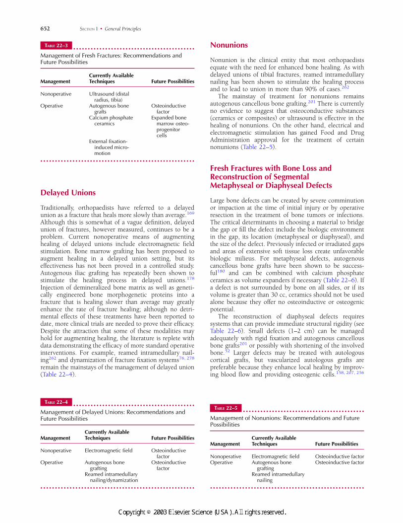
Delayed Unions
Traditionally, orthopaedists have referred to a delayedunion as a fracture that heals more slowly than average.169
Although this is somewhat of a vague definition, delayedunion of fractures, however measured, continues to be aproblem. Current nonoperative means of augmentinghealing of delayed unions include electromagnetic fieldstimulation. Bone marrow grafting has been proposed toaugment healing in a delayed union setting, but itseffectiveness has not been proved in a controlled study.Autogenous iliac grafting has repeatedly been shown tostimulate the healing process in delayed unions.178
Injection of demineralized bone matrix as well as geneti-cally engineered bone morphogenetic proteins into afracture that is healing slower than average may greatlyenhance the rate of fracture healing; although no detri-mental effects of these treatments have been reported todate, more clinical trials are needed to prove their efficacy.Despite the attraction that some of these modalities mayhold for augmenting healing, the literature is replete withdata demonstrating the efficacy of more standard operativeinterventions. For example, reamed intramedullary nail-ing262 and dynamization of fracture fixation systems76, 278
remain the mainstays of the management of delayed union(Table 22–4).
Nonunions
Nonunion is the clinical entity that most orthopaedistsequate with the need for enhanced bone healing. As withdelayed unions of tibial fractures, reamed intramedullarynailing has been shown to stimulate the healing processand to lead to union in more than 90% of cases.262
The mainstay of treatment for nonunions remainsautogenous cancellous bone grafting.201 There is currentlyno evidence to suggest that osteoconductive substances(ceramics or composites) or ultrasound is effective in thehealing of nonunions. On the other hand, electrical andelectromagnetic stimulation has gained Food and DrugAdministration approval for the treatment of certainnonunions (Table 22–5).
Fresh Fractures with Bone Loss andReconstruction of SegmentalMetaphyseal or Diaphyseal Defects
Large bone defects can be created by severe comminutionor impaction at the time of initial injury or by operativeresection in the treatment of bone tumors or infections.The critical determinants in choosing a material to bridgethe gap or fill the defect include the biologic environmentin the gap, its location (metaphyseal or diaphyseal), andthe size of the defect. Previously infected or irradiated gapsand areas of extensive soft tissue loss create unfavorablebiologic milieus. For metaphyseal defects, autogenouscancellous bone grafts have been shown to be success-ful180 and can be combined with calcium phosphateceramics as volume expanders if necessary (Table 22–6). Ifa defect is not surrounded by bone on all sides, or if itsvolume is greater than 30 cc, ceramics should not be usedalone because they offer no osteoinductive or osteogenicpotential.
The reconstruction of diaphyseal defects requiressystems that can provide immediate structural rigidity (seeTable 22–6). Small defects (1–2 cm) can be managedadequately with rigid fixation and autogenous cancellousbone grafts201 or possibly with shortening of the involvedbone.52 Larger defects may be treated with autologouscortical grafts, but vascularized autologous grafts arepreferable because they enhance local healing by improv-ing blood flow and providing osteogenic cells.158, 207, 256
TABLE 22–3z z z z z z z z z z z z z z z z z z z z z z z z z z z z z z z z z z z z z z z z z z
Management of Fresh Fractures: Recommendations andFuture Possibilities
ManagementCurrently AvailableTechniques Future Possibilities
Nonoperative Ultrasound (distalradius, tibia)
Operative Autogenous bonegrafts
Osteoinductivefactor
Calcium phosphateceramics
Expanded bonemarrow osteo-progenitorcells
External fixation-induced micro-motion
z z z z z z z z z z z z z z z z z z z z z z z z z z z z z z z z z z z z z z z z z z z z z z z z z z z z z
TABLE 22–4z z z z z z z z z z z z z z z z z z z z z z z z z z z z z z z z z z z z z z z z z z
Management of Delayed Unions: Recommendations andFuture Possibilities
ManagementCurrently AvailableTechniques Future Possibilities
Nonoperative Electromagnetic field Osteoinductivefactor
Operative Autogenous bonegrafting
Osteoinductivefactor
Reamed intramedullarynailing/dynamization
z z z z z z z z z z z z z z z z z z z z z z z z z z z z z z z z z z z z z z z z z z z z z z z z z z z z z
TABLE 22–5z z z z z z z z z z z z z z z z z z z z z z z z z z z z z z z z z z z z z z z z z z
Management of Nonunions: Recommendations and FuturePossibilities
ManagementCurrently AvailableTechniques Future Possibilities
Nonoperative Electromagnetic field Osteoinductive factorOperative Autogenous bone
graftingOsteoinductive factor
Reamed intramedullarynailing
z z z z z z z z z z z z z z z z z z z z z z z z z z z z z z z z z z z z z z z z z z z z z z z z z z z z z
652 SECTION I • General Principles
#6818 @ l 1/ id/CLS b k /GRP d /JOB b /DIV h22 8/29/02 12 P 14/22 P 652 COLOR T#6818 @ l 1/ id/CLS b k /GRP d /JOB b /DIV h22 8/29/02 12 P 14/22 P 652 BLACK T
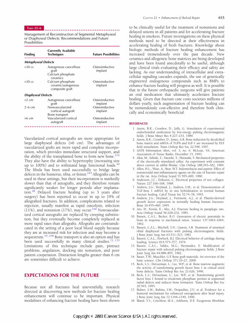
Vascularized cortical autografts are more appropriate forlarge diaphyseal defects (>6 cm). The advantages ofvascularized grafts are more rapid and complete incorpo-ration, which provides immediate structural support, andthe ability of the transplanted bone to form new bone.276
They also have the ability to hypertrophy (increasing sizeup to 100%) and to withstand large functional loads.49
The fibula has been used successfully to bridge largedefects in the humerus, tibia, or femur.245 Allografts can beused in these settings, but their incorporation is markedlyslower than that of autografts, and they therefore remainsignificantly weaker for longer periods after implanta-tion.98 Delayed fracture healing (up to 3 years aftersurgery) has been reported to occur in up to 19% ofallografted fractures. In addition, complications related torejection, usually manifest as rapid osteolysis, infection(11%), and nonunion (17%), may occur.166 Nonvascular-ized cortical autografts are replaced by creeping substitu-tion, but they eventually become completely replaced atmore rapid rates than allografts. Allografts are contraindi-cated in the setting of a poor local blood supply becausethey are at increased risk for infection and may become asequestrum.45, 258 Bone transport is also an option and hasbeen used successfully in many clinical studies.72, 170
Limitations of this technique include pain, pintractproblems, angulation, docking site nonunion, and poorpatient cooperation. Distraction lengths greater than 6 cmare sometimes difficult to achieve.
EXPECTATIONS FOR THE FUTUREz z z z z z z z z z z z z z z z z z z z z z z z z z z z z z z z z z z z z z z z z z z z z z z z z z z z z z z z z z z
Because not all fractures heal uneventfully, researchdirected at discovering new methods for fracture healingenhancement will continue to be important. Physicalmodalities of enhancing fracture healing have been shown
to be clinically useful for the treatment of nonunions anddelayed unions in all patients and for accelerating fracturehealing in smokers. Future investigations on these physicalmethods need to be directed at their effectiveness inaccelerating healing of fresh fractures. Knowledge aboutbiologic methods of fracture healing enhancement hasincreased tremendously over the past decade. Newceramics and allogeneic bone matrices are being developedand have been found anecdotally to be useful, althoughlarge clinical trials evaluating their efficacy and safety arelacking. As our understanding of intracellular and extra-cellular signaling cascades expands, the use of geneticallyengineered endogenous compounds such as BMPs toenhance fracture healing will progress as well. It is possiblethat in the future orthopaedic surgeons will give patientsan oral medication that ultimately accelerates fracturehealing. Given that fracture care costs societies millions ofdollars yearly, such augmentation of fracture healing canbe tremendously cost-effective and therefore both clini-cally and economically beneficial.
REFERENCES
1. Aaron, R.K.; Ciombor, D.; Jolly, G. Stimulation of experimentalendochondral ossification by low-energy pulsing electromagneticfields. J Bone Miner Res 4:227–233, 1989.
2. Aaron, R.K.; Ciombor, D.; Jones, A.R. Bone induction by decalcifiedbone matrix and mRNA of TGFb and IGF-1 are increased by ELFfield stimulation. Trans Orthop Res Soc 22:548, 1997.
3. AATB Information Alert, vol. 3, no. 6. McLean, VA, AmericanAssociation of Tissue Banks, December 15, 1993.
4. Akai, M.; Yabuki, T.; Tateishi, T.; Shirasaki, Y. Mechanical propertiesof the electrically stimulated callus: An experiment with constantdirect current in rabbit fibulae. Clin Orthop 188:293–302, 1984.
5. Allen, H.L.; Wase, A.; Bear, W.T. Indomethacin and aspirin: Effect ofnonsteroidal anti-inflammatory agents on the rate of fracture repairin the rat. Acta Orthop Scand 51:595–600, 1980.
6. Anderson, J.C.; Eriksson, C. Electrical properties of wet collagen.Nature 227:166–168, 1968.
7. Andrew, J.G.; Hoyland, J.; Andrew, S.M.; et al. Demonstration ofTGF-beta 1 mRNA by in situ hybridization in normal humanfracture healing. Calcif Tissue Int 52:74–78, 1993.
8. Andrew, J.G.; Hoyland, J.; Freemont, A.J.; et al. Platelet-derivedgrowth factor expression in normally healing human fractures.Bone 16:455–460, 1995.
9. Aro, H.; Eerola, E.; Aho, A.J. Fracture healing in paraplegic rats.Acta Orthop Scand 56:228–232, 1985.
10. Bassett, C.A.L.; Becker, R.O. Generation of electric potentials inbone in response to mechanical stress. Science 137:1063–1064,1962.
11. Bassett, C.A.L.; Mitchell, S.N.; Gaston, S.R. Treatment of ununitedtibial diaphyseal fractures with pulsing electromagnetic fields.J Bone Joint Surg Am 63:511–523, 1981.
12. Bassett, C.A.L.; Pawluck, R.J. Electrical behavior of cartilage duringloading. Science 814:575–577, 1974.
13. Bassett, C.A.L.; Valdes, M.G.; Hernandez, E. Modification offracture repair with selected pulsing electromagnetic fields. J BoneJoint Surg Am 64:888–895, 1982.
14. Bauer, T.W.; Muschler, G.F. Bone graft materials. An overview of thebasic science. Clin Orthop 371:10–27, 2000.
15. Beck, L.S.; DeGuzman, L.; Lee, W.P.; et al. Bone marrow augmentsthe activity of transforming growth factor beta 1 in critical sizedbone defects. Trans Orthop Res Soc 21:626, 1996.
16. Beck, L.S.; DeGuzman, L.; Lee, W.P.; et al. Transforming growthfactor beta 1 bound to tricalcium phosphate persists at segmentalradial defects and induces bone formation. Trans Orthop Res Soc20:593, 1995.
17. Bidner, S.M.; Rubins, I.M.; Desjardins, J.V.; et al. Evidence for ahumoral mechanism for enhanced osteogenesis after head injury.J Bone Joint Surg Am 72:1144–1149, 1990.
18. Bland, Y.S.; Critchlow, M.A.; Ashhurst, D.E. Exogenous fibroblast
TABLE 22–6z z z z z z z z z z z z z z z z z z z z z z z z z z z z z z z z z z z z z z z z z z
Management of Reconstruction of Segmental Metaphysealor Diaphyseal Defects: Recommendations and FuturePossibilities
FindingCurrently AvailableTechniques Future Possibilities
Metaphyseal Defects
<30 cc Autogenous cancellousgraft
Calcium phosphateceramics
Osteoinductiveimplant
>30 cc Calcium phosphateceramic/autogenouscomposite graft
Osteoinductiveimplant
Diaphyseal Defects
<2 cm Autogenous cancellousgraft
Osteoinductiveimplant
2–6 cm Nonvascularizedcortical autograft
Bone transport
Osteoinductiveimplant
>6 cm Vascularized corticalautograft
Osteoinductiveimplant
z z z z z z z z z z z z z z z z z z z z z z z z z z z z z z z z z z z z z z z z z z z z z z z z z z z z z
653CHAPTER 22 • Enhancement of Skeletal Repair
#6818 @ l 1/ id/CLS b k /GRP d /JOB b /DIV h22 8/29/02 12 P 15/22 P 653 COLOR T#6818 @ l 1/ id/CLS b k /GRP d /JOB b /DIV h22 8/29/02 12 P 15/22 P 653 BLACK T
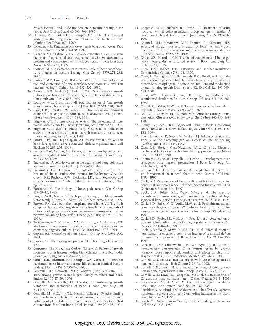
growth factors-1 and -2 do not accelerate fracture healing in therabbit. Acta Orthop Scand 66:543–548, 1995.
19. Blenman, P.R.; Carter, D.S.; Beaupre, G.S. Role of mechanicalloading in the progressive ossification of the fracture callus.J Orthop Res 7:398–407, 1989.
20. Bolander, M.E. Regulation of fracture repair by growth factors. ProcSoc Exp Biol Med 200:165–170, 1992.
21. Bolander, M.E.; Balian, G. The use of demineralized bone matrix inthe repair of segmental defects: Augmentation with extracted matrixproteins and a comparison with autologous grafts. J Bone Joint SurgAm 68:1264–1274, 1986.
22. Bostrom, M.P.G.; Camacho, N.P. Potential role of bone morphoge-netic proteins in fracture healing. Clin Orthop 355S:274–282,1998.
23. Bostrom, M.P.; Lane, J.M.; Berberian, W.S.; et al. Immunolocaliza-tion and expression of bone morphogenetic proteins 2 and 4 infracture healing. J Orthop Res 13:357–367, 1995.
24. Bostrom, M.P.; Saleh, K.J.; Einhorn, T.A. Osteoinductive growthfactors in preclinical fracture and long bone defects models. OrthopClin North Am 30:647–658, 1999.
25. Bourque, W.T.; Gross, M.; Hall, B.K. Expression of four growthfactors during fracture repair. Int J Dev Biol 37:573–579, 1993.
26. Boyd, H.B.; Lipinski, S.W.; Wiley, J.H. Observations on non-unionof the shaft of long bones, with statistical analysis of 842 patients.J Bone Joint Surg Am 43:159–168, 1961.
27. Brighton, C.T. Current concepts review: The treatment of non-unions with electricity. J Bone Joint Surg Am 63:847–851, 1981.
28. Brighton, C.T.; Black, J.; Friedenberg, Z.B.; et al. A multicenterstudy of the treatment of non-union with constant direct current.J Bone Joint Surg Am 62:2–13, 1981.
29. Bruder, S.P.; Fink, D.J.; Caplan, A.I. Mesenchymal stem cells inbone development: Bone repair and skeletal regeneration. J CellBiochem 56:283–294, 1994.
30. Bucholz, R.W.; Carlton, A.; Holmes, R. Interporous hydroxyapatiteas a bone graft substitute in tibial plateau fractures. Clin Orthop240:53–62, 1989.
31. Buckwalter, J.A. Activity vs. rest in the treatment of bone, soft tissueand joint injuries. Iowa Orthop J 15:29–42, 1995.
32. Buckwalter, J.A.; Einhorn, T.A.; Bolander, M.E.; Cruess, R.L.Healing of the musculoskeletal tissues. In: Rockwood, C.A., Jr.;Green, D.P.; Bucholz, R.W.; Heckman, J.D., eds. Rockwood andGreen’s Fractures in Adults. Philadelphia, J.B. Lippincott, 1996,pp. 261–304.
33. Burchardt, H. The biology of bone graft repair. Clin Orthop174:28–42, 1983.
34. Burgess, W.H.; Maciag, T. The heparin-binding (fibroblast) growthfactor family of proteins. Annu Rev Biochem 58:575–606, 1989.
35. Burwell, R.G. Studies in the transplantation of bone: VII. The freshcomposite homograft-autograft of cancellous bone: An analysis offactors leading to osteogenesis in marrow transplants and inmarrow-containing bone grafts. J Bone Joint Surg Br 46:110–140,1964.
36. Buschmann, M.D.; Gluzband, Y.A.; Grodzinky, A.J.; Hunziker, E.B.Mechanical compression modulates matrix biosynthesis inchondrocyte/agarose culture. J Cell Sci 108:1497–1508, 1995.
37. Caplan, A.I. Mesenchymal stem cells. J Orthop Res 9:641–650,1991.
38. Caplan, A.I. The mesengenic process. Clin Plast Surg 21:429–435,1994.
39. Carpenter, J.E.; Hipp, J.A.; Gerhart, T.N.; et al. Failure of growthhormone to alter fracture healing biomechanics in a rabbit model.J Bone Joint Surg Am 74:359–367, 1992.
40. Carter, D.R.; Blenman, P.R.; Beaupre, G.S. Correlations betweenmechanical stress history and tissue differentiation in initial fracturehealing. J Orthop Res 6:736–748, 1988.
41. Centrella, M.; Horowitz, M.C.; Wozney, J.M.; McCarthy, T.L.Transforming growth factor-ß gene family members and bone.Endocr Rev 15:27–39, 1994.
42. Centrella, M.; McCarthy, T.L.; Canalis, E. Transforming growthfactor-beta and remodeling of bone. J Bone Joint Surg Am73:1418–1428, 1991.
43. Centrella, M.; McCarthy, T.L.; Kusmik, W.F.; et al. Relative bindingand biochemical effects of heterodynamic and homodynamicisoforms of platelet-derived growth factor in osteoblast-enrichedcultures from foetal rat bone. J Cell Physiol 146:420–426, 1991.
44. Chapman, M.W.; Bucholz, R.; Cornell, C. Treatment of acutefractures with a collagen-calcium phosphate graft material: Arandomized clinical trial. J Bone Joint Surg Am 79:495–502,1997.
45. Charnell, M.J.; McAndrew, M.P.; Thomas, R.; Schwartz, H.S.Structural allografts for reconstruction of lower extremity openfractures with ten centimeters or more of acute segmental defects.J Orthop Trauma 9:222–226, 1995.
46. Chase, W.S.; Herndon, C.H. The fate of autogenous and homeoge-neous bone grafts: A historical review. J Bone Joint Surg Am37:809–841, 1955.
47. Chen, C.S.; Ingber, D.E. Tensegrity and mechanoregulation.Osteoarthritis Cartilage 7:81–94, 1999.
48. Chen, P.; Carrington, J.L.; Hammonds, R.G.; Reddi, A.H. Stimula-tion of chondrogenesis in limb bud mesoderm cells by recombinanthuman bone morphogenetic protein 2B (BMP-2B) and modulationby transforming growth factor-ß1 and ß2. Exp Cell Res 195:509–515, 1991.
49. Chew, W.Y.C.; Low, C.K.; Tan, S.K. Long term results of freevascularized fibular grafts. Clin Orthop Rel Res 311:258–261,1995.
50. Chiroff, R.; Weber, J.; White, E. Tissue ingrowth of replamineformimplants. J Biomed Mater Res 9:29–45, 1975.
51. Chu, C.R.; Convery, F.R.; Akeson, W.H. Articular cartilage trans-plantation. Clinical results in the knee. Clin Orthop 360:159–168,1999.
52. Cierny, G.; Zorn, K.E. Segmental tibial defects: Comparingconventional and Ilizarov methodologies. Clin Orthop 301:118–123, 1994.
53. Claes, L.; Augat, P.; Suger, G.; Wilke, H.J. Influence of size andstability of the osteotomy gap on success of fracture healing.J Orthop Res 15:577–584, 1997.
54. Claes, L.E.; Heigele, C.A.; Neidlinger-Wilke, C.; et al. Effects ofmechanical factors on the fracture healing process. Clin Orthop355:S132–S147, 1998.
55. Connolly, J.; Guse, R.; Lippiello, L.; Dehne, R. Development of anosteogenic bone marrow preparation. J Bone Joint Surg Am71:684–691, 1989.
56. Constantz, B.R.; Ison, I.C.; Fulmer, M.T.; et al. Skeletal repair by insitu formation of the mineral phase of bone. Science 267:1796–1799, 1995.
57. Cook, S.D. Acceleration of bone healing with OP-1 in a caninenoncritical size defect model. Abstract. Second International OP-1Conference, Boston, MA, 1997.
58. Cook, S.D.; Baffes, G.C.; Wolfe, M.W.; et al. The effect ofrecombinant human osteogenic protein-1 on healing of largesegmental bone defects. J Bone Joint Surg Am 76:827–838, 1994.
59. Cook, S.D.; Baffes, G.C.; Wolfe, M.W.; et al. Recombinant humanbone morphogenetic protein-7 induces healing in a caninelong-bone segmental defect model. Clin Orthop 301:302–312,1994.
60. Cook, S.D.; Ryaby, J.P.; McCabe, J.; Frey, J.J.; et al. Acceleration oftibia and distal radius fracture healing in patients who smoke. ClinOrthop 337:198–207, 1997.
61. Cook, S.D.; Wolfe, M.W.; Salkeld, S.L.; et al. Effect of recombi-nant human osteogenic protein-1 on healing of segmental defectsin non-human primates. J Bone Joint Surg Am 77:734–750,1995.
62. Copeland, K.C.; Underwood, L.E.; Van Wyk, J.J. Induction ofimunoreactive somatomedin C in human serum by growthhormone: Dose response relationships and effects on chromato-graphic profiles. J Clin Endocrinol Metab 50:690–697, 1980.
63. Cornell, C.N. Initial clinical experience with use of collagraft as abone graft substitute. Tech Orthop 7:55–63, 1992.
64. Cornell, C.N.; Lane, J.M. Current understanding of osteoconduc-tion in bone regeneration. Clin Orthop 355:S267–S273, 1998.
65. Cornell, C.N.; Lane, J.M.; Chapman, M.; et al. Multicenter trial ofCollagraft as bone graft substitute. J Orthop Trauma 5:1–8, 1991.
66. Court-Brown, C.; McQueen, M. Compartment syndrome delaystibial union. Acta Orthop Scand 58:249–252, 1987.
67. Critchlow, M.A.; Bland, Y.S.; Ashhurst, D.E. The effect of exogenoustransforming growth factor-beta 2 on healing fractures in the rabbit.Bone 16:521–527, 1995.
68. Czech, M.P. Signal transmission by the insulin-like growth factors.Cell 59:235–238, 1989.
654 SECTION I • General Principles
#6818 @ l 1/ id/CLS b k /GRP d /JOB b /DIV h22 8/29/02 12 P 16/22 P 654 COLOR T#6818 @ l 1/ id/CLS b k /GRP d /JOB b /DIV h22 8/29/02 12 P 16/22 P 654 BLACK T
69. den Boer, F.C.; Bramer, J.A.M.; Blokhius, T.J.; et al. The effect ofrecombinant human osteogenic protein-1 on the healing of freshclosed diaphyseal fracture. Abstract. Second International OP-1Conference, Boston, MA, 1997.
70. Dennis, J.E.; Caplan, A.I. Porous ceramic vehicles for rat marrow–derived (Rattus norvegicus) osteogenic cell delivery: Effects ofpretreatment with fibronectin or laminin. J Oral Implant 19:106–115, 1993.
71. Diduch, D.R.; Coe, M.R.; Joyner, C.; et al. Two cell lines from bonemarrow that differ in terms of collagen synthesis, osteogeniccharacteristics, and matrix mineralization. J Bone Joint Surg Am75:92–105, 1993.
72. DiPasquale, D.; Ochsner, M.G.; Kelly, A.M.; Maloney, D.M. TheIlizarov method for complex fracture non-unions. J Trauma37:629–634, 1994.
73. Doi, K.; Oda, T.; Soo-Heong, T.; Nanda, V. Free vascularized bonegraft for non-union of the scaphoid. J Hand Surg [Am] 25:507–519,2000.
74. Duarte, L.R. The stimulation of bone growth by ultrasound. ArchOrthop Trauma Surg 101:153–159, 1983.
75. Duffy, G.P.; Wood, M.B.; Rock, M.G.; Sim, F.H. Vascularized freefibular transfer combined with autografting for the management offracture non-unions associated with radiation therapy. J Bone JointSurg Am 82:544–554, 2000.
76. Egger, E.L.; Gottsauner, F.; Palmer, J.; et al. Effects of axialdynamization on bone healing. J Trauma 34:185–192, 1993.
77. Einhorn, T.A. Current concepts review: Enhancement of fracture-healing. J Bone Joint Surg Am 77:940–956, 1995.
78. Einhorn, T.A.; Bonnarens, F.; Burnstein, A.H. The contributions ofdietary protein and mineral to the healing of experimental fractures:A biomechanical study. J Bone Joint Surg Am 68:1389–1395, 1986.
79. Einhorn, T.A.; Lane, J.M.; Burstein, A.H.; et al. The healing ofsegmental bone defects induced by demineralized bone matrix: Aradiographic and biomechanical study. J Bone Joint Surg Am66:274–279, 1984.
80. Einhorn, T.A.; Majeska, R.J.; Oloumi, G.; et al. Enhancement ofexperimental fracture healing with a local percutaneous injection ofrhBMP-2. Abstract. American Academy of Orthopaedic SurgeonsAnnual Meeting, San Francisco, California, 1997.
81. Einhorn, T.A.; Majeska, R.J.; Rush, E.B.; et al. The expression ofcytokine activity by fracture callus. J Bone Miner Res 10:1272–1281, 1995.
82. Elder, S.; Frankenburg, E.; Goulet, J.; et al. Biomechanicalevaluation of calcium phosphate cement–augmented fixation ofunstable intertrochanteric fractures. J Orthop Trauma 14:386–393,2000.
83. Ellies, L.G.; Nelson, D.G.; Featherstone, J.D. Crystallographicstructure and surface morphology of sintered carbonated apatites.J Biomed Mater Res 22:541–553, 1988.
84. Emami, A.; Petren-Mallmin, M.; Larsson, S. No effect of low-intensity ultrasound on healing time of intramedullary fixed tibialfractures. J Orthop Trauma 13:252–257, 1999.
85. Enneking, W.F.; Burchardt, H.; Puhl, J.J.; Piotrowski, G. Physicaland biological aspects of repair in dog cortical bone transplants.J Bone Joint Surg Am 57:237–252, 1975.
86. Fitzsimmons, R.J.; Ryaby, J.T.; Magee, F.P.; Baylink, D.J. IGF-IIreceptor number is increased in TE-85 cells by low amplitude, lowfrequency, electromagnetic field exposure. J Bone Miner Res10:812–819, 1995.
87. Fitzsimmons, R.J.; Ryaby, J.T.; Magee, F.P.; et al. Combined magneticfields increase IGF-II secretion in osteoblast cultures. Endocrinol-ogy 136:3100–3106, 1995.
88. Fitzsimmons, R.J.; Ryaby, J.T.; Mohan, S.; et al. Combined magneticfields increase insulin-like growth factor II in TE85 humanosteosarcoma bone cell cultures. Endocrinology 136:3100–3106,1995.
89. Frankenburg, E.P.; Goldstein, S.A.; Bauer, T.W; et al. Biomechanicaland histological evaluation of a calcium phosphate cement. J BoneJoint Surg Am 80:1112–1124, 1998.
90. Frayssinet, P.; Mathon, D.; Lerch, A.; et al. Osseointegration ofcomposite calcium phosphate bioceramics. J Biomed Mater Res50:125–130, 2000.
91. Fredericks, D.C.; Nepola, J.V.; Baker, J.T.; et al. Effects of pulsedelectromagnetic fields on bone healing in a rabbit tibial osteotomymodel. J Orthop Trauma 14:93–100, 2000.
92. Friedenberg, Z.B.; Andrews, E.T.; Smolenski, B.I.; et al. Bonereaction to varying amounts of direct current. Surg Gynecol Obstet131:894–899, 1970.
93. Friedenberg, Z.B.; Brighton, C.T. Bioelectric potentials in bone.J Bone Joint Surg Am 48:915–923, 1966.
94. Friedenstein, A.J.; Chailakhyan, R.K.; Gerasimov, U.V. Bone marrowosteogenic stem cells in vitro cultivation and transplantation indiffusion chambers. Cell Tissue Kinet 20:263–272, 1987.
95. Friedlaender, G. Treatment of established tibial non-unions usingrecombinant osteogenic protein-1 (OP-1). Abstract. Second Inter-national OP-1 Conference, Boston, MA, 1997.
96. Friedlaender, G.E. Bone grafts: The basic science rationale forclinical applications. J Bone Joint Surg Am 69:786–790, 1987.
97. Friedlaender, G.E. Immune responses to osteochondral allografts:Current knowledge and future directions. Clin Orthop 174:58–68,1983.
98. Friedlaender, G.E.; Goldberg, V.M., eds. Bone and CartilageAllografts: Biology and Clinical Applications. Park Ridge, IL,American Academy of Orthopaedic Surgeons, 1991.
99. Frolik, C.A.; Ellis, L.F.; Williams, D.C. Isolation and characteriza-tion of insulin-like growth factor II from human bone. BiochemBiophys Res Comm 151:1011–1018, 1988.
100. Fukada, E.; Yasuda, I. On the piezoelectric effect of bone. J PhysiolSoc Jpn 12:121–128, 1957.
101. Gabl, M.; Reinhart, C.; Lutz, M.; et al. Vascularized bone graft fromthe iliac crest for the treatment of non-union of the proximal partof the scaphoid with an avascular fragment. J Bone Joint Surg Am81:1414–1428, 1999.
102. Garg, N.K.; Gaur, S.; Sharma, S. Percutaneous autogenous bonemarrow grafting in 20 cases of ununited fracture. Acta OrthopScand 64:671–672, 1993.
103. Garland, D.E.; Toder, L. Fractures of the tibial diaphysis in adultswith head injuries. Clin Orthop 150:198–202, 1980.
104. Gazdag, A.R.; Lane, J.M.; Glaser, D.; Forster, R.A. Alternatives toautogenous bone graft: Efficacy and indications. J Am Acad OrthopSurg 3:1–8, 1995.
105. Geesink, R.G.T.; Hoefnagels, N.H.M.; Bulstra, S.K. Osteogenicactivity of OP-1 bone morphogenetic protein (BMP-7) in a humanfibular defect. J Bone Joint Surg Br 81:710–718, 1999.
106. Gerhart, T.N.; Kirker-Head, C.A.; Kriz, M.J. Healing segmentalfemoral defects in sheep using recombinant human bone morpho-genetic protein. Clin Orthop 293:317–326, 1993.
107. Glowacki, J.; Kaban, L.B.; Murray, J.E.; et al. Application of thebiological principle of induced osteogenesis for craniofacial defects.Lancet 1:959–963, 1981.
108. Goodman, S.B.; Bauer, T.W.; Carter, D.; et al. Norian SRS cementaugmentation in hip fracture treatment. Laboratory and initialclinical results. Clin Orthop 348:42–50, 1998.
109. Goodship, A.E.; Kenwright, J. The influence of induced micro-movement upon the healing of experimental fractures. J Bone JointSurg Br 67:650–655, 1985.
110. Goodwin, C.B.; Brighton, C.T.; Guyer, R.D.; et al. A double-blindstudy of capacitively coupled electrical stimulation as an adjunct tolumbar spinal fusions. Spine 24:1349–1356, 1999.
111. Goshima, J.; Goldberg, V.M.; Caplan, A.I. The origin of boneformed in composite grafts of porous calcium phosphate ceramicloaded with marrow cells. Clin Orthop 269:274–283, 1991.
112. Graves, D.T.; Antoniades, H.N.; Williams, S.R.; et al. Evidence forfunctional platelet-derived growth factor receptors on MG-63human osteosarcoma cells. Cancer Res 44:2966–2970, 1984.
113. Gray, M.L.; Pizzanelli, A.M.; Grodzinsky, A.J.; et al. Mechanical andphysicochemical determinants of the chondrocyte biosyntheticresponse. J Orthop Res 6:777–792, 1988.
114. Grodzinsky, A.J. Electromechanical and physicochemical proper-ties of connective tissue. Crit Rev Biomed Eng 9:133–199,1983.
115. Grodzinsky, A.J.; Lipshitz, H.; Glimcher, M.J. Electromechanicalproperties of articular cartilage during compression and stressrelaxation. Nature 275:448–450, 1978.
116. Guilak, F. Compression-induced changes in the shape and vol-ume of the chondrocyte nucleus. J Biomech 28:1529–1541,1995.
117. Hartshorne, E. On the cause of pseudoarthrosis and especially thatform of it sometimes called supernumerary joint. Am J Med1:121–156, 1841.
655CHAPTER 22 • Enhancement of Skeletal Repair
#6818 @ l 1/ id/CLS b k /GRP d /JOB b /DIV h22 8/29/02 12 P 17/22 P 655 COLOR T#6818 @ l 1/ id/CLS b k /GRP d /JOB b /DIV h22 8/29/02 12 P 17/22 P 655 BLACK T
118. Hauschka, P.V.; Mavrakos, A.E.; Iafrati, M.D.; et al. Growth factorsin bone matrix: Isolation of multiple types of affinity chroma-tography on heparin-sepharose. J Biol Chem 261:12665–12674,1986.
119. Hayashi, K.; Ishidou, Y.; Yonemori, K.; et al. Expression andlocalization of bone morphogenetic proteins (BMPs) and BMPreceptors in ossification of the ligamentum flavum. Bone 21:23–30,1997.
120. Heckman, J.D.; Aufdemorte, T.B.; Athanasiou, K.A.; et al. Treatmentof acute ostectomy defects in the dog radius with rhTGF-ß1. TransOrthop Res Soc 20:590, 1995.
121. Heckman, J.D.; Boyan, B.D.; Aufdemorte, T.B.; et al. The use ofbone morphogenetic protein in the treatment of non-union in acanine model. J Bone Joint Surg Am 73:750–764, 1991.
122. Heckman, J.D.; Ehler, W.; Brooks, B.P.; et al. Bone morphogeneticprotein but not transforming growth factor-ß enhances boneformation in canine diaphyseal non-unions implanted with abiodegradable composite polymer. J Bone Joint Surg Am 81:1717–1729, 1999.
123. Heckman, J.D.; Ryaby, J.P.; McCabe, J.; et al. Acceleration of tibialfracture-healing by noninvasive low-intensity pulsed ultrasound.J Bone Joint Surg Am 76:26–34, 1994.
124. Heiple, K.G.; Chase, S.W.; Herndon, C.H. A comparative study ofthe healing process following different types of bone transplanta-tion. J Bone Joint Surg Am 45:1593–1616, 1963.
125. Heldin, C.H.; Ostman, A.; Ronnstrand, L. Signal transduction viaplatelet-derived growth factor receptors. Biochim Biophys Acta1378:F79–F113, 1998.
126. High, W. Effects of orally administered prostaglandin E1 on corticalbone turnover in adult dogs: A histomorphometric study. Bone8:363–374, 1987.
127. Hogan, B.L.M. Bone morphogenetic proteins: Multifunctionalregulators of vertebrate development. Genes Dev 10:1580–1594,1996.
128. Ilizarov, G.A. The tension-stress effect on the genesis and growth oftissues: Part I. Clin Orthop 238:249–281, 1989.
129. Ingber, D.E. Tensegrity: The architectural basis of cellular mecha-notransduction. Annu Rev Physiol 59:575–599, 1997.
130. Ingram, R.T.; Bonde, S.K.; Riggs, B.L.; et al. Effects of transforminggrowth factor beta (TGF-ß) and 1,25 dihydroxyvitamin D3 on thefunction, cytochemistry, and morphology of normal humanosteoblast-like cells. Differentiation 55:153–163, 1994.
131. Ishidou, Y.; Kitajima, I.; Obama, H.; et al. Enhanced expression oftype I receptors for bone morphogenetic proteins during boneformation. J Bone Miner Res 10:1651–1659, 1995.
132. Jensen, S.S.; Aaboe, M.; Pinholt, E.M.; et al. Tissue reaction andmaterial characteristics of four bone substitutes. Int J OralMaxillofac Implants 11:55–56, 1996.
133. Jingushi, S.; Heydemann, A.; Kana, S.K.; et al. Acidic fibroblastgrowth factor (aFGF) injection stimulates cartilage enlargement andinhibits cartilage gene expression in rat fracture healing. J OrthopRes 8:364–371, 1990.
134. Joyce, M.E.; Jingushi, S.; Bolander, M.E. Transforming growthfactor-beta in the regulation of fracture repair. Orthop Clin NorthAm 21:199–209, 1990.
135. Joyce, M.E.; Nemeth, G.G.; Jingushi, S.; et al. Expression andlocalization of transforming growth factor-ß in a model of fracturehealing. Proceedings of the 35th Annual Meeting of the OrthopedicResearch Society. Orthop Trans 13:460, 1989.
136. Jupiter, J.B.; Winters, S.; Sigman, S.; et al. Repair of five distal radiusfractures with an investigational cancellous bone cement: Apreliminary report. J Orthop Trauma 11:110–116, 1997.
137. Kaban, L.B.; Mulliken, J.B.; Glowacki, J. Treatment of jaw defectswith demineralized bone implants. J Oral Maxillofac Surg 40:623–626, 1982.
138. Kawata, A.; Mikuni-Takagaki, Y. Mechanotransduction in stretchedosteocytes: Temporal expression of immediate early and othergenes. Biochem Biophys Res Commun 246:404–408, 1998.
139. Ke, H.Z.; Li, M.; Jee, W.S. Prostaglandin E2 prevents ovariectomy-induced cancellous bone loss in rats. Bone Miner 19:45–62, 1992.
140. Ke, H.Z.; Li, X.J.; Jee, W.S. Partial loss of anabolic effect ofprostaglandin E2 on bone after its withdrawal in rats. Bone12:173–183, 1991.
141. Kenwright, J; Goodship, A.R. Controlled mechanical stimulation inthe treatment of tibial fractures. Clin Orthop 241:36–47, 1989.
142. Kenwright, J.; Richardson, J.B.; Cunningham, J.L.; et al. Axialmovement and tibial fractures. J Bone Joint Surg Br 73:654–659,1991.
143. Kenwright, J.; Richardson, J.B.; Goodship, A.E.; et al. Effect ofcontrolled axial microenvironment on healing of tibial fractures.Lancet 2:1185–1187, 1986.
144. Kershaw, C.J.; Cunningham, J.L.; Kenwright, J. Tibial externalfixation, weight bearing, and fracture movement. Clin Orthop293:28–36, 1993.
145. Kim, Y.J.; Grodzinsky, A.J.; Plaas, A.H. Compression of cartilageresults in differential effects on biosynthetic pathways for aggrecan,link protein and hyaluronan. Arch Biochem Biophys 328:331–340,1996.
146. Kingsley, D.M. The transforming growth factor beta superfamily:New members, new receptors, and new genetic tests of function indifferent organisms. Genes Dev 8:133–146, 1994.
147. Kirchen, M.E.; O’Connor, K.M.; Gruber, H.E.; et al. Effects ofmicrogravity on bone healing in a rat fibular osteotomy model. ClinOrthop 318:231–242, 1995.
148. Kirker-Head, C.A.; Gerhart, T.N.; Armstrong, R.; et al. Healing boneusing recombinant human bone morphogenetic protein 2 andcopolymer. Clin Orthop 349:205–217, 1998.
149. Kirker-Head, C.A.; Gerhart, T.N.; Schelling, S.H.; et al. Long termhealing of bone using recombinant human bone morphogeneticprotein 2. Clin Orthop 318:222–230, 1995.
150. Kopylov, P.; Runnqvist, K.; Jonsson, K.; Aspenberg, P. NorianSRS versus external fixation in redisplaced distal radial fractures.A randomized study in 40 patients. Acta Orthop Scand 70:1–5,1999.
151. Koskinen, E.V.S.; Lindholm, R.V.; Nieminen, R.A.; et al. Humangrowth hormone in delayed union and non-union of fracture. IntOrthop 1:317–322, 1978.
152. Kristiansen, T.K.; Ryaby, J.P.; McCabe, J; et al. Accelerated healing ofdistal radial fractures with the use of specific, low-intensityultrasound. A multicenter, prospective, randomized, double-blindplacebo-controlled study. J Bone Joint Surg Am 79:961–973, 1997.
153. Kurioka, K.; Umeda, M.; Teranobu, O.; Komori, T. Effect of variousproperties of hydroxyapatite ceramics on osteoconduction andstability. Kobe J Med Sci 45:149–163, 1999.
154. Kurz, L.T.; Garfin, S.R.; Booth, R.E. Harvesting autogenous iliacbone grafts: A review of complications and techniques. Spine14:1324–1331, 1989.
155. Kushwaha, V.P.; Garland, D.G. Extremity fractures in the patientwith a traumatic brain injury. J Am Acad Orthop Surg 6:298–307,1998.
156. Kwiatkowski, T.C.; Hanley, E.N.; Ramp, W.K. Cigarette smokingand its orthopaedic consequences. Am J Orthop 9:590–595,1996.
157. Ladd, A.L.; Pliam, N.B. Use of bone-graft substitutes in distal radiusfractures. J Am Acad Orthop Surg 7:279–290, 1999.
158. Lai-Toh, C.; Jupiter, J.B. The infected non-union of the tibia. ClinOrthop 315:176–191, 1995.
159. Law, H.T.; Annan, I.; McCarthy, I.D.; et al. The effect of inducedelectric currents on bone after experimental osteotomy in sheep.J Bone Joint Surg Br 67:463–469, 1985.
160. Lee, F.Y.; Hazan, E.J.; Gebhardt, M.C.; Mankin, H.J. Experimentalmodel for allograft incorporation and allograft fracture repair.J Orthop Res 18:303–306, 2000.
161. Lein, P.; Johnson, M.; Guo, X.; et al. Osteogenic protein-1 inducesdendritic growth in rat sympathetic neurons. Neuron 15:597–605,1995.
162. Lente, R.W. Cases of ununited fracture treated by electricity. N YState J Med 5:317–319, 1850.
163. Lind, M.; Schumacker, B.; Soballe, K.; et al. Transforming growthfactor-ß enhances fracture healing in rabbit tibiae. Acta OrthopScand 64:553–556, 1993.
164. Lotke, P.A.; Black, J.; Richardson S. Electromechanical properties inhuman articular cartilage. J Bone Joint Surg Am 56:1040–1048,1974.
165. Macey, L.R.; Kana, S.M.; Jingushi, S.; et al. Defects of early fracturehealing in experimental diabetes. J Bone Joint Surg Am 71:722–733, 1989.
166. Mankin, H.J.; Gebhardt, M.C.; Jennings, L.C.; et al. Long-termresults of allograft replacement in the management of bone tumors.Clin Orthop 324:86–97, 1996.
656 SECTION I • General Principles
#6818 @ l 1/ id/CLS b k /GRP d /JOB b /DIV h22 8/29/02 12 P 18/22 P 656 COLOR T#6818 @ l 1/ id/CLS b k /GRP d /JOB b /DIV h22 8/29/02 12 P 18/22 P 656 BLACK T
167. Marden, L.J.; Fan, R.S.P.; Pierce, G.F.; et al. Platelet derived growthfactor inhibits bone regeneration induced by osteogenin, a bonemorphogenetic protein, in rat craniotomy defects. J Clin Invest92:2897–2905, 1993.
168. Marks, S.C., Jr.; Miller, S.C. Effects of prostaglandins on theskeleton. In: Hollinger, J.; Seyfer, A.E., eds. Portland BoneSymposium 1993. Portland, Oregon Health Sciences University,1993, pp. 39–60.
169. Marsh, J.L.; Buckwalter, J.A.; Evarts, C.M. Non-union, delayedunion, malunion and avascular necrosis. In: Epps, C.H., ed.Complications in Orthopaedic Surgery. Philadelphia, J.B. Lippin-cott, 1994, pp. 183–211.
170. Marsh, J.L.; Prokuski, L.; Biermann, J.S. Chronic infected tibialnon-unions with bone loss: Conventional techniques versus bonetransport. Clin Orthop 301:139–146, 1994.
171. Massague, J. TGF-B signaling: Receptors, transducers, and madproteins. Cell 85:947–950, 1996.
172. Matyas, J.R.; Anton, M.G.; Shrive, N.G.; Frank, C.B. Stress governstissue phenotype at the femoral insertion of the rabbit MCL.J Biomech 28:147–157, 1995.
173. Mayr, E.; Frankel, V.; Ruter, A. Ultrasound—An alternative healingmethod for non-unions. Arch Orthop Trauma Surg 120:1–8,2000.
174. Mazue, G.; Bertolero, F.; Garofano, L.; et al. Experience with thepreclinical assessment of basic fibroblast growth factor (bFGF).Toxicol Lett 64:329–338, 1992.
175. McKibbin, B. The biology of fracture healing in long bones. J BoneJoint Surg Br 60:150–162, 1978.
176. McLeod, K.J.; Rubin, C.T. The effect of low-frequency electri-cal fields on osteogenesis. J Bone Joint Surg Am 74:920–929,1992.
177. Meadows, T.H.; Bronk, J.T.; Chao, E.Y.S.; Kelly, P.J. Effect of weightbearing on healing of cortical defects in the canine tibia. J BoneJoint Surg Am 72:1074–1080, 1990.
178. Meiser, K.; Segal, D.; Whitelaw, G.P. The role of bone grafting in thetreatment of delayed unions and non-unions of the tibia. OrthopRev 19:260–271, 1990.
179. Mooney, V.; Nickel, V.L.; Harvey, J.P., Jr.; et al. Cast-brace treatmentfor fractures of the distal part of the femur: A prospective controlledstudy of one hundred and fifty patients. J Bone Joint Surg Am52:1563–1578, 1970.
180. Moore, T.M.; Patzakis, M.J.; Harvey, J.P. Tibial plateau fractures:Definition, demographics, treatment rationale, and long-termresults of closed traction management of operative reduction.J Orthop Trauma 1:97–119, 1987.
181. Mulliken, J.B.; Glowacki, J.; Kaban, L.B.; et al. Use of demineralizedallogeneic bone implants for the correction of maxillocraniofacialdeformities. Ann Surg 194:366–372, 1981.
182. Muschler, G.F.; Lane, J.M. Orthopaedic surgery. In: Habal, M.B.;Reddi, A.H., eds. Bone Grafts and Bone Substitutes. Philadelphia,W.B. Saunders, 1992, pp. 375–407.
183. Mustoe, T.A.; Glenn, F.P.; Thomason, A.; et al. Accelerated healingof incisional wounds in rats induced by transforming growth factor.Science 237:1333–1336, 1987.
184. Nagai, M.; Ota, M. Pulsating electromagnetic field stimulates mRNAexpression of bone morphogenetic protein-2 and-4. J Dent Res73:1601–1605, 1994.
185. Nakamura, T.; Hara, Y.; Tagawa, M.; et al. Recombinant humanfibroblast growth factor accelerates fracture healing by enhancingcallus remodeling in experimental dog tibial fracture. J Bone MinerRes 13:942–949, 1988.
186. Nakase, T.; Nomura, S.; Yoshikawa, H.; et al. Transient andlocalized expression of bone morphogenetic protein 4 messengerRNA during fracture healing. J Bone Miner Res 9:651–659, 1994.
187. Nash, T.J.; Howlett, C.R.; Steele, J.; et al. Effect of platelet-derivedgrowth factor on tibial osteotomies in rabbits. Bone 15:203–208,1994.
188. Neufeld, G.; Gospodarowicz, D. Basic and acidic fibroblast growthfactors interact with the same cell surface receptors. J Biol Chem261:5631–5637, 1986.
189. Nicoll, E.A. Fractures of the tibial shaft: A survey of 705 cases.J Bone Joint Surg Br 46:373–387, 1964.
190. Nielson, H.M.; Andreassen, T.T.; Ledet, T.; Oxlund, H. Localinjection of TGF-beta increases the strength of tibial fractures in therat. Acta Orthop Scand 65:37–41, 1994.
191. Norrdin, R.W.; Shih, M. Systemic effects of prostaglandin E2 onvertebral trabecular remodeling in beagles used in a healing study.Calcif Tissue Int 42:363–368, 1988.
192. Northmore-Ball, M.D.; Wood, M.R.; Meggitt, B.E. A biomechanicalstudy of the effects of growth hormone in experimental fracturehealing. J Bone Joint Surg Br 62:391–396, 1980.
193. Ogawa, Y.; Schmidt, D.K.; Dasch, J.R.; et al. Purification andcharacterization of transforming growth factor-ß2.3 and ß1.2 het-erodimers from bovine bone. J Biol Chem 267:2325–2328, 1992.
194. Onishi, T.; Ishidou, T.; Nagamine, K.; et al. Distinct andoverlapping patterns of localization of bone morphogenetic protein(BMP) family members and a BMP type II receptor during fracturehealing in rats. Bone 22:605–612, 1998.
195. Oonishi, H.; Kushitani, S.; Yasukawa, E.; et al. Particulate bioglasscompared with hydroxyapatite as a bone graft substitute. ClinOrthop 334:316–325, 1997.
196. Oreffo, R.O.; Mundy, G.R.; Seyedin, S.M.; Bonewald, L.F. Activationof the latent bone derived TGF beta complex by isolated osteoclasts.Biochem Biophys Res Commun 158:817–823, 1989.
197. O’Sullivan, M.E.; Chao, E.Y.S.; Kelly, P.J. Current concepts review:The effects of fixation on fracture healing. J Bone Joint Surg Am71:306–310, 1989.
198. Owan, I.; Burr, D.B.; Turner, C.H.; et al. Mechanotransduction inbone: Osteoblasts are more responsive to fluid forces thanmechanical strain. Am J Physiol 273:C810–C815, 1997.
199. Owen, M. Lineage of osteogenic cells and their relationship to thestromal system. In: Peck, W.A., ed. Bone and Mineral Research, 3rded. New York, Elsevier, 1985, pp. 1–25.
200. Parvizi, J.; Wu, C.C.; Lewallen, D.G.; et al. Low-intensity ultra-sound stimulates proteoglycan synthesis in rat chondrocytes byincreasing aggrecan gene expression. J Orthop Res 17:488–494,1999.
201. Patzakis, M.J.; Scilaris, T.A.; Chon, J.; et al. Results of bone graftingfor infected tibial non-union. Clin Orthop Rel Res 315:192–198,1995.
202. Pelker, R.R; Friedlaender, G.E.; Markham, T.C.; et al. Effects offreezing and freeze drying on the biomechanical properties of ratbone. J Orthop Res 1:405–411, 1984.
203. Perkins, R.; Skirving, A.P. Callus formation and the rate of healingof femoral fractures in patients with head injuries. J Bone Joint SurgBr 69:521–524, 1987.
204. Perren, S.M. Physical and biological aspects of fracture healing withspecial reference to internal fixation. Clin Orthop 138:138–195,1979.
205. Peterson, D.R.; Glancy, T.P.; Bacon-Clarke, R.; et al. A study ofdelivery timing and duration on the transforming growth factorbeta 1 induced healing of critical-size long bone defects. Abstract.J Bone Miner Res 12:S304, 1997.
206. Pilla, A.A.; Mont, M.A.; Nasser, P.R.; et al. Noninvasive low-intensity pulsed ultrasound accelerates bone healing in the rabbit.J Orthop Trauma 4:246–253, 1990.
207. Pirela-Cruz, M.A.; DeCoster, T.A. Vascularized bone grafts. Ortho-paedics 17:407–412, 1994.
208. Plenk, H.; Hollmann, K.; Wilfert, K.H. Experimental bridging ofosseous defects in rats by the implantation of Kiel bone containingfresh autologous marrow. J Bone Joint Surg Br 54:735–743, 1972.
209. Poplich, L.S.; Salfeld, S.L.; Rueger, D.C.; et al. Critical andnoncritical size defects healing with osteogenic protein-1. Abstract.Trans Orthop Res Soc 22:600, 1997.
210. Praemer, A.; Furner, S.; Rice, D.P. Musculoskeletal injuries. In:Musculoskeletal Conditions in the United States. Park Ridge, IL,American Academy of Orthopaedic Surgeons, 1992, pp. 85–124.
211. Radomsky, M.L.; Aufdemorte, T.B.; Swain, L.D.; et al. Novelformulation of fibroblast growth factor-2 in a hyaluronan gelaccelerates fracture healing in nonhuman primates. J Orthop Res17:607–614, 1999.
212. Radomsky, M.L.; Thompson, A.Y.; Spiro, R.C.; et al. Potential roleof fibroblast growth factor in enhancement of fracture healing. ClinOrthop 355S:283–293, 1998.
213. Rajaram, S.; Baylink, D.J.; Mohan, S. Insulin-like growth factorbinding proteins in serum and other biological fluids: Regulationand functions. Endocr Rev 18:801–831, 1997.
214. Raschke, M.J.; Bail, H.; Windhagen, H.J.; et al. Recombinant growthhormone accelerates bone regenerate consolidation in distractionosteogenesis. Bone 24:81–88, 1999.
657CHAPTER 22 • Enhancement of Skeletal Repair
#6818 @ l 1/ id/CLS b k /GRP d /JOB b /DIV h22 8/29/02 12 P 19/22 P 657 COLOR T#6818 @ l 1/ id/CLS b k /GRP d /JOB b /DIV h22 8/29/02 12 P 19/22 P 657 BLACK T
215. Reddi, A.H. Extracellular matrix and development. In: Piez, K.A.;Reddi, A.H., eds. Extracellular Matrix Biochemistry. New York,Elsevier, 1984, pp. 375–412.
216. Reher, P.; Doan, N.; Bradnock, B.; et al. Effect of ultrasound on theproduction of IL-8, basic FGF and VEGF. Cytokine 11:416–423,1999.
217. Riedel, G.E.; Valentin-Opran, A. Clinical evaluation of rhBMP-2/ACS in orthopedic trauma: A progress report. Orthopedics22:663–665, 1999.
218. Riggins, R.S.; Simanonok, C.; Lewis, D.W.; Smith, A.H. Weightbearing: Its lack of effect on fracture healing. Int Orthop9:199–203, 1985.
219. Ro, J.; Sudman, E.; Marton, P.F. Effect of indomethacin on fracturehealing in rats. Acta Orthop Scand 47:588–599, 1976.
220. Roberts, A.B.; Anzano, M.A.; Lamb, L.C.; et al. New class oftransforming growth factors potentiated by epidermal growthfactor: Isolation from non-neoplastic tissues. Proc Natl Acad SciU S A 78:5339–5343, 1981.
221. Robey, P.G.; Young, M.F.; Flanders, K.C.; et al. Osteoblastssynthesize and respond to transforming growth factor-ß in vitro.J Cell Biol 105:457–463, 1987.
222. Robinson, D.; Alk, D.; Sandbank, J.; et al. Inflammatory reactionsassociated with a calcium sulfate bone substitute. Ann Transplant4:91–97, 1999.
223. Rosen, D.M.; Stempien, S.A.; Thompson, A.Y.; et al. Transforminggrowth factor-ß modulates the expression of osteoblasts andchondrocyte phenotypes in vitro. J Cell Physiol 134:337–346,1988.
224. Rosier, R.N.; O’Keefe, R.J.; Hicks, D.G. The potential role oftransforming growth factor beta in fracture healing. Clin Orthop355:S294–S300, 1998.
225. Ross, R.; Raines, E.W.; Boewn-Pope, D. The biology of plateletderived growth factor. Cell 46:155–169, 1986.
226. Rothman, R.H.; Klemek, J.S.; Toton, J.J. The effect of iron deficiencyanemia on fracture healing. Clin Orthop 77:276–283, 1971.
227. Rubin, C.; Gross, T.; Qin, Y.X.; et al. Differentiation of thebone-tissue remodeling response to axial and torsional loading inthe turkey ulna. J Bone Joint Surg Am 78:1523–1533, 1996.
228. Sakou, T. Bone morphogenetic proteins: From basic studies toclinical approaches. Bone 22:591–603, 1998.
229. Salama, R.; Burwell, R.G.; Dickson, I.R. Recombined grafts of boneand marrow: The beneficial effect upon osteogenesis of impregnat-ing xenograft (heterograft) bone with autologous red marrow.J Bone Joint Surg Br 55:402–417, 1973.
230. Salama, R.; Weissman, S.L. The clinical use of combined xenograftsof bone and autologous red marrow: A preliminary report. J BoneJoint Surg Br 60:111–115, 1978.
231. Sanchez-Sotelo, J.; Munuera, L.; Madero, R. Treatment of fracturesof the distal radius with a remodellable bone cement: A prospective,randomized study using Norian SRS. J Bone Joint Surg Br82:856–863, 2000.
232. Sandberg, M.M.; Aro, H.T.; Vuorio, E.I. Gene expression duringbone repair. Clin Orthop 289:292–312, 1993.
233. Sarker, A.B.; Nashimuddin, A.N.; Islam, K.M.; et al. Effect of PEMFon fresh fracture-healing in rat tibia. Bangladesh Med Res CouncBull 19:103–112, 1993.
234. Sarmiento, A. A functional below-the-knee cast for tibial fractures.J Bone Joint Surg Am 49:855–875, 1967.
235. Sarmineto, A. On the behavior of closed tibial fractures: Clinical/radiological correlations. J Orthop Trauma 14:199–205, 2000.
236. Sarmiento, A.; Schaeffer, J.F.; Beckerman, L.; et al. Fracture healingin rat femora as affected by functional weightbearing. J Bone JointSurg Am 59:369–375, 1977.
237. Sarmiento, A.; Sharpe, F.E.; Ebramzadeh, E.; et al. Factorsinfluencing outcome of closed tibial fractures treated with func-tional bracing. Clin Orthop 315:8–24, 1995.
238. Sartoris, D.J.; Gershuni, D.H.; Akeson, W.H.; et al. Corallinehydroxyapatite bone graft substitutes: Preliminary report of radio-graphic evaluation. Radiology 159:133–137, 1986.
239. Schildhauer, T.A.; Bauer, T.W.; Josten, C.; Muhr, G. Open reductionand augmentation of internal fixation with an injectable skeletalcement for the treatment of complex calcaneal fractures. J OrthopTrauma 14:309–317, 2000.
240. Sciadini, M.F.; Dawson, J.M.; Berman, L.M.; et al. Dose-responsecharacteristics of recombinant human bone morphogenetic
protein-2 in a canine segmental defect model. Trans Orthop Res Soc20:594, 1995.
241. Scott, G.; King, J.B. A prospective, double-blind trial of electricalcapacitive coupling in the treatment of non-union of long bones.J Bone Joint Surg Am 76:820–826, 1994.
242. Sebecic, B.; Gabelica, V.; Patrlj, L.; Sosa, T. Percutaneous autologousbone marrow grafting on the site of tibial delayed union. Croat MedJ 40:429–432, 1999.
243. Shah, N.M.; Groves, A.K.; Anderson, D.J. Alternative neural crestcell fates are instructively promoted by TGFB superfamily member.Cell 85:331–343, 1996.
244. Shamos, M.H.; Lavine, L.S. Piezoelectricity as a fundamentalproperty of biological tissues. Nature 212:267–268, 1967.
245. Shapiro, M.S.; Endrizzi, D.P.; Cannon, R.M.; Dick, H.M. Treatmentof tibial defects and non-unions using ispilateral vascularizedfibular transposition. Clin Orthop 296:207–212, 1993.
246. Sharma, S.; Garg, N.K.; Veliath, A.J.; et al. Percutaneous bone-marrow grafting of osteotomies and bony defects in rabbits. ActaOrthop Scand 63:166–169, 1992.
247. Sharrard, W.J.W. A double-blind trial of pulsed electromagneticfields for delayed union of tibial fractures. J Bone Joint Surg Br72:347–355, 1990.
248. Silverstein, P. Smoking and wound healing. Am J Med 93(1A):22S–24S, 1992.
249. Sim, F.H.; Frassica, F.J. Use of allografts following resection oftumors of the musculoskeletal system. Instr Course Lect 42:405–413, 1993.
250. Smith, R. Head injury, fracture healing and callus. J Bone Joint SurgBr 69:518–520, 1987.
251. Smith, S.R.; Bronk, J.T.; Kelly, P.J. Effects of fixation on fractureblood flow. Orthop Trans 11:294–295, 1987.
252. Solheim, E.; Pinholt, E.M.; Bang, O.G.; et al. Inhibition ofheterotopic osteogenesis in rats by a new bio-erodable system forlocal delivery of indomethacin. J Bone Joint Surg 74:705–712,1992.
253. Sone, K.; Tashiro, M.; Fujinaga, R.; et al. Long-term low-doseprostaglandin E1 administration. J Pediatr 97:866–867, 1980.
254. Spencer, R.F. The effect of head injury on fracture healing: Aquantitative assessment. J Bone Joint Surg Br 69:525–528, 1987.
255. Sporn, M.B.; Roberts, A.B. Peptide growth factors are multifunc-tional. Nature 332:217–219, 1988.
256. Springfield, D.S. Autogenous bone grafts: Nonvascular and vascu-lar. Orthopaedics 15:1237–1241, 1992.
257. Stevenson, S.; Cunningham, N.; Toth, J.; et al. The effect ofosteogenin (a bone morphogenetic protein) on the formation ofbone in orthotopic segmental defects in rats. J Bone Joint Surg Am76:827–838, 1994.
258. Stoffelen, D.; Lammens, J.; Fabry, G. Resection of a periostealosteosarcoma and reconstruction using the Ilizarov technique ofsegmental transport. J Hand Surg [Br] 18:144–146, 1993.
259. Sudman, E.; Bang, G. Indomethacin-induced inhibition of haver-sian remodelling in rabbits. Acta Orthop Scand 50:621–627, 1979.
260. Talley-Ronsholdt, D.J.; Lajiness, E.; Nagodawithana, K. Transform-ing growth factor-beta inhibition of mineralization by neonatal ratosteoblasts in monolayer and collagen gel culture. In Vitro Cell DevBiol Anim 31:274–282, 1995.
261. Tashjian, A.H.; Hohmann, E.L.; Antoniades, H.N.; et al. Platelet-derived growth factor stimulates bone resorption via a prostaglan-din mediated mechanism. Endocrinology 111:118–124, 1982.
262. Templeman, D.; Thomas, M.; Vasecka, T.; Kyle, R. Exchangereamed intramedullary nailing for delayed union and non-union ofthe tibia. Clin Orthop 315:56–63, 1995.
263. Thies, R.S.; Bauduy, M.; Ashton, B.A.; et al. Recombinant humanmorphogenetic protein-2 induces osteoblastic differentiation inW-20–17 stromal cells. Endocrinology 130:1318–1324, 1992.
264. Toma, C.D.; Ashkar, S.; Gray, M.L.; et al. Signal transduction ofmechanical stimuli is dependent on microfilament integrity:Identification of osteopontin as a mechanically induced gene inosteoblasts. J Bone Miner Res 12:1626–1636, 1997.
265. Trippel, S.B. Growth factors as therapeutic agents. Instr Course Lect46:473–476, 1997.
266. Turek, T.; Bostrom, M.P.G.; Camacho, N.P.; et al. The acceleration offracture healing in a rabbit ulnar osteotomy model with bonemorphogenetic protein 2. Abstract. Trans Orthop Res Soc 22:526,1997.
658 SECTION I • General Principles
#6818 @ l 1/ id/CLS b k /GRP d /JOB b /DIV h22 8/29/02 12 P 20/22 P 658 COLOR T#6818 @ l 1/ id/CLS b k /GRP d /JOB b /DIV h22 8/29/02 12 P 20/22 P 658 BLACK T
267. Ueda, K.; Saito, A.; Nakano, H.; et al. Cortical hyperostosisfollowing long-term administration of prostaglandin E1 in infantswith cyanotic congenital heart disease. J Pediatr 97:834–836,1980.
268. Uhthoff, H.K.; Rahn, B.A. Healing patterns of metaphysealfractures. Clin Orthop 160:295–303, 1981.
269. Upton, J.; Boyajian, M.; Mulliken, J.B.; Glowacki, J. The use ofdemineralized xenogeneic bone implants to correct phalangealdefects: A case report. J Hand Surg [Am] 9:388–391, 1984.
270. Urist, M.R. Bone: Formation by autoinduction. Science 150:893–899, 1965.
271. Urist, M.R. Bone transplants and implants. In: Urist, M.R., ed.Fundamental and Clinical Bone Physiology. Philadelphia, J.B.Lippincott, 1980, pp. 331–368.
272. Urist, M.R.; Strates, B.S. Bone morphogenetic protein. J Dent Res50:1392–1406, 1971.
273. Vaes, G. Cellular biology and biochemical mechanism of boneresorption: A review of recent developments on the formation,activation, and mode of action of osteoclasts. Clin Orthop231:239–271, 1988.
274. Walsh, W.R.; Sherman, P.; Howlett, C.R.; et al. Fracture healing inthe rat osteopenia model. Clin Orthop 342:218–227, 1997.
275. Wang, S.J.; LeWallen, D.G.; Bolander, M.E.; et al. Low intensityultrasound treatment increases strength in a rat femoral fracturemodel. J Orthop Res 12:40–47, 1994.
276. Weiland, A.J. Current concepts review: Vascularized free bonetransplant. J Bone Joint Surg Am 63:166–169, 1981.
277. Wergedal, J.E.; Matsuyama, T.; Strong, D.D. Differentiation ofnormal human bone cells by transforming growth factor-beta and1,25 (OH)2 vitamin D3. Metabolism 41:42–48, 1992.
278. Wiss, D.A.; Stetson, W.B. Unstable fractures of the tibia treated withreamed intramedullary interlocking nail. Clin Orthop 315:56–63,1995.
279. Wolfe, S.W.; Pike, L.; Slade, J.F.; Katz, L.D. Augmentation of distalradius fracture fixation with coralline hydroxyapatite bone graftsubstitute. J Hand Surg [Am] 24:816–827, 1999.
280. Wolff, J. Das Gaesetz der Transformation der Knochen. Berlin, A.Hirchwald, 1892.
281. Wozney, J.M.; Rosen, V.; Celeste, A.J.; et al. Novel regulators of boneformation: Molecular clones and activities. Science 242:1528–1534, 1988.
282. Xavier, C.A.M.; Duarte, L.R. Treatment of non-unions by ultra-sound stimulation: First clinical applications. Presented at themeeting of the Latin-American Orthopaedic Association and at theannual meeting of the American Academy of Orthopaedic Sur-geons, San Francisco, CA, January 25, 1987.
283. Xie, M.H.; Holcomb, I.; Deuel, B.; et al. FGF-19, a novel fibroblastgrowth factor with unique specificity for FGFR4. Cytokine11:729–735, 1999.
284. Yamaguchi, A.; Katagiri, T.; Ikeda, T.; et al. Recombinant humanbone morphogenetic protein-2 stimulates osteoblastic maturationand inhibits myogenic differentiation in vitro. J Cell Biol 113:681–687, 1991.
285. Yang, K.H.; Parvizi, J.; Wang, S.J.; Lewallen, D.G.; et al. Exposureto low-intensity ultrasound increases aggrecan gene expression in arat femur fracture model. J Orthop Res 14:802–809, 1996.
286. Yasko, A.W.; Lane, J.M.; Fellinger, E.J.; et al. The healing ofsegmental bone defects induced by recombinant human morpho-genetic protein (rhBMP-2). A radiographic, histological and biome-chanical study in rats. J Bone Joint Surg Am 74:659–670, 1992.
287. Yasuda, I. [Fundamental aspects of fracture treatment]. J Kyoto MedSoc 4:395–406, 1953.
288. Yetkinler, D.N.; Ladd, A.L.; Poser, R.D.; et al. Biomechanicalevaluation of fixation of intra-articular fractures of the distal part ofthe radius in cadavers: Kirschner wires compared with calcium-phosphate bone cement. J Bone Joint Surg Am 81:391–399, 1999.
289. Yonemori, K.; Imamura, T.; Ishidou, Y.; et al. Bone morphogeneticprotein receptors and activin receptors are highly expressed inossified ligament tissues of patients with ossification of the posteriorlongitudinal ligament. Am J Pathol 150:1335–1347, 1997.
290. Younger, E.M.; Chapman, M.W. Morbidity at bone graft donor sites.J Orthop Trauma 3:192–195, 1989.
291. Zaidemberg, C.; Siebert, J.W.; Angrigiani, C. A new vascularizedbone graft for scaphoid non-union. J Hand Surg [Am] 16:474–478,1991.
292. Zegzula, H.D.; Buck D.C.; Brekke J.; et al. Bone formation with useof rhBMP-2 (recombinant human bone morphogenetic protein-2).J Bone Joint Surg Am 79:1778–1790, 1997.
293. Zhuang, H.; Wang, W.; Seldes, R.M.; et al. Electrical stimulationinduces the level of TGF-B1 mRNA in osteoblastic cells by amechanism involving calcium/calmodulin pathway. Biochem Bio-phys Res Comm 237:225–229, 1997.
659CHAPTER 22 • Enhancement of Skeletal Repair
#6818 @ l 1/ id/CLS b k /GRP d /JOB b /DIV h22 8/29/02 12 P 21/22 P 659 COLOR T#6818 @ l 1/ id/CLS b k /GRP d /JOB b /DIV h22 8/29/02 12 P 21/22 P 659 BLACK T

#6818 @ l 1/ id/CLS b k /GRP d /JOB b /DIV h22 8/29/02 12 P 22/22 P 660 COLOR BLANK T#6818 @ l 1/ id/CLS b k /GRP d /JOB b /DIV h22 8/29/02 12 P 22/22 P 660 BLACK BLANK T