Embed Size (px)
Citation preview
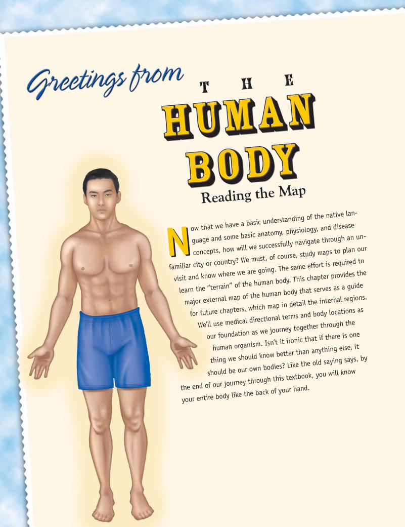
Now that we have a basic understanding of the native lan-
guage and some basic anatomy, physiology, and disease
concepts, how will we successfully navigate through an un-
familiar city or country? We must, of course, study maps to plan our
visit and know where we are going. The same effort is required to
learn the “terrain” of the human body. This chapter provides the
major external map of the human body that serves as a guide
for future chapters, which map in detail the internal regions.
We’ll use medical directional terms and body locations as
our foundation as we journey together through the
human organism. Isn’t it ironic that if there is one
thing we should know better than anything else, it
should be our own bodies? Like the old saying says, by
the end of our journey through this textbook, you will know
your entire body like the back of your hand.
N
ch02.1.qxp 4/21/08 11:07 AM Page 30
LEARNING OBJECTIVES
At the end of your journey through this chapter, you will be able to:
E List and describe the various body positions
E Define the body planes and associated
directional terms
E Locate and describe the body cavities and
their respective organs
E List and describe the anatomical divisions of
the abdominal region
E Identify and locate the various body regions
E Explain the various imaging techniques to
view the body
E Describe situations in which body position
can help or hinder the disease process
MULTIMEDIA APPLICATIONS
CD-ROM Interactive Exercises
E Additional video information on body
positions, 2-1
E Videos on ultrasound, MRIs, and other
diagnostic imaging, 2-2
E Interactive drag-and-drop exercise to rein-
force learning of body cavities, 2-3
E Interactive drag-and-drop exercise to rein-
force learning of anterior and posterior
body regions, 2-4
E Interactive games and puzzles, 2-5
www.prenhall.com/colbert
E Professional Profiles:
• Radiologic Technology
• Surgical Technology
E Related Internet Links
E Additional Review Questions
ch02.1.qxp 4/21/08 11:07 AM Page 31
THE MAP OF THE HUMAN BODY
When reading a map, you need certain universal directional terms, such asnorth, east, south and west. Maps of specific regions can include more detailsabout that region, making it easier to explore. Likewise, scientists have createdstandardized body directional terms and split the body into distinct regions,sections, and cavities so that we can more clearly and rapidly locate and discussanatomical features. Having certain anatomical landmarks on the body alsoprovides needed points of reference for assessment and surgical procedures. Forexample, the spinal cord is a major anatomical landmark for many structures inthe center of our bodies.
If a patient states, “I have pain in my stomach,” what does that really tellyou? Location of pain can help in determining what is wrong with a patient. Itis helpful to know the type of pain (dull, sharp, or stabbing) and exactly wherein that region the pain is located to help determine its cause. For example, painin the general stomach area can indicate a variety of problems, such as an ulcer,heart attack, appendicitis, indigestion, or liver problems. Knowing the exactregion can help a clinician better determine the exact problem.
Body Positions
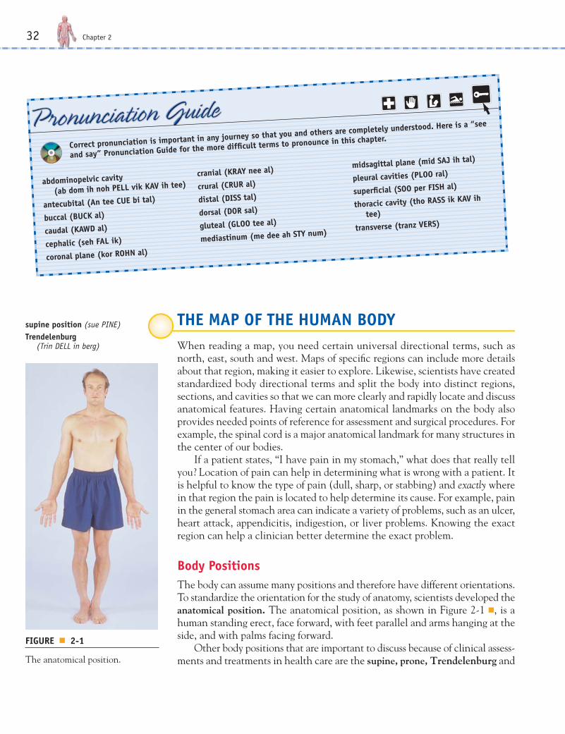
The body can assume many positions and therefore have different orientations.To standardize the orientation for the study of anatomy, scientists developed theanatomical position. The anatomical position, as shown in Figure 2-1 ■, is ahuman standing erect, face forward, with feet parallel and arms hanging at theside, and with palms facing forward.
Other body positions that are important to discuss because of clinical assess-ments and treatments in health care are the supine, prone, Trendelenburg and
32 Chapter 2
Correct pronunciation is important in any journey so that you and others are completely understood. Here is a “see
and say” Pronunciation Guide for the more difficult terms to pronounce in this chapter.
abdominopelvic cavity
(ab dom ih noh PELL vik KAV ih tee)
antecubital (An tee CUE bi tal)
buccal (BUCK al)
caudal (KAWD al)
cephalic (seh FAL ik)
coronal plane (kor ROHN al)
cranial (KRAY nee al)
crural (CRUR al)
distal (DISS tal)
dorsal (DOR sal)
gluteal (GLOO tee al)
mediastinum (me dee ah STY num)
midsagittal plane (mid SAJ ih tal)
pleural cavities (PLOO ral)
superficial (SOO per FISH al)
thoracic cavity (tho RASS ik KAV ih
tee)
transverse (tranz VERS)
FIGURE ■ 2-1
The anatomical position.
supine position (sue PINE)Trendelenburg
(Trin DELL in berg)
ch02.1.qxp 4/21/08 11:07 AM Page 32
The Human Body: Reading the Map 33
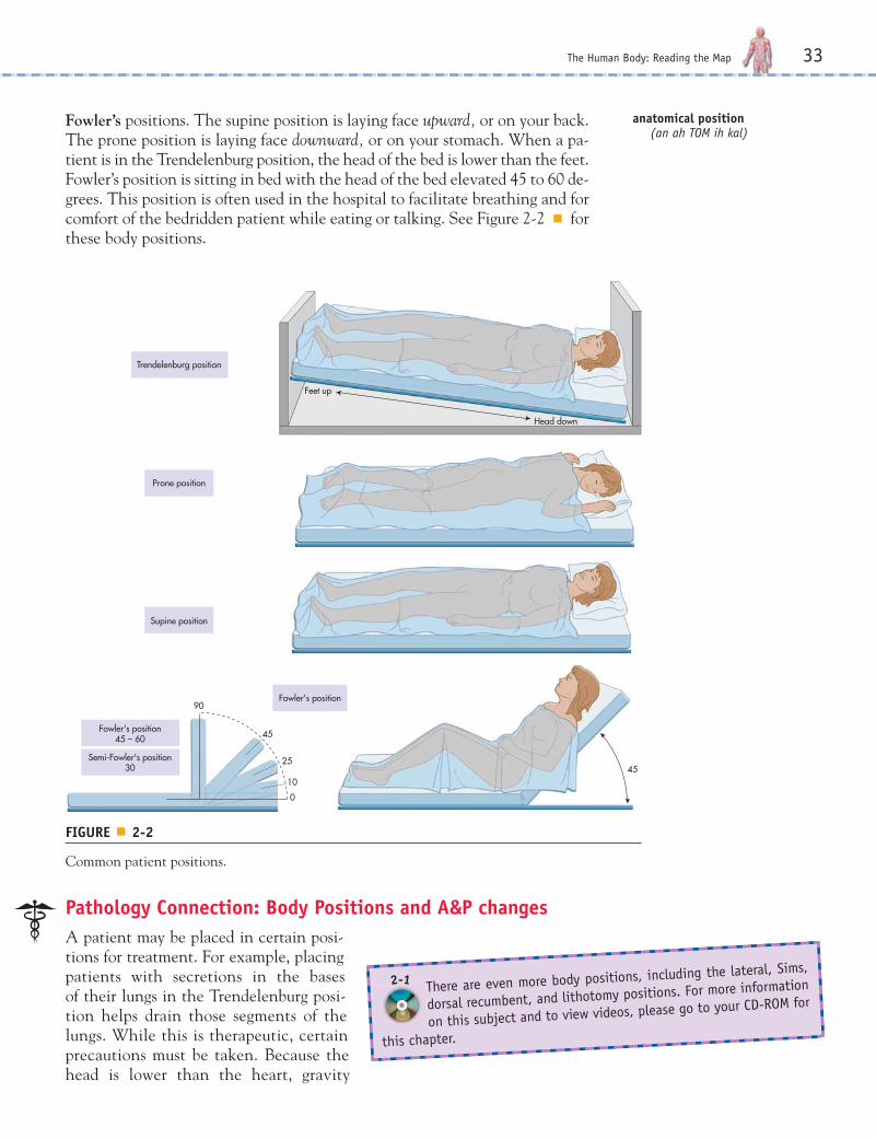
Fowler’s positions. The supine position is laying face upward, or on your back.The prone position is laying face downward, or on your stomach. When a pa-tient is in the Trendelenburg position, the head of the bed is lower than the feet.Fowler’s position is sitting in bed with the head of the bed elevated 45 to 60 de-grees. This position is often used in the hospital to facilitate breathing and forcomfort of the bedridden patient while eating or talking. See Figure 2-2 ■ forthese body positions.
Prone position
Trendelenburg position
Supine position
Fowler's position
Fowler's position45 – 60
Semi-Fowler's position30
0
10
25
45
45
Feet up
Head down
90
FIGURE ■ 2-2
Common patient positions.
Pathology Connection: Body Positions and A&P changes
A patient may be placed in certain posi-tions for treatment. For example, placingpatients with secretions in the bases of their lungs in the Trendelenburg posi-tion helps drain those segments of thelungs. While this is therapeutic, certainprecautions must be taken. Because thehead is lower than the heart, gravity
There are even more body positions, including the lateral, Sims,
dorsal recumbent, and lithotomy positions. For more information
on this subject and to view videos, please go to your CD-ROM for
this chapter.
2-1
anatomical position(an ah TOM ih kal)
ch02.1.qxp 4/21/08 11:07 AM Page 33
increases the blood flow and therefore the intracranial pressure. This positionmay be contraindicated in patients with cerebral injury or bleeding. Also,patients are more prone to aspirate (take in) vomitus into their lungs and there-fore patients should not eat within 2-4 hours of being placed in the Trendelenburgpostion.
Another example: when a patient experiences severe heart failure, the neckveins become filled with extra blood due to the backup of fluid into the venoussystem. This is called Jugular Venous Distension (JVD). If the patient expe-riences engorged neck veins while in the upright sitting position, this indicatessevere heart failure. The physiologic reason is that the pressure of the "backed-up" venous blood has become greater than the effects of gravity.
Sometimes when a person quickly arises from a seated position he or she be-comes weak and dizzy. This may be a sign of orthostatic hypotension. When theperson is seated, the blood pressure may be low, but the brain is still receivingadequate blood flow (perfusion) for normal functioning. When the personstands, however, the heart has to pump harder against gravity to send the bloodto the brain. If the heart cannot compensate, the pressure becomes even lowerand the person experiences weakness and/or dizziness due to lack of cerebralperfusion. These kinds of postural changes in blood pressure will be a problemfor Ray, our spinal cord patient introduced to you in the Common Case Studyat the end of Chapter 1, due to a dysfunction of his nervous system when proppedup in his wheelchair.
Some people sleep better when they prop themselves up with several pillowsin bed. Such patients are experiencing orthopnea, in which it is easier to breathein a more upright position than lying flat. This is because when a person sits up-right, gravity assists the diaphragm in the downward movement needed for in-spiration. You’ll learn more on how the diaphragm functions in Chapter 13 onthe respiratory system.
34 Chapter 2
TEST YOUR KNOWLEDGE 2-1
Complete the following:
1. Try standing in the anatomical position.
2. Give the best body position (prone, supine, orFowler’s position) for the following circumstances:
a. Getting a back massage ____________________
b. Eating in a hospital bed ____________________
c. Watching television in bed __________________
d. Watching the stars at night _________________
3. A person experiencing orthopnea would breathebetter in which position?___________________________________________
4. What position would be contraindicated for someonewho just had eye surgery?___________________________________________
ch02.1.qxp 4/21/08 11:07 AM Page 34

Body Planes and Directional Terms
Sometimes it is necessary to divide the body or even an organ or tissue sampleinto specific sections to further examine it. A plane is an imaginary line drawnthrough the body or organ to separate it into specific sections. For example, inFigure 2-3 ■, we see the transverse plane or horizontal plane, dividing the bodyinto top (superior) and bottom (inferior) sections. This can also be called cross-sectioning the body. Cross-sectioning is often done with tissue and organ sam-ples to further examine internal structures.
The Human Body: Reading the Map 35
Transverse (tranz VERS)
cranial (KRAY nee al)cranio = skull
cephalic (seh FAL ik)cephalo = toward the head
caudal (KAWD al)cauda = tail
-ic and -al are adjective endings thatmean “pertaining to”
Spleen
Transverse plane
Superior(cranial orcephalic)
Inferior(caudal)
Liver
Spinal cord
Stomach
Subcutaneousfat layer
Aorta
FIGURE ■ 2-3
Transverse plane and a cross-sectional view of the upper abdominal region.
Notice in Figure 2-3 that certain directional terms can be used to describeareas divided by the transverse plane. One more analogy that relates to a mapis the concept of a reference point. If you were traveling from Colorado toFlorida, you would have to travel in a southeasterly direction. Colorado is yourstarting point and serves as your reference point. However, if you were travel-ing from Florida to Colorado, you would travel in a northwesterly direction be-cause Florida is now your point of reference. In Figure 2-3, you can see thatsuperior (cranial or cephalic) means toward the head or upper body and inferior(caudal) means away from the head or toward the lower part of the body. Anybody part can be either superior or inferior depending upon your referencepoint. For example, the knee is superior to the ankle if the ankle is the refer-ence point. Turning this around, the ankle is inferior to the knee if the knee isthe reference point. Two other terms from this illustration are cranial, whichrefers to the skull, and caudal, which refers to body parts near the tail (tailbone).
ch02.1.qxp 4/21/08 11:07 AM Page 35
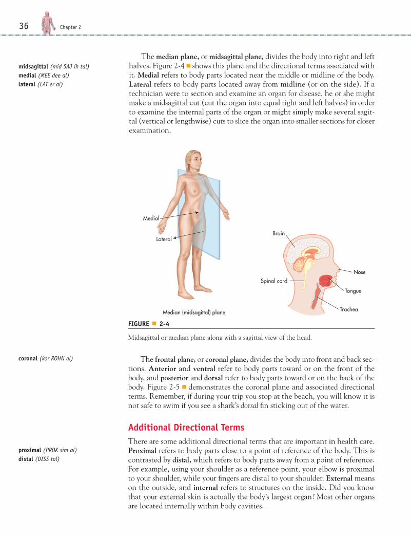
The median plane, or midsagittal plane, divides the body into right and lefthalves. Figure 2-4 ■ shows this plane and the directional terms associated withit. Medial refers to body parts located near the middle or midline of the body.Lateral refers to body parts located away from midline (or on the side). If atechnician were to section and examine an organ for disease, he or she mightmake a midsagittal cut (cut the organ into equal right and left halves) in orderto examine the internal parts of the organ or might simply make several sagit-tal (vertical or lengthwise) cuts to slice the organ into smaller sections for closerexamination.
36 Chapter 2
midsagittal (mid SAJ ih tal)medial (MEE dee al)lateral (LAT er al)
coronal (kor ROHN al)
proximal (PROK sim al)distal (DISS tal)
Median (midsagittal) plane
Lateral
Medial
Brain
Nose
Tongue
Trachea
Spinal cord
FIGURE ■ 2-4
Midsagittal or median plane along with a sagittal view of the head.
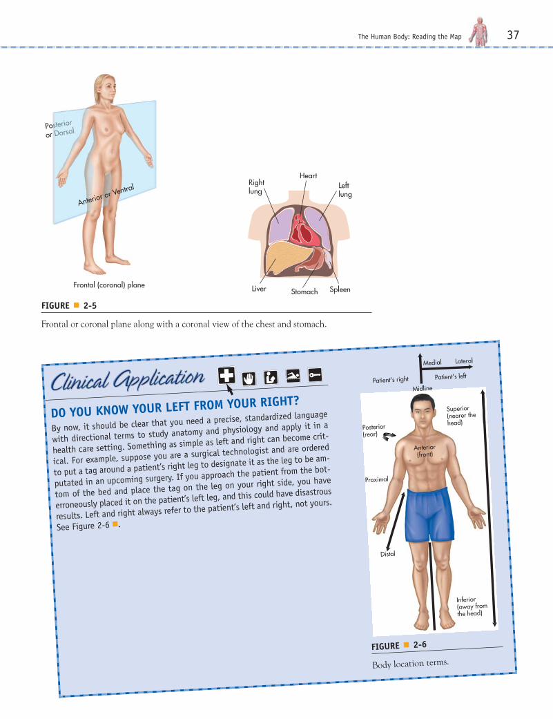
The frontal plane, or coronal plane, divides the body into front and back sec-tions. Anterior and ventral refer to body parts toward or on the front of thebody, and posterior and dorsal refer to body parts toward or on the back of thebody. Figure 2-5 ■ demonstrates the coronal plane and associated directionalterms. Remember, if during your trip you stop at the beach, you will know it isnot safe to swim if you see a shark’s dorsal fin sticking out of the water.
Additional Directional Terms
There are some additional directional terms that are important in health care.Proximal refers to body parts close to a point of reference of the body. This iscontrasted by distal, which refers to body parts away from a point of reference.For example, using your shoulder as a reference point, your elbow is proximalto your shoulder, while your fingers are distal to your shoulder. External meanson the outside, and internal refers to structures on the inside. Did you knowthat your external skin is actually the body’s largest organ? Most other organsare located internally within body cavities.
ch02.1.qxp 4/21/08 11:07 AM Page 36
The Human Body: Reading the Map 37
Frontal (coronal) plane
Posterior
or Dorsal
Anterior or VentralHeart
Left lung
Rightlung
Liver Stomach Spleen
FIGURE ■ 2-5
Frontal or coronal plane along with a coronal view of the chest and stomach.
DO YOU KNOW YOUR LEFT FROM YOUR RIGHT?
By now, it should be clear that you need a precise, standardized language
with directional terms to study anatomy and physiology and apply it in a
health care setting. Something as simple as left and right can become crit-
ical. For example, suppose you are a surgical technologist and are ordered
to put a tag around a patient’s right leg to designate it as the leg to be am-
putated in an upcoming surgery. If you approach the patient from the bot-
tom of the bed and place the tag on the leg on your right side, you have
erroneously placed it on the patient’s left leg, and this could have disastrous
results. Left and right always refer to the patient’s left and right, not yours.
See Figure 2-6 ■.
Medial
Midline
Anterior(front)
Patient's right
Posterior(rear)
Proximal
Distal
Inferior(away fromthe head)
Superior(nearer thehead)
Patient's left
Lateral
FIGURE ■ 2-6
Body location terms.
ch02.1.qxp 4/21/08 11:07 AM Page 37

An embolism is a sudden obstruction of a blood vessel by debris that can includeblood clots (thrombi), plaques, bacteria, cancer cells, fat from bone marrow,and air bubbles.
Superficial means toward or at the bodysurface. When a clinician draws blood fromyou, he or she looks for superficial veinsthat are easy to see and easy to access withthe needle. Deep means away from thebody surface. The large veins in your legsare deep veins and are more protectedthan superficial veins because injury tothem can be more critical to survival thancan injury to a smaller, superficial bloodvessel. These deep leg veins are a com-mon site for blood clots (thrombi) to formthat can then break away and travel toareas such as the lungs (pulmonary em-boli) or brain (cerebral emboli) and blockvital blood flow. Central refers to loca-tions around the center of the body(torso and head), and peripheral refersto the extremities (arms and legs) or sur-rounding or outer regions. Table 2-1 pro-vides a summary of directional terms.
38 Chapter 2
CENTRAL VERSUS PERIPHERAL CYANOSIS
cyanosis (sigh ah NOH siss)
cyano = blue
osis = condition of
Cyanosis is a condition of bluish colored skin that is usually the result of
low levels of oxygen in the blood. Peripheral cyanosis presents as bluish
fingers and toes and may indicate the need for oxygen therapy depending
on the condition of the patient. Peripheral cyanosis is sometimes difficult
to detect in people with dark skin. Central cyanosis is much more serious
and presents as bluish discoloration of the torso and inside the mouth.
See Figure 2-7 ■, which illustrates central and peripheral cyanosis.
PERIPHERAL CYANOSIS
CENTRAL CYANOSIS
FIGURE ■ 2-7
Contrast of central versus peripheral cyanosis.
www.prenhall.com/colbert
Surgical technologists must have a command of medical direc-
tional terms used during surgical procedures. If you are inter-
ested in learning more about this profession, please visit the Web site for
this chapter.
embolism (EM bow lizm)
ch02.1.qxp 4/21/08 11:07 AM Page 38
The Human Body: Reading the Map 39
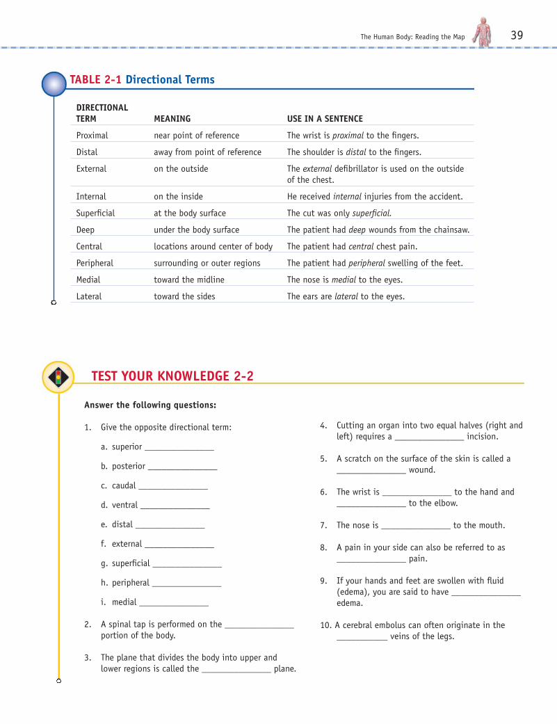
DIRECTIONAL TERM MEANING USE IN A SENTENCE
Proximal near point of reference The wrist is proximal to the fingers.
Distal away from point of reference The shoulder is distal to the fingers.
External on the outside The external defibrillator is used on the outside of the chest.
Internal on the inside He received internal injuries from the accident.
Superficial at the body surface The cut was only superficial.
Deep under the body surface The patient had deep wounds from the chainsaw.
Central locations around center of body The patient had central chest pain.
Peripheral surrounding or outer regions The patient had peripheral swelling of the feet.
Medial toward the midline The nose is medial to the eyes.
Lateral toward the sides The ears are lateral to the eyes.
TABLE 2-1 Directional Terms
TEST YOUR KNOWLEDGE 2-2
Answer the following questions:
1. Give the opposite directional term:
a. superior _______________
b. posterior _______________
c. caudal _______________
d. ventral _______________
e. distal _______________
f. external _______________
g. superficial _______________
h. peripheral _______________
i. medial _______________
2. A spinal tap is performed on the _______________portion of the body.
3. The plane that divides the body into upper andlower regions is called the _______________ plane.
4. Cutting an organ into two equal halves (right andleft) requires a _______________ incision.
5. A scratch on the surface of the skin is called a_______________ wound.
6. The wrist is _______________ to the hand and_______________ to the elbow.
7. The nose is _______________ to the mouth.
8. A pain in your side can also be referred to as_______________ pain.
9. If your hands and feet are swollen with fluid(edema), you are said to have _______________edema.
10. A cerebral embolus can often originate in the___________ veins of the legs.
ch02.1.qxp 4/21/08 11:07 AM Page 39

Body Cavities
The body has two large spaces or cavities that house andprotect organs. Located in the back of the body are thedorsal cavities and in the front, the ventral cavities. Fig-ure 2-8 ■ illustrates these cavities. The larger anteriorcavity is subdivided into two main cavities called thethoracic cavity and abdominopelvic cavity. These cavi-ties are physically separated by the large, dome-shapedmuscle called the diaphragm, which is used for breathing.The thoracic cavity contains the heart, lungs, and largeblood vessels. The heart has its own small cavity calledthe pericardial cavity. The abdominopelvic cavity con-tains the digestive organs, such as the stomach, intes-tines, liver, gallbladder, pancreas, and spleen in the upperor abdominal portion. The lower portion, called thepelvic cavity, contains the urinary and reproductive or-gans and the last part of the large intestine. A posterioror dorsal cavity is located in the back of the body andconsists of the cranial cavity, which houses the brain, andthe spinal cavity, which contains the spinal cord.There are also smaller body cavities that designate spe-cific areas, and these are further explored in upcomingchapters. For example, the nasal cavity is the space be-hind the nose, the oral or buccal cavity is the spacewithin the mouth, and the orbital cavity houses the eyes.
40 Chapter 2
thoracic cavity(thoh RASS ik KAV ih tee)
abdominopelvic cavity (ab dom ihnoh PELL vik KAV ih tee)
cranial cavity(KRAY nee al KAV ih tee)
spinal cavity (SPY nal KAV ih tee)
Spinalcavity
Pleuralcavity
Diaphragm
Abdominopelviccavity
Abdominalcavity
Pelviccavity
Pericardialcavity
Cranialcavity
Cranial cavity
Spinal cavity
Dorsalcavities
Ventralcavities
POSTERIOR
ANTERIOR
Abdominalcavity
Pelviccavity
Thoracic cavity
Pericardial cavity
Abdomino-pelviccavity
FIGURE ■ 2-8
Main body cavities.
ch02.1.qxp 4/21/08 11:07 AM Page 40
The Human Body: Reading the Map 41
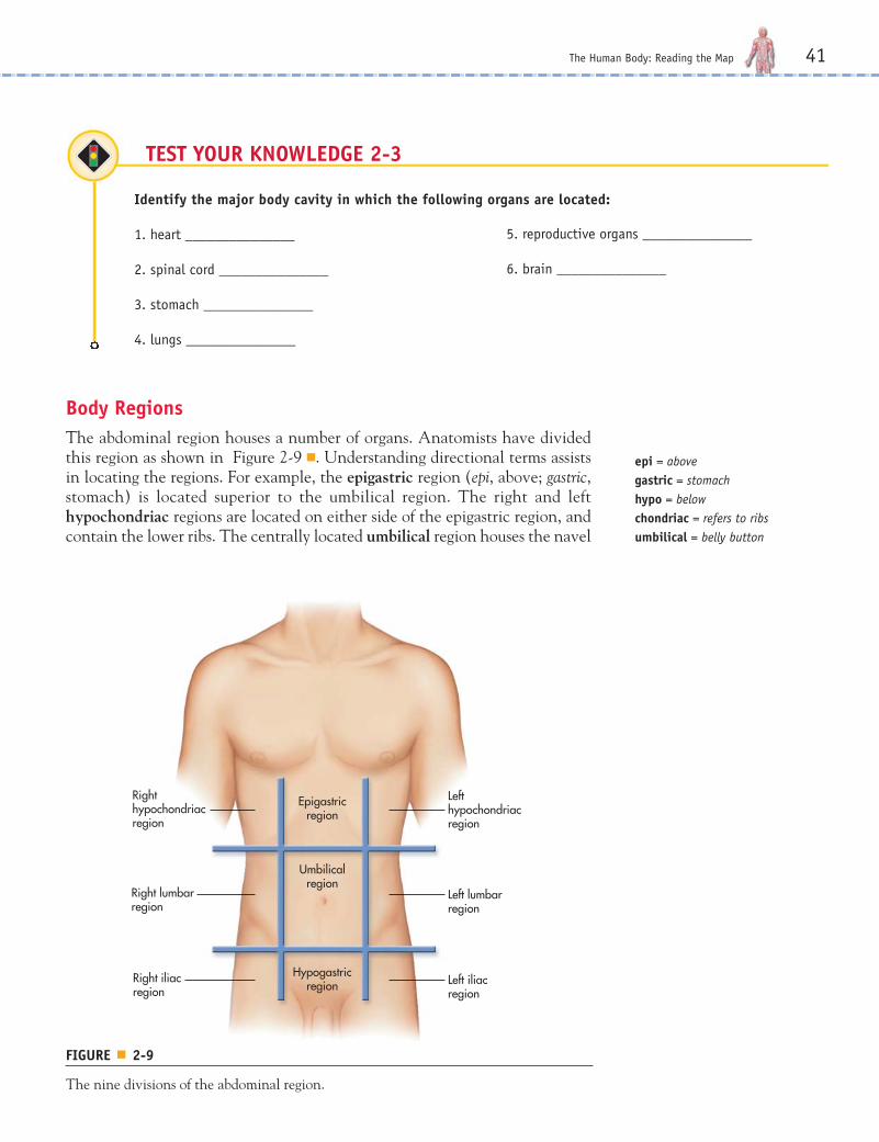
Body Regions
The abdominal region houses a number of organs. Anatomists have dividedthis region as shown in Figure 2-9 ■. Understanding directional terms assistsin locating the regions. For example, the epigastric region (epi, above; gastric,stomach) is located superior to the umbilical region. The right and lefthypochondriac regions are located on either side of the epigastric region, andcontain the lower ribs. The centrally located umbilical region houses the navel
TEST YOUR KNOWLEDGE 2-3
Identify the major body cavity in which the following organs are located:
1. heart _______________
2. spinal cord _______________
3. stomach _______________
4. lungs _______________
5. reproductive organs _______________
6. brain _______________
epi = abovegastric = stomachhypo = belowchondriac = refers to ribsumbilical = belly button
Umbilicalregion
Left lumbarregion
Epigastricregion
Hypogastricregion
Lefthypochondriacregion
Left iliacregion
Right lumbarregion
Righthypochondriacregion
Right iliacregion
FIGURE ■ 2-9
The nine divisions of the abdominal region.
ch02.1.qxp 4/21/08 11:07 AM Page 41
42 Chapter 2
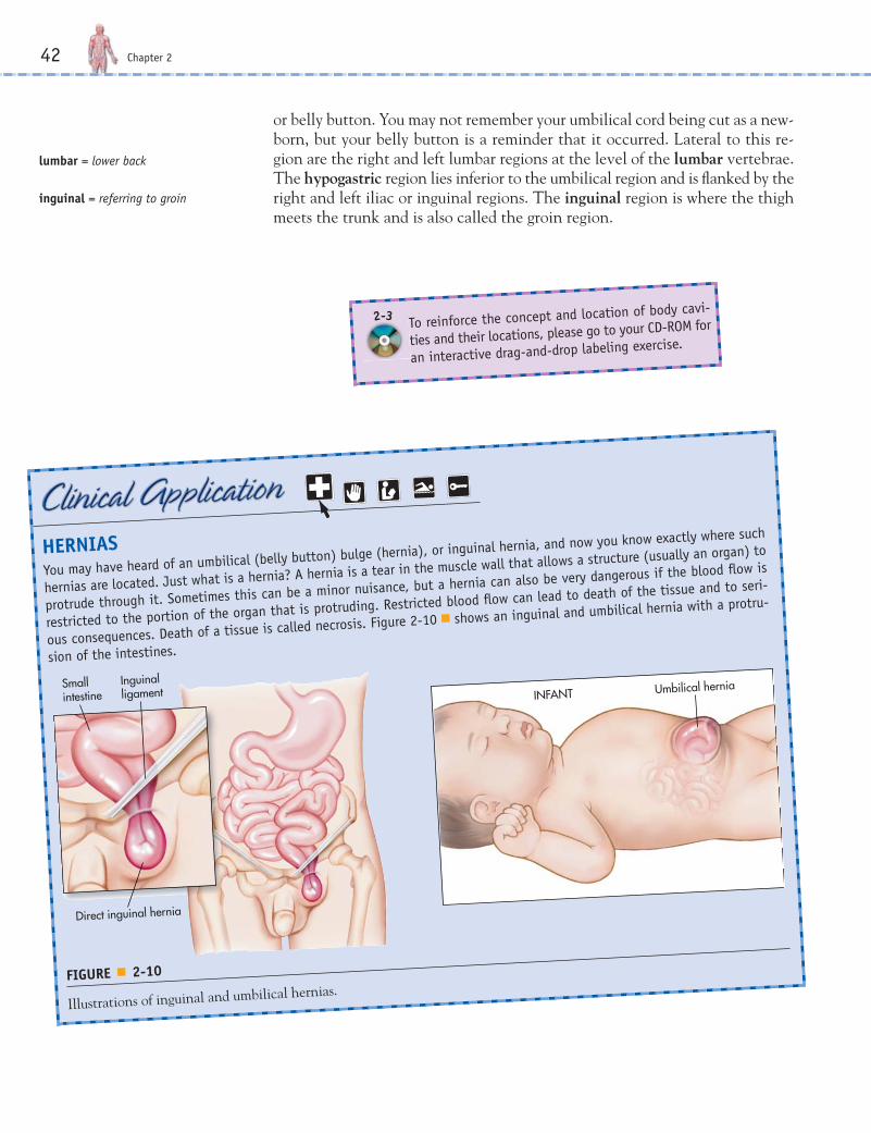
HERNIAS
You may have heard of an umbilical (belly button) bulge (hernia), or inguinal hernia, and now you know exactly where such
hernias are located. Just what is a hernia? A hernia is a tear in the muscle wall that allows a structure (usually an organ) to
protrude through it. Sometimes this can be a minor nuisance, but a hernia can also be very dangerous if the blood flow is
restricted to the portion of the organ that is protruding. Restricted blood flow can lead to death of the tissue and to seri-
ous consequences. Death of a tissue is called necrosis. Figure 2-10 ■ shows an inguinal and umbilical hernia with a protru-
sion of the intestines.
Umbilical herniaINFANT
Smallintestine
Inguinalligament
Direct inguinal hernia
FIGURE ■ 2-10
Illustrations of inguinal and umbilical hernias.
or belly button. You may not remember your umbilical cord being cut as a new-born, but your belly button is a reminder that it occurred. Lateral to this re-gion are the right and left lumbar regions at the level of the lumbar vertebrae.The hypogastric region lies inferior to the umbilical region and is flanked by theright and left iliac or inguinal regions. The inguinal region is where the thighmeets the trunk and is also called the groin region.
To reinforce the concept and location of body cavi-
ties and their locations, please go to your CD-ROM for
an interactive drag-and-drop labeling exercise.
2-3
lumbar = lower back
inguinal = referring to groin
ch02.1.qxp 4/21/08 11:07 AM Page 42

The Human Body: Reading the Map 43
THE CENTRAL LANDMARK: THE SPINAL COLUMN
The spinal or vertebral column is a major, centrally located anatomical landmark
and has five sets of vertebrae (spinal bones) labeled for the body region (see Fig-
ure 2-11 ■). The seven cervical (C) vertebrae are located in the neck; the 12 tho-
racic (T) vertebrae are located in the chest; the five lumbar (L) vertebrae are located
in the lower back; and the five fused sacral (S) vertebrae (sacrum) are located near
the final coccyx vertebra (tailbone). For example, the T5 vertebra is used to help lo-
cate on a chest X-ray the area where the right and left lung begin to branch. You’ll
learn even more about the spinal column and cord in Chapter 6, “The Skeletal Sys-
tem,” and Chapter 9, “The Nervous System.”
Cervical1-7
Thoracic1-12
Lumbar1-5
Sacrum
Coccyx
FIGURE ■ 2-11
The spinal column.
ch02.1.qxp 4/21/08 11:07 AM Page 43
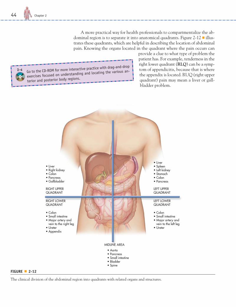
A more practical way for health professionals to compartmentalize the ab-dominal region is to separate it into anatomical quadrants. Figure 2-12 ■ illus-trates these quadrants, which are helpful in describing the location of abdominalpain. Knowing the organs located in the quadrant where the pain occurs can
provide a clue to what type of problem thepatient has. For example, tenderness in theright lower quadrant (RLQ) can be a symp-tom of appendicitis, because that is wherethe appendix is located. RUQ (right upperquadrant) pain may mean a liver or gall-bladder problem.
44 Chapter 2
• Liver• Right kidney• Colon• Pancreas• Gallbladder
• Colon• Small intestine• Major artery and vein to the right leg• Ureter• Appendix
• Liver• Spleen• Left kidney• Stomach• Colon• Pancreas
• Colon• Small intestine• Major artery and vein to the left leg• Ureter
RIGHT UPPER QUADRANT
RIGHT LOWERQUADRANT
• Aorta• Pancreas• Small intestine• Bladder• Spine
MIDLINE AREA
LEFT UPPERQUADRANT
LEFT LOWERQUADRANT
FIGURE ■ 2-12
The clinical division of the abdominal region into quadrants with related organs and structures.
Go to the CD-ROM for more interactive practice with drag-and-drop
exercises focused on understanding and locating the various an-
terior and posterior body regions.
2-4
ch02.1.qxp 4/21/08 11:07 AM Page 44

There are additional body regionsthat further aid in locating areas andstructures. For example, what if youwere asked to obtain an axillary tem-perature on an infant? Just where is thebrachial or femoral pulse? What part ofthe body does carpal tunnel syndromeaffect? See Figure 2-13 ■ for other com-mon body regions and parts that are dis-cussed in later chapters. In addition,review Table 2-2 for further practical ex-amples of the medical importance of thevarious body regions.
The Human Body: Reading the Map 45
PSOAS TEST
The psoas (SOH as) test—with its strange name—is one way to help determine if a
patient has appendicitis. The patient is placed in a supine position and instructed
to raise his or her right leg while the practitioner places a hand on the patient’s
right thigh and gives a slight opposing downward force. If the patient has appen-
dicitis, he or she will usually experience pain in the right lower quadrant.
Orbital
Oral
Nasal
Buccal
Sternal
Axillary
Brachial
Antebrachial
Antecubital
Carpal
Umbilical Inguinal
Pubic
Patellar
Digital
PedalPlantar
Gluteal
Caudal
Occipital
Lumbar
Abdominal
Femoral
Cranial (Cephalic)
Popliteal
Cervical
FIGURE ■ 2-13
Anterior and posterior body regions.
ch02.1.qxp 4/21/08 11:07 AM Page 45
46 Chapter 2
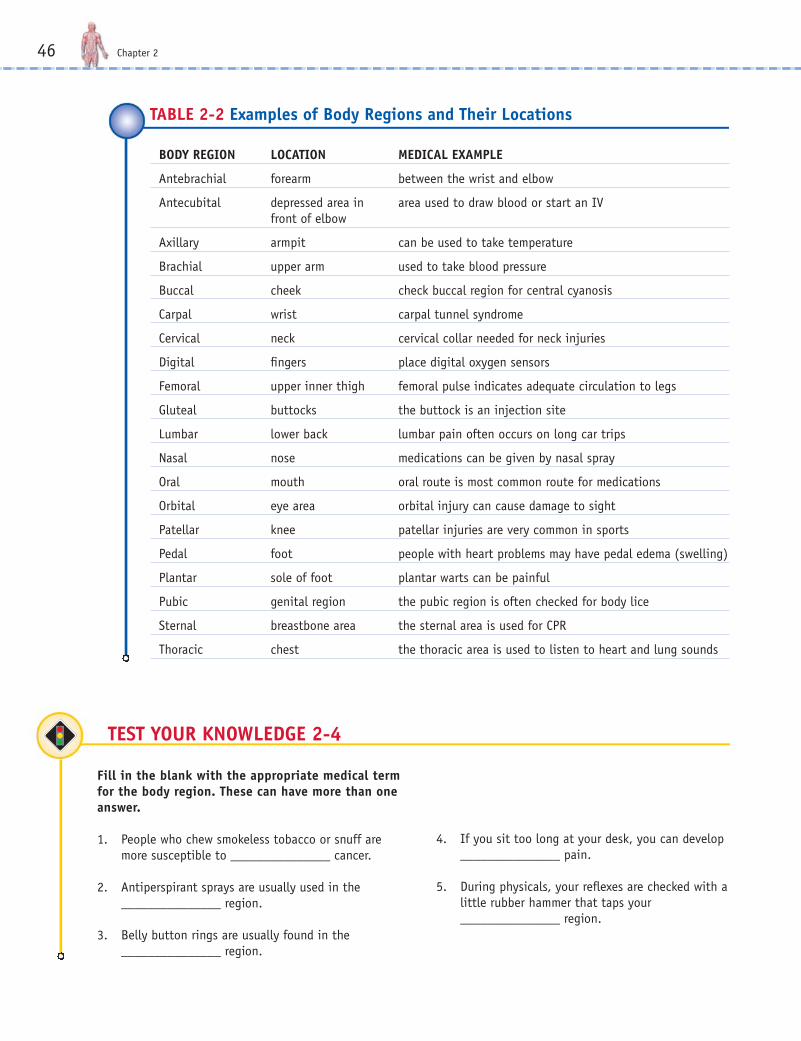
BODY REGION LOCATION MEDICAL EXAMPLE
Antebrachial forearm between the wrist and elbow
Antecubital depressed area in area used to draw blood or start an IVfront of elbow
Axillary armpit can be used to take temperature
Brachial upper arm used to take blood pressure
Buccal cheek check buccal region for central cyanosis
Carpal wrist carpal tunnel syndrome
Cervical neck cervical collar needed for neck injuries
Digital fingers place digital oxygen sensors
Femoral upper inner thigh femoral pulse indicates adequate circulation to legs
Gluteal buttocks the buttock is an injection site
Lumbar lower back lumbar pain often occurs on long car trips
Nasal nose medications can be given by nasal spray
Oral mouth oral route is most common route for medications
Orbital eye area orbital injury can cause damage to sight
Patellar knee patellar injuries are very common in sports
Pedal foot people with heart problems may have pedal edema (swelling)
Plantar sole of foot plantar warts can be painful
Pubic genital region the pubic region is often checked for body lice
Sternal breastbone area the sternal area is used for CPR
Thoracic chest the thoracic area is used to listen to heart and lung sounds
TABLE 2-2 Examples of Body Regions and Their Locations
TEST YOUR KNOWLEDGE 2-4
Fill in the blank with the appropriate medical termfor the body region. These can have more than oneanswer.
1. People who chew smokeless tobacco or snuff aremore susceptible to _______________ cancer.
2. Antiperspirant sprays are usually used in the_______________ region.
3. Belly button rings are usually found in the_______________ region.
4. If you sit too long at your desk, you can develop_______________ pain.
5. During physicals, your reflexes are checked with alittle rubber hammer that taps your_______________ region.
ch02.1.qxp 4/21/08 11:07 AM Page 46
RADIOLOGY: THE SCIENCE OF VIEWING THE BODY
While Chapter 5 is devoted to diagnostic testing, diagnostic imaging fits wellwithin this chapter on body positions and locations. Positions are importantin the radiologic sciences for obtaining a proper internal view of body structures.X-rays are a form of high-energy radiation that penetrate the body and gives atwo-dimensional view of the bones, air, and tissues in the body. The standardX-ray (much like a photograph) can be computer-enhanced to give much greaterdetail and contrast and to allow for a more realistic three-dimensional view.For example, if a standard chest X-ray showed a golf ball–sized tumor in thelung, you would have no idea of its actual depth because it would look flat, likea quarter. Computed tomography (CT) scanning uses a narrowly focused X-raybeam that circles rapidly around the body. The computer constructs thin-sliceimages and combines them to give much greater detail and allow for a morethree-dimensional view, much like a loaf of sliced bread gives a better idea ofthe total shape of the loaf than does a single slice. The CT scan reveals thetrue depth of the quarter-shaped tumor shown on the regular X-ray. A mag-netic resonance imager (MRI) produces even greater detail of tissue structures,even down to individual nerve bundles. Another possible advantage of the MRIis a decrease in radiation exposure.
X-rays (Radiograph or Roentgenogram)
The most common type of radiologic diagnostic modality is the X-ray. An X-ray is produced by passing X-ray beams through a specific area of the body andthen exposing a photographic film to those rays that successfully pass throughthat area. As a result, the developed film or digital image will possess an imageof varying degrees of dark and light, depending on the densities found in thebody that the X-rays passed through. Less dense areas, such as the lungs whichcontain a lot of air, allow the X-rays to pass through easily and show up on thefilm as a dark (radiolucent) region. Bones are much denser and allow fewer X-rays to pass through them. As a result, the denser bones appear as light (radiopaque) structures on the film. The densities of the other body parts aresomewhere between these examples, and vary in their lightness or darkness onthe film. In general there are four densities that can be found in the body:
1. Air is the least dense, and therefore the most radiolucent, showing up asblack on an X-ray film.
2. Tissue/Fat densities can depend the on the thickness of the tissue and/or theamount of fat present. The thicker the layer, the more radiation is absorbed,and the lighter the image.
3. Water density can be represented by blood, or by edema as a result of tissueinjury or inflammatory processes. Water density is a mid-range density: moredense than air and less dense than bone.
4. Bone/metal is the highest density, absorbing the greatest amount of radiation.Since less radiation passes through and onto the film, the film image is whiteor lighter than the other densities.
The Human Body: Reading the Map 47
ch02.1.qxp 4/21/08 11:07 AM Page 47

One of the problems with an X-ray film is that it is a one-dimensional view ofa specific area. As a result, the patient has to be positioned differently in rela-tionship to the X-ray machine and film depending on what the physician islooking for. For example, it would be difficult to see a small lung tumor locatedright behind the heart if you were looking at a frontal view of the patient. Every attempt should be made to position the patient’s area to be X-rayed asclose to the film as possible. This will help insure that the structures appear asclear and as close to actual size as possible on the X-ray. The following are thethree main views normally used in the clinical setting:
• Posteroanterior (PA) This standard position places the patient in an uprightposition with the chest placed in front of the X-ray film. The X-ray beamtravels from the machine, through the patient’s posterior region, out the pa-tient’s anterior, and onto the film. The shoulders are commonly rotated for-ward to move the shoulder blades away from the lungs if there is a questionabout any lung pathology. Generally, the distance between the X-ray ma-chine and the film is six feet.
• Anteroposterior (AP) In this position, the patient’s posterior is against theX-ray film. The X-ray beam travels through the patient’s anterior region, outthe posterior, and onto the film. The distance between the X-ray machineand the film is 48 inches. This is the position commonly seen in the hospitalwhere a portable X-ray machine is brought to the patient’s room if he or sheis too ill to be taken to the Radiology department. The patient is usually in asitting position on the bed.
• Lateral: This position is often done as a complement to the PA image. Thelateral view is done to eliminate interfering organs or structures in front ofother structures of concern, such as a tumor. The object in question is placedas close to the film as possible. For example, if there is a suspected lesion inthe left lung behind the heart, the left side of the patient is placed next to thefilm. This would be a left lateral view. This view prevents the heart from ob-structing the view of the suspected tumor and, in conjunction with a PA X-ray, gives a more 3D sense of the tumor.
Computerized Tomography (CT or CAT Scan)
As previously stated, one of the problems with an X-ray is that it provides aone-dimensional picture. The CT scan provides a high-resolution series of crosssectional “slices” (much like a loaf of sliced bread) of the body. As a result, itcreates a three-dimensional view of structures in the body as well as tissue struc-tures within solid organs . This allows the clinician to determine the exact lo-cation, size, and shape of a suspected tumor in the body, which would not bepossible with only an X-ray. The downside to a CT scan is the high level of ra-diation exposure required. A single CT scan is the equivalent of hundreds ofchest X-rays in terms of radiation exposure.
48 Chapter 2
ch02.1.qxp 4/21/08 11:07 AM Page 48

Magnetic Resonance Imaging (MRI)
Instead of utilizing radiation, this diagnostic tool uses magnetic energy to pro-duce cross-sectional images of body structures. The clarity of these images isgenerally much better than those from X-rays and is often better than a CTscan. However, there are some drawbacks. Since an intense magnetic field isused, certain steel aneurysm clips or prosthetic heart valves may be adverselyaffected, even to the point of shifting their position in the body. Patients mustalso be completely still or there will be a loss in resolution. Open MRIs havebeen developed to decrease the feelings of claustrophobia experienced by someindividuals in a closed MRI which takes place inside a very small tunnel.
Ultrasound (Sonography)
Much more sensitive than a regular X-ray, ultrasound uses sound waves to dis-tinguish structures in the body, much like SONAR or fish finders in fishingboats. Ultrasound studies also allow for body activities to be viewed in real time.Due to this feature, the actions of heart valves can be observed, or fetal devel-opment and movement can be monitored, for example. No radiation exposureis involved in these studies. In the past, there had been some concern about po-tential damage to a developing fetus due to the sound waves used during thesestudies, but no evidence has been found that this is a hazard. Figure 2-14 ■ con-trasts X-ray, CT, MRI, and ultrasound.
The Human Body: Reading the Map 49
Go to the CD-ROM to view videos concerning ultrasound, CT, MRI,
and other diagnostic imaging techniques used to visualize the in-
terior of the body.
2-2
www.prenhall.com/colbert
If you are interested in learning more about the profession of
radiologic technology, please visit this chapter’s Web site.
ch02.1.qxp 4/21/08 11:07 AM Page 49
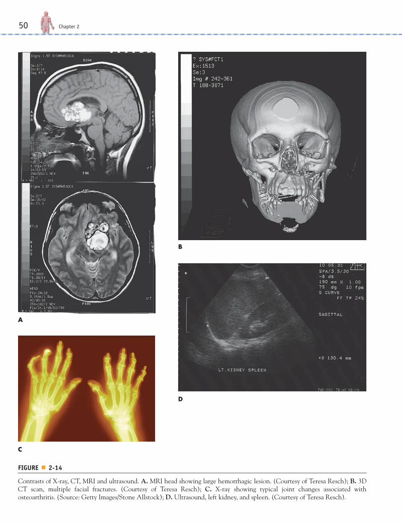
FIGURE ■ 2-14
Contrasts of X-ray, CT, MRI and ultrasound. A. MRI head showing large hemorrhagic lesion. (Courtesy of Teresa Resch); B. 3DCT scan, multiple facial fractures. (Courtesy of Teresa Resch); C. X-ray showing typical joint changes associated withosteoarthritis. (Source: Getty Images/Stone Allstock); D. Ultrasound, left kidney, and spleen. (Courtesy of Teresa Resch).
50 Chapter 2
A
B
D
C
ch02.1.qxp 4/21/08 11:07 AM Page 50
The Human Body: Reading the Map 51
SUMMARY
E The body can assume many different positions, and to standardize the study of anatomy, scientists oftenreference the anatomical position. In the anatomical position, the person stands with face and toes for-ward, hands at sides, and palms facing forward. Other positions, such as the prone, supine, and Fowlerpositions, are used in health care for assessment and treatment.
E The body can be divided into different sections along different planes. For example, the transverse orhorizontal plane divides the body into superior and inferior sections. The median or midsagittal planedivides the body into equal right and left halves. The sagittal plane divides the body into right and leftparts. The frontal or coronal plane divides the body into anterior and posterior sections.
E Directional terms such as internal and external, proximal and distal, superficial and deep, central andperipheral help us to navigate the body.
E It is important to remember that directions such as right and left are referenced from the patient’s per-spective and not yours.
E The body has several cavities that house anatomical structures (mainly organs). For example, the cra-nial cavity houses the brain, the thoracic cavity houses the heart and lungs, the abdominopelvic cavityhouses the digestive and reproductive organs, and the spinal cavity houses (guess what) the spinal cord.
E The body has many specific regions. For example, the umbilical region is found around your navel, orbelly button, and the femoral region is located in the upper inner thigh area.
E The directional terms, anatomical landmarks, body regions, and body cavities are all important to knowso that health care professionals can communicate in specific terms that leave no room for confusion.
E Detailed images of the internal structures of the body can be obtained from radiologic studies such asX-rays, CT scans, MRIs, and ultrasound.
ch02.1.qxp 4/21/08 11:07 AM Page 51

COMMON CASE STUDY
Ray’s Story
Ray’s quadriplegia will require extensive radiologic studies throughout the rest of his life. Given thefollowing scenarios, give your best diagnostic answer.
a. Since this was initially a neck injury, on what specific portion of the spinal column would you focus your studies?
_______________________________________________________________________________________
b. Due to being bedridden and inactive, Ray will be breathing in a monotonous shallow manner even with theassistance of a mechanical ventilator and therefore not fully exercising his respiratory system. This can lead tolung collapse and pneumonia. Which body cavity would you focus your study?
_______________________________________________________________________________________
c. Ray will be more prone to accidental falls while being assisted in daily activities of living. What could happenthat would require radiologic studies?
_______________________________________________________________________________________
52 Chapter 2
A 50-year-old female patient presents with sternal pain radiating to the left brachial area. Peripheral
cyanosis is noted in the digital areas, and she exhibits pedal edema. No epigastric pain is noted. She
reports that she became dizzy and fell, bruising the right orbital region, and she received superficial cuts
to the right patellar region. The physician orders an IV to be started in the left antecubital space. Please
answer the following questions in common lay terms.
a. Where would you suggest placing a bandage?
______________________________________________________________________________
b. Where did her pain begin?
______________________________________________________________________________
c. Where does the pain move to?
______________________________________________________________________________
d. Does she have stomach pain?
______________________________________________________________________________
e. Where will the IV be started?
______________________________________________________________________________
f. What part of her body is swollen?
______________________________________________________________________________
ch02.1.qxp 4/21/08 11:07 AM Page 52
1. A massage therapist would ask you to assumewhich position for a back massage?a. proneb. supinec. Fowler’sd. lotus
2. Which of the following is not in theabdominopelvic cavity?a. stomachb. liverc. reproductive organsd. heart
3. Carpal tunnel syndrome occurs in what regionof the body?a. headb. cheekc. armpitd. wrist
4. The midsagittal plane divides the body into:a. top and bottomb. front and backc. upper and lowerd. left and right
5. An organ contained in the RLQ would be:a. appendixb. heartc. lungsd. brain
6. The type of radiologic diagnostic tool that cangive you real time action shots:a. CT scansb. chest X-raysc. PET scansd. ultrasound
The Human Body: Reading the Map 53
REVIEW QUESTIONS
Multiple Choice
Fill in the Blank
1. A standard position in which a person stands erect, face forward, with feet parallel, arms at sides, and palmsforward is called the ______________ position.
2. The ______________ position is laying face upward and on your back.
3. The mouth is located ______________ to the nose, whereas the nose is located ______________ to the mouth.
4. The organ found in the cranial cavity is the ______________.
5. ______________ indicates blueness of the extremities and therefore affects the peripheral areas of the body.
6. A person who gets weak and dizzy when he or she quickly stands may be suffering from ___________________
ch02.1.qxp 4/21/08 11:07 AM Page 53
Short Answer
1. List the organs found in the abdominal cavity.____________________________________________________________________________________________________________________________________________________________________________________________________________________________________________________________________________________________________________________________________________________________
2. Contrast the differences between the prone, supine, and Fowler’s positions.____________________________________________________________________________________________________________________________________________________________________________________________________________________________________________________________________________________________________________________________________________________________
3. List and describe two specific body regions that are found on the lower extremities._____________________________________________________________________________________________________________________________________________________________________________________________________________________________________________________________________
4. List and describe the location of the nine abdominal regions using directional terms._____________________________________________________________________________________________________________________________________________________________________________________________________________________________________________________________________
Suggested Activities
1. Using a white t-shirt, draw and label the abdominal quadrants and the related organs.
2. Play Pin the Tail on the Donkey by guiding the blindfolded person using only medical directional terms.
54 Chapter 2
Now that you have completed your journey through this chapter,
please go to the CD-ROM for interactive games and puzzles con-
cerning the medical terms and concepts contained in this chapter.
By playing the games you will reinforce your learning of medical terminol-
ogy in a fun way.
2-5
ch02.1.qxp 4/21/08 11:07 AM Page 54

ch02.1.qxp 4/21/08 11:07 AM Page 55